Article

Metagenomic Shotgun Sequencing Analysis of Canalicular Concretions in Lacrimal Canaliculitis Cases

1
Department of Ophthalmology, Toho University, 6-11-1 Omori-nishi, Ota-ku, Tokyo 143-8541, Japan
2
Hyogo Prefectural Amagasaki General Medical Center, 2-17-77 Higashi-nanba cho, Amagasaki 661-0892, Japan
3
Department of Ophthalmology, School of Medicine, The Jikei University, 3-19-18 Shinbashi-nishi, Minato-ku, Tokyo 105-8471, Japan
4
Department of Ophthalmology, School of Medicine, Saitama University, 38 Morohongo Moroyama-machi, Iruma-gun, Saitama 350-0495, Japan
5
Department of Microbiology and Infectious Diseases, School of Medicine, Toho University, 6-11-1 Omori-nishi, Ota-ku, Tokyo 143-8541, Japan
*
Author to whom correspondence should be addressed.
Curr. Issues Mol. Biol. 2021, 43(2), 676-686; https://0-doi-org.brum.beds.ac.uk/10.3390/cimb43020049
Submission received: 10 May 2021 / Revised: 6 July 2021 / Accepted: 9 July 2021 / Published: 12 July 2021
(This article belongs to the Section Bioinformatics and Systems Biology)

Abstract

:
Lacrimal canaliculitis is a rare infection of the lacrimal canaliculi with canalicular concretions formed by aggregation of organisms. Metagenomic shotgun sequencing analysis using next-generation sequencing has been used to detect pathogens directly from clinical samples. Using this technology, we report cases of successful pathogen detection of canalicular concretions in lacrimal canaliculitis cases. We investigated patients with primary lacrimal canaliculitis examined in the eye clinics of four hospitals from February 2015 to July 2017. Eighteen canalicular concretion specimens collected from 18 eyes of 17 patients were analyzed by shotgun metagenomics sequencing using the MiSeq platform (Illumina). Taxonomic classification was performed using the GenBank NT database. The canalicular concretion diversity was characterized using the Shannon diversity index. This study included 18 eyes (17 patients, 77.1 ± 6.1 years): 82.4% were women with lacrimal canaliculitis; canalicular concretions were obtained from 12 eyes using lacrimal endoscopy and six eyes using canaliculotomy with curettage. Sequencing analysis detected bacteria in all samples (Shannon diversity index, 0.05–1.47). The following genera of anaerobic bacteria (>1% abundance) were identified: Actinomyces spp. in 15 eyes, Propionibacterium spp., Parvimonas spp. in 11 eyes, Prevotella spp. in 9 eyes, Fusobacterium spp. in 6 eyes, Selenomonas spp. in 5 eyes, Aggregatibacter spp. in 3 eyes, facultative and aerobic bacteria such as Streptococcus spp. in 13 eyes, Campylobacter spp. in 6 eyes, and Haemophilus spp. in 3 eyes. The most common combinations were Actinomyces spp. and Streptococcus spp. and Parvinomonas spp. and Streptococcus spp., found in 10 cases. Pathogens were identified successfully using metagenomic shotgun sequencing analysis in patients with canalicular concretions. Canalicular concretions are polymicrobial with anaerobic and facultative, aerobic bacteria.

1. Introduction

Lacrimal canaliculitis, an infectious inflammation of the lacrimal canaliculus, often remains undiagnosed for extended periods as a result of its rarity and variety. Patients with lacrimal canaliculitis typically present with red eye, epiphora, and punctal discharge. Lacrimal canaliculitis can be primary or secondary. In 1854, Von Graefe first reported primary canaliculitis, which leads to concretions with lacrimal canaliculus, and in 1878, Israel, who described human Actinomycosis, considered it to be often associated with Actinomyces species (spp.). Secondary canaliculitis is often a complication of punctal or intercanalicular plug insertion for treating dry eye [1].
The presence of concretions may shield the bacteria from the antibiotics [2], thus promoting resistance and an inadequate treatment response. Hence, early diagnosis and prompt surgical management are important. Many studies have summarized the clinical characteristics and treatment of primary canaliculitis, but few have investigated primary canaliculitis with concretions.
Previous reports on the culture of lacrimal duct stones showed that Actinomyces was detected in 5% to 44.4% [3,4,5,6,7,8,9,10], while pathologic evaluation showed a discrepancy of more than 90%, depending on the test. The reasons for this are that traditional bacterial culture and identification are both time-consuming and laborious, while some anaerobic bacteria can be difficult to isolate and identify.
Consequently, the history of canalicular concretion studies is long, and the pathophysiology remains unknown. However, metagenomic shotgun sequencing analysis using next-generation sequencing (NGS) was introduced recently and enables the identification of all bacterial species with their taxonomic classification [11,12,13,14,15,16]. This method has been applied successfully to detailed analysis of the microbiome. The term NGS is generically used to indicate the two main sequencing methods: the marker gene sequencing approach (targeted-amplicon sequencing) and the shotgun approach. Targeted-amplicon sequencing is used mainly in microbiome analysis with taxonomic purposes. However, the shotgun approach to sequencing is performed across random fragments of all DNA in a given sample and also can be used in cases with an unknown microbial target [15]. In addition, shotgun metagenomics facilitates the simultaneous study of viruses, bacteriophages, archaea, and eukaryotes [17]. NGS does not require target-specific primers; the entire genome of a pathogen is sequenced randomly and directly from clinical samples. As a result, some studies have used NGS for pathogen identification in culture-negative cases [18]. Li et al. [11] reported 16 cases of infectious keratitis and four controls in formalin-fixed corneas analyzed by NGS to identify pathogens from corneal specimens. Doan et al. [19] reported that metagenomic DNA sequencing was highly concordant in cases of uveitis and was superior to pathogen-directed polymerase chain reaction. These previous studies established the feasibility of using metagenomics to investigate bacteria, fungi, amoeba, and viruses associated with pathogenic ocular infections.
Therefore, we believe that NGS is the best method for biome analysis of canalicular concretions. No studies have reported the use of NGS to detect the pathogens causing canalicular concretions with lacrimal canaliculitis. In this first study, we describe the results of using NGS for metagenomic shotgun sequencing analysis of canalicular concretion samples from primary lacrimal canaliculitis; this is the first report to clarify the composition of the canalicular concretions.

2. Materials and Methods

2.1. Subject Recruitment

This prospective study was approved by the Ethics Committee of the Faculty of Medicine, Toho University School of Medicine (No. 27019) and was conducted in accordance with the principles of the Declaration of Helsinki. Written informed consent was obtained from all the studied subjects for sample collection and subsequent analyses.

2.2. Sampling of Canalicular Concretions

We investigated patients with primary lacrimal canaliculitis who were examined in Toho University Omori Medical Center Hospital, Amagasaki Medical Center Hospital, Jikei University Hospital, Saitama Medical Center Hospital from February 2015 to July 2017. Exclusion criteria for this study were secondary canaliculitis such as punctal plug placement, facial surgery or trauma, allergies, and recurrent cases. In this study, we analyzed the pathogens of canalicular concretions. Because lacrimal stones (concretion or dacryoliths) can occur in any part of the lacrimal system, we enrolled only patients with primary canalicular concretions in this study.
Specimens such as canalicular concretion were collected and they were placed in Eppendorf tubes and stored in a freezer at −80 °C until DNA extraction. All specimens were frozen and sent to the Department of Microbiology and Infectious Diseases at the Faculty of Medicine, Toho University School of Medicine.

2.3. DNA Extraction and Shotgun Metagenomic Sequencing Analysis

After the specimens were treated with achromopeptidase, nucleotides were extracted using a Recover All Total Nucleic Acid Isolation Kit for FFPE (Life Technologies, Thermo Fisher Scientific Inc., Waltham, MA, USA). Nextra XT DNA Library Preparation Kit (Illumina, Inc., San Diego, CA, USA) was used in preparation for shotgun sequencing DNA libraries. Shotgun metagenomic sequencing for 150 bases of the single end was performed using a MiSeq platform (Illumina, Inc.). Skewer (version 0.1.126) was used for trimming the adapter sequence to less than a Phred quality score (Q) of 15 for low-quality sequences [20]. Human genome sequences were subtracted using the Burrows-Wheeler Aligner with the “MEM” option with the human genome GRCh37.p13 (GenBank assembly accession: GCA_000001405.14) as a mapping reference and SAM tools (version 1.3) [21,22]. Reads with human genome subtracted were analyzed by a MEGABLAST search against the GenBank nt and WGS databases (ftp://ftp.ncbi.nlm.nih.gov/blast/db/) (Access date: 12 May 2016), downloaded in May 2016, followed by metagenomic browser MEGAN5 (http://ab.inf.uni-tuebingen.de/software/megan5) (Access date: 10 September 2015) [23]. The human genome was subtracted from the whole genome, and the subtracted genome was considered the microbial genome (bacterial, eukaryotic, and viral genome). Effective bacterial genera were defined as having an abundance >1.0%.

3. Results

3.1. Patients’ Characteristics

Seventeen patients (18 eyes; 82.4% women patients) were enrolled in this prospective study. The mean age at presentation was 77.1 ± 6.1 years (range, 68–89 years). In six cases, the canalicular concretion was on the upper right side, in one case on the lower right side, in five cases on the upper left side, and in six cases on the lower left side. The sample collection method using lacrimal passage endoscopy was performed in 12 cases and in six cases canaliculotomy with curettage. Before sample collection, antibacterial eye drops were used in all samples, and in 6 of 16 cases, antibacterial internal drugs were used (Table 1). Microbiology laboratory culture results of removed canalicular concretion are shown (Table 2).

3.2. Metagenomic Shotgun Sequencing Analysis

Samples from canalicular concretion from primary lacrimal canaliculitis underwent metagenomic shotgun sequencing analysis. Although most reads were of human origin, nonhuman quality-filtered microbial sequence reads/sample were obtained for subsequent analysis. The proportion of sequence reads with significant hits to bacteria, viruses, fungi, and Eukaryota are summarized in Table 3. In total, the genome minimal number of reads/sample was 27,552; the maximal number was 1,148,458 (average ± standard deviation (SD) 403,011 ± 371,050). Overall, 18 of 18 cases were bacteria-positive (minimal to maximal range detected, 21,000 to 698,597 reads; relative abundance, 18.6–92.1%), virus-positive in 2 of 18 cases (maximal, 33 reads), and fungus-positive 14 of 18 cases (maximal 10,061 reads). All cases were Eukaryota-positive and detected in 91 to 3524 reads.

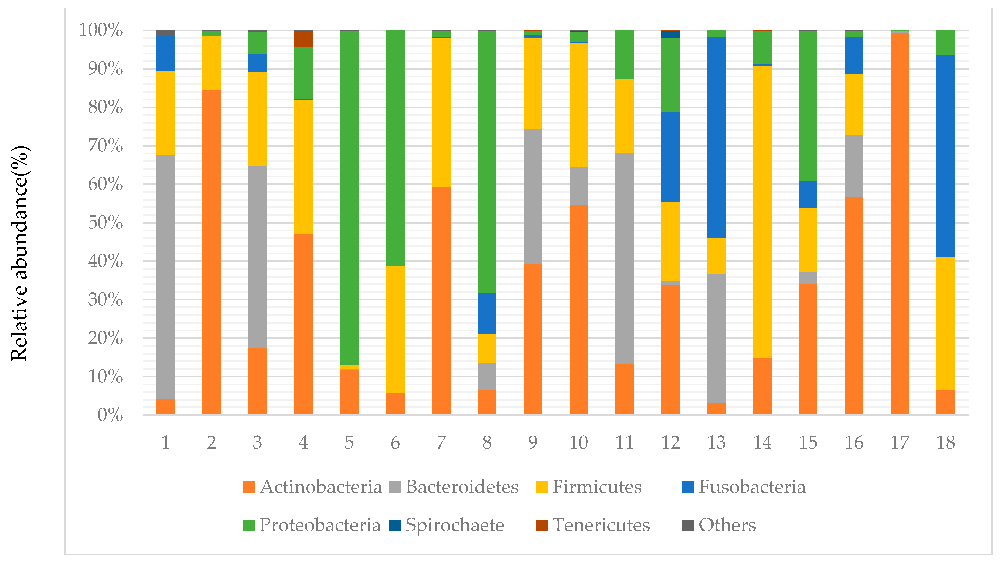
3.3. Taxonomy of Canalicular Concretions and Identification of Bacterial Phyla and Shannon Index

Figure 1 shows that the most abundant phyla were Firmicutes (26.2%), Bacteroidetes (26.1%), Actinobacteria (18.1%), Proteobacteria (16.8%), and Fusobacteria (12.4%), accounting for 99.6% of the total reads. The remaining two phyla, in relatively low abundance (0.4% total), were Spirochaete and Tenericutes. The most prevalent phyla were Actinobacteria, which were found in all 18 samples. Firmicutes and Proteobacteria were the second most prevalent, detected in 17 samples; Bacteroidetes was detected in 16 samples; Fusobacteria was detected in 14 samples. The mean Shannon index was 0.97 ± 0.362 (range, 0.047–1.472).

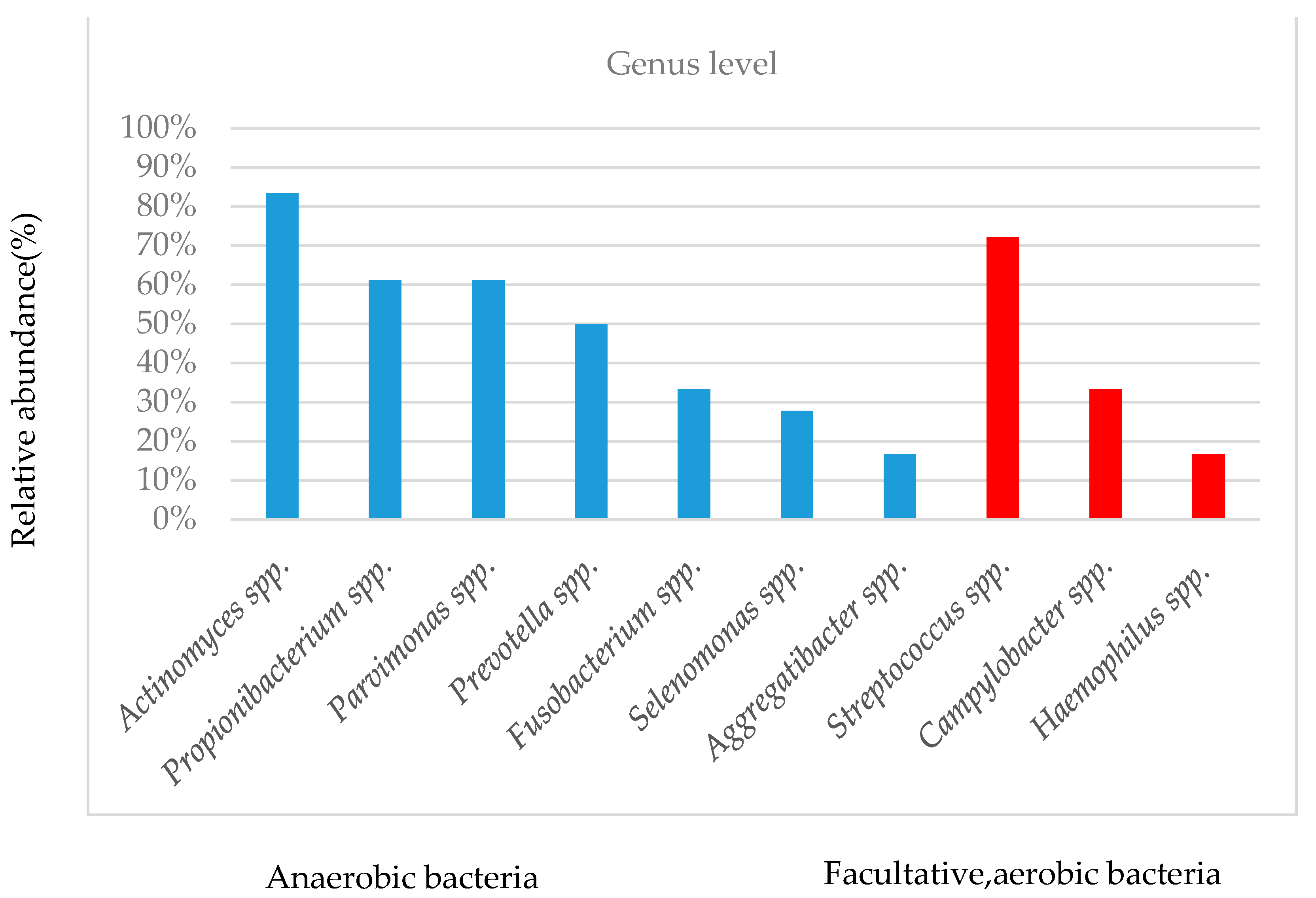
3.4. Taxonomy of Canalicular Concretions and Identification of Bacterial Genera

Table 4 provides the number of detected genera in each sample and the number of detected bacterial genera with an abundance >1.0%.
Among the anaerobic bacteria, the most prevalent genera were Actinomyces spp. (15/18 eyes, relative abundance, 1.1–54.1%), Propionibacterium spp. (11/18 eyes, relative abundance, 5.5–97.9%), Parvimonas spp. (11/18 eyes, relative abundance, 2.5–35.4%), Prevotella spp. (9/18 eyes; abundance, 2.8–52.7%), Fusobacterium spp. (6/18 eyes, abundance, 4.6–52.5%), Selenomonas spp. (5/18 eyes, abundance, 1.0–18.6%), and Aggregatibacter spp. (3/18 eyes, abundance, 1.1–5.7%). In facultative, aerobic bacteria, the most prevalent genera were Streptococcus spp. (13/18 eyes, relative abundance, 2.3–61.9%), Campylobacter spp. (6/18 eyes, relative abundance, 3.2–18.5%), and Haemophilus spp. (3/18 eyes, relative abundance, 1.0–78.9%). The top 10 species were classified as anaerobic or facultative, aerobic bacteria, as shown in Figure 2. The relative abundance of the prevalent genera varied significantly depending on individual and sample types.
The most common combinations were Actinomyces spp. and Streptococcus spp. in 10 eyes, Parvinomonas spp. and Streptococcus in 10 eyes, followed by Actinomyces spp. and Parvinomonas spp., Actinomyces spp. and Prevotella spp., Actinomyces spp. and Propionibacterium spp. (combination of two genera), and Actinomyces spp. and Parvinomonas spp. and Streptococcus spp. (combination of three genera), found in eight eyes.

3.5. The Treatment Results

After specimen collection, almost all of the canalicular concretions were removed by lacrimal endoscopy or canaliculotomy with curettage. Recurrence of canaliculitis was observed in one eye in sample 7 (Table 5).

4. Discussion

Although lacrimal canaliculitis has been analyzed in culture and pathology, this is the first report using metagenomic shotgun sequencing analysis. The results of our metagenomic shotgun sequencing analysis were used to investigate the canalicular concretions in primary canaliculitis. All evaluated canalicular concretions were bacteria-positive and a few samples were virus- and fungi-positive.
Few studies have investigated the use of metagenomic shotgun sequencing analysis to detect the pathogens causing lacrimal infection such as canaliculitis.
In the current study, Firmicutes was the most dominant phylum. In addition, Bacteroidetes, Actinobacteria, Proteobacteria, and Fusobacteria were present in abundant levels. Actinobacteria was detected in all samples. No previous reports have used NGS to analyze canalicular concretions. The results of conjunctival sac culture [24,25,26] consisted of Proteobacteria, Firmicutes, and Actinobacteria detected at the phylum level. These similar structures were not found in ocular diseases and were similar to the microbiome structure of endodontic diseases in the oral cavity [27]. It is anatomically relevant that the lacrimal drainage system comprises the puncta, canaliculi, lacrimal sac, and nasolacrimal duct, the last of which opens into the inferior nasal meatus [28].
At the gene level, metagenomic analysis showed that the average detected number of bacteria was 5.7, assuming that 1% or more was detected. Comparing the present results with the NGS results, few studies have reported on canalicular concretions, but in dacryocystitis [29], Staphylococcus, Streptococcus, Veillonella, Haemophilus, and Stenotrophomonas were detected. Comparing the current results with the culture pathological (only Actinomyces spp.) results, anaerobic bacteria such as Actinomyces spp. were detected in 15 eyes, while in the culture results, Actinomyces spp. was detected in 5% to 44.4% [3,4,5,6,7,8,9,10], and in the pathological results was detected in 93.8% to 100% [9,30]; Propionibacterium spp. was detected in 11 eyes, but in the culture results, it was undetectable; Parvimonas spp. was detected in 11 eyes, but in the culture results, it was undetectable; Prevotella spp. in nine eyes and 2.2% in culture results [5]; Fusobacterium spp. in six eyes and 28.6% in culture results [10]; Selenomonas spp. in five eyes, and Aggregatibacter spp. in three eyes, but in culture results, it was undetectable. Facultative bacteria such as Streptococcus spp. were detected in 13 eyes, but culture results showed 10.6% to 60% [3,5,8,10]. Aerobic bacteria such as Campylobacter spp. were detected in six eyes but were undetected in the culture results. Haemophilus spp. was detected in three eyes and was found at 22.2% to 28.6% [3,10] in culture results. In contrast, Staphylococcus spp. detected by culture tests ranged from 10% to 53.3% [3,4,5,6,7], while none was detected in our NGS results. The difference between these results is that antimicrobial eye drops and oral antimicrobial medication were used in almost all cases, and anaerobic media with a 5% carbon dioxide atmosphere was used for at least 4 to 6 days, as described by Hussain et al. [10] and Briscoe et al. [9].
The most prevalent genus was Actinomyces spp. For a long time, Actinomyces spp. was the most common pathogen in lacrimal canaliculitis [10]. Most published studies and case reports on canaliculitis have detailed an attempt to determine the causative organism either by culture or histopathological examination. Perumal et al. [30] reported that they often present in histopathological sections of canalicular concretions and further found that a relatively large percentage of filamentous bacteria (Actinomyces spp.) and non-filamentous bacteria (both Gram-positive and Gram-negative) can be seen on pathological analysis. They also proposed that canalicular concretions harbored mixed infections seen on histopathologic analysis. Similarly, Actinomyces spp. was a mixed-bacterial infection that coexisted with aerobic and anaerobic bacteria such as in internal medicine diseases [31] and periodontitis [32].
Similarly, Streptococcus spp. also has been detected in culture in from 10% to 60% [3,5,7,8,9], which is consistent with our results. Streptococcus spp. are clinically important Gram-positive bacteria that can cause a wide variety of diseases in human. The initial step in establishing a bacterial infection is adhesion of the organism to the epithelium of the host. Streptococcus spp. use multiple adhesins to attach to host cells [33].
Although Propionibacterium spp. is a common ocular surface microflora and may not be recognized as a pathogen because it has been non-pathogenic until now, it has been detected in endophthalmitis [34,35], suggesting either that it may not have been considered a causative agent or that it can become pathogenic by coexisting with other bacteria [36].
According to our metagenomic analysis, common bacterial combinations were Actinomyces spp. and Streptococcus spp., Parvinomonas spp. and Streptococcus spp. found in 10 cases, followed by Actinomyces spp. and Parvinomonas spp., Actinomyces spp. and Prevotella spp., Actinomyces spp. and Propionibacterium spp., and the triple combination of Actinomyces spp., Parvinomonas spp., and Streptococcus spp. in eight cases.
Furthermore, when the bacteria detected by metagenomic analysis and bacterial combinations were examined in detail, interestingly, they were similar to those found in oral gingivitis and periodontitis, as well as at the phyla level.
Periodontal diseases occur when a bacterial biofilm (dental plaque) adheres to the boundary between the teeth and gingiva, causing chronic inflammation and progressively destroying the periodontal tissue that supports the teeth. This allows the first bacteria (early colonizers), such as Streptococcus spp., to attach to the teeth, colonize, and grow. After some growth of early colonizers, the biofilm becomes more compliant with other bacterial species, known as late colonizers such as Actinomyces spp., Fusobacterium spp., Prevotella spp., Parvimonas spp., and Aggregatibacter spp. This provides the conditions for cell–cell interactions of both anaerobic bacteria and facultative, aerobic bacteria. In brief, periodontitis is caused by mixed-species communities rather than by individual pathogens working in isolation [37]. Bacterial biofilms cause chronic infections because they show increased tolerance to antibiotics and disinfectant chemicals as well as resisting phagocytosis and other components of the body’s defense system [38]. Above all, Actinomyces spp. and Prevotella spp. maintain or establish biofilm complexity [39]. It is also important that, anatomically, the lacrimal duct connects the ocular surface to the nose.
The current findings highlight the important role of anaerobic bacteria in canalicular concretions. We hypothesized that lacrimal canaliculitis results from a combination of bacteria (anaerobic and facultative, aerobic) in a closed environment of lacrimal canaliculi, and matures to form a biofilm. The formation of biofilms by bacteria may lead to drug resistance and the development of intractable diseases.
Furthermore, as a result of metagenomic analysis, a small amount of fungi (1.04%) also was detected in addition to bacteria. Recently, a strong association was reported between the presence of Candida and periodontal diseases [40]. Vécsei et al. [8] also reported fungi in 23% of cases in culture. We suggested that a few cases of canalicular concretions formed by co-infection with bacteria and fungi.
Lacrimal canaliculitis is often delayed in detection due to a lack of symptoms [2]. Recent studies using optical coherence tomography (OCT) have been able to detect several anatomical elements of the lacrimal gland (ducts, lobules, parenchyma, and acini) [41]. In addition, it has been observed by OCT and other techniques that lacrimal duct obstruction causes changes in tear fluid dynamics [42,43]. This may help in the diagnosis of lacrimal duct inflammation such as canaliculitis by using OCT in the future, and is expected to be a subject of future research.
In the present case, only one of the 18 eyes had recurrence. We believe that the results of shotgun sequencing do not contribute to the choice of antibiotics for the treatment of lacrimal canalicular concretions. The results of this study suggest that canaliculitis is often caused by anaerobic bacteria, and canaliculotomy with curettage or using lacrimal endoscope is a better approach than antibiotic therapy.
Periodontal disease has been associated with systemic vascular diseases such as cerebral infarction and myocardial infarction [44]. If the bacterial composition of lacrimal canaliculitis is similar to that of periodontal disease, it is necessary to elucidate the pathophysiology and establish therapeutic agents.
The results of the current study should be interpreted with caution because of some limitations, one of which is that DNA sequencing alone cannot detect RNA viruses (e.g., rubella). Another limitation was the small sample size. A major limitation of metagenomic analysis is the chance that the results would represent contaminants. In addition, it was difficult to determine if the organisms detected in the metagenomic analysis were pathogenic or nonpathogenic agents. We defined the causative organisms as those detected more than 1% of the time, but there was no cut-off value. Finally, we did not conduct a chemical analysis of the composition of the canalicular concretions, such as calcium, or proteins, such as trefoil factor peptides and mucins.

5. Conclusions

Pathogens were identified successfully using metagenomic shotgun sequencing analysis in patients with canalicular concretions. Canalicular concretions are polymicrobial with anaerobic and facultative, aerobic bacteria.

Author Contributions

Conceptualization, Y.O., C.M. and Y.H.; methodology, Y.O. and K.A.; validation, Y.O. and K.A.; formal analysis, K.A.; investigation, Y.O., C.M., Y.S., S.G., S.I. and Y.H.; resources, Y.I., K.T. and Y.H.; data curation, K.A.; writing—original draft preparation, Y.O.; writing—review and editing, Y.I. and T.S.; visualization, Y.O.; supervision, K.T. and Y.H.; project administration, Y.H.; funding acquisition, Y.H. All authors have read and agreed to the published version of the manuscript.

Funding

This research received no external funding.

Institutional Review Board Statement

The study was conducted according to the guidelines of the Declaration of Helsinki and approved by the Institutional Review Board of Toho University School of Medicine (No. 27019, The 17 September 2015).

Informed Consent Statement

Informed consent was obtained from all subjects involved in the study.

Conflicts of Interest

The authors declare no conflict of interest.

Abbreviations

CNScoagulase-negative staphylococci
SDstandard deviation
spp.species
PCRpolymerase chain reaction
NGSnext-generation sequencing
DNAdeoxyribonucleic acid
RNAribonucleic acid

References

  1. Freedman, J.R.; Markert, M.S.; Cohen, A.J. Primary and secondary lacrimal canaliculitis: A review of literature. Surv. Ophthalmol. 2011, 56, 336–347. [Google Scholar] [CrossRef] [PubMed]
  2. Singh, M.; Gautam, N.; Agarwal, A.; Kaur, M. Primary lacrimal canaliculitis-A clinical entity often misdiagnosed. J. Curr. Ophthalmol. 2018, 30, 87–90. [Google Scholar] [CrossRef]
  3. Lin, S.C.; Kao, S.C.; Tsai, C.C.; Cheng, C.Y.; Kau, H.C.; Hsu, W.M.; Lee, S.M. Clinical characteristics and factors associated the outcome of lacrimal canaliculitis. Acta Ophthalmol. 2011, 89, 759–763. [Google Scholar] [CrossRef] [PubMed]
  4. Xiang, S.; Lin, B.; Pan, Q.; Zheng, M.; Qin, X.; Wang, Y.; Zhang, Z. Clinical features and surgical outcomes of primary canaliculitis with concretions. Medicine 2017, 96, e6188. [Google Scholar] [CrossRef] [PubMed]
  5. Gogandy, M.; Al-Sheikh, O.; Chaudhry, I. Clinical features and bacteriology of lacrimal canaliculitis in patients presenting to a tertiary eye care center in the Middle East. Saudi J. Ophthalmol. 2014, 28, 31–35. [Google Scholar] [CrossRef] [Green Version]
  6. Anand, S.; Hollingworth, K.; Kumar, V.; Sandramouli, S. Canaliculitis: The incidence of long-term epiphora following canaliculotomy. Orbit 2004, 23, 19–26. [Google Scholar] [CrossRef] [PubMed]
  7. Kim, U.R.; Wadwekar, B.; Prajna, L. Primary canaliculitis: The incidence, clinical features, outcome and long-term epiphora after snip-punctoplasty and curettage. Saudi J. Ophthalmol. 2015, 29, 274–277. [Google Scholar] [CrossRef] [PubMed] [Green Version]
  8. Vécsei, V.P.; Huber-Spitzy, V.; Arocker-Mettinger, E.; Steinkogler, F.J. Canaliculitis: Difficulties in diagnosis, differential diagnosis and comparison between conservative and surgical treatment. Ophthalmologica 1994, 208, 314–317. [Google Scholar] [CrossRef]
  9. Briscoe, D.; Edelstein, E.; Zacharopoulos, I.; Keness, Y.; Kilman, A.; Zur, F.; Assia, E.I. Actinomyces canaliculitis: Diagnosis of a masquerading disease. Graefes. Arch. Clin. Exp. Ophthalmol. 2004, 242, 682–686. [Google Scholar] [CrossRef]
  10. Hussain, I.; Bonshek, R.E.; Loudon, K.; Armstrong, M.; Tullo, A.B. Canalicular infection caused by Actinomyces. Eye 1993, 7, 542–544. [Google Scholar] [CrossRef]
  11. Li, Z.; Breitwieser, F.P.; Lu, J.; Jun, A.S.; Asnaghi, L.; Salzberg, S.L.; Eberhart, C.G. Identifying Corneal Infections in Formalin-Fixed Specimens Using Next Generation Sequencing. Investig. Ophthalmol. Vis. Sci. 2018, 59, 280–288. [Google Scholar] [CrossRef]
  12. Shigeyasu, C.; Yamada, M.; Aoki, K.; Ishii, Y.; Tateda, K.; Yaguchi, T.; Hori, Y. Metagenomic analysis for detecting Fusarium solani in a case of fungal keratitis. J. Infect. Chemother. 2018, 24, 664–668. [Google Scholar] [CrossRef]
  13. Seitzman, G.D.; Thulasi, P.; Hinterwirth, A.; Chen, C.; Shantha, J.; Doan, T. Capnocytophaga Keratitis: Clinical Presentation and Use of Metagenomic Deep Sequencing for Diagnosis. Cornea 2019, 38, 246–248. [Google Scholar] [CrossRef] [PubMed]
  14. Borroni, D.; Romano, V.; Kaye, S.B.; Somerville, T.; Napoli, L.; Fasolo, A.; Ferrari, S. Metagenomics in ophthalmology: Current findings and future prospectives. BMJ Open Ophthalmol. 2019, 4, e000248. [Google Scholar] [CrossRef] [PubMed]
  15. Gallon, P.; Parekh, M.; Ferrari, S.; Fasolo, A.; Ponzin, D.; Borroni, D. Metagenomics in ophthalmology: Hypothesis or real prospective? Biotechnol. Rep. 2019, 23, e00355. [Google Scholar] [CrossRef]
  16. Doan, T.; Wilson, M.R.; Crawford, E.D.; Chow, E.D.; Khan, L.M.; Knopp, K.A.; DeRisi, J.L. Illuminating uveitis: Metagenomic deep sequencing identifies common and rare pathogens. Genome Med. 2016, 8, 90. [Google Scholar] [CrossRef] [Green Version]
  17. Norman, J.M.; Handley, S.A.; Virgin, H.W. Kingdom-agnostic metagenomics and the importance of complete characterization of enteric microbial communities. Gastroenterology 2014, 146, 1459–1469. [Google Scholar] [CrossRef]
  18. Deurenberg, R.H.; Bathoorn, E.; Chlebowicz, M.A.; Couto, N.; Ferdous, M.; García-Cobos, S.; Rossen, J.W. Application of next generation sequencing in clinical microbiology and infection prevention. J. Biotechnol. 2017, 243, 16–24. [Google Scholar] [CrossRef]
  19. Doan, T.; Acharya, N.R.; Pinsky, B.A.; Sahoo, M.K.; Chow, E.D.; Banaei, N.; DeRisi, J.L. Metagenomic DNA Sequencing for the Diagnosis of Intraocular Infections. Ophthalmology 2017, 124, 1247–1248. [Google Scholar] [CrossRef] [Green Version]
  20. Jiang, H.; Lei, R.; Ding, S.W.; Zhu, S. Skewer: A fast and accurate adapter trimmer for next-generation sequencing paired-end reads. BMC Bioinform. 2014, 15, 182. [Google Scholar] [CrossRef]
  21. Li, H.; Durbin, R. Fast and accurate short read alignment with Burrows-Wheeler transform. Bioinformatics 2009, 25, 1754–1760. [Google Scholar] [CrossRef] [Green Version]
  22. Li, H.; Handsaker, B.; Wysoker, A.; Fennell, T.; Ruan, J.; Homer, N.; Durbin, R. The Sequence Alignment/Map format and SAMtools. Bioinformatics 2009, 25, 2078–2079. [Google Scholar] [CrossRef] [PubMed] [Green Version]
  23. Huson, D.H.; Mitra, S.; Ruscheweyh, H.J.; Weber, N.; Schuster, S.C. Integrative analysis of environmental sequences using MEGAN4. Genome Res. 2011, 21, 1552–1560. [Google Scholar] [CrossRef] [Green Version]
  24. Huang, Y.; Yang, B.; Li, W. Defining the normal core microbiome of conjunctival microbial communities. Clin. Microbiol. Infect. 2016, 22, 643.e7–643.e12. [Google Scholar] [CrossRef] [PubMed] [Green Version]
  25. Dong, Q.; Brulc, J.M.; Iovieno, A.; Bates, B.; Garoutte, A.; Miller, D.; Shestopalov, V.I. Diversity of bacteria at healthy human conjunctiva. Investig. Ophthalmol. Vis. Sci. 2011, 52, 5408–5413. [Google Scholar] [CrossRef] [Green Version]
  26. Ozkan, J.; Nielsen, S.; Diez-Vives, C.; Coroneo, M.; Thomas, T.; Willcox, M. Temporal Stability and Composition of the Ocular Surface Microbiome. Sci. Rep. 2017, 7, 9880. [Google Scholar] [CrossRef] [Green Version]
  27. Shin, J.M.; Luo, T.; Lee, K.H.; Guerreiro, D.; Botero, T.M.; McDonald, N.J.; Rickard, A.H. Deciphering Endodontic Microbial Communities by Next-generation Sequencing. J. Endod. 2018, 44, 1080–1087. [Google Scholar] [CrossRef]
  28. Salmon, J.F.; Kanski, J.J. Kanski’s Clinical Ophthalmology: A Systematic Approach, 9th ed.; Elsevier Limited: Edinburgh, UK, 2020; p. 100. [Google Scholar]
  29. Eguchi, H.; Hotta, F.; Kuwahara, T.; Imaohji, H.; Miyazaki, C.; Hirose, M.; Shimomura, Y. Diagnostic Approach to Ocular Infections Using Various Techniques From Conventional Culture to Next-Generation Sequencing Analysis. Cornea 2017, 36, S46–S52. [Google Scholar] [CrossRef] [PubMed]
  30. Perumal, B.; Carlson, J.A.; Meyer, D.R. A Pathological Analysis of Canaliculitis Concretions: More Than Just Actinomyces. Scientifica 2016. [Google Scholar] [CrossRef] [Green Version]
  31. Pulverer, G.; Schütt-Gerowitt, H.; Schaal, K.P. Human cervicofacial actinomycoses: Microbiological data for 1997 cases. Clin. Infect. Dis. 2003, 37, 490–497. [Google Scholar] [CrossRef]
  32. Dentino, A.; Lee, S.; Mailhot, J.; Hefti, A.F. Principles of periodontology. Periodontol 2000, 61, 16–53. [Google Scholar] [CrossRef] [Green Version]
  33. Moschioni, M.; Pansegrau, W.; Barocchi, M.A. Adhesion determinants of the Streptococcus species. Microb. Biotechnol. 2010, 3, 370–388. [Google Scholar] [CrossRef] [Green Version]
  34. Ormerod, L.D.; Puklin, J.E.; Giles, C.L. Chronic Propionibacterium acnes endophthalmitis as a cause of intermediate uveitis. Ocul. Immunol. Inflamm. 1997, 5, 67–68. [Google Scholar] [CrossRef]
  35. Abrahams, I.W. Propionibacterium acnes endophthalmitis: An unusual manner of presentation. J. Cataract. Refract. Surg. 1989, 15, 698–701. [Google Scholar] [CrossRef]
  36. Sousa, V.; Nibali, L.; Spratt, D.; Dopico, J.; Mardas, N.; Petrie, A.; Donos, N. Peri-implant and periodontal microbiome diversity in aggressive periodontitis patients: A pilot study. Clin. Oral Implant. Res. 2017, 28, 558–570. [Google Scholar] [CrossRef]
  37. Daniluk, T.; Tokajuk, G.; Cylwik-Rokicka, D.; Rozkiewicz, D.; Zaremba, M.L.; Stokowska, W. Aerobic and anaerobic bacteria in subgingival and supragingival plaques of adult patients with periodontal disease. Adv. Med. Sci. 2006, 51, 81–85. [Google Scholar] [PubMed]
  38. Høiby, N.; Bjarnsholt, T.; Givskov, M.; Molin, S.; Ciofu, O. Antibiotic resistance of bacterial biofilms. Int. J. Antimicrob. Agents 2010, 35, 322–332. [Google Scholar] [CrossRef] [PubMed] [Green Version]
  39. Valm, A.M.; Mark Welch, J.L.; Rieken, C.W.; Welch, J.L.M.; Rieken, C.W.; Hasegawa, Y.; Sogin, M.L.; Oldenbourg, R.; Borisy, G.G. Systems-level analysis of microbial community organization through combinatorial labeling and spectral imaging. Proc. Natl. Acad. Sci. USA 2011, 108, 4152–4157. [Google Scholar] [CrossRef] [Green Version]
  40. Suresh Unniachan, A.; Krishnavilasom Jayakumari, N.; Sethuraman, S. Association between Candida species and periodontal disease: A systematic review. Curr. Med. Mycol. 2020, 6, 63–68. [Google Scholar] [CrossRef] [PubMed]
  41. Doh, S.H.; Kim, E.C.; Chung, S.Y.; Kim, M.S.; Chung, S.K.; Shin, M.C.; Hwang, H.S. Optical Coherence Tomography Imaging of Human Lacrimal Glands: An In Vivo Study. Ophthalmology 2015, 122, 2364–2366. [Google Scholar]
  42. Napoli, P.E.; Nioi, M.; d’Aloja, E.; Fossarello, M. The Bull′s Eye Pattern of the Tear Film in Humans during Visual Fixation on En-Face Optical Coherence Tomography. Sci. Rep. 2019, 9, 1413. [Google Scholar] [CrossRef]
  43. Napoli, P.E.; Nioi, M.; Mangoni, L.; Gentile, P.; Braghiroli, M.; d′Aloja, E.; Fossarello, M. Fourier-Domain OCT Imaging of the Ocular Surface and Tear Film Dynamics: A Review of the State of the Art and an Integrative Model of the Tear Behavior During the Inter-Blink Period and Visual Fixation. J. Clin. Med. 2020, 9, 668. [Google Scholar] [CrossRef] [PubMed] [Green Version]
  44. Larvin, H.; Kang, J.; Aggarwal, V.R.; Pavitt, S.; Wu, J. Risk of incident cardiovascular disease in people with periodontal disease: A systematic review and meta-analysis. Clin. Exp. Dent. Res. 2021, 7, 109–122. [Google Scholar] [CrossRef] [PubMed]
Figure 1. Distribution of bacteria phyla identified by metagenomic analysis of 18 samples obtained from canaliculitis concretions.
Cimb 43 00049 g001
Figure 2. Top 10 bacterial species with the most frequent appearance.
Cimb 43 00049 g002
Table 1. Patient characteristics.
Sample
Mean age (years ± SD), n = 1877.1 ± 6.1
No. women (%), n = 1814 (82.4%)
Side of involvement
Upper right; lower right; upper left; lower left, eyes (%)
6 eyes; 1 eye; 5 eyes; 6 eyes
Sample collection method using lacrimal passage endoscope or
curettage, eyes (%) n = 18
12 eyes; 6 eyes
Before sample collection, antibacterial eye drops were used, eyes (%) n = 1818/18
Before sample collection, antibacterial internal drugs were used, eyes (%), n = 16 (unknown in 2 eyes)6/16
Table 2. Summary of microbiology laboratory culture of canalicular concretions.
SampleMicrobiology Laboratory Culture
1Prevotella melaninogenica, Fusobacterium nucleatum, Streptococcus milleri
2Actinomyces spp., Streptococcus constellatus
3No culture test
4Actinomyces odontolyticus, Eikenella corrodens
5Actinomyces spp. Haemophilus parainfluenzae
6Streptococcus spp.
7Peptostereptococcus spp., α-streptococcus
8Negative culture for anaerobic bacteria
9Negative culture for anaerobic bacteria
10CNS, Coynebacteirum spp.
11No culture test
12Propionibacterium spp.
13No culture test
14Streptococcus milleri group Peptostreptococcus spp.
15Haemophilus influenzae
16Haemophilus influenzae
17Actinomyces israelii
18Streptococcus milleri group, CNS
Table 3. Read summary of metagenomic shotgun sequencing analysis of canalicular concretions.
SampleTotal GenomeBacteria%Virus%Fungi%Eukaryota%
1968,005698,59772.20010,0611.0416030.17
2118,76226,54822.400130.0119381.63
3974,992181,29918.6001200.0118030.18
4166,88558,75035.200210.019590.57
577,72048,10761.900100.0117582.26
6260,161222,24685.4000023400.90
7103,63955,69853.700150.0115461.49
8180,213129,31471.80000910.05
91,087,307451,32641.5001430.017600.07
10209,08567,15832.100410.0214960.72
111,148,458501,68143.7001100.016930.06
12191,096118,84662.200150.0115530.81
1378,33465,92684.2000013521.73
14273,766140,08251.200340.0135241.29
15413,328153,52337.1330.01630.0223170.56
16336,017126,40637.600350.0115640.47
1727,55221,00076.220.0120.01277310.06
18638,880588,39692.1000012430.19
Table 4. Number of detected genera in each sample and number of detected bacterial genera of 1% or more abundance (ratio of anaerobic bacteria to facultative, aerobic bacteria).
SampleNumber of Detected Bacterial GeneraNumber of Detected Bacterial Genera, 1% or More (Ratio of Anaerobic Bacteria to Facultative, Aerobic Bacteria)
1135 (4:1)
21394 (3:1)
338012 (10:2)
42337 (3:4)
5993 (2:1)
6164(2:2)
71035 (4:1)
8547(6:1)
91098 (7:1)
102048 (7:1)
11824 (2:2)
12687 (5:2)
13455 (4:1)
14957 (5:2)
151577 (5:2)
161194 (4:0)
17212 (2:0)
18 154(2:2)
Average ± SD108.4 ± 90.05.7 ± 2.3
Table 5. The treatment results of canaliculitis.
SampleTreatment Results
1No recurrence
2No recurrence
3No recurrence
4No recurrence
5No recurrence
6No recurrence
7Recurrence
8No recurrence
9No recurrence
10No recurrence
11No recurrence
12No recurrence
13No recurrence
14No recurrence
15No recurrence
16No recurrence
17No recurrence
18No recurrence
Publisher’s Note: MDPI stays neutral with regard to jurisdictional claims in published maps and institutional affiliations.

