Intrahepatic Cholestasis of Pregnancy: A Case Study of the Rare Onset in the First Trimester

 ,
,  and
and 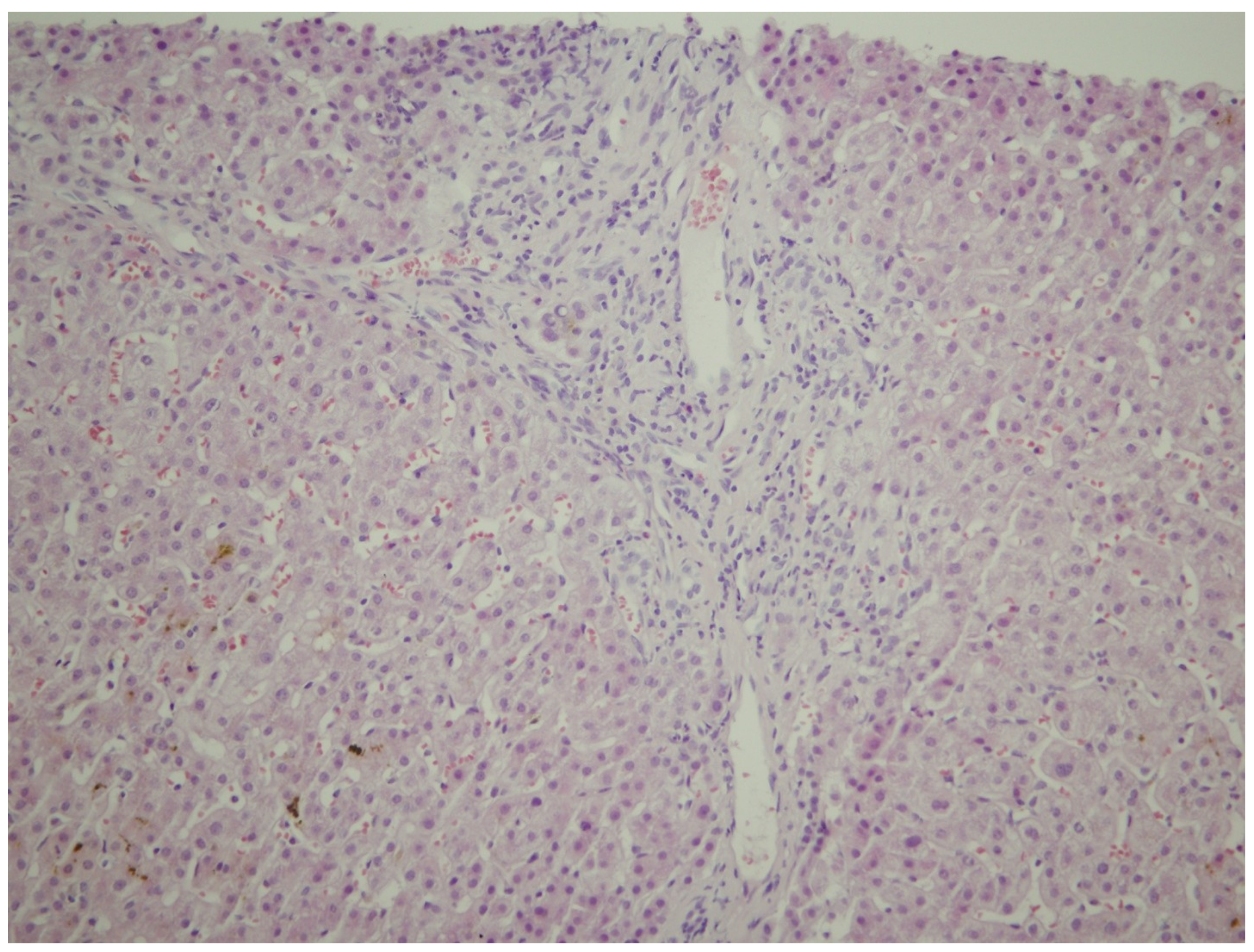
Abstract
:1. Introduction
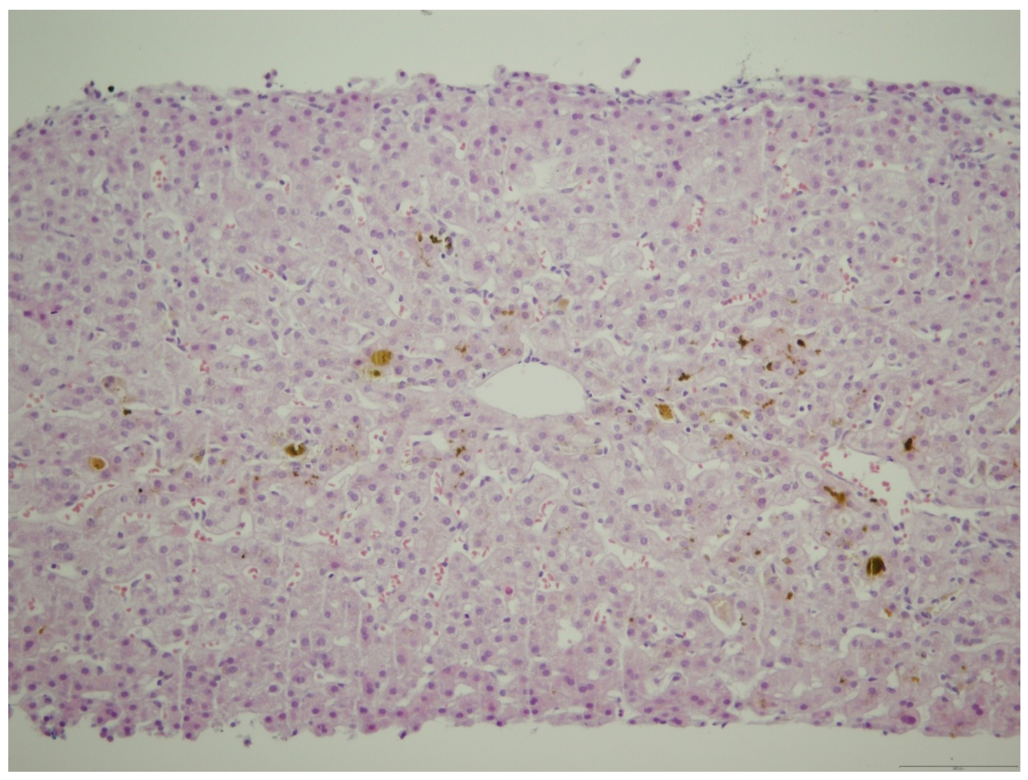
2. Case Report
3. Discussion
4. Conclusions
Author Contributions
Funding
Acknowledgments
Conflicts of Interest
References
- Ahlfeld, F. Berichte und Arbeiten aus der Geburtshilflich-Gynaekologischen Klinik zu Giessen; Bokus: Leipzig, Germany, 1883. [Google Scholar]
- Glantz, A.; Marschall, H.U.; Mattsson, L.A. Intrahepatic cholestasis of pregnancy: Relationships between bile acid levels and fetal complication rates. Hepatology 2004, 40, 467–474. [Google Scholar] [CrossRef] [PubMed]
- Saleh, M.M.; Abdo, K.R. Intrahepatic cholestasis of pregnancy: Review of the literature and evaluation of current evidence. J. Womens Health 2007, 16, 833–841. [Google Scholar] [CrossRef] [PubMed]
- Geenes, V.; Williamson, C. Intrahepatic cholestasis of pregnancy. World J. Gastroenterol. 2009, 15, 2049–2066. [Google Scholar] [CrossRef] [PubMed] [Green Version]
- Ozkan, S.; Ceylan, Y.; Ozkan, O.V.; Yildirim, S. Review of a challenging clinical issue: Intrahepatic cholestasis of pregnancy. World J. Gastroenterol. 2015, 21, 7134–7141. [Google Scholar] [CrossRef] [PubMed]
- Marathe, J.A.; Lim, W.H.; Metz, M.P.; Scheil, W.; Dekker, G.A.; Hague, W.M. A retrospective cohort review of intrahepatic cholestasis of pregnancy in a South Australian population. Eur. J. Obstet. Gynecol. Reprod. Biol. 2017, 218, 33–38. [Google Scholar] [CrossRef] [PubMed]
- Reyes, H. Review: Intrahepatic cholestasis. A puzzling disorder of pregnancy. J. Gastroenterol. Hepatol. 1997, 12, 211–216. [Google Scholar] [CrossRef] [PubMed]
- Joshi, D.; James, A.; Quaglia, A.; Westbrook, R.H.; Heneghan, M.A. Liver disease in pregnancy. Lancet 2010, 375, 594–605. [Google Scholar] [CrossRef]
- Sepulveda, W.H.; Gonzalez, C.; Cruz, M.A.; Rudolph, M.I. Vasoconstrictive effect of bile acids on isolated human placental chorionic veins. Eur. J. Obstet. Gynecol. Reprod. Biol. 1991, 42, 211–215. [Google Scholar] [CrossRef]
- Williamson, C.; Gorelik, J.; Eaton, B.M.; Lab, M.; de Swiet, M.; Korchev, Y. The bile acid taurocholate impairs rat cardiomyocyte function: A proposed mechanism for intra-uterine fetal death in obstetric cholestasis. Clin. Sci. 2001, 100, 363–369. [Google Scholar] [CrossRef]
- Zecca, E.; De Luca, D.; Marras, M.; Caruso, A.; Bernardini, T.; Romagnoli, C. Intrahepatic cholestasis of pregnancy and neonatal respiratory distress syndrome. Pediatrics 2006, 117, 1669–1672. [Google Scholar] [CrossRef]
- Puljic, A.; Kim, E.; Page, J.; Esakoff, T.; Shaffer, B.; LaCoursiere, D.Y.; Caughey, A.B. The risk of infant and fetal death by each additional week of expectant management in intrahepatic cholestasis of pregnancy by gestational age. Am. J. Obstet. Gynecol. 2015, 212, e1–e5. [Google Scholar]
- Mackillop, L.; Williamson, C. Liver disease in pregnancy. Postgrad. Med. J. 2010, 86, 160–164. [Google Scholar] [CrossRef] [PubMed]
- Maldonado, M.; Alhousseini, A.; Awadalla, M.; Idler, J.; Welch, R.; Puder, K.; Patwardhan, M.; Gonik, B. Intrahepatic Cholestasis of Pregnancy Leading to Severe Vitamin K Deficiency and Coagulopathy. Case Rep. Obstet. Gynecol. 2017, 2017, 5646247. [Google Scholar] [CrossRef] [PubMed]
- Marschall, H.U.; Wikstrom Shemer, E.; Ludvigsson, J.F.; Stephansson, O. Intrahepatic cholestasis of pregnancy and associated hepatobiliary disease: A population-based cohort study. J. Hepatol 2013, 58, 1385–1391. [Google Scholar] [CrossRef] [PubMed]
- Pathak, B.; Sheibani, L.; Lee, R.H. Cholestasis of pregnancy. Obstet. Gynecol. Clin. N. Am. 2010, 37, 269–282. [Google Scholar] [CrossRef] [PubMed]
- Diken, Z.; Usta, I.M.; Nassar, A.H. A clinical approach to intrahepatic cholestasis of pregnancy. Am. J. Perinatol. 2014, 31, 1–8. [Google Scholar] [CrossRef] [PubMed]
- Paternoster, D.M.; Fabris, F.; Palu, G.; Santarossa, C.; Bracciante, R.; Snijders, D.; Floreani, A. Intra-hepatic cholestasis of pregnancy in hepatitis C virus infection. Acta Obstet. Gynecol. Scand. 2002, 81, 99–103. [Google Scholar]
- Gonzalez, M.C.; Reyes, H.; Arrese, M.; Figueroa, D.; Lorca, B.; Andresen, M.; Segovia, N.; Molina, C.; Arce, S. Intrahepatic cholestasis of pregnancy in twin pregnancies. J. Hepatol. 1989, 9, 84–90. [Google Scholar] [CrossRef]
- Koivurova, S.; Hartikainen, A.L.; Karinen, L.; Gissler, M.; Hemminki, E.; Martikainen, H.; Tuomivaara, L.; Jarvelin, M.R. The course of pregnancy and delivery and the use of maternal healthcare services after standard IVF in Northern Finland 1990–1995. Hum. Reprod. 2002, 17, 2897–2903. [Google Scholar] [CrossRef]
- Heinonen, S.; Kirkinen, P. Pregnancy outcome with intrahepatic cholestasis. Obstet. Gynecol. 1999, 94, 189–193. [Google Scholar]
- Hubschmann, A.G.; Orzechowski, K.M.; Berghella, V. Severe First Trimester Recurrent Intrahepatic Cholestasis of Pregnancy: A Case Report and Literature Review. AJP Rep. 2016, 6, e38–e41. [Google Scholar] [PubMed]
- Keitel, V.; Vogt, C.; Haussinger, D.; Kubitz, R. Combined mutations of canalicular transporter proteins cause severe intrahepatic cholestasis of pregnancy. Gastroenterol 2006, 131, 624–629. [Google Scholar] [CrossRef] [PubMed]
- Williamson, C.; Geenes, V. Intrahepatic cholestasis of pregnancy. Obstet. Gynecol. 2014, 124, 120–133. [Google Scholar] [CrossRef] [PubMed]
- Gabzdyl, E.M.; Schlaeger, J.M. Intrahepatic cholestasis of pregnancy: A critical clinical review. J. Perinat. Neonatal Nurs. 2015, 29, 41–50. [Google Scholar] [CrossRef] [PubMed]
- Reyes, H.; Sjovall, J. Bile acids and progesterone metabolites in intrahepatic cholestasis of pregnancy. Ann. Med. 2000, 32, 94–106. [Google Scholar] [CrossRef] [PubMed]
- Heinonen, S.; Eloranta, M.L.; Heiskanen, J.; Punnonen, K.; Helisalmi, S.; Mannermaa, A.; Hiltunen, M. Maternal susceptibility locus for obstetric cholestasis maps to chromosome region 2p13 in Finnish patients. Scand. J. Gastroenterol. 2001, 36, 766–770. [Google Scholar] [CrossRef] [PubMed]
- Savander, M.; Ropponen, A.; Avela, K.; Weerasekera, N.; Cormand, B.; Hirvioja, M.L.; Riikonen, S.; Ylikorkala, O.; Lehesjoki, A.E.; Williamson, C.; et al. Genetic evidence of heterogeneity in intrahepatic cholestasis of pregnancy. Gut 2003, 52, 1025–1029. [Google Scholar] [CrossRef] [PubMed] [Green Version]
- Soroka, C.J.; Boyer, J.L. Biosynthesis and trafficking of the bile salt export pump, BSEP: Therapeutic implications of BSEP mutations. Mol. Asp. Med. 2014, 37, 3–14. [Google Scholar] [CrossRef] [PubMed]
- Invernizzi, P. Intrahepatic cholestasis of pregnancy: A further important step in dissecting its genetic architecture. Dig. Liver Dis. 2013, 45, 266–267. [Google Scholar] [CrossRef]
- Floreani, A.; Caroli, D.; Lazzari, R.; Memmo, A.; Vidali, E.; Colavito, D.; D’Arrigo, A.; Leon, A.; Romero, R.; Gervasi, M.T. Intrahepatic cholestasis of pregnancy: New insights into its pathogenesis. J. Matern. Fetal. Neonatal Med. 2013, 26, 1410–1415. [Google Scholar] [CrossRef]
- Yi, P.; Yin, N.; Zheng, Y.; Jiang, H.; Yu, X.; Yan, Y.; Liu, Q.; Xiao, F.; Li, L. Elevated plasma levels of hypermethylated RASSF1A gene sequences in pregnant women with intrahepatic cholestasis. Cell Biochem. Biophys. 2013, 67, 977–981. [Google Scholar] [CrossRef] [PubMed]
- Reyes, H.; Gonzalez, M.C.; Ribalta, J.; Aburto, H.; Matus, C.; Schramm, G.; Katz, R.; Medina, E. Prevalence of intrahepatic cholestasis of pregnancy in Chile. Ann. Intern. Med. 1978, 88, 487–493. [Google Scholar] [CrossRef] [PubMed]
- Mays, J.K. The active management of intrahepatic cholestasis of pregnancy. Curr. Opin. Obstet. Gynecol. 2010, 22, 100–103. [Google Scholar] [CrossRef] [PubMed]
- Estiu, M.C.; Frailuna, M.A.; Otero, C.; Dericco, M.; Williamson, C.; Marin, J.J.G.; Macias, R.I.R. Relationship between early onset severe intrahepatic cholestasis of pregnancy and higher risk of meconium-stained fluid. PLoS ONE 2017, 12, e0176504. [Google Scholar] [CrossRef] [PubMed]
- Geenes, V.; Chappell, L.C.; Seed, P.T.; Steer, P.J.; Knight, M.; Williamson, C. Association of severe intrahepatic cholestasis of pregnancy with adverse pregnancy outcomes: A prospective population-based case-control study. Hepatology 2014, 59, 1482–1491. [Google Scholar] [CrossRef] [PubMed]
- Turnpenny, P.D.; Ellard, S. Alagille syndrome: Pathogenesis, diagnosis and management. Eur. J. Hum. Genet. 2012, 20, 251–257. [Google Scholar] [CrossRef] [PubMed]
- Oude Elferink, R.P.; Paulusma, C.C.; Groen, A.K. Hepatocanaliculartransport defects: Pathophysiologic mechanisms of rare diseases. Gastroenterol 2006, 130, 908–925. [Google Scholar] [CrossRef]
- Bolukbas, F.F.; Bolukbas, C.; Aygun, C.; Ignak, S.; Ergul, E.; Yazicioglu, M. Intrahepatic Cholestasis of Pregnancy: Spontaneous vs in vitro Fertilization. Eur. J. Hepato Gastroenterol. 2017, 7, 126–129. [Google Scholar] [CrossRef]
- Papacleovoulou, G.; Abu-Hayyeh, S.; Nikolopoulou, E.; Briz, O.; Owen, B.M.; Nikolova, V.; Ovadia, C.; Huang, X.; Vaarasmaki, M.; Baumann, M.; et al. Maternal cholestasis during pregnancy programs metabolic disease in offspring. J. Clin. Investig. 2013, 123, 3172–3181. [Google Scholar] [CrossRef] [Green Version]
- Binder, T.; Salaj, P.; Zima, T.; Vitek, L. Randomized prospective comparative study of ursodeoxycholic acid and S-adenosyl-L-methionine in the treatment of intrahepatic cholestasis of pregnancy. J. Perinat. Med. 2006, 34, 383–391. [Google Scholar] [CrossRef]
- Glantz, A.; Marschall, H.U.; Lammert, F.; Mattsson, L.A. Intrahepatic cholestasis of pregnancy: A randomized controlled trial comparing dexamethasone and ursodeoxycholic acid. Hepatology 2005, 42, 1399–1405. [Google Scholar] [CrossRef] [PubMed]
- Hirvioja, M.L.; Tuimala, R.; Vuori, J. The treatment of intrahepatic cholestasis of pregnancy by dexamethasone. Br. J. Obstet. Gynaecol. 1992, 99, 109–111. [Google Scholar] [CrossRef] [PubMed]
- Marschall, H.U.; Wagner, M.; Zollner, G.; Fickert, P.; Diczfalusy, U.; Gumhold, J.; Silbert, D.; Fuchsbichler, A.; et al. Complementary stimulation of hepatobiliary transport and detoxification systems by rifampicin and ursodeoxycholic acid in humans. Gastroenterology 2005, 129, 476–485. [Google Scholar] [CrossRef] [PubMed]
- Geenes, V.; Chambers, J.; Khurana, R.; Shemer, E.W.; Sia, W.; Mandair, D.; Elias, E.; Marschall, H.U.; et al. Rifampicin in the treatment of severe intrahepatic cholestasis of pregnancy. Eur. J. Obstet. Gynecol. Reprod. Biol. 2015, 189, 59–63. [Google Scholar] [CrossRef] [PubMed]


© 2019 by the authors. Licensee MDPI, Basel, Switzerland. This article is an open access article distributed under the terms and conditions of the Creative Commons Attribution (CC BY) license (http://creativecommons.org/licenses/by/4.0/).
Share and Cite
Stulic, M.; Culafic, D.; Boricic, I.; Stojkovic Lalosevic, M.; Pejic, N.; Jankovic, G.; Milovanovic, T.; Culafic-Vojinovic, V.; Vlaisavljevic, Z.; Culafic, M. Intrahepatic Cholestasis of Pregnancy: A Case Study of the Rare Onset in the First Trimester. Medicina 2019, 55, 454. https://0-doi-org.brum.beds.ac.uk/10.3390/medicina55080454
Stulic M, Culafic D, Boricic I, Stojkovic Lalosevic M, Pejic N, Jankovic G, Milovanovic T, Culafic-Vojinovic V, Vlaisavljevic Z, Culafic M. Intrahepatic Cholestasis of Pregnancy: A Case Study of the Rare Onset in the First Trimester. Medicina. 2019; 55(8):454. https://0-doi-org.brum.beds.ac.uk/10.3390/medicina55080454
Chicago/Turabian StyleStulic, Milos, Djordje Culafic, Ivan Boricic, Milica Stojkovic Lalosevic, Nina Pejic, Goran Jankovic, Tamara Milovanovic, Violeta Culafic-Vojinovic, Zeljko Vlaisavljevic, and Milica Culafic. 2019. "Intrahepatic Cholestasis of Pregnancy: A Case Study of the Rare Onset in the First Trimester" Medicina 55, no. 8: 454. https://0-doi-org.brum.beds.ac.uk/10.3390/medicina55080454