Review

On the Quest for In Vitro Platelet Production by Re-Tailoring the Concepts of Megakaryocyte Differentiation

by
Patricia Martínez-Botía
1,2,
Andrea Acebes-Huerta
1,
Jerard Seghatchian
3 and
Laura Gutiérrez
1,2,*
1
Platelet Research Lab, Instituto de Investigación Sanitaria del Principado de Asturias (ISPA), 33011 Oviedo, Spain
2
Department of Medicine, University of Oviedo, 33003 Oviedo, Spain
3
International Consultancy in Strategic Safety/Quality Improvements of Blood-Derived Bioproducts and Suppliers Quality Audit/Inspection, London NW3 3AA, UK
*
Author to whom correspondence should be addressed.
Submission received: 5 November 2020 / Revised: 26 November 2020 / Accepted: 30 November 2020 / Published: 3 December 2020
(This article belongs to the Special Issue Platelet Production and Function in Health and Disease)

Abstract

:
The demand of platelet transfusions is steadily growing worldwide, inter-donor variation, donor dependency, or storability/viability being the main contributing factors to the current global, donor-dependent platelet concentrate shortage concern. In vitro platelet production has been proposed as a plausible alternative to cover, at least partially, the increasing demand. However, in practice, such a logical production strategy does not lack complexity, and hence, efforts are focused internationally on developing large scale industrial methods and technologies to provide efficient, viable, and functional platelet production. This would allow obtaining not only sufficient numbers of platelets but also functional ones fit for all clinical purposes and civil scenarios. In this review, we cover the evolution around the in vitro culture and differentiation of megakaryocytes into platelets, the progress made thus far to bring the culture concept from basic research towards good manufacturing practices certified production, and subsequent clinical trial studies. However, little is known about how these in vitro products should be stored or whether any safety measure should be implemented (e.g., pathogen reduction technology), as well as their quality assessment (how to isolate platelets from the rest of the culture cells, debris, microvesicles, or what their molecular and functional profile is). Importantly, we highlight how the scientific community has overcome the old dogmas and how the new perspectives influence the future of platelet-based therapy for transfusion purposes.

1. Background: Platelet Transfusion over the Years

Platelet (PLT) transfusions were originally given in a therapeutic manner to those patients suffering from bleeding episodes, as it occurs in congenital thrombopathic patients or in patients suffering from acute hemorrhage caused by traumatic injury or hemorrhagic complications derived from surgery [1,2]. The use of PLT transfusions in a prophylactic manner, i.e., to prevent bleeding, has increased over the past years, and it is advised for those patients at risk of severe bleeding, as it occurs in onco-hematologic patients, whose condition and treatment induce a reduction and dysfunction of PLTs [3,4]. Over the last decade, there has been a steady, worldwide increase in the demand of PLT concentrates (PCs) for transfusion [5,6], which is concordant with the improvement of PLT disorder diagnosis and management and a growing, aging population with its concomitant increase in the incidence of onco-hematological diseases [7,8].
Currently, the source of PLTs comes uniquely from donations. The half-life of PLTs is short, thus their storage time or shelf life is limited to a few days [9]. They are generally stored shaking at room temperature in order to prevent loss of PLT functional integrity, which also poses a risk of bacterial or viral contamination [10,11]. While the implementation of pathogen reduction treatments (PRTs) has allowed an extension of the shelf life and a safer transfusion product, thus enabling blood centers to better manage their PC supplies, the demand is still causing pressure to find alternative solutions to increase the availability of PLTs for transfusion [12,13]. This is in part due to the well-known fact that long-term stored PCs start presenting signs of decay, the so named PLT storage lesion [14], which directly conditions the efficacy of PLT transfusions and seems to be enhanced by certain PRTs [15,16,17]. Briefly, the implementation of PRTs allows for longer storage times and a safer product, however, the tendency to provide hospitals with long-term stored PCs (in order to better manage the PC pool at blood banks) results in less efficient transfusions, which in turn calls for repetitive PLT transfusions per patient, all leading to an increase in the costs [18].
In this regard, efforts are being made at different levels in order to meet the demand of PLT transfusions. At the level of PCs, there are several studies focusing on the effect of the different PRT methods on the mentioned PLT storage lesion as to identify the ones that are compatible with a better preservation of PLT functionality. Other studies, for example, aim at the development of storage methods that would extend PLT lifespan in PCs without disturbing their functionality (either at 4 °C or cryopreserved) [19].
In parallel, the possibility to boost endogenous PLT production with exogenous or recombinant thrombopoietin (TPO), or TPO receptor agonists, has provided an alternative approach to PLT transfusion in certain patients or clinical situations. Nevertheless, this therapeutic approach is incompatible with the set of patients with higher PLT transfusion demands (i.e., onco-hematological), rendering transfusions non-substitutable in terms of efficacy and even safety [20].
In this review, we focus on the efforts put into producing PLTs in vitro, which involves the growth and the differentiation of primary megakaryocytes (MKs) or megakaryocytic lines, with the aim to develop current good manufacturing practices (cGMP)-grade PLTs in a large-scale manner, with the ultimate goal of overcoming the aforementioned shortage of PCs from donations. We recently reported the variables that we think are important to consider in this regard [21], and in the present review, we expand on those issues, since we believe they are not always acknowledged despite their tremendous impact on the desired functional PLT product.

2. In Vitro Megakaryocyte (and Platelet) Production: The Story Thus Far

2.1. Thrombopoietin and other Factors Influencing Megakaryopoiesis

PLTs are the smallest anucleate components of the blood and play a capital role not only in the maintenance of hemostasis but also in angiogenesis, cancer, embryonic development, inflammation, and immunity [22]. They derive from bone marrow (BM) MKs, who undergo a complex differentiation process driven mainly by the actions of the hormone TPO [23].
The discovery of TPO opened the door to not only the development of therapeutic approaches (as evidenced by the use of TPO receptor agonists of first and second generations in several pathologies of different etiology to induce PLT production in patients, thus avoiding the use of purified or recombinant human TPO), but also to the possibility to culture MKs in vitro [24,25]. The first proof of TPO as an MK stimulating factor was obtained from colony-forming experiments, which showed that TPO alone produces near maximal numbers of MK colony-forming units (as compared to other mixtures using other growth factors required for the expansion of early hematopoietic precursors, such as stem cell factor (SCF)) [26]. However, the TPO receptor, MPL, is not restricted to the MK committed precursor cells but is also expressed in hematopoietic stem cells (HSCs) [27,28], where it contributes to the regulation of their self-renewal, proliferation, and differentiation, in addition to its actions on MK committed progenitors [29,30,31]. That is why, when used in in vitro culture, and depending on the source material, it is frequent to obtain a highly heterogeneous cell culture enriched in MKs at different stages of differentiation as opposed to other hematopoietic lineages that can be grown in a highly pure and synchronous manner when adding the specific growth factors (erythroid cultures, B-cells, T-cells, neutrophils, macrophages, etc.) [32,33].
Several studies have pinpointed the role of TPO at the early, but not terminal, stages of MK maturation, where other factors such as stromal cell-derived factor 1 alpha (SDF1α), fibroblast growth factor 4 (FGF4), and sphingosine-1-phosphate (SP1) might play a role [34,35,36]. Furthermore, there is still residual megakaryopoiesis in Tpo-deficient mice, suggesting the existence of alternative, TPO-independent mechanisms of MK generation [37]. In addition, there seems to be a difference in the processes driving steady state MK differentiation as compared to the process of MK differentiation and PLT production under subjacent inflammation, which has been recently unraveled, and where even PLTs display a distinct functional profile [38,39].
Nowadays, a number of MK culture methods still consider the addition of several of these factors, along with a variety of cytokines. Some of the most common ones include members of the β common cytokine family or colony-stimulating factors (CSF), such as granulocyte-macrophage CSF (GM-CSF) and interleukine (IL)-3, and of the IL-6 family (IL-6 and IL-11), IL-9, erythropoietin (EPO), and cytokines produced by BM stromal cells, such as SCF and/or FMS-like tyrosine kinase 3 ligand (Flt3-L) [40,41].
Gene knockout mouse models have shown that absence of these cytokines of inflammatory nature does not affect MK development but rather acts in a synergic manner with TPO, stimulating MK maturation at the progenitor level [42,43,44]. However, it is important to acknowledge that adding some of these cytokines to the culture would create an inflammatory environment that directly affects megakaryopoiesis and its PLT produce [45]. None of the cells during this process are isolated entities but rather maintain a constant dialogue with their surroundings, taking up queues and reacting to them. Therefore, MK differentiation and PLTs produced under an inflammatory environment display a distinct phenotype compared to steady state conditions, as evidenced in recent studies of mouse models of sepsis [38]. In those cases, where there is high clearance and thus demand of PLTs, the conventional model of PLT production to replenish their number in the circulation is not sufficient. It is in this pathological situation that pro-inflammatory cytokines (such as IL-1α) induce a rapid MK rupture into PLTs, thus meeting that demand [46].
At present, there is still not a consensus on which factors or culture systems (soluble or matrix-based, static or dynamic) are needed in order to efficiently mimic physiological PLT production in vitro and to provide steady state competent PLTs at the functional level.

2.2. The Source Material and Its Developmental Stage

MK culture pursues the goal of understanding the process of megakaryopoiesis itself, not only to be able to faithfully mimic it but also to recapitulate a disease (phenocopy) in order to provide therapeutic solutions or phenotype rescuing [47]. Since the discovery of TPO, the scientific community has been able to successfully culture MKs and PLTs from a variety of HSC sources of both mice and human origin from fetal liver, umbilical cord blood (CB), and from both neonatal and adult peripheral blood (PB) and BM [48]. When using any of these sources, it is important to bear in mind the major developmental, phenotypical, and transcriptional differences in megakaryopoiesis between cells from fetal/neonatal and adult origin, which undoubtedly render PLTs with different phenotypes and functional characteristics that could have an impact in the final transfusion product [49]. Despite the experience acquired in the field from studies using mouse models, the development of in vitro culture methods for PLT production with cells of human origin has proven a difficult task, partially due to the difficulties to comprehensively characterize megakaryopoiesis in human [50,51].
During hematopoiesis, cells from fetal/neonatal sources yield larger numbers of small, low ploidy MKs, although mature in granule content [52]. This therefore leads to reduced levels of PLT production per MK. However, this seemingly negative liability balances out eventually due to the fact that HSCs and MK progenitors have a higher proliferative potential than their adult counterparts [53].
These and other phenotypical differences may reside in the composition of the niches at the various developmental stages and are due to the interplay between environmental factors [54], such as differential secretion of cytokines from stromal and sinusoidal endothelium cells from the fetal liver or osteoclasts and osteoblasts from the BM, distinctive responses to growth factors (included TPO), and cell-inherent mechanisms. As for the latter, in a study comparing the transcriptome at different stages of development, it showed that genes from fetal MKs were enriched in pathways such as angiogenesis, transforming growth factor β (TGFβ) signaling, and extracellular matrix (ECM), contrary to what would be expected, and emerged in adult MKs, that is, pathways related to PLT classical functions in hemostasis [52].
This shift towards non-conventional pathways was later translated into a production of PLTs that differed from the ones generated during steady state, adult megakaryopoiesis. Fetal/neonatal PLTs have shown noticeable different activation responses compared to adult PLTs that points to a hyporeactive phenotype in response to physiological agonists such as collagen, which is complemented by an enhanced response to mechanical stimuli and von Willebrand factor (vWF) receptor-mediated responses, together with the procoagulant properties of neonate blood [55]. Moreover, their transcriptional profiles display noticeable differences in protein metabolism, and little is known regarding the plethora of non-hemostatic functions assigned in recent years to PLTs and how this would influence PLT transfusion in infants/neonates vs. adults [56].

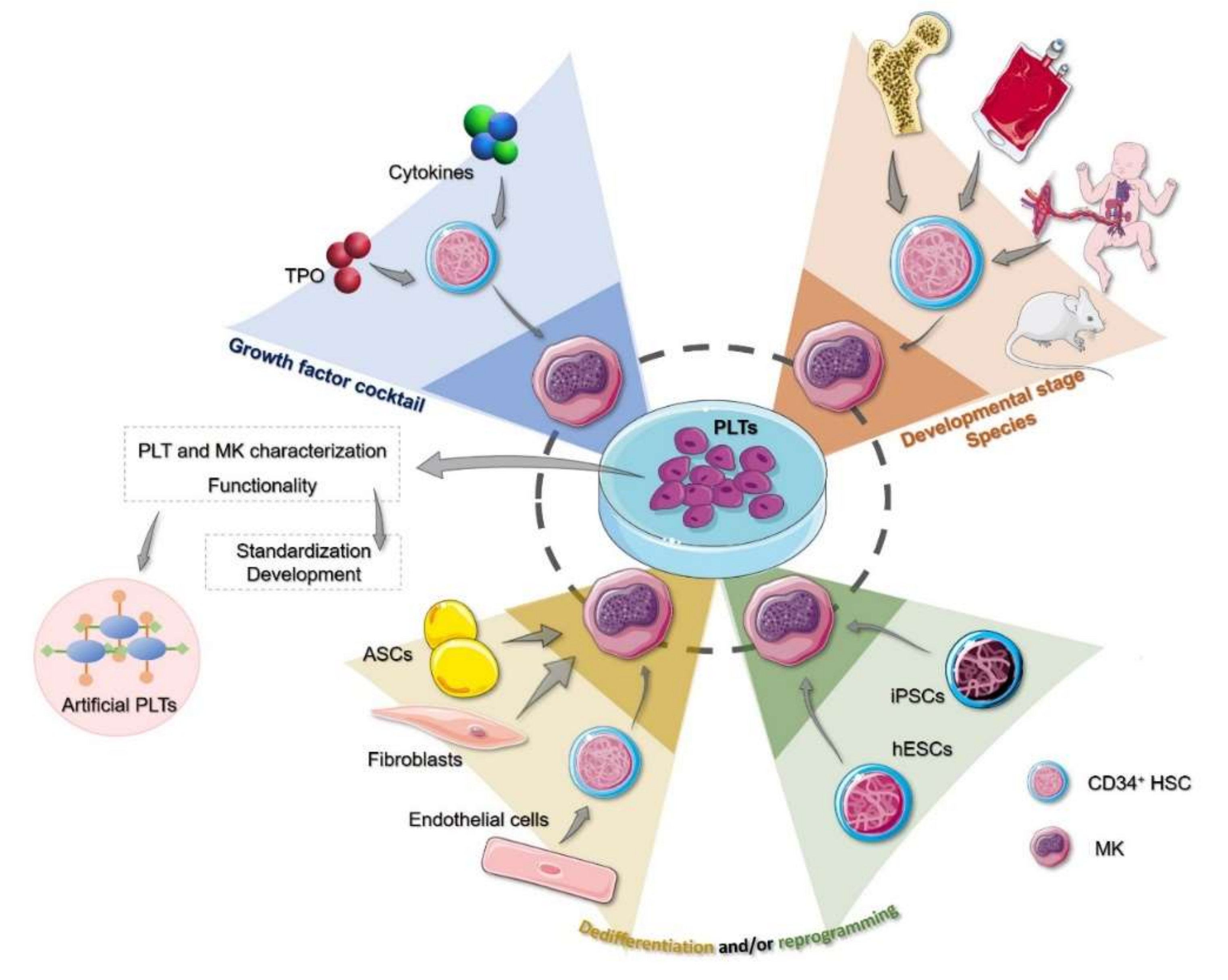
3. Human Cell Sources to Produce In Vitro Megakaryocytes and Platelets

The idea behind the production of PLTs in vitro demands a methodology of MK in vitro culture that takes advantage of a source material that guarantees proper differentiation and PLT production in sufficient numbers. However, there is still no agreement on this matter and, currently, different source materials are being employed, which we discuss below.

3.1. Hematopoietic Stem Cells and Precursors

The ex vivo production of PLTs focused first on the differentiation of primary human CD34+ HSCs, which have been successfully isolated from CB [57], PB [58] and, albeit rarely, from BM [32].
CB CD34+ cells, although limited due to their availability and authorized access, have been the most widely used source for MK culture in vitro. Subsequent developments and modifications in the growth conditions allowed for the culture of MKs from both PB and BM [59]. However, as it was noticed in the previous section, and in spite of presenting certain advantages compared to other HSC sources, differentiation of CB-derived MKs differ from their adult counterparts (e.g., PB or BM) in a way that may affect the functionality of the resulting PLTs [60,61].
PB CD34+ cells, although easy to obtain with non-invasive techniques and relatively minor processing, are still limited to ethical regulations and donations. While the relative frequency of CD34+ cells is lower than in CB samples [62], it can be enriched by G-CSF-induced mobilization of CD34+ progenitors from the BM to the PB [63]. In addition, other whole blood processed material may provide a more enriched CD34+ cell fraction, such as leuko-depletion filters and buffy coats from routine donations [64,65]. On this line, it has been shown that MKs can be differentiated from the human peripheral blood mononuclear cell (PMBCs) fraction without CD34+ cell enrichment (e.g., sorting). With this method, early hematopoietic precursors commit to the MK lineage in vitro given adequate growth conditions, and cells are unable to undergo this differentiation and eventually die in the culture [41]. This opens the possibility of avoiding cell sorting and thus reducing the costs of the process.
BM CD34+ cells, while they might represent the most appropriate source for recapitulating physiological MK differentiation from a given patient ex vivo, require invasive techniques for harvesting and seem unable to sustain the culture of MKs in large quantities and therefore may be used for other purposes than PLT production (e.g., disease-related basic or translational research) [33,60].
In any case, it is important to bear in mind that CD34+ sorted cells do not constitute a pure HSC population, however enriched. Furthermore, the purity of the starting material does not guarantee a pure, homogeneous, and synchronous culture, as mentioned before.
Lastly, although basic and translational research into megakaryopoiesis and PLT production in vitro is highly dependent on isolated HSCs, either from adult or fetal origin, and PBMCs, primary cells are not immortalized and thus have limited self-proliferation, making them unfit to meet the demands of the PLT number required for (a single) transfusion [66]. However, they can still be used, offering an inexpensive and accessible source, and more importantly, a source for rare phenotypes or compatible autologous stem cells.

3.2. Cell Reprogramming

The need to overcome the aforementioned limitations, namely self-renewal and PLT yield, drove some groups to develop cultures using the so-called human pluripotent stem cells (hPSCs), which comprise human embryonic stem cells (hESCs) [61] and the recently discovered human induced pluripotent stem cells (iPSCs) [67]. Both can be differentiated towards any cell type given the adequate growth conditions. In this case, it has been proven that they both are able to differentiate into MKs and produce PLT-like particles [68]. This directed differentiation via signals administered in the form of cytokine cocktails may lead to asynchronous cultures of suboptimal purity [69]. This means that some of these cells may persist after the differentiation has occurred, and given their embryonic nature, they have the potential to form teratomas [70]. However, although the collected PLTs can be irradiated to prevent remaining hPSCs from causing any damage, a large number of apoptotic cells and vesicles in the supernatant may elicit an immune response if transfused [71].
Other limitations associated with these cell sources are the ethical concerns raised by hESCs and the fact that both of them require sophisticated experimental setups that make them highly expensive to maintain, especially if we consider that the current PLT yields obtained from iPSCs, although high, are still not comparable to the amount of PLTs contained in a PC [72]. In addition, their culture still requires serum and feeder cells, which makes them unsuitable to be produced abiding cGMP guidelines [61,73]. Lastly, and more importantly, the impact of the developmental stage of these cell sources should not be overlooked, as commented upon before, since the use of both hESCs and iPSCs involves an embryonic hematopoietic status when reprogramming cells towards MKs.
Despite all of the above, iPSCs are widely regarded as the best solution for the scientific community to solve the problem of producing ex vivo, cGMP-ready PLT transfusions [74]. They present with advantages that outweigh their drawbacks, such as being an inexhaustible, self-renewable source of MKs and PLTs [72]. They can also be genetically modified to, for example, match any major histocompatibility group or, contrariwise, to be devoid of HLA antigens to give rise to universal PLTs, minimizing the risk of refractoriness and alloimmunization [75]. In addition, studies have shown that it is possible to drive these cultures using feeder- and serum-free mediums [76,77]. As for concerns regarding culture purity, ongoing efforts such as forward programming seem to be closing in what mechanisms might drive fate decisions in MK differentiation [78].
Lastly, although currently a seemingly viable option, iPSCs still have a long way to go in terms of proving their capacity to produce fully functional PLTs with the right morphology, surface receptors, and adequate half-lives in sufficient numbers and at accessible costs.

3.3. Non-Hematopoietic Sources

Fibroblasts, as well as endothelial and adipose tissue-derived stromal cells (ASCs) have been used as source material to generate MKs in vitro. Endothelial cells can be driven towards MK differentiation through an HSC transition stage upon adequate stimulation [79]. Likewise, fibroblasts, which also express MPL, can be coerced into becoming MKs when transfused with certain factors [80]; however, ASCs seem to perform better overall. In a first approach, a study conducted by Ono-Uruga et al. found a simple and cost-effective way to differentiate ASCs to MKs by means of endogenous TPO, and without genetic manipulation or feeder cells [81]. Resulting PLTs were found to be functional, with normal PLT surface marker expression and in numbers comparable to those obtained with iPSCs. The major drawbacks present in this study were that only a subpopulation of ASCs was able to differentiate into a MK lineage, and that they were non self-renewable. Nonetheless, a recent study by the same group aimed to overcome said limitations by developing an ASC line (ASCL) and thus turning the original source into a donor-independent one [82]. However, although similar in hemostatic function to in vivo PLTs, ASCL-derived ones were found to be slightly hyperreactive (as opposed to the PLTs produced from ASCs), and the fact that it is not an immortalized cell line would make it necessary to produce new ones regularly, which in turn would result in inter-batch variability.
The developmental stage of the source material is not the only parameter that might influence the characteristics of the final PLT produce. The choice and the possibilities to develop PLT production culture methods from either primary or reprogrammed cells must be taken into account, for which we should know and describe their differentiation characteristics and functional properties of the produced PLTs.

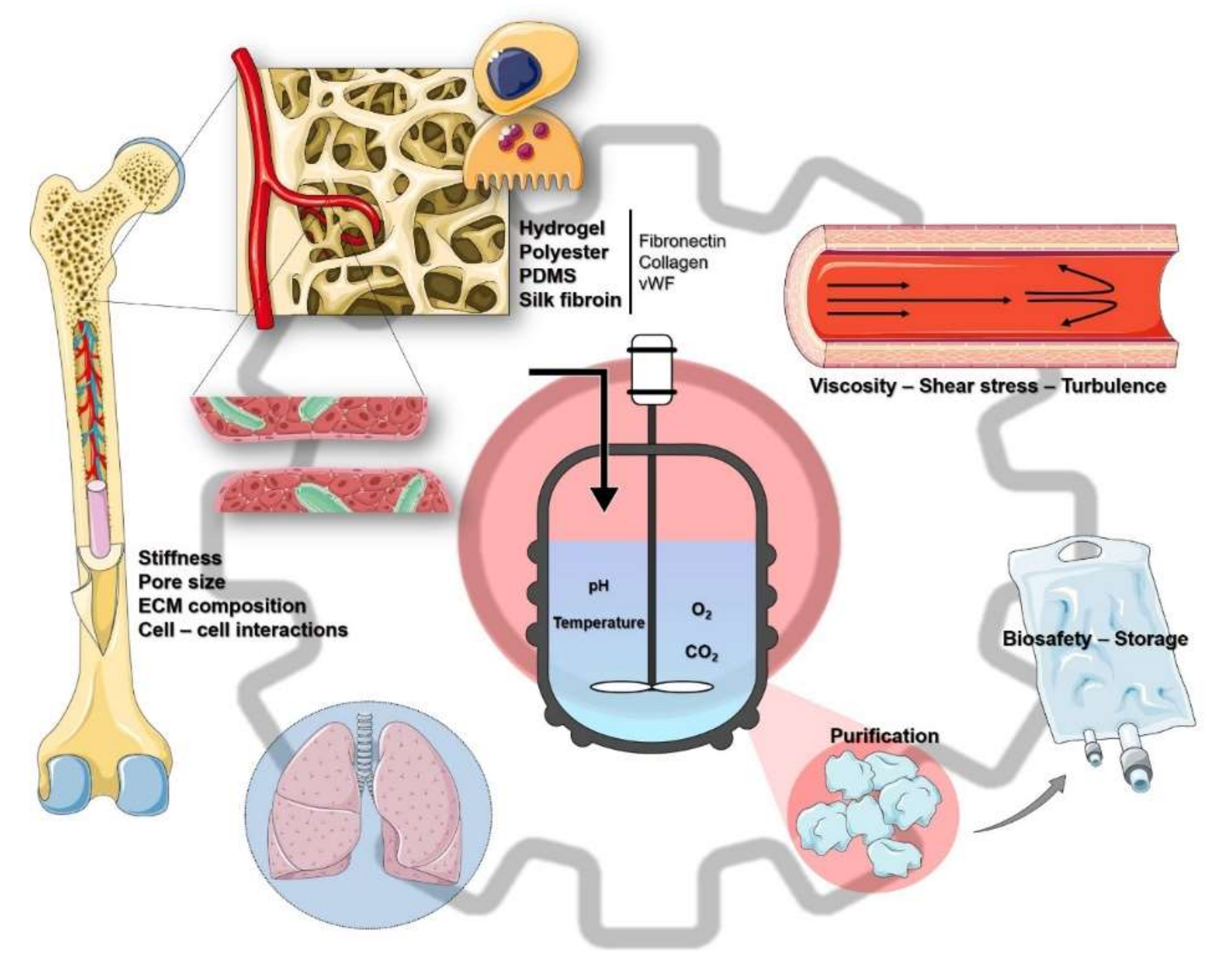
4. The Culture Engineering

Another aspect of concern regarding in vitro PLT production that should not be overlooked is the physical characteristics of the culture. Currently, PLT yields obtained in vitro are far from those encountered in physiological conditions, and it is mainly due to an inefficient release from in vitro cultured MKs, which adds to the lack of synchronicity and controllable MK terminal differentiation. Ongoing efforts are focusing on overcoming this bottleneck by developing bioengineering techniques that would reproduce the complex interactions that occur within the BM [83,84]. Whether the culture is performed in a static or a dynamic system or by implementing a three-dimensional (3D) matrix or scaffold is a matter of discussion, and developments are being done in this regard.
Adult hematopoiesis occurs in the BM, a complex 3D environment that exerts constraints on the development of the resident cells, and thus the architectures of the stromal niche, the endothelium, and the circulation are determinant in healthy PLT production [85,86]. Although the dynamics of such an environment are still poorly understood, efforts must be made to distance ourselves from the classical suspension culture and aim towards mimicking, as closely as possible, the physiological (soluble factors, cell types) and the physical (stiffness, rheology) aspects that characterize the BM niche [87]. Recreating such an environment has proven to improve in vitro MK differentiation and proplatelet formation [88].
Lastly, other added difficulties stemming from the in vitro culturing of MKs are caused by the heterogeneity, despite the enrichment of MKs, and the asynchronous nature of MK cell differentiation, plus other events such as cell death, vesiculation, etc. This makes it difficult to harvest the produced PLTs all at once while remaining pure, free of debris and vesicles, and maintaining their functional integrity. Current methods developed to tackle this issue and to separate PLTs from MKs, progenitors, or debris include serial centrifugations at different time points [63]. This technique, however, might result in yield reduction, PLT activation, and it is time-consuming and difficult to standardize to meet cGMP requirements. Recently, a procedure involving a spinning-membrane filtration device was developed that ensures the return of a pure population of PLTs with high yield and maintains PLT functional integrity [89]. Furthermore, contaminating MKs were able to resume proplatelet formation and PLT production when returned to culture conditions.

4.1. Bioreactors

Ex vivo PLT production has come a long way since MKs were first cultured, and bioreactors represent state-of-the-art technology aimed towards the goal of manufacturing PLTs in sufficient numbers to replace donor-derived PLTs [90]. They have the advantage of being easily adaptable and of functioning independently of the cell source used. Most of them can also be scaled-up, to a greater or lesser extent, to accommodate large numbers of MKs and thus be commercially and clinically suitable.
In the last years, the main effort to recapitulate the BM environment has focused on developing microfluidic bioreactors to faithfully mimic key physiological characteristics, such as the ECM composition, BM stiffness, soluble factors, and blood vessel architecture, which includes tissue-specific microvascular endothelium, endothelial cell contacts, and circulatory shear stress [91]. They constitute the most suitable method for large-scale production due to its scalability, handling, control of cell density, uniform nutrient distribution, and of culture conditions (e.g., pH, temperature, and O2 and CO2 concentrations) [92,93].
When MKs are exposed to a microenvironment that reproduces the essential characteristics of their native niche, it is expected that MK maturation, proplatelet formation, and shedding of PLTs will occur appropriately. When switching from 2D to 3D cultures, there is an increment of the surface area, which in turn allows for more interactions between MKs and their proplatelets and the surrounding co-cultured cells or matrix scaffolds. To achieve this kind of environment, several scaffolds have been used, made of hydrogel [94], polyester, or polydimethylsiloxane (PDMS) [95], coated with ECM proteins (e.g., fibronectin, collagen, vWF), and perfused with media containing specific growth factors and cytokines [91].
A major concern regarding these scaffolds is the biocompatibility and the scalability of the materials. Di Buduo et al. first aimed at tackling this issue by using a silk fibroin sponge [96], which was further developed by designing a microtube-like structure made of PDMS, coated with such a sponge [97]. This structure was also covered on the inside by endothelial cells, vascular endothelial growth factor (VEGF), and other ECM-mimicking components. At the same time, they considered other factors that affected the rheology of this novel material, such as stiffness, and introduced a flow composed of red blood cells to modulate viscosity and shear stress [97].
On this line, several studies have shown the importance of an optimal control of the physical attributes of the bioreactor. While hypoxia and hypothermia may help in the expansion and the differentiation of MKs [92,98], respectively, it has been shown that control of the flow rate (i.e., shear stress) favors rapid proplatelet formation and PLT production [99]. On this line, Thon et al. manufactured a chip-based microfluidic bioreactor that, apart from demonstrating the optimal rate of shear stress by using parallel flows, also incorporated certain aspects of the BM environment, such as stiffness, pore size, ECM composition, and endothelial cell contacts [100]. Other approaches that build on this attribute in order to control PLT release are a microfluidic device that retains MKs in vWF-coated micropillars [101], developed by Blin et al., and a two-chamber nanofiber membrane-based bioreactor with two perpendicular flows [102], developed by Avanzi et al. However, there is still no consensus on whether it is best to use dual or single flows to increase production [103] and whether a confluent system where different flow angles correlated with different PLT yields may be more effective [95].
Lastly, Ito et al., prompted by the long-lasting issue of the insufficient PLT numbers generated by bioreactors, have recently identified turbulence as a key physical factor in the management of large-scale PLT production [104]. By conducting BM in vivo imaging and particle image velocimetry, they observed that turbulent forces were only present around proplatelet-carrying MKs and thus were able to simulate this condition in a bioreactor and, by means of scaling-up, to produce near-clinical numbers of PLTs.
However, a deeper characterization of the PLT produce is necessary in order to assure its interchangeability with their donor-derived counterparts [105]. Once PLTs are obtained, they must be concentrated, purified, washed, and suspended in a preserving solution compatible with transfusion, which represents a challenge due to the asynchronous and the heterogeneous nature of these cultures. Furthermore, this enrichment is especially important to avoid debris, microvesicles, and other particles that carry some resemblance to PLTs, but are not able to perform their usual tasks, although they might be capable of adhering to certain substrates, thus potentially impoverishing the quality of the product.
Although some groups have characterized, to a greater or lesser extent, the PLTs generated under their specific culture conditions, a consensus on what to do exactly is still lacking [96,104,106]. We propose, first, a PLT standardization protocol encompassed within the production method, consisting of a series of functional and characterization tests, with the aim of determining that the final products are, in fact, viable PLTs that exert their expected function (e.g., immunophenotyping, viability tests, morphological and molecular characterization, adhesion, degranulation, and aggregation assays) (see Table 1). This, in turn, will provide insights into the manufacturing process, allowing its targeted improvement. Secondly, once a standard procedure is consolidated, a selection of assays could be settled for the quality control of every subsequent manufactured batch to assure the quality and the function of the in vitro derived PLTs (see Table 1 and Figure 1).
As an additional consideration, the production method should be as brief as possible, given that the in vitro produced PLTs would face the same storage limitations as their donor-derived counterparts, and should be readily available in cases of emergency. The quality of the product through storage should also be determined. Lastly, one significant issue that would undoubtedly hinder the efforts towards its widespread use is the extremely high production cost per unit. Laboratory-generated PLTs ultimately must be priced competitively relative to donor-derived PLTs in order to be effectively implemented in a clinical setting.

4.2. Using the Lungs as In Vivo Bioreactors

Another organ, besides the BM, where MKs have been shown to reside and shed PLTs, is the lung [108]. Thus, an alternative and already tested approach that would abolish the need to produce in vitro PLTs is turning the lungs into in vivo bioreactors by intravenously infusing ex vivo cultured MKs and using the pulmonary bed as the location of PLT production. In a first approach, murine MKs of BM or fetal liver origin were infused into mice and shown to be able to release functional PLTs [109]. In a second approach, a similar experiment was performed, but in this case, human CD34+ cells from the BM were xenoinfused into immunodeficient mice, which became entrapped in the pulmonary microvasculature [110]. A small percentage was also found in the spleen, while there were no signs of entrapment in either the BM, the liver, the heart, or the brain. PLTs released under these circumstances were comparable to donor-derived PLTs in terms of size, granule distribution, half-life, and surface marker expression but were released in a delayed manner and were rapidly cleared from the circulation. As for their functionality, although similar, they showed reduced convulxin (CVX) responsiveness and incorporation to thrombi [110].
However, this approach, albeit promising, poses its own risks that may outweigh its benefits, since the number of mature MKs that would have to be injected to achieve the PLT numbers provided by an apheresis unit could potentially cause capillary obstruction, and their extruded nucleus may elicit inflammation and autoimmune responses [110]. Another issue would be the time it takes for these transfused MKs to produce PLTs (approximately 24 h) versus direct infusion of PLTs. In addition, if genetically modified iPSCs were to be used as source, the risk of tumorigenesis would have to be addressed, although it has been shown that radiation of MKs might not affect either proplatelet formation and shedding or PLT functionality [111,112]. Lastly, these studies were performed in murine models, and thus any conclusion as to whether this could be a viable option in humans would have to wait, although their safety and their tolerability have already been tested in phase I clinical trials with positive results [113].

4.3. Artificial Platelets and Applications beyond Transfusion Medicine

The development of artificial PLTs seems a promising alternative for the acute bleeding patients requiring immediate transfusions, as these hybrid synthetic PLTs are compatible with all blood groups, comply with transfusion safety requirements, are biodegradable, easy to manufacture, allow for longer storage lives, and their production costs are more reduced than those of in vitro PLT production (considering a single application unit) [114]. The ultimate goal of these artificial PLTs is to recreate their functions of adhesion and aggregation as well as to mimic their morphology and physical properties, therefore overcoming the aforementioned limitations in the obtention of physiological-like PLTs.
There are currently several approaches to the manufacture of these pseudo-PLTs, such as the thrombosomes and the infusible PLT membrane, which make use only of the membrane and its associated receptors [115,116]. Other methods consist of adorning the surface of albumin microparticles, red blood cells, or synthetic polymers with fibrinogen or fibrinogen-mimetic peptides [117,118,119]. More recent developments include the use of the latter in combination with vWF- and collagen-binding peptides to coat the surface of liposomal nanoconstructs [120].
Nonetheless, the use of in vitro-generated PLTs might extend beyond their application in transfusion medicine. PLTs and artificial PLTs can be converted into drug carriers, given their ability to target and migrate to sites of injuries or disease [121,122,123]. In addition, their newly-identified functions in immune modulation, inflammation, cancer, and tissue regeneration [124,125,126] open the door to the in vitro production of fine-tuned PLTs, both at the receptor and the cargo levels, that can potentially modulate these pathological states [127,128,129].

5. Conclusions

In recent years, there have been enormous advances towards the manufacturing of PLTs ex vivo in order to meet the rising demand of PLT transfusions and to be less dependent on donations. Such advances have come from both the field of the in vitro culture of MKs, in terms of cell sources and cytokine cocktails, and the developments regarding growth conditions that mimic the BM niche (Figure 1). Bioreactors coupled with immortalized cell lines, namely iPSCs capable of undergoing megakaryopoiesis, might be the closest the scientific community has gotten to replicating the process ex vivo, with PLT yields similar to those found in PCs from donations (Figure 2).
Despite these advances, there are still hurdles that must be overcome. Costs and production times are too high (up to 26 days), and added to the fact that their shelf life is the same as their donated counterparts, this makes them unsuitable in cases of emergency, where large numbers of PLTs are needed in a very short period of time. Furthermore, a more thorough characterization of both MKs and PLTs produced in vitro is required [130]. Many studies have focused on the PLT yield while obviating either their morphology, their protein surface expression, or their functionality both in vitro and in vivo. On this line, these ex vivo PLTs have yet to be fully tested in humans. Clinical trials have only focused thus far on their safety and tolerability, meaning further trials to look into efficacy, function, and other specific variables must be performed. It is also important to distinguish between those patients where a PLT transfusion is essential and those where TPO administration or alternative methods, such as artificial PLTs, might suffice.
Considering as well the natural PLT distinct functional profiles due to the developmental stage (infant vs. adult) or the health status of an individual (healthy or with subjacent inflammation), it opens the question as to whether in vitro produced PLTs from whichever source, method, and conditions should be suitable for a given specific patient and clinical circumstances. Furthermore, we lack knowledge on whether in vitro produced PLTs may be prone to acquiring PLT storage lesions in the same way as donor PLTs, whether they can be stored in the same conditions and for the same time, and most importantly, the question arises as to how we set production protocols to not only abide cGMP regulations but also, regarding transfusion safety, whether these in vitro produced PLTs require PRTs and how that would affect their integrity.
Lastly, there are currently a plethora of protocols regarding MK differentiation and PLT production. They all differ in the starting cell source, growth conditions, and physical forces applied within the 3D culture, and while this variety of methods has greatly aided in the refinement of every step of megakaryopoiesis, it also makes it difficult to reach a consensus on what protocol to use. Paving the way to standardization will position ourselves closer to production of cGMP-grade PLTs and their final implementation in the clinical practice.

Author Contributions

Writing–original draft preparation and figures, P.M.-B.; review and editing, A.A.-H., J.S., L.G.; conceptualization, L.G. and J.S. All authors have read and agreed to the published version of the manuscript.

Funding

This work is partially supported by a Ramón y Cajal Fellowship (RYC-2013-12587, Ministerio de Economía y Competitividad–Spain) to L.G., an I+D Excellence Research Project (SAF2017-85489-P, Ministerio de Economía y Competitividad–Spain–and Fondos FEDER) to L.G. and P.M.-B., a Severo Ochoa Grant (PA-20-PF-BP19-014) to P.M.-B. and a postdoctoral ISPA 2018 intramural fellowship to A.A.-H.

Acknowledgments

Images from both figures are part of Servier Medical Art (SMART) by Servier and are licensed under CC BY 3.0.

Conflicts of Interest

The authors declare no conflict of interest.

References

  1. Deutsch, V.R.; Tomer, A. Megakaryocyte development and platelet production. Br. J. Haematol. 2006, 134, 453–466. [Google Scholar] [CrossRef] [PubMed]
  2. Slichter, S.J. Platelet transfusion therapy. Hematol. Oncol. Clin. North. Am. 2007, 21, 697–729. [Google Scholar] [CrossRef]
  3. Blajchman, M.A.; Slichter, S.J.; Heddle, N.M.; Murphy, M.F. New Strategies for the Optimal Use of Platelet Transfusions. Hematology 2008, 2008, 198–204. [Google Scholar] [CrossRef] [PubMed] [Green Version]
  4. Wandt, H.; Schaefer-Eckart, K.; Wendelin, K.; Pilz, B.; Wilhelm, M.; Thalheimer, M.; Mahlknecht, U.; Ho, A.; Schaich, M.; Kramer, M.; et al. Therapeutic platelet transfusion versus routine prophylactic transfusion in patients with haematological malignancies: An open-label, multicentre, randomised study. Lancet 2012, 380, 1309–1316. [Google Scholar] [CrossRef]
  5. Estcourt, L.J. Why has demand for platelet components increased? A review. Transfus. Med. 2014, 24, 260–268. [Google Scholar] [CrossRef] [PubMed]
  6. Yazer, M.H.; Shaz, B.; Seheult, J.N.; Apelseth, T.O.; De Korte, D.; Devin, G.; Devine, D.; Doncaster, C.; Field, S.; Flanagan, P.; et al. Trends in platelet distributions from 2008 to 2017: A survey of twelve national and regional blood collectors. Vox Sang. 2020. [Google Scholar] [CrossRef]
  7. Storch, E.K.; Custer, B.S.; Jacobs, M.R.; Menitove, J.E.; Mintz, P.D. Review of current transfusion therapy and blood banking practices. Blood Rev. 2019, 38, 100593. [Google Scholar] [CrossRef]
  8. Williamson, L.M.; Devine, D.V. Challenges in the management of the blood supply. Lancet 2013, 381, 1866–1875. [Google Scholar] [CrossRef]
  9. Flint, A.W.; McQuilten, Z.K.; Irwin, G.; Rushford, K.; Haysom, H.E.; Wood, E.M. Is Platelet Expiring Out of Date? A Systematic Review. Transfus. Med. Rev. 2020, 34, 42–50. [Google Scholar] [CrossRef]
  10. Allain, J.P.; Bianco, C.; Blajchman, M.A.; Brecher, M.E.; Busch, M.; Leiby, D.; Lin, L.; Stramer, S. Protecting the blood supply from emerging pathogens: The role of pathogen inactivation. Transfus. Med. Rev. 2005, 19, 110–126. [Google Scholar] [CrossRef]
  11. Strassel, C.; Gachet, C.; Lanza, F. On the way to in vitro platelet production. Transfus. Clin. Biol. 2018, 25, 220–227. [Google Scholar] [CrossRef]
  12. Salunkhe, V.; Van der Meer, P.F.; De Korte, D.; Seghatchian, J.; Gutierrez, L. Development of blood transfusion product pathogen reduction treatments: A review of methods, current applications and demands. Transfus. Apher. Sci. 2015, 52, 19–34. [Google Scholar] [CrossRef]
  13. Jimenez-Marco, T.; Garcia-Recio, M.; Girona-Llobera, E. Our experience in riboflavin and ultraviolet light pathogen reduction technology for platelets: From platelet production to patient care. Transfusion 2018, 58, 1881–1889. [Google Scholar] [CrossRef] [Green Version]
  14. Devine, D.V.; Serrano, K. The platelet storage lesion. Clin. Lab. Med. 2010, 30, 475–487. [Google Scholar] [CrossRef]
  15. Zeddies, S.; De Cuyper, I.M.; Van der Meer, P.F.; Daal, B.B.; De Korte, D.; Gutierrez, L.; Thijssen-Timmer, D.C. Pathogen reduction treatment using riboflavin and ultraviolet light impairs platelet reactivity toward specific agonists in vitro. Transfusion 2014, 54, 2292–2300. [Google Scholar] [CrossRef]
  16. Garraud, O.; Lozano, M. Pathogen inactivation/reduction technologies for platelet transfusion: Where do we stand? Transfus. Clin. Biol. 2018, 25, 165–171. [Google Scholar] [CrossRef]
  17. Magron, A.; Laugier, J.; Provost, P.; Boilard, E. Pathogen reduction technologies: The pros and cons for platelet transfusion. Platelets 2018, 29, 2–8. [Google Scholar] [CrossRef]
  18. Slichter, S.J.; Davis, K.; Enright, H.; Braine, H.; Gernsheimer, T.; Kao, K.-J.; Kickler, T.; Lee, E.; McFarland, J.; McCullough, J.; et al. Factors affecting posttransfusion platelet increments, platelet refractoriness, and platelet transfusion intervals in thrombocytopenic patients. Blood 2005, 105, 4106–4114. [Google Scholar] [CrossRef] [Green Version]
  19. Johnson, L.; Tan, S.; Wood, B.; Davis, A.; Marks, D.C. Refrigeration and cryopreservation of platelets differentially affect platelet metabolism and function: A comparison with conventional platelet storage conditions. Transfusion 2016, 56, 1807–1818. [Google Scholar] [CrossRef]
  20. Desborough, M.; Estcourt, L.J.; Doree, C.; Trivella, M.; Hopewell, S.; Stanworth, S.J.; Murphy, M.F. Alternatives, and adjuncts, to prophylactic platelet transfusion for people with haematological malignancies undergoing intensive chemotherapy or stem cell transplantation. Cochrane Database Syst. Rev. 2016, Cd010982. [Google Scholar] [CrossRef] [Green Version]
  21. Martínez-Botía, P.; Acebes-Huerta, A.; Seghatchian, J.; Gutiérrez, L. In vitro platelet production for transfusion purposes: Where are we now? Transfus. Apher. Sci. 2020, 59, 102864. [Google Scholar] [CrossRef]
  22. Franco, A.T.; Corken, A.; Ware, J. Platelets at the interface of thrombosis, inflammation, and cancer. Blood 2015, 126, 582–588. [Google Scholar] [CrossRef] [Green Version]
  23. Kaushansky, K. Historical review: Megakaryopoiesis and thrombopoiesis. Blood 2008, 111, 981–986. [Google Scholar] [CrossRef]
  24. Ghanima, W.; Cooper, N.; Rodeghiero, F.; Godeau, B.; Bussel, J.B. Thrombopoietin receptor agonists: Ten years later. Haematologica 2019, 104, 1112–1123. [Google Scholar] [CrossRef] [Green Version]
  25. Choi, E.; Nichol, J.; Hokom, M.; Hornkohl, A.; Hunt, P. Platelets generated in vitro from proplatelet-displaying human megakaryocytes are functional. Blood 1995, 85, 402–413. [Google Scholar] [CrossRef] [Green Version]
  26. Kaushansky, K.; Lok, S.; Holly, R.D.; Broudy, V.C.; Lin, N.; Bailey, M.C.; Forstrom, J.W.; Buddle, M.M.; Oort, P.J.; Hagen, F.S.; et al. Promotion of megakaryocyte progenitor expansion and differentiation by the c-Mpl ligand thrombopoietin. Nature 1994, 369, 568–571. [Google Scholar] [CrossRef]
  27. Terskikh, A.V.; Miyamoto, T.; Chang, C.; Diatchenko, L.; Weissman, I.L. Gene expression analysis of purified hematopoietic stem cells and committed progenitors. Blood 2003, 102, 94–101. [Google Scholar] [CrossRef]
  28. Ninos, J.M.; Jefferies, L.C.; Cogle, C.R.; Kerr, W.G. The thrombopoietin receptor, c-Mpl, is a selective surface marker for human hematopoietic stem cells. J. Transl. Med. 2006, 4, 9. [Google Scholar] [CrossRef] [Green Version]
  29. Yoshihara, H.; Arai, F.; Hosokawa, K.; Hagiwara, T.; Takubo, K.; Nakamura, Y.; Gomei, Y.; Iwasaki, H.; Matsuoka, S.; Miyamoto, K.; et al. Thrombopoietin/MPL signaling regulates hematopoietic stem cell quiescence and interaction with the osteoblastic niche. Cell Stem Cell 2007, 1, 685–697. [Google Scholar] [CrossRef] [Green Version]
  30. Nakamura-Ishizu, A.; Matsumura, T.; Stumpf, P.S.; Umemoto, T.; Takizawa, H.; Takihara, Y.; O’Neil, A.; Majeed, A.; MacArthur, B.D.; Suda, T. Thrombopoietin Metabolically Primes Hematopoietic Stem Cells to Megakaryocyte-Lineage Differentiation. Cell Rep. 2018, 25, 1772–1785.e1776. [Google Scholar] [CrossRef] [Green Version]
  31. Nakamura-Ishizu, A.; Suda, T. Multifaceted roles of thrombopoietin in hematopoietic stem cell regulation. Ann. N. Y. Acad. Sci. 2019. [Google Scholar] [CrossRef]
  32. De Bruyn, C.; Delforge, A.; Martiat, P.; Bron, D. Ex vivo expansion of megakaryocyte progenitor cells: Cord blood versus mobilized peripheral blood. Stem Cells Dev. 2005, 14, 415–424. [Google Scholar] [CrossRef]
  33. Van den Oudenrijn, S.; Von dem Borne, A.E.; De Haas, M. Differences in megakaryocyte expansion potential between CD34(+) stem cells derived from cord blood, peripheral blood, and bone marrow from adults and children. Exp. Hematol. 2000, 28, 1054–1061. [Google Scholar] [CrossRef]
  34. Niswander, L.M.; Fegan, K.H.; Kingsley, P.D.; McGrath, K.E.; Palis, J. SDF-1 dynamically mediates megakaryocyte niche occupancy and thrombopoiesis at steady state and following radiation injury. Blood 2014, 124, 277–286. [Google Scholar] [CrossRef] [PubMed] [Green Version]
  35. Avecilla, S.T.; Hattori, K.; Heissig, B.; Tejada, R.; Liao, F.; Shido, K.; Jin, D.K.; Dias, S.; Zhang, F.; Hartman, T.E.; et al. Chemokine-mediated interaction of hematopoietic progenitors with the bone marrow vascular niche is required for thrombopoiesis. Nat. Med. 2004, 10, 64–71. [Google Scholar] [CrossRef] [PubMed]
  36. Zhang, L.; Orban, M.; Lorenz, M.; Barocke, V.; Braun, D.; Urtz, N.; Schulz, C.; Von Bruhl, M.L.; Tirniceriu, A.; Gaertner, F.; et al. A novel role of sphingosine 1-phosphate receptor S1pr1 in mouse thrombopoiesis. J. Exp. Med. 2012, 209, 2165–2181. [Google Scholar] [CrossRef] [PubMed]
  37. Ng, A.P.; Kauppi, M.; Metcalf, D.; Hyland, C.D.; Josefsson, E.C.; Lebois, M.; Zhang, J.G.; Baldwin, T.M.; Di Rago, L.; Hilton, D.J.; et al. Mpl expression on megakaryocytes and platelets is dispensable for thrombopoiesis but essential to prevent myeloproliferation. Proc. Natl. Acad. Sci. USA 2014, 111, 5884–5889. [Google Scholar] [CrossRef] [Green Version]
  38. Assinger, A.; Schrottmaier, W.C.; Salzmann, M.; Rayes, J. Platelets in Sepsis: An Update on Experimental Models and Clinical Data. Front. Immunol. 2019, 10. [Google Scholar] [CrossRef]
  39. Müller-Newen, G.; Stope, M.B.; Kraus, T.; Ziegler, P. Development of platelets during steady state and inflammation. J. Leukoc. Biol. 2017, 101, 1109–1117. [Google Scholar] [CrossRef]
  40. Reems, J.A.; Pineault, N.; Sun, S. In vitro megakaryocyte production and platelet biogenesis: State of the art. Transfus. Med. Rev. 2010, 24, 33–43. [Google Scholar] [CrossRef] [Green Version]
  41. Salunkhe, V.; Papadopoulos, P.; Gutiérrez, L. Culture of Megakaryocytes from Human Peripheral Blood Mononuclear Cells. Bioprotocol 2015, 5, e1639. [Google Scholar] [CrossRef]
  42. Kaushansky, K.; Broudy, V.C.; Lin, N.; Jorgensen, M.J.; McCarty, J.; Fox, N.; Zucker-Franklin, D.; Lofton-Day, C. Thrombopoietin, the Mp1 ligand, is essential for full megakaryocyte development. Proc. Natl. Acad. Sci. USA 1995, 92, 3234–3238. [Google Scholar] [CrossRef] [Green Version]
  43. Gainsford, T.; Nandurkar, H.; Metcalf, D.; Robb, L.; Begley, C.G.; Alexander, W.S. The residual megakaryocyte and platelet production in c-mpl-deficient mice is not dependent on the actions of interleukin-6, interleukin-11, or leukemia inhibitory factor. Blood 2000, 95, 528–534. [Google Scholar] [CrossRef]
  44. Ku, H.; Yonemura, Y.; Kaushansky, K.; Ogawa, M. Thrombopoietin, the ligand for the Mpl receptor, synergizes with steel factor and other early acting cytokines in supporting proliferation of primitive hematopoietic progenitors of mice. Blood 1996, 87, 4544–4551. [Google Scholar] [CrossRef]
  45. Couldwell, G.; Machlus, K.R. Modulation of megakaryopoiesis and platelet production during inflammation. Thromb. Res. 2019, 179, 114–120. [Google Scholar] [CrossRef]
  46. Nishimura, S.; Nagasaki, M.; Kunishima, S.; Sawaguchi, A.; Sakata, A.; Sakaguchi, H.; Ohmori, T.; Manabe, I.; Italiano, J.E., Jr.; Ryu, T.; et al. IL-1alpha induces thrombopoiesis through megakaryocyte rupture in response to acute platelet needs. J. Cell Biol. 2015, 209, 453–466. [Google Scholar] [CrossRef] [Green Version]
  47. Wilcox, D.A. Megakaryocyte- and megakaryocyte precursor–related gene therapies. Blood 2016, 127, 1260–1268. [Google Scholar] [CrossRef] [Green Version]
  48. Avanzi, M.P.; Mitchell, W.B. Ex vivo production of platelets from stem cells. Br. J. Haematol. 2014, 165, 237–247. [Google Scholar] [CrossRef]
  49. Davenport, P.; Liu, Z.J.; Sola-Visner, M. Changes in megakaryopoiesis over ontogeny and their implications in health and disease. Platelets 2020, 1–8. [Google Scholar] [CrossRef]
  50. Strassel, C.; Eckly, A.; Léon, C.; Moog, S.; Cazenave, J.P.; Gachet, C.; Lanza, F. Hirudin and heparin enable efficient megakaryocyte differentiation of mouse bone marrow progenitors. Exp. Cell Res. 2012, 318, 25–32. [Google Scholar] [CrossRef]
  51. Italiano, J.E., Jr.; Lecine, P.; Shivdasani, R.A.; Hartwig, J.H. Blood platelets are assembled principally at the ends of proplatelet processes produced by differentiated megakaryocytes. J. Cell Biol. 1999, 147, 1299–1312. [Google Scholar] [CrossRef]
  52. Bluteau, O.; Langlois, T.; Rivera-Munoz, P.; Favale, F.; Rameau, P.; Meurice, G.; Dessen, P.; Solary, E.; Raslova, H.; Mercher, T.; et al. Developmental changes in human megakaryopoiesis. J. Thromb. Haemost. 2013, 11, 1730–1741. [Google Scholar] [CrossRef]
  53. Liu, Z.J.; Italiano, J., Jr.; Ferrer-Marin, F.; Gutti, R.; Bailey, M.; Poterjoy, B.; Rimsza, L.; Sola-Visner, M. Developmental differences in megakaryocytopoiesis are associated with up-regulated TPO signaling through mTOR and elevated GATA-1 levels in neonatal megakaryocytes. Blood 2011, 117, 4106–4117. [Google Scholar] [CrossRef] [Green Version]
  54. Slayton, W.B.; Wainman, D.A.; Li, X.M.; Hu, Z.; Jotwani, A.; Cogle, C.R.; Walker, D.; Fisher, R.C.; Wingard, J.R.; Scott, E.W.; et al. Developmental differences in megakaryocyte maturation are determined by the microenvironment. Stem Cells 2005, 23, 1400–1408. [Google Scholar] [CrossRef]
  55. Margraf, A.; Nussbaum, C.; Sperandio, M. Ontogeny of platelet function. Blood Adv. 2019, 3, 692–703. [Google Scholar] [CrossRef] [Green Version]
  56. Caparros-Perez, E.; Teruel-Montoya, R.; Lopez-Andreo, M.J.; Llanos, M.C.; Rivera, J.; Palma-Barqueros, V.; Blanco, J.E.; Vicente, V.; Martinez, C.; Ferrer-Marin, F. Comprehensive comparison of neonate and adult human platelet transcriptomes. PLoS ONE 2017, 12, e0183042. [Google Scholar] [CrossRef] [Green Version]
  57. Tao, H.; Gaudry, L.; Rice, A.; Chong, B. Cord blood is better than bone marrow for generating megakaryocytic progenitor cells. Exp. Hematol. 1999, 27, 293–301. [Google Scholar] [CrossRef]
  58. Guerriero, R.; Testa, U.; Gabbianelli, M.; Mattia, G.; Montesoro, E.; Macioce, G.; Pace, A.; Ziegler, B.; Hassan, H.J.; Peschle, C. Unilineage megakaryocytic proliferation and differentiation of purified hematopoietic progenitors in serum-free liquid culture. Blood 1995, 86, 3725–3736. [Google Scholar] [CrossRef] [Green Version]
  59. De Bruyn, C.; Delforge, A.; Lagneaux, L.; Bron, D. Characterization of CD34+ subsets derived from bone marrow, umbilical cord blood and mobilized peripheral blood after stem cell factor and interleukin 3 stimulation. Bone Marrow Transplant. 2000, 25, 377–383. [Google Scholar] [CrossRef] [Green Version]
  60. Miyazaki, R.; Ogata, H.; Iguchi, T.; Sogo, S.; Kushida, T.; Ito, T.; Inaba, M.; Ikehara, S.; Kobayashi, Y. Comparative analyses of megakaryocytes derived from cord blood and bone marrow. Br. J. Haematol. 2000, 108, 602–609. [Google Scholar] [CrossRef]
  61. Gaur, M.; Kamata, T.; Wang, S.; Moran, B.; Shattil, S.J.; Leavitt, A.D. Megakaryocytes derived from human embryonic stem cells: A genetically tractable system to study megakaryocytopoiesis and integrin function. J. Thromb. Haemost. 2006, 4, 436–442. [Google Scholar] [CrossRef] [PubMed]
  62. Herbein, G.; Sovalat, H.; Wunder, E.; Baerenzung, M.; Bachorz, J.; Lewandowski, H.; Schweitzer, C.; Schmitt, C.; Kirn, A.; Hénon, P. Isolation and identification of two CD34+ cell subpopulations from normal human peripheral blood. Stem Cells 1994, 12, 187–197. [Google Scholar] [CrossRef] [PubMed]
  63. Panuganti, S.; Schlinker, A.C.; Lindholm, P.F.; Papoutsakis, E.T.; Miller, W.M. Three-stage ex vivo expansion of high-ploidy megakaryocytic cells: Toward large-scale platelet production. Tissue Eng. Part A 2013, 19, 998–1014. [Google Scholar] [CrossRef] [PubMed] [Green Version]
  64. Ivanovic, Z.; Duchez, P.; Morgan, D.A.; Hermitte, F.; Lafarge, X.; Chevaleyre, J.; Praloran, V.; Dazey, B.; Vezon, G.; Boiron, J.M. Whole-blood leuko-depletion filters as a source of CD 34+ progenitors potentially usable in cell therapy. Transfusion 2006, 46, 118–125. [Google Scholar] [CrossRef]
  65. Six, K.R.; Sicot, G.; Devloo, R.; Feys, H.B.; Baruch, D.; Compernolle, V. A comparison of haematopoietic stem cells from umbilical cord blood and peripheral blood for platelet production in a microfluidic device. Vox Sang. 2019, 114, 330–339. [Google Scholar] [CrossRef]
  66. Lambert, M.P.; Sullivan, S.K.; Fuentes, R.; French, D.L.; Poncz, M. Challenges and promises for the development of donor-independent platelet transfusions. Blood 2013, 121, 3319–3324. [Google Scholar] [CrossRef]
  67. Takahashi, K.; Tanabe, K.; Ohnuki, M.; Narita, M.; Ichisaka, T.; Tomoda, K.; Yamanaka, S. Induction of pluripotent stem cells from adult human fibroblasts by defined factors. Cell 2007, 131, 861–872. [Google Scholar] [CrossRef] [Green Version]
  68. Takayama, N.; Eto, K. Pluripotent stem cells reveal the developmental biology of human megakaryocytes and provide a source of platelets for clinical application. Cell. Mol. Life Sci. 2012, 69, 3419–3428. [Google Scholar] [CrossRef] [Green Version]
  69. Focosi, D.; Amabile, G.; Di Ruscio, A.; Quaranta, P.; Tenen, D.G.; Pistello, M. Induced pluripotent stem cells in hematology: Current and future applications. Blood Cancer J. 2014, 4, e211. [Google Scholar] [CrossRef] [Green Version]
  70. Lee, A.S.; Tang, C.; Rao, M.S.; Weissman, I.L.; Wu, J.C. Tumorigenicity as a clinical hurdle for pluripotent stem cell therapies. Nat. Med. 2013, 19, 998–1004. [Google Scholar] [CrossRef] [Green Version]
  71. Peng, Y.; Martin, D.A.; Kenkel, J.; Zhang, K.; Ogden, C.A.; Elkon, K.B. Innate and adaptive immune response to apoptotic cells. J. Autoimmun. 2007, 29, 303–309. [Google Scholar] [CrossRef] [PubMed] [Green Version]
  72. Nakamura, S.; Takayama, N.; Hirata, S.; Seo, H.; Endo, H.; Ochi, K.; Fujita, K.; Koike, T.; Harimoto, K.; Dohda, T.; et al. Expandable megakaryocyte cell lines enable clinically applicable generation of platelets from human induced pluripotent stem cells. Cell Stem Cell 2014, 14, 535–548. [Google Scholar] [CrossRef] [PubMed] [Green Version]
  73. Takayama, N.; Nishikii, H.; Usui, J.; Tsukui, H.; Sawaguchi, A.; Hiroyama, T.; Eto, K.; Nakauchi, H. Generation of functional platelets from human embryonic stem cells in vitro via ES-sacs, VEGF-promoted structures that concentrate hematopoietic progenitors. Blood 2008, 111, 5298–5306. [Google Scholar] [CrossRef] [PubMed] [Green Version]
  74. Hansen, M.; Von Lindern, M.; Van den Akker, E.; Varga, E. Human-induced pluripotent stem cell-derived blood products: State of the art and future directions. FEBS Lett. 2019, 593, 3288–3303. [Google Scholar] [CrossRef]
  75. Figueiredo, C.; Goudeva, L.; Horn, P.A.; Eiz-Vesper, B.; Blasczyk, R.; Seltsam, A. Generation of HLA-deficient platelets from hematopoietic progenitor cells. Transfusion 2010, 50, 1690–1701. [Google Scholar] [CrossRef] [Green Version]
  76. Pick, M.; Azzola, L.; Osborne, E.; Stanley, E.G.; Elefanty, A.G. Generation of megakaryocytic progenitors from human embryonic stem cells in a feeder- and serum-free medium. PLoS ONE 2013, 8, e55530. [Google Scholar] [CrossRef] [Green Version]
  77. Feng, Q.; Shabrani, N.; Thon, J.N.; Huo, H.; Thiel, A.; Machlus, K.R.; Kim, K.; Brooks, J.; Li, F.; Luo, C.; et al. Scalable Generation of Universal Platelets from Human Induced Pluripotent Stem Cells. Stem Cell Rep. 2014, 3, 817–831. [Google Scholar] [CrossRef] [Green Version]
  78. Moreau, T.; Evans, A.L.; Vasquez, L.; Tijssen, M.R.; Yan, Y.; Trotter, M.W.; Howard, D.; Colzani, M.; Arumugam, M.; Wu, W.H.; et al. Large-scale production of megakaryocytes from human pluripotent stem cells by chemically defined forward programming. Nature Commun. 2016, 7, 11208. [Google Scholar] [CrossRef]
  79. Lis, R.; Karrasch, C.C.; Poulos, M.G.; Kunar, B.; Redmond, D.; Duran, J.G.B.; Badwe, C.R.; Schachterle, W.; Ginsberg, M.; Xiang, J.; et al. Conversion of adult endothelium to immunocompetent haematopoietic stem cells. Nature 2017, 545, 439–445. [Google Scholar] [CrossRef]
  80. Ono, Y.; Wang, Y.; Suzuki, H.; Okamoto, S.; Ikeda, Y.; Murata, M.; Poncz, M.; Matsubara, Y. Induction of functional platelets from mouse and human fibroblasts by p45NF-E2/Maf. Blood 2012, 120, 3812–3821. [Google Scholar] [CrossRef] [Green Version]
  81. Ono-Uruga, Y.; Tozawa, K.; Horiuchi, T.; Murata, M.; Okamoto, S.; Ikeda, Y.; Suda, T.; Matsubara, Y. Human adipose tissue-derived stromal cells can differentiate into megakaryocytes and platelets by secreting endogenous thrombopoietin. J. Thromb. Haemost. 2016, 14, 1285–1297. [Google Scholar] [CrossRef] [PubMed] [Green Version]
  82. Tozawa, K.; Ono-Uruga, Y.; Yazawa, M.; Mori, T.; Murata, M.; Okamoto, S.; Ikeda, Y.; Matsubara, Y. Megakaryocytes and platelets from a novel human adipose tissue-derived mesenchymal stem cell line. Blood 2019, 133, 633–643. [Google Scholar] [CrossRef] [PubMed]
  83. Pinho, S.; Frenette, P.S. Haematopoietic stem cell activity and interactions with the niche. Nat. Rev. Mol. Cell Biol. 2019, 20, 303–320. [Google Scholar] [CrossRef] [PubMed]
  84. Li, Z.; Li, L. Understanding hematopoietic stem-cell microenvironments. Trends Biochem. Sci. 2006, 31, 589–595. [Google Scholar] [CrossRef] [PubMed]
  85. Gurkan, U.A.; Akkus, O. The mechanical environment of bone marrow: A review. Ann. Biomed. Eng. 2008, 36, 1978–1991. [Google Scholar] [CrossRef]
  86. Junt, T.; Schulze, H.; Chen, Z.; Massberg, S.; Goerge, T.; Krueger, A.; Wagner, D.D.; Graf, T.; Italiano, J.E., Jr.; Shivdasani, R.A.; et al. Dynamic visualization of thrombopoiesis within bone marrow. Science 2007, 317, 1767–1770. [Google Scholar] [CrossRef] [Green Version]
  87. Di Buduo, C.A.; Kaplan, D.L.; Balduini, A. In vitro generation of platelets: Where do we stand? Transfus. Clin. Biol. 2017, 24, 273–276. [Google Scholar] [CrossRef]
  88. Aguilar, A.; Pertuy, F.; Eckly, A.; Strassel, C.; Collin, D.; Gachet, C.; Lanza, F.; Leon, C. Importance of environmental stiffness for megakaryocyte differentiation and proplatelet formation. Blood 2016, 128, 2022–2032. [Google Scholar] [CrossRef] [Green Version]
  89. Schlinker, A.C.; Radwanski, K.; Wegener, C.; Min, K.; Miller, W.M. Separation of in-vitro-derived megakaryocytes and platelets using spinning-membrane filtration. Biotechnol. Bioeng. 2015, 112, 788–800. [Google Scholar] [CrossRef] [Green Version]
  90. Ingavle, G.; Shabrani, N.; Vaidya, A.; Kale, V. Mimicking megakaryopoiesis in vitro using biomaterials: Recent advances and future opportunities. Acta Biomater. 2019, 96, 99–110. [Google Scholar] [CrossRef]
  91. Thon, J.N.; Dykstra, B.J.; Beaulieu, L.M. Platelet bioreactor: Accelerated evolution of design and manufacture. Platelets 2017, 28, 472–477. [Google Scholar] [CrossRef] [PubMed]
  92. Lasky, L.C.; Sullenbarger, B. Manipulation of oxygenation and flow-induced shear stress can increase the in vitro yield of platelets from cord blood. Tissue Eng. Part. C Methods 2011, 17, 1081–1088. [Google Scholar] [CrossRef] [PubMed]
  93. Kropp, C.; Kempf, H.; Halloin, C.; Robles-Diaz, D.; Franke, A.; Scheper, T.; Kinast, K.; Knorpp, T.; Joos, T.O.; Haverich, A.; et al. Impact of Feeding Strategies on the Scalable Expansion of Human Pluripotent Stem Cells in Single-Use Stirred Tank Bioreactors. Stem. Cells Transl. Med. 2016, 5, 1289–1301. [Google Scholar] [CrossRef] [PubMed] [Green Version]
  94. Sullenbarger, B.; Bahng, J.H.; Gruner, R.; Kotov, N.; Lasky, L.C. Prolonged continuous in vitro human platelet production using three-dimensional scaffolds. Exp. Hematol. 2009, 37, 101–110. [Google Scholar] [CrossRef] [Green Version]
  95. Nakagawa, Y.; Nakamura, S.; Nakajima, M.; Endo, H.; Dohda, T.; Takayama, N.; Nakauchi, H.; Arai, F.; Fukuda, T.; Eto, K. Two differential flows in a bioreactor promoted platelet generation from human pluripotent stem cell-derived megakaryocytes. Exp. Hematol. 2013, 41, 742–748. [Google Scholar] [CrossRef] [Green Version]
  96. Di Buduo, C.A.; Wray, L.S.; Tozzi, L.; Malara, A.; Chen, Y.; Ghezzi, C.E.; Smoot, D.; Sfara, C.; Antonelli, A.; Spedden, E.; et al. Programmable 3D silk bone marrow niche for platelet generation ex vivo and modeling of megakaryopoiesis pathologies. Blood 2015, 125, 2254–2264. [Google Scholar] [CrossRef] [Green Version]
  97. Tozzi, L.; Laurent, P.A.; Di Buduo, C.A.; Mu, X.; Massaro, A.; Bretherton, R.; Stoppel, W.; Kaplan, D.L.; Balduini, A. Multi-channel silk sponge mimicking bone marrow vascular niche for platelet production. Biomaterials 2018, 178, 122–133. [Google Scholar] [CrossRef]
  98. Pineault, N.; Boucher, J.F.; Cayer, M.P.; Palmqvist, L.; Boyer, L.; Lemieux, R.; Proulx, C. Characterization of the effects and potential mechanisms leading to increased megakaryocytic differentiation under mild hyperthermia. Stem Cells Dev. 2008, 17, 483–493. [Google Scholar] [CrossRef]
  99. Dunois-Lardé, C.; Capron, C.; Fichelson, S.; Bauer, T.; Cramer-Bordé, E.; Baruch, D. Exposure of human megakaryocytes to high shear rates accelerates platelet production. Blood 2009, 114, 1875–1883. [Google Scholar] [CrossRef] [Green Version]
  100. Thon, J.N.; Mazutis, L.; Wu, S.; Sylman, J.L.; Ehrlicher, A.; Machlus, K.R.; Feng, Q.; Lu, S.; Lanza, R.; Neeves, K.B.; et al. Platelet bioreactor-on-a-chip. Blood 2014, 124, 1857–1867. [Google Scholar] [CrossRef] [Green Version]
  101. Blin, A.; Le Goff, A.; Magniez, A.; Poirault-Chassac, S.; Teste, B.; Sicot, G.; Nguyen, K.A.; Hamdi, F.S.; Reyssat, M.; Baruch, D. Microfluidic model of the platelet-generating organ: Beyond bone marrow biomimetics. Sci. Rep. 2016, 6, 21700. [Google Scholar] [CrossRef] [Green Version]
  102. Avanzi, M.P.; Oluwadara, O.E.; Cushing, M.M.; Mitchell, M.L.; Fischer, S.; Mitchell, W.B. A novel bioreactor and culture method drives high yields of platelets from stem cells. Transfusion 2016, 56, 170–178. [Google Scholar] [CrossRef] [PubMed]
  103. Martinez, A.F.; McMahon, R.D.; Horner, M.; Miller, W.M. A uniform-shear rate microfluidic bioreactor for real-time study of proplatelet formation and rapidly-released platelets. Biotechnol. Prog. 2017, 33, 1614–1629. [Google Scholar] [CrossRef] [PubMed]
  104. Ito, Y.; Nakamura, S.; Sugimoto, N.; Shigemori, T.; Kato, Y.; Ohno, M.; Sakuma, S.; Ito, K.; Kumon, H.; Hirose, H.; et al. Turbulence Activates Platelet Biogenesis to Enable Clinical Scale Ex Vivo Production. Cell 2018, 174, 636–648.e618. [Google Scholar] [CrossRef] [PubMed] [Green Version]
  105. Heazlewood, S.Y.; Nilsson, S.K.; Cartledge, K.; Be, C.L.; Vinson, A.; Gel, M.; Haylock, D.N. Progress in bio-manufacture of platelets for transfusion. Platelets 2017, 28, 649–656. [Google Scholar] [CrossRef]
  106. Do Sacramento, V.; Mallo, L.; Freund, M.; Eckly, A.; Hechler, B.; Mangin, P.; Lanza, F.; Gachet, C.; Strassel, C. Functional properties of human platelets derived in vitro from CD34+ cells. Sci. Rep. 2020, 10, 914. [Google Scholar] [CrossRef] [Green Version]
  107. Josefsson, E.C.; White, M.J.; Dowling, M.R.; Kile, B.T. Platelet life span and apoptosis. Methods Mol. Biol. 2012, 788, 59–71. [Google Scholar] [CrossRef]
  108. Lefrançais, E.; Ortiz-Muñoz, G.; Caudrillier, A.; Mallavia, B.; Liu, F.; Sayah, D.M.; Thornton, E.E.; Headley, M.B.; David, T.; Coughlin, S.R.; et al. The lung is a site of platelet biogenesis and a reservoir for haematopoietic progenitors. Nature 2017, 544, 105–109. [Google Scholar] [CrossRef]
  109. Fuentes, R.; Wang, Y.; Hirsch, J.; Wang, C.; Rauova, L.; Worthen, G.S.; Kowalska, M.A.; Poncz, M. Infusion of mature megakaryocytes into mice yields functional platelets. J. Clin. Invest. 2010, 120, 3917–3922. [Google Scholar] [CrossRef] [Green Version]
  110. Wang, Y.; Hayes, V.; Jarocha, D.; Sim, X.; Harper, D.C.; Fuentes, R.; Sullivan, S.K.; Gadue, P.; Chou, S.T.; Torok-Storb, B.J.; et al. Comparative analysis of human ex vivo-generated platelets vs megakaryocyte-generated platelets in mice: A cautionary tale. Blood 2015, 125, 3627–3636. [Google Scholar] [CrossRef] [Green Version]
  111. Eicke, D.; Baigger, A.; Schulze, K.; Latham, S.L.; Halloin, C.; Zweigerdt, R.; Guzman, C.A.; Blasczyk, R.; Figueiredo, C. Large-scale production of megakaryocytes in microcarrier-supported stirred suspension bioreactors. Sci. Rep. 2018, 8, 10146. [Google Scholar] [CrossRef] [PubMed] [Green Version]
  112. Monzen, S.; Osuda, K.; Miyazaki, Y.; Hayashi, N.; Takahashi, K.; Kashiwakura, I. Radiation sensitivities in the terminal stages of megakaryocytic maturation and platelet production. Radiat Res. 2009, 172, 314–320. [Google Scholar] [CrossRef] [PubMed]
  113. Xi, J.; Zhu, H.; Liu, D.; Nan, X.; Zheng, W.; Liu, K.; Shi, W.; Chen, L.; Lv, Y.; Yan, F.; et al. Infusion of megakaryocytic progenitor products generated from cord blood hematopoietic stem/progenitor cells: Results of the phase 1 study. PLoS ONE 2013, 8, e54941. [Google Scholar] [CrossRef] [PubMed] [Green Version]
  114. Modery-Pawlowski, C.L.; Tian, L.L.; Pan, V.; McCrae, K.R.; Mitragotri, S.; Sen Gupta, A. Approaches to synthetic platelet analogs. Biomaterials 2013, 34, 526–541. [Google Scholar] [CrossRef]
  115. Barroso, J.; Osborne, B.; Teramura, G.; Pellham, E.; Fitzpatrick, M.; Biehl, R.; Yu, A.; Pehta, J.; Slichter, S.J. Safety evaluation of a lyophilized platelet-derived hemostatic product. Transfusion 2018, 58, 2969–2977. [Google Scholar] [CrossRef]
  116. Nasiri, S. Infusible platelet membrane as a platelet substitute for transfusion: An overview. Blood Transfus. 2013, 11, 337–342. [Google Scholar] [CrossRef]
  117. Levi, M.; Friederich, P.W.; Middleton, S.; De Groot, P.G.; Wu, Y.P.; Harris, R.; Biemond, B.J.; Heijnen, H.F.; Levin, J.; Ten Cate, J.W. Fibrinogen-coated albumin microcapsules reduce bleeding in severely thrombocytopenic rabbits. Nat. Med. 1999, 5, 107–111. [Google Scholar] [CrossRef]
  118. Coller, B.S.; Springer, K.T.; Beer, J.H.; Mohandas, N.; Scudder, L.E.; Norton, K.J.; West, S.M. Thromboerythrocytes. In vitro studies of a potential autologous, semi-artificial alternative to platelet transfusions. J. Clin. Invest. 1992, 89, 546–555. [Google Scholar] [CrossRef]
  119. Bertram, J.P.; Williams, C.A.; Robinson, R.; Segal, S.S.; Flynn, N.T.; Lavik, E.B. Intravenous hemostat: Nanotechnology to halt bleeding. Sci. Transl. Med. 2009, 1, 11ra22. [Google Scholar] [CrossRef] [Green Version]
  120. Hickman, D.A.; Pawlowski, C.L.; Shevitz, A.; Luc, N.F.; Kim, A.; Girish, A.; Marks, J.; Ganjoo, S.; Huang, S.; Niedoba, E.; et al. Intravenous synthetic platelet (SynthoPlate) nanoconstructs reduce bleeding and improve ’golden hour’ survival in a porcine model of traumatic arterial hemorrhage. Sci. Rep. 2018, 8, 3118. [Google Scholar] [CrossRef]
  121. Xu, P.; Zuo, H.; Chen, B.; Wang, R.; Ahmed, A.; Hu, Y.; Ouyang, J. Doxorubicin-loaded platelets as a smart drug delivery system: An improved therapy for lymphoma. Sci. Rep. 2017, 7, 42632. [Google Scholar] [CrossRef] [PubMed]
  122. Díaz, A.; Saxena, V.; González, J.; David, A.; Casañas, B.; Carpenter, C.; Batteas, J.D.; Colón, J.L.; Clearfield, A.; Hussain, M.D. Zirconium phosphate nano-platelets: A novel platform for drug delivery in cancer therapy. Chem. Commun. 2012, 48, 1754–1756. [Google Scholar] [CrossRef] [PubMed]
  123. Girish, A.; Hickman, D.A.; Banerjee, A.; Luc, N.; Ma, Y.; Miyazawa, K.; Sekhon, U.D.S.; Sun, M.; Huang, S.; Sen Gupta, A. Trauma-targeted delivery of tranexamic acid improves hemostasis and survival in rat liver hemorrhage model. J. Thromb. Haemost. 2019, 17, 1632–1644. [Google Scholar] [CrossRef] [PubMed]
  124. Morrell, C.N.; Aggrey, A.A.; Chapman, L.M.; Modjeski, K.L. Emerging roles for platelets as immune and inflammatory cells. Blood 2014, 123, 2759–2767. [Google Scholar] [CrossRef] [Green Version]
  125. Leblanc, R.; Peyruchaud, O. Metastasis: New functional implications of platelets and megakaryocytes. Blood 2016, 128, 24–31. [Google Scholar] [CrossRef]
  126. Nurden, A.T. Platelets, inflammation and tissue regeneration. Thromb. Haemost. 2011, 105 (Suppl. 1), S13–S33. [Google Scholar] [CrossRef]
  127. Rayes, J.; Watson, S.P.; Nieswandt, B. Functional significance of the platelet immune receptors GPVI and CLEC-2. J. Clin. Invest. 2019, 129, 12–23. [Google Scholar] [CrossRef] [Green Version]
  128. Lam, F.W.; Vijayan, K.V.; Rumbaut, R.E. Platelets and Their Interactions with Other Immune Cells. Compr. Physiol. 2015, 5, 1265–1280. [Google Scholar] [CrossRef] [Green Version]
  129. Acebes-Huerta, A.; Arias-Fernández, T.; Bernardo, Á.; Muñoz-Turrillas, M.C.; Fernández-Fuertes, J.; Seghatchian, J.; Gutiérrez, L. Platelet-derived bio-products: Classification update, applications, concerns and new perspectives. Transfus. Apher. Sci. 2020, 59, 102716. [Google Scholar] [CrossRef]
  130. Sim, X.; Poncz, M.; Gadue, P.; French, D.L. Understanding platelet generation from megakaryocytes: Implications for in vitro-derived platelets. Blood 2016, 127, 1227–1233. [Google Scholar] [CrossRef] [Green Version]
Figure 1. Variables conditioning the in vitro culture of MKs for PLT production: growth factors and cellular sources. Blue triangle: the appropriate growth factor cocktail aims at the differentiation of precursors into mature MKs, however, there is no consensus on the cocktail composition. Orange triangle: the source of hematopoietic precursors used to date are from adult or fetal origin, human or mouse, however, while acknowledged, we sit at the tip of the iceberg regarding the comprehensive knowledge of the differences on megakaryopoiesis and PLTs derived from these different sources. Green and yellow triangles: reprogramming and dedifferentiation approaches. iPSCs or hESCs may provide advantages to primary cultures due to their immortality, while they present ethical concerns (green triangle). Other approaches include dedifferentiation and/or reprogramming of non-hematopoietic cells, such as endothelial cells, ASCs, and fibroblasts (yellow triangle). No comprehensive characterization of megakaryopoiesis and PLTs has been done on these models. Red circle: artificial PLTs constitute a viable option only in certain conditions (e.g., trauma). In summary, before any implementation in a clinical setting, a full characterization of both MKs and PLTs must be performed to ensure the correct functionality and safety of this product. This characterization will, at the same time, contribute to the standardization and the improvement of culture methods. TPO, thrombopoietin; MK, megakaryocyte; PLT, platelet; HSC, hematopoietic stem cell; hESC, human embryonic stem cell; iPSC, induced pluripotent stem cell; ASC, adipose tissue-derived stromal cells.
Medicina 56 00671 g001
Figure 2. The culture engineering. Bioreactors aim to mimic the complex 3D structure and interactions that occur within the human BM. For this purpose, it is important to control the physical characteristics of the BM (e.g., stiffness, cell–cell interactions) by using biocompatible materials (e.g., PDMS, silk fibroin, coated with collagen) and recapitulating its architecture, including the characteristics of the blood vessels (i.e., rheology). The resulting PLTs have to be isolated from the rest of the culture and stored until their use under the proper conditions, thus their safety is guaranteed and their functionality untouched. Lastly, it has been shown that the lungs may constitute another source of PLTs when MKs become trapped in the microvasculature and could be used as in vivo bioreactors, transfusing MKs that produce PLTs after homing to the lungs. ECM, extracellular matrix; PDMS, polyldimethylsiloxane; vWF, von Willebrand factor.
Medicina 56 00671 g002
Table 1. Proposed characterization and functional PLT tests that would comprise the standardization phase and/or the quality control per se. WB, western blot; ELISA, enzyme-linked immunosorbent assay; qRT-PCR, real-time quantitative reverse transcription polymerase chain reaction.
Standardization PhaseQuality Control
PLT characterizationImmunophenotyping–comprehensive receptor expression (flow cytometry)Immunophenotyping selected receptors
Morphology (size, granularity; flow cytometry, e-microscopy)Morphology (flow cytometry)
Viability/Apoptosis tests (mitochondrial function, caspase activity) [107]If relevant from standardization, mitochondrial function
ProteomicsIf relevant from standardization, selected proteins (WB, ELISA, Luminex)
TranscriptomicsIf relevant from standardization, selected transcripts (qRT-PCR)
PLT functionAdhesion (comprehensive substrate panel)The ideal would be to minimize functional tests, when the characterization reaches a certain quality level as derived from the standardization phase
Degranulation (comprehensive agonist panel)
Aggregation (comprehensive agonist panel)Aggregation (selected agonists)
Thrombi formation (perfusion assays in vitro)
In vivo transfusion assays
Publisher’s Note: MDPI stays neutral with regard to jurisdictional claims in published maps and institutional affiliations.

