
Habituation of Somatosensory Evoked Potentials in Patients with Alzheimer’s Disease and Those with Vascular Dementia

 ,
,  , , ,
, , ,
Abstract
:1. Introduction
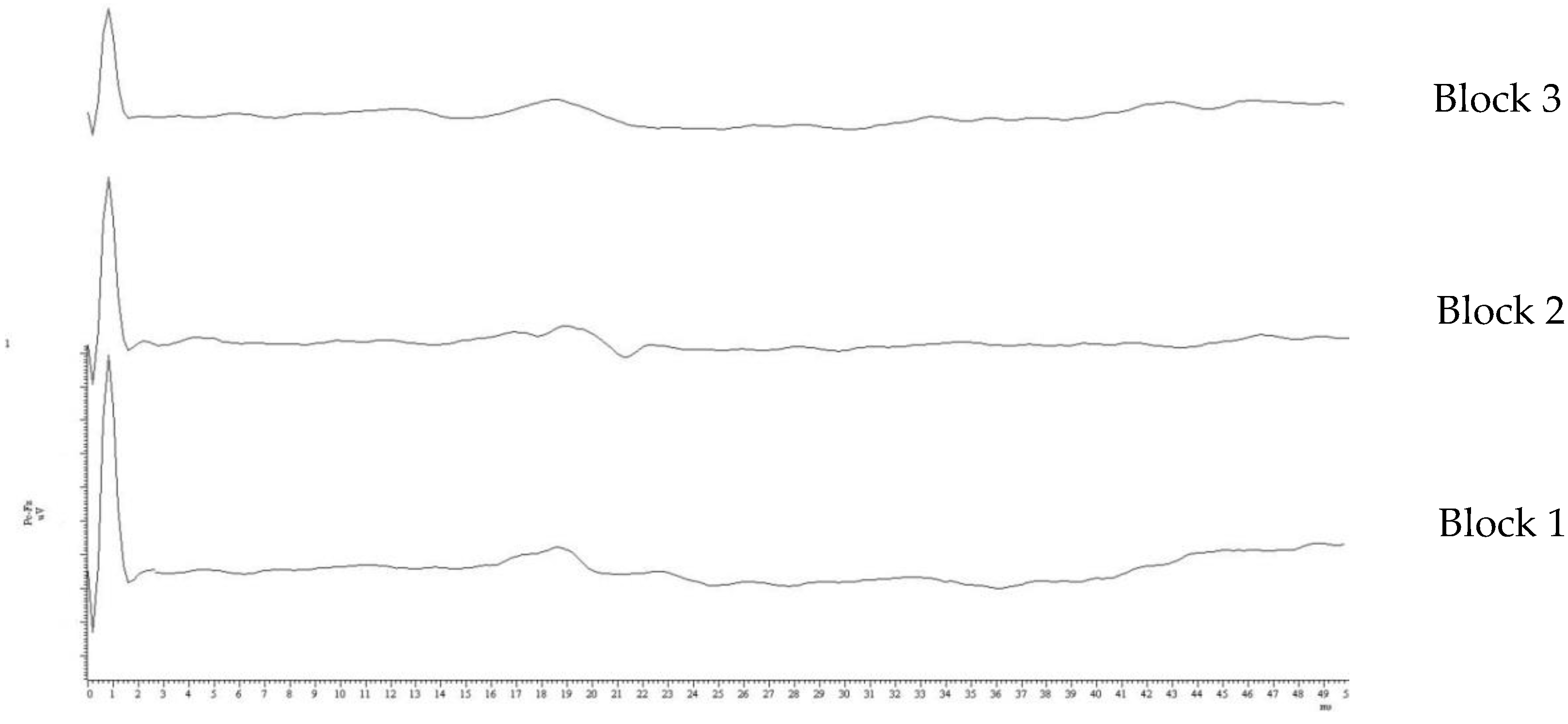
2. Methods
2.1. Subjects
2.2. Data Acquisition
2.3. Statistical Analysis
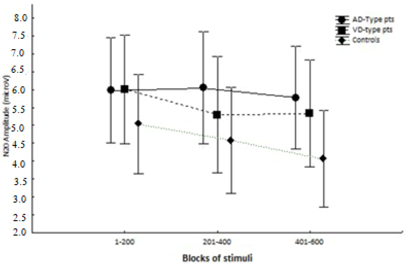
3. Results
4. Discussion
5. Conclusions
Author Contributions
Funding
Institutional Review Board Statement
Informed Consent Statement
Data Availability Statement
Conflicts of Interest
References
- World Health Organization. Global Action Plan on the Public Health Response to Dementia 2017–2025; World Health Organization: Geneva, Switzerland, 2017. [Google Scholar]
- van der Flier, W.M.; Scheltens, P. Epidemiology and risk factors of dementia. J. Neurol. Neurosurg. Psychiatry 2005, 76, v2–v7. [Google Scholar] [CrossRef] [Green Version]
- Haass, C.; Selkoe, D.J. Soluble protein oligomers in neurodegeneration: Lessons from the Alzheimer’s amyloid β-peptide. Nat. Rev. Mol. Cell Biol. 2007, 8, 101–112. [Google Scholar] [CrossRef]
- O’Brien, J.T.; Erkinjuntti, T.; Reisberg, B.; Roman, G.; Sawada, T.; Pantoni, L.; Bowler, J.V.; Ballard, C.; DeCarli, C.; Gorelick, P.B.; et al. Vascular cognitive impairment. Lancet Neurol. 2003, 2, 89–98. [Google Scholar] [CrossRef]
- Davies, C.; Mann, D.; Sumpter, P.; Yates, P. A quantitative morphometric analysis of the neuronal and synaptic content of the frontal and temporal cortex in patients with Alzheimer’s disease. J. Neurol. Sci. 1987, 78, 151–164. [Google Scholar] [CrossRef]
- Perdahl, E.; Adolfsson, R.; Alafuzoff, I.; Albert, K.A.; Nestler, E.J.; Greengard, P.; Winblad, B. Synapsin I (protein I) in different brain regions in senile dementia of Alzheimer type and in multiinfarct dementia. J. Neural Transm. 1984, 60, 133–141. [Google Scholar] [CrossRef]
- Sze, C.-I.; Troncoso, J.C.; Kawas, C.; Moution, P.; Price, D.L.; Martin, L.J. Loss of the Presynaptic Vesicle Protein Synaptophysin in Hippocampus Correlates with Cognitive Decline in Alzheimer Disease. J. Neuropathol. Exp. Neurol. 1997, 56, 933–944. [Google Scholar] [CrossRef] [Green Version]
- Sinclair, L.I.; Tayler, H.; Love, S. Synaptic protein levels altered in vascular dementia. Neuropathol. Appl. Neurobiol. 2015, 41, 533–543. [Google Scholar] [CrossRef] [Green Version]
- Thompson, R.F. Habituation: A history. Neurobiol. Learn. Mem. 2009, 92, 127–134. [Google Scholar] [CrossRef]
- Carew, T.J.; Pinsker, H.M.; Kandel, E.R. Long-Term Habituation of a Defensive Withdrawal Reflex in Aplysia. Sci. 1972, 175, 451–454. [Google Scholar] [CrossRef]
- Callaway, E., III. Habituation of averaged evoked potentials in man. Physiol. Substr. 1973, 153–174. [Google Scholar]
- Coppola, G.; Currà, A.; Di Lorenzo, C.; Parisi, V.; Gorini, M.; Sava, S.L.; Schoenen, J.; Pierelli, F. Abnormal cortical responses to somatosensory stimulation in medication-overuse headache. BMC Neurol. 2010, 10, 126. [Google Scholar] [CrossRef] [Green Version]
- Serrano-Pozo, A.; Frosch, M.P.; Masliah, E.; Hyman, B.T. Neuropathological Alterations in Alzheimer Disease. Cold Spring Harb. Perspect. Med. 2011, 1, a006189. [Google Scholar] [CrossRef]
- Loeb, C. Clinical criteria for the diagnosis of vascular dementia. Eur. Neurol. 1988, 28.2, 87–92. [Google Scholar] [CrossRef]
- Jackson, C.E.; Snyder, P.J. Electroencephalography and event-related potentials as biomarkers of mild cognitive impairment and mild Alzheimer’s disease. Alzheimer’s Dement. 2008, 4, 137–143. [Google Scholar] [CrossRef]
- Antczak, J.; Rusin, G.; Słowik, A. Transcranial Magnetic Stimulation as a Diagnostic and Therapeutic Tool in Various Types of Dementia. J. Clin. Med. 2021, 10, 2875. [Google Scholar] [CrossRef]
- McKhann, G.M.; Knopman, D.S.; Chertkow, H.; Hyman, B.T.; Jack, C.R.; Kawas, C.H.; Klunkk, W.E.; Koroshetzl, W.J.; Manlym, J.J.; Mayeux, R.; et al. The diagnosis of dementia due to Alzheimer’s disease: Recommendations from the National Institute on Aging-Alzheimer’s Association workgroups on diagnostic guidelines for Alzheimer’s disease. Alzheimers Dement 2011, 7, 263–269. [Google Scholar] [CrossRef] [Green Version]
- Roman, G.C.; Tatemichi, T.K.; Erkinjuntti, T.; Cummings, J.L.; Masdeu, J.C.; Garcia, J.H.; Amaducci, L.; Orgogozo, J.-M.; Brun, A.; Hofman, A.; et al. Vascular dementia: Diagnostic criteria for research studies: Report of the NINDS-AIREN International Workshop. Neurology 1993, 43, 250. [Google Scholar] [CrossRef]
- Abbruzzese, G.; Reni, L.; Cocito, L.; Ratto, S.; Favale, E. Short-latency somatosensory evoked potentials in degenerative and vascular dementia. J. Neurol. Neurosurg. Psychiatry 1984, 47, 1034–1037. [Google Scholar] [CrossRef] [Green Version]
- Tachibana, H.; Takeda, M.; Okuda, B.; Kawabata, K.; Nishimura, H.; Kodama, N.; Iwamoto, Y.; Sugita, M. Multimodal evoked potentials in Alzheimer’s disease and Binswanger’s disease. J. Geriatr. Psychiatry Neurol. 1996, 9, 7–12. [Google Scholar] [CrossRef]
- Rosén, I.; Gustafson, L.; Risberg, J. Multichannel EEG Frequency Analysis and Somatosensory-Evoked Potentials in Patients with Different Types of Organic Dementia. Dement. Geriatr. Cogn. Disord. 1993, 4, 43–49. [Google Scholar] [CrossRef]
- Stephen, J.M.; Montaño, R.; Donahue, C.H.; Adair, J.C.; Knoefel, J.; Qualls, C.; Hart, B.; Ranken, D.; Aine, C.J. Somatosensory responses in normal aging, mild cognitive impairment, and Alzheimer’s disease. J. Neural Transm. 2009, 117, 217–225. [Google Scholar] [CrossRef] [PubMed] [Green Version]
- Tsiptsios, I.; Fountoulakis, K.N.; Sitzoglou, K.; Papanicolaou, A.; Phokas, K.; Fotiou, F.; Kaprinis, G.S. Clinical and neuroimaging correlates of abnormal short-latency Somatosensory Evoked Potentials in elderly vascular dementia patients: A psychophysiological exploratory study. Ann. Gen. Psychiatry 2003, 2, 8. [Google Scholar] [CrossRef] [PubMed] [Green Version]
- McCarberg, B.; Peppin, J. Pain Pathways and Nervous System Plasticity: Learning and Memory in Pain. Pain Med. 2019, 20, 2421–2437. [Google Scholar] [CrossRef]
- Apkarian, A.V.; Bushnell, M.C.; Treede, R.-D.; Zubieta, J.-K. Human brain mechanisms of pain perception and regulation in health and disease. Eur. J. Pain 2005, 9, 463. [Google Scholar] [CrossRef]
- van Kooten, J.; Smalbrugge, M.; van der Wouden, J.C.; Stek, M.L.; Hertogh, C.M. Prevalence of pain in nursing home residents: The role of dementia stage and dementia subtypes. J. Am. Med. Dir. Assoc. 2017, 18, 522–527. [Google Scholar] [CrossRef]
- Fletcher, P.D.; Downey, L.E.; Golden, H.L.; Clark, C.; Slattery, C.F.; Paterson, R.W.; Rohrer, J.; Schott, J.; Rossor, M.; Warren, J.D. Pain and temperature processing in dementia: A clinical and neuroanatomical analysis. Brain 2015, 138, 3360–3372. [Google Scholar] [CrossRef] [PubMed]
- Shigihara, Y.; Hoshi, H.; Fukasawa, K.; Ichikawa, S.; Kobayashi, M.; Sakamoto, Y.; Negishi, K.; Haraguchi, R.; Konno, S. Resting-State Magnetoencephalography Reveals Neurobiological Bridges Between Pain and Cognitive Impairment. Pain Ther. 2021, 10, 349–361. [Google Scholar] [CrossRef]
- Whitlock, E.L.; Diaz-Ramirez, L.G.; Glymour, M.M.; Boscardin, W.J.; Covinsky, K.E.; Smith, A.K. Association Between Persistent Pain and Memory Decline and Dementia in a Longitudinal Cohort of Elders. JAMA Intern. Med. 2017, 177, 1146–1153. [Google Scholar] [CrossRef] [Green Version]
- Braak, H.; Braak, E. Neuropathological stageing of Alzheimer-related changes. Acta Neuropathol. 1991, 82, 239–259. [Google Scholar] [CrossRef]
- Teipel, S.J.; Stahl, R.; Dietrich, O.; Schoenberg, S.O.; Perneczky, R.; Bokde, A.L.; Reiser, M.F.; Möller, H.-J.; Hampel, H. Multivariate network analysis of fiber tract integrity in Alzheimer’s disease. NeuroImage 2007, 34, 985–995. [Google Scholar] [CrossRef]
- Hsiao, F.J.; Chen, W.T.; Wang, Y.J.; Yan, S.H.; Lin, Y.Y. Altered source-based EEG coherence of resting-state sensorimotor network in early-stage Alzheimer’s disease compared to mild cognitive impairment. Neurosci. Lett. 2014, 558, 47–52. [Google Scholar] [CrossRef]
- Babiloni, C.; Blinowska, K.; Bonanni, L.; Cichocki, A.; De Haan, W.; Del Percio, C.; Dubois, B.; Escudero, J.; Fernández, A.; Frisoni, M.; et al. What electrophysiology tells us about Alzheimer’s disease: A window into the synchronization and connectivity of brain neurons. Neurobiol. Aging 2020, 85, 58–73. [Google Scholar] [CrossRef]
- D’Antonio, F.; De Bartolo, M.I.; Ferrazzano, G.; Trebbastoni, A.; Amicarelli, S.; Campanelli, A.; de Lena, C.; Berardelli, A.; Conte, A. Somatosensory Temporal Discrimination Threshold in Patients with Cognitive Disorders. J. Alzheimer’s Dis. 2019, 70, 425–432. [Google Scholar] [CrossRef] [Green Version]
- Di Lazzaro, V.; Oliviero, A.; Tonali, P.A.; Marra, C.; Daniele, A.; Profice, P.; Saturno, E.; Pilato, F.; Masullo, C.; Rothwell, J.C. Noninvasive in vivo assessment of cholinergic cortical circuits in AD using transcranial magnetic stimulation. Neurology 2002, 59, 392–397. [Google Scholar] [CrossRef]
- Nardone, R.; Bergmann, J.; Kronbichler, M.; Kunz, A.; Klein, S.; Caleri, F.; Tezzon, F.; Ladurner, G.; Golaszewski, S. Abnormal short latency afferent inhibition in early Alzheimer’s disease: A transcranial magnetic demonstration. J. Neural Transm. 2008, 115, 1557–1562. [Google Scholar] [CrossRef] [PubMed]
- Battaglia, F.; Wang, H.-Y.; Ghilardi, M.F.; Gashi, E.; Quartarone, A.; Friedman, E.; Nixon, R.A. Cortical Plasticity in Alzheimer’s Disease in Humans and Rodents. Biol. Psychiatry 2007, 62, 1405–1412. [Google Scholar] [CrossRef] [PubMed]
- Menon, U.; Kelley, R.E. Chapter 2 Subcortical Ischemic Cerebrovascular Dementia. Int. Rev. Neurobiol. 2009, 84, 21–33. [Google Scholar] [CrossRef]
- Fu, Z.; Iraji, A.; Caprihan, A.; Adair, J.C.; Sui, J.; Rosenberg, G.A.; Calhouna, V.D. In search of multimodal brain alterations in Alzheimer’s and Binswanger’s disease. Neuroimage Clin. 2020, 26, 101937. [Google Scholar] [CrossRef] [PubMed]
- Bella, R.; Cantone, M.; Lanza, G.; Ferri, R.; Vinciguerra, L.; Puglisi, V.; Pennisi, M.; Ricceri, R.; Di Lazzaro, V.; Pennisi, G. Cholinergic circuitry functioning in patients with vascular cognitive impairment—No dementia. Brain Stimul. 2016, 9, 225–233. [Google Scholar] [CrossRef]
- Groves, P.M.; Thompson, R.F. Habituation: A dual-process theory. Psychol Rev. 1970, 77, 419–450. [Google Scholar] [CrossRef]
- Chui, H.C. Subcortical ischemic vascular dementia. Neurol. Clin. 2007, 25, 717–740. [Google Scholar] [CrossRef] [PubMed] [Green Version]
- Rosenberg, G.A. Binswanger’s disease: Biomarkers in the inflammatory form of vascular cognitive impairment and dementia. J. Neurochem. 2018, 144, 634–643. [Google Scholar] [CrossRef] [PubMed]


| Patient | Grp | Age | Sex | Education (Years) | MMSE | Memory Enhancing Drugs |
|---|---|---|---|---|---|---|
| 1 | AD-type | 76 | F | 5 | 18 | Donepezil 10 mg Memantine 20 mg |
| 2 | AD-type | 79 | M | 13 | 22 | None |
| 3 | AD-type | 62 | M | 5 | 20 | Galantamine 16 mg |
| 4 | AD-type | 75 | F | 8 | 22 | Rivastigmine 9 mg |
| 5 | AD-type | 68 | F | 13 | 21 | None |
| 6 | AD-type | 78 | F | 0 | 20 | Galantamine 16 mg |
| 7 | AD-type | 78 | F | 3 | 22 | Donepezil 5 mg |
| 8 | AD-type | 80 | M | 16 | 21 | Memantine 20 mg |
| 9 | AD-type | 65 | F | 5 | 19 | None |
| 10 | AD-type | 68 | F | 13 | 22 | Rivastigmine patch 4.5 mg |
| 11 | AD-type | 79 | F | 5 | 20 | Donepezil 5 mg |
| 12 | AD-type | 82 | F | 5 | 18 | None |
| 13 | AD-type | 74 | F | 5 | 21 | Galantamine 16 mg |
| 14 | AD-type | 77 | F | 5 | 18 | Rivastigmine patch 13.3 mg Memantine 20 mg |
| 15 | AD-type | 80 | F | 9 | 20 | Galantamine 16 mg |
| 16 | VD-type | 86 | F | 5 | 18 | |
| 17 | VD-type | 70 | F | 5 | 21 | |
| 18 | VD-type | 74 | M | 5 | 19 | |
| 19 | VD-type | 75 | M | 5 | 22 | |
| 20 | VD-type | 82 | M | 5 | 22 | |
| 21 | VD-type | 84 | F | 5 | 18 | |
| 22 | VD-type | 85 | M | 5 | 20 | |
| 23 | VD-type | 83 | F | 17 | 22 | |
| 24 | VD-type | 73 | M | 8 | 19 | |
| 25 | VD-type | 80 | F | 3 | 21 | |
| 26 | VD-type | 69 | F | 5 | 19 | |
| 27 | VD-type | 80 | F | 4 | 18 | |
| 28 | VD-type | 89 | F | 5 | 18 | |
| 29 | VD-type | 77 | M | 5 | 22 |
| ORIENTATION | MEMORY | ACE-R | FLUENCIES | |||||||||||||||||
|---|---|---|---|---|---|---|---|---|---|---|---|---|---|---|---|---|---|---|---|---|
| Patient | MMSE | Temporal | Spatial | Personal | Raven | Digit for | Digit back | Rey Immediate | Rey Delayed | Prose Immediate | Prose Delayed | Oblivion | Orientation | Memory | Fluency | Language | Visuo-Spatial | Tot. | Phonemic | Semantic |
| 1 | 18 | 74 | +/- | - | 13 | 8 | 7 | 23 | 9 | 60 | ||||||||||
| 2 | 22 | 96 | + | + | 4 | 3 | 20 | 0 | 1 | 0 | 1 | 25 | 10 | |||||||
| 3 | 20 | 94 | + | - | 3.3 | 2 | 1.3 | 13 | 7 | 5 | 16 | 6 | 47 | 15 | 9 | |||||
| 4 | 22 | 94 | + | + | 24 | 4 | 2 | 3.3 | 0 | 3.3 | 13 | 15 | 6 | 21 | 12 | 67 | 15 | 11 | ||
| 5 | 21 | 90 | + | +/- | 18 | 1 | 27 | 7 | ||||||||||||
| 6 | 20 | 87 | + | - | 12 | 9 | 9 | 21 | 10 | 61 | ||||||||||
| 7 | 22 | 98 | + | + | 21 | 3 | 2 | 22 | 2 | 5.3 | 2.3 | 3 | 16 | 16 | 6 | 23 | 13 | 74 | 18 | 9 |
| 8 | 21 | 88 | + | + | 28 | 4.6 | 1 | 3.6 | 14 | 18 | 9 | 25 | 14 | 76 | 23 | 12 | ||||
| 9 | 19 | 83 | - | +/- | 14 | 4 | 0 | 4 | 9 | 8 | 6 | 17 | 4 | 44 | 11 | 9 | ||||
| 10 | 22 | 100 | + | + | 30 | 5 | 4 | 32 | 3 | 5 | 2.2 | 2.8 | 17 | 11 | 9 | 26 | 15 | 78 | 39 | 11 |
| 11 | 20 | 90 | +/- | - | 18 | 3 | 5 | 3 | 2 | 1 | 8 | |||||||||
| 12 | 18 | 82 | - | - | 11 | 11 | 8 | 21 | 11 | 62 | ||||||||||
| 13 | 21 | 90 | +/- | +/- | 12 | 13 | 6 | 21 | 9 | 61 | 17 | 9 | ||||||||
| 14 | 18 | 92 | +/- | +/- | 13 | 10 | 7 | 20 | 11 | 61 | ||||||||||
| 15 | 20 | 76 | + | + | 18 | 11 | 7 | 8 | 17 | 10 | 53 | 24 | 11 | |||||||
| 16 | 18 | 77 | - | - | 10 | 8 | 6 | 21 | 13 | 58 | ||||||||||
| ORIENTATION | MEMORY | ACE-R | FLUENCIES | |||||||||||||||||
|---|---|---|---|---|---|---|---|---|---|---|---|---|---|---|---|---|---|---|---|---|
| Patient | MMSE | Temporal | Spatial | Personal | Raven | Digit for | Digit back | Rey Immediate | Rey Delayed | Prose Immediate | Prose Delayed | Oblivion | Orientation | Memory | Fluency | Language | Visuo-Spatial | Tot. | Phonemic | Semantic |
| 17 | 21 | 91 | + | - | 16 | 4 | 2 | 9 | 1 | 0 | 0 | 0 | 7 | 8 | ||||||
| 18 | 19 | 89 | + | +/- | 12 | 11 | 8 | 22 | 13 | 66 | ||||||||||
| 19 | 22 | 94 | + | + | 14 | 4 | 0 | 4 | 15 | 11 | 6 | 20 | 9 | 61 | 18 | 10 | ||||
| 20 | 22 | 100 | + | + | 20 | 5 | 3 | 3.1 | 3 | 1.1 | 16 | 13 | 7 | 24 | 14 | 72 | 18 | 9 | ||
| 21 | 18 | 88 | + | +/- | 5 | 4 | 17 | 0 | 3 | 0 | 3 | 25 | 13 | |||||||
| 22 | 20 | 100 | + | + | 17 | 3 | 0 | 3 | 15 | 8 | 8 | 20 | 9 | 60 | 22 | 12 | ||||
| 23 | 22 | 100 | + | + | 27 | 5 | 3 | 18 | 0 | 2.2 | 2 | 0 | 18 | 11 | 8 | 25 | 14 | 76 | 10 | 12 |
| 24 | 19 | 86 | + | +/- | 3 | 2 | 19 | 3 | 2 | 2 | 0 | 9 | 5 | |||||||
| 25 | 21 | 91 | +/- | + | 13 | 14 | 5 | 20 | 11 | 63 | 12 | 10 | ||||||||
| 26 | 19 | 76 | + | - | 13 | 10 | 7 | 22 | 10 | 62 | ||||||||||
| 27 | 18 | 78 | +/- | +/- | 12 | 10 | 6 | 18 | 12 | 58 | ||||||||||
| 28 | 18 | 75 | - | - | 12 | 8 | 7 | 22 | 14 | 63 | ||||||||||
| 29 | 22 | 90 | + | + | 29 | 3 | 0 | 3 | 13 | 10 | 6 | 20 | 13 | 62 | 10 | 13 | ||||
Publisher’s Note: MDPI stays neutral with regard to jurisdictional claims in published maps and institutional affiliations. |
© 2021 by the authors. Licensee MDPI, Basel, Switzerland. This article is an open access article distributed under the terms and conditions of the Creative Commons Attribution (CC BY) license (https://creativecommons.org/licenses/by/4.0/).
Share and Cite
Currà, A.; Marinelli, L.; Cotellessa, F.; Mori, L.; Avanti, C.; Greco, D.; Gorini, M.; Missori, P.; Fattapposta, F.; Trompetto, C. Habituation of Somatosensory Evoked Potentials in Patients with Alzheimer’s Disease and Those with Vascular Dementia. Medicina 2021, 57, 1364. https://0-doi-org.brum.beds.ac.uk/10.3390/medicina57121364
Currà A, Marinelli L, Cotellessa F, Mori L, Avanti C, Greco D, Gorini M, Missori P, Fattapposta F, Trompetto C. Habituation of Somatosensory Evoked Potentials in Patients with Alzheimer’s Disease and Those with Vascular Dementia. Medicina. 2021; 57(12):1364. https://0-doi-org.brum.beds.ac.uk/10.3390/medicina57121364
Chicago/Turabian StyleCurrà, Antonio, Lucio Marinelli, Filippo Cotellessa, Laura Mori, Chiara Avanti, Daniela Greco, Manuela Gorini, Paolo Missori, Francesco Fattapposta, and Carlo Trompetto. 2021. "Habituation of Somatosensory Evoked Potentials in Patients with Alzheimer’s Disease and Those with Vascular Dementia" Medicina 57, no. 12: 1364. https://0-doi-org.brum.beds.ac.uk/10.3390/medicina57121364