Article

Sleep Quality Improvement Enhances Neuropsychological Recovery and Reduces Blood Aβ42/40 Ratio in Patients with Mild–Moderate Cognitive Impairment

1
Department of Geriatrics, Jianghan Oilfield General Hospital, The Yangtze University School of Medicine, Qianjiang 433121, China
2
Hubei Clinical Research Center of Dementia and Cognitive Impairment, Wuhan 434300, China
3
Health Science Center, The Yangtze University School of Medicine, Jingzhou 434023, China
4
Department of Clinical Laboratory, Jianghan Oilfield General Hospital, The Yangtze University School of Medicine, Qianjiang 433121, China
5
Division of Research & Education Administration, Jianghan Oilfield General Hospital, The Yangtze University School of Medicine, Qianjiang 433121, China
6
Department of Urology, The University of Kansas Medical Center, Kansas City, KS 66160, USA
7
Department of Nursing, Jianghan Oilfield General Hospital, The Yangtze University School of Medicine, Qianjiang 433121, China
*
Authors to whom correspondence should be addressed.
Submission received: 18 October 2021 / Revised: 29 November 2021 / Accepted: 1 December 2021 / Published: 15 December 2021
(This article belongs to the Topic Age-Related Neurodegenerative Diseases and Stroke)

Abstract

:
Background and objectives: Alzheimer’s disease is a progressive brain degeneration and is associated with a high prevalence of sleep disorders. Amyloid β peptide-42/40 (Aβ42/40) and Tau-pT181 are the core biomarkers in cerebrospinal fluid and blood. Accumulated data from studies in mouse models and humans demonstrated an aberrant elevation of these biomarkers due to sleep disturbance, especially sleep-disordered breathing (SDB). However, it is not clear if sleep quality improvement reduces the blood levels of Ab42/40 ratio and Tau-pT181 in Alzheimer’s disease patients. Materials and Methods: In this prospective study, a longitudinal analysis was conducted on 64 patients with mild–moderate cognition impairment (MCI) due to Alzheimer’s disease accompanied by SDB. Another 33 MCI cases without sleep-disordered breathing were included as the control group. Sleep quality was assessed using the Pittsburgh Sleep Quality Index (PSQI) score system. Neuropsychological assessments were conducted using the Montreal Cognitive Assessment (MoCA), Geriatric Depression Scale (GDS), Clinical Dementia Rating (CDR), 24-h Hamilton Rating Scale for Depression (HRSD-24), and Hamilton Anxiety Rating Scale (HAMA) scoring systems. Aβ42, Aβ40, and Tau-pT181 protein levels in blood specimens were measured using ELISA assays. All patients received donepezil treatment for Alzheimer’s disease. SDB was managed with continuous pressure ventilation. Results: A significant correlation was found among PSQI, HRSD-24, HAMA, Aβ42/40 ratio, and Tau-pT181 level in all cases. In addition, a very strong and negative correlation was discovered between education level and dementia onset age. Compared to patients without SDB (33 non-SD cases), patients with SDB (64 SD cases) showed a significantly lower HRSD-24 score and a higher Aβ42/40 ratio Tau-pT181 level. Sleep treatment for patients with SDB significantly improved all neuropsychological scores, Aβ42/40 ratio, and Tau-pT181 levels. However, 11 patients did not completely recover from a sleep disorder (PSQI > 5 post-treatment). In this subgroup of patients, although HAMA score and Tau-pT181 levels were significantly reduced, MoCA and HRSD-24 scores, as well as Aβ42/40 ratio, were not significantly improved. ROC analysis found that the blood Aβ42/40 ratio held the highest significance in predicting sleep disorder occurrence. Conclusions: This is the first clinical study on sleep quality improvement in Alzheimer’s disease patients. Sleep quality score was associated with patient depression and anxiety scores, as well as Aβ42/40 ratio and Tau-pT181 levels. A complete recovery is critical for fully improving all neuropsychological assessments, Aβ42/40 ratio, and Tau-pT181 levels. Blood Aβ42/40 ratio is a feasible prognostic factor for predicting sleep quality.

1. Introduction

Alzheimer’s disease (AD) is a progressive neuronal degenerative disorder, and nearly 50 million people live with dementia worldwide—75% are Alzheimer’s disease patients [1]. AD is the sixth leading cause of death in the US, accounting for more than 122 K deaths in 2018, more than breast cancer and prostate cancer combined [2]. It is estimated that 5.8 million Americans age 65 and older have Alzheimer’s disease this year, and the number is projected to be 13.8 million by 2050, according to the Alzheimer’s Association (www.alz.org accessed on 29 November 2021). Therefore, there is a desperate need for medical breakthroughs to prevent, slow, or cure Alzheimer’s disease.
Alzheimer’s disease has two pathophysiological hallmarks in the brain: interstitial deposition of insoluble amyloid-β (Aβ) peptides and intracellular aggregation of hyperphosphorylated Tau proteins [3]. Aβ deposition and Tau protein aggregation are long-term and slow processes, starting 20 years before any noticeable symptoms, a so-called prodromal phase [4,5]. Aβ peptides are physiologically processed from their precursor protein, mainly in neuronal cells after sequential proteolytic cleavage by β-secretase and γ-secretase [5,6]. There are four significant isoforms of Aβ peptides (38, 40, 42, and 43), which are detectable in brain interstitial fluid (ISF), cerebrospinal fluid (CSF), and blood plasma [7]. Aβ40 and Aβ42 peptides are more abundant than others. The Aβ42 peptide is less soluble due to two extra hydrophobic amino acid residues at the C-terminus, rendering it more prone to deposition than Aβ40 [8]. Currently, the levels of Aβ42/40 ratio in patient CSF and blood are implicated as disease biomarkers in the clinic [7,9]. Tau protein is a crucial component of microtubule assembly in axons, and its function is regulated by phosphorylation on multiple residues, including threonine 181 (Tau-pT181) [10]. Like Aβ peptides, Tau protein and its pT181 form are also detectable in CSF and blood specimens; their levels are associated with disease progression [11].
In recent years, sleep disorders have been linked to Alzheimer’s disease progression in addition to the typical symptoms of progressive loss of memory, speech, and cognition [12,13]. A high prevalence (25–66%) of Alzheimer’s disease patients was reported to exhibit various sleep disorders, including sleep-disordered breathing (SDB) [14,15]. In contrast, the incidence of sleep disorders was only 18.3–27.6% in elderly adults without cognition impairment [14]. These sleep behavior changes began in the prodromal phase of Alzheimer’s disease, while patients only suffered from mild cognitive impairment (MCI), possibly due to amyloid/Tau pathology before cognition decline [14]. Recent studies showed that more than half of the AD patients also are suffering from SDB [16]. Sleep deprivation or disturbances increased Aβ peptides and Tau proteins in cerebrospinal fluid (CSF) compared to normal sleep controls [9,17,18,19] due to high production of Aβ peptides [20], which was supported by studies from transgenic mouse models of Alzheimer’s disease [21].
Interestingly, sleep treatment with continuous positive airway pressure in SDB patients significantly improved cognition scores in patients with Alzheimer’s disease [22]. On the other hand, a healthy sleep cycle was shown to facilitate Aβ clearance from the brain tissue [23]. These studies suggest a bidirectional relationship between sleep disorders and AD progression [14,24]. However, it is unknown if sleep quality improvement would reduce or slow cognition impairment in Alzheimer’s disease patients.
The purpose of this study was to examine the effect of sleep quality improvement on neuropsychological symptoms and blood levels of Aβ peptides and Tau proteins in Alzheimer’s disease patients with mild–moderate cognition impairments. We were also interested in identifying potential risk factors that provide prognostic feasibility for sleep quality prediction in Alzheimer’s disease patients. We compared the sleep quality (PSQI) score with a longitudinal approach, neuropsychological parameters (Montreal Cognitive Assessment (MoCA), Geriatric Depression Scale (GDS), Clinical Dementia Rating (CDR), 24-h Hamilton Rating Scale for Depression (HRSD-24) and Hamilton Anxiety Rating Scale (HAMA)), blood Aβ42/40 ratio, and Tau-pT181 proteins before and after a 6-month sleep treatment in 64 MCI patients with sleep-disordered breathing. Our results revealed that PSQI scores significantly correlated with HRSD-24 and HAMA scores, as well as Aβ42/42 ratio and Tau-pT181 levels, but not with MoCA, GDS, or CDR scores. A significant improvement was achieved after sleep treatment for PSQI, MoCA, HRSD-24, HAMA scores, and Aβ42/40 ratio and Tau-pT181 levels. Most interestingly, a complete recovery of sleep quality improved neuropsychological scores, Aβ42/40 ratio, and Tau-pT181 levels. However, MoCA and HRSD-24 scores, plus Aβ42/40 ratio, were not significantly enhanced in the patient who did not completely recover (PSQI > 5 post-treatment). ROC analysis identified Aβ42/40 ratio as the most potent predicting factor for sleep disorder occurrence. Our study demonstrated for the first time, as the authors are aware, that sleep quality improvement can enhance the neuropsychological status and reduce blood Aβ42/40 ratio and Tau-pT181 levels in MCI patients due to Alzheimer’s disease.

2. Materials and Methods

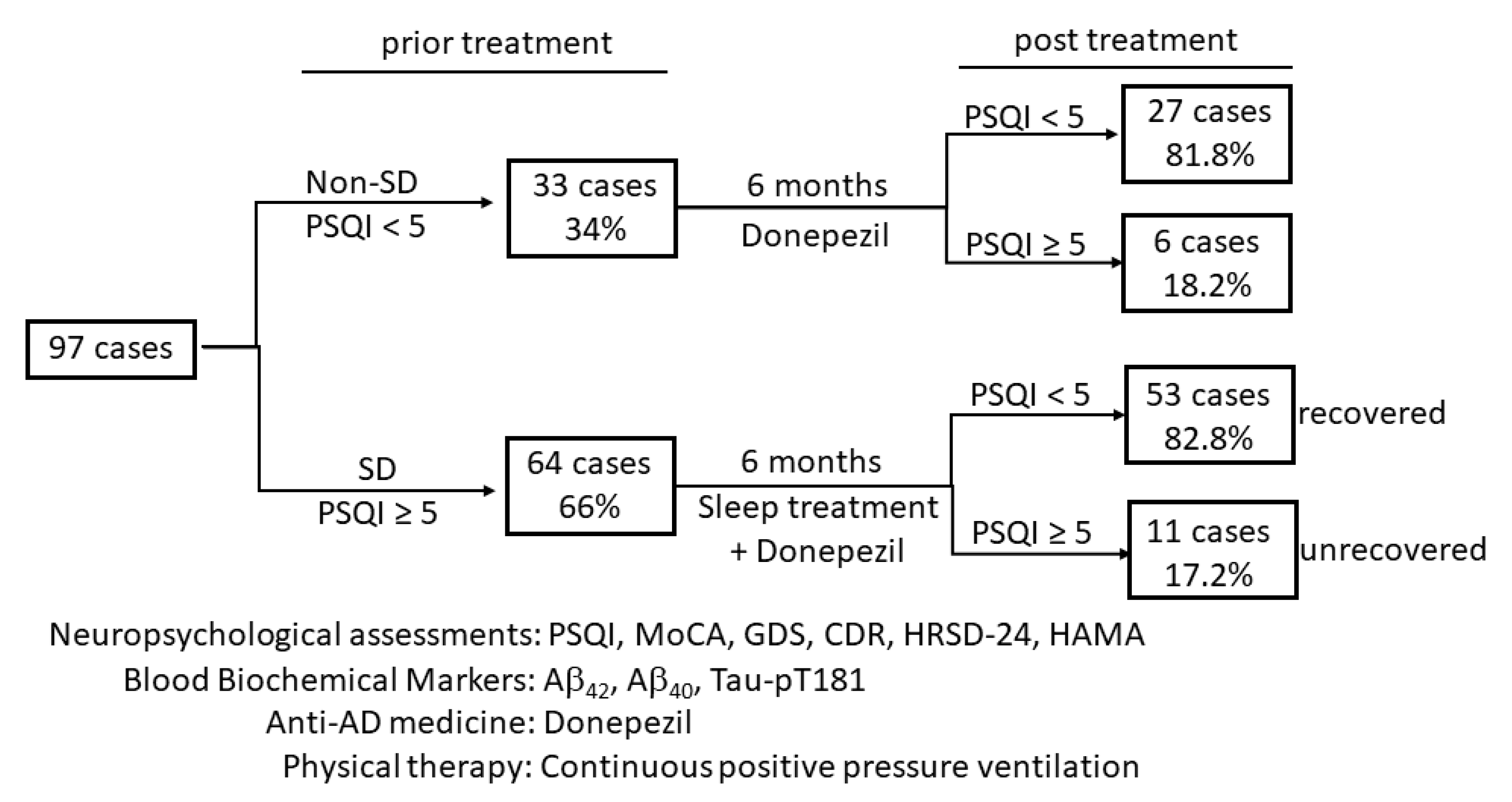
2.1. Study Design and Patients

A prospective longitudinal study was designed to examine the effect of sleep quality improvement on neuropsychological behaviors and the changes of blood Aβ42/40 ratio and Tau-pT181 levels in patients with mild cognitive impairment due to Alzheimer’s disease accompanied with sleep-disordered breathing. Patients were recruited at the Memory and Sleep Clinic at the Jianghan Oilfield General Hospital from February 2017 to December 2019. The diagnosis of AD dementia was made based on the core diagnostic criteria developed by the National Institute on Aging-Alzheimer’s Association in 2011 [25]. The diagnostic criteria for sleep-disordered breathing in AD patients were based on the definition of “Dementia-related sleep disorders” in the International Classification of Sleep Disorders guidelines [26] and the clinical diagnostic criteria for Alzheimer’s disease-related sleep disorders [27]. Other inclusion criteria include no severe dysfunctions or lesions of the heart, lung, liver, kidney, and other vital organs and the ability to complete relevant neuropsychological assessment and auxiliary examination. Exclusion criteria included: (1) severe dementia; (2) other types of cognitive impairment, including vascular dementia, Parkinson’s disease, frontotemporal dementia, Lewy body dementia; (3) a history of severe mental illness; (4) association with a severely debilitating illness, infectious disease, painful condition or other diseases that may affect the quality of sleep, such as chronic obstructive pulmonary disease, stroke, heart failure, kidney failure, severe cerebrovascular disease, epilepsy; and (5) severe physical movement disorder.

2.1.1. General Procedures

The study protocol was reviewed and approved by the Ethics Committee of the Jiangshan Oilfield General Hospital (study ethical code number 2017016, approval date 20170226). All the patients and their immediate family members were informed with a written consent form, and the patient’s signatures were obtained before enrollment. This study was conducted according to the principles stated in the Declaration of Helsinki [28].
After diagnosis and recruitment, all patients were managed by dedicated research nurses responsible for collecting demographic data, medical history, physical examination, behavioral and neuropsychological assessment, biochemical specimen collection, and regular follow-up. Structured questionnaires were used to assess patient medical history, gender, age, onset age of the disease, course of the disease, years of education, marital status, the living situation at home, and patient support level on the enrollment day. Patients were requested to spend one night at the hospital, where peripheral blood specimens were collected in the morning before daytime activity.

2.1.2. Sleep Quality Evaluation

The Pittsburgh Sleep Quality Index (PSQI) questionnaire (a Chinese version) was used to evaluate patients’ overall sleep quality [29,30]. There were 18 items on the scale divided into seven sub-items: subjective sleep quality, time to sleep, sleep time, sleep efficiency, night sleep disturbance, sleep drug use, and daytime dysfunction. Each item’s score is 0–3 points, and the total score is 0–21 points. A PSQI score at or above five was set as the cutoff value for a sleep disorder.

2.1.3. Assessment of Neuropsychological Status

The Montreal Cognitive Assessment Scale (MoCA) was used to assess patient cognitive function [31]. There were seven cognitive domains: visuospatial and executive function, naming, delayed recall, attention, language, abstraction, and orientation. A total score of less than 26 was considered as cognitive impairment.
The Clinical Dementia Assessment Scale (CDR) was used to provide a global evaluation of the severity of dementia [32]. A CDR score of 1 was classified as mild, 2 as moderate, and 3 as severe. The Global Deterioration Scale (GDS) was used to assess the extent and progress of dementia [33], which provides an overview of a patient who has degenerative dementia. The GDS scale was divided into seven levels and was completed by interviewing the patients and their caregivers. A GDS score of 1 indicates no cognitive impairment; 2 indicates a very mild cognitive impairment, 3 as mild, 4 as moderate, 5 as severe, 6 as very severe, and 7 as worst impairment.
The 24-item Hamilton Rating Scale for Depression (HRSD-24) was utilized to assess patient depressive symptoms [34]. In the HRSD-24 version, a total score of 8 or less was classified as no depression, 9–20 as suspicious depression, 21–35 as moderate or mild depression, and above 35 as severe depression.
The Hamilton Anxiety Scale (HAMA) was used to evaluate patient anxiety, and there were 14 items on this scale [35]. The scale adopts a 5-point scoring method ranging from 0 to 4 points. A total score less than 7 indicated no anxiety, 7–13 was classified as possible anxiety, 14–20 was anxiety, 21–28 was classified as significant anxiety, above 29 was classified as severe anxiety.

2.2. Measurement of Blood Levels of Amyloid Peptides and Tau-pT181 Proteins

Amyloid-β and Tau-pT181 levels in blood specimens were measured using the enzyme-linked immunosorbent assay (ELISA) methods. The ELISA kits for Aβ40 (KHB3481), Aβ42 (KHB3441), and Tau-pT181 proteins (KHO0631) were obtained from Invitrogen (Carlsbad, CA, USA). All blood specimens were collected between 6:00 and 9:00 a.m. in a fasted state. Heparin anticoagulant blood was collected by a vacuum tube and centrifuged at 3000 rpm for 10 min for plasma separation. The samples were then aliquoted and stored at −80 °C. ELISA assays were conducted within one week at three repeats for each specimen.

2.3. The Intervention of Sleep Disorders and Dementia

All patients enrolled in this study received a standard anti-dementia medicine donepezil (5–10 mg, QN). Patients with sleep-disordered breathing were managed by continuous positive pressure ventilation.

2.4. Data Collection and Statistical Analysis

Data of biometrics, behavioral and neuropsychological assessments, and Amyloid-β and Tau-pT181 tests were obtained at the enrollment before treatment and at the end of the 6-month treatment. Statistical analysis was performed using SPSS software (version 28.0, Chicago, IL, USA) and the figure graphs were generated using the GraphPad Prism software (version 9.0.0, San Diego, CA, USA). Statistical comparison among multiple groups was conducted using two-tail ANOVA analysis. The comparison of the parameters between pre- and post-treatment was analyzed using a paired t-test. Pearson correlation analysis was used to determine the correlation among all parameters. The Receiver Operator Characteristic (ROC) analysis was used to identify a prediction factor for sleep quality disorder.

3. Results

3.1. Patients Population and Clinical Parameters

A total of 97 AD-related dementia patients were enrolled in this study, of whom 33 patients without sleep-disordered breath were included as the control group, and 64 cases with sleep-disordered breathing were set as the treatment group (Figure 1). All biometric data are summarized in Table 1. All subjects had a CDR score of 1–2 (mild–moderate dementia) and a MoCA score of 12–26 (cognition impairment). Sixty-three patients (64.9%) were scored as suspicious depression (HRSD-24 score 9–20). Fifty-three (54.6%) patients were scored as anxiety (HAMA score 14–18), and 42 (43.3%) patients were scored as possible anxiety (HAMA score 7–13). Interestingly, a negative and robust correlation (Persons r = −0.573) was discovered between the education level and dementia onset age (Table 2), indicating that higher educated patients tended to have an early onset age of mild–moderate dementia.

3.2. Sleep Quality Was Associated with Neuropsychological Symptoms and Blood Biomarkers

We first assessed the entire group (97 cases) for the correlation of sleep quality using the PQSI score with the neuropsychological status and blood biomarkers. As shown in Table 2, PQSI score significantly correlated with HAMA and HRSD-24 scores, as well as the Blood Aβ42/40 ratio and Tau-pT181 level (Table 2). Further analysis revealed a significant correlation among Tau-pT181 level, Aβ42/40 ratio, HAMA, and HRSD-24 scores. These data indicate that sleep quality is associated with depression and anxiety behavior, as well as blood biomarkers in MCI patients due to Alzheimer’s disease.
We then compared neuropsychological scores and blood biomarkers between patients with or without sleep-disordered breathing conditions (non-SD vs. SD cases). As shown in Figure 2, HRSD-24 score, Aβ42/40 ratio, and Tau-pT181 levels were significantly higher in patients with sleep-disordered breathing (SD cases) than those in non-SD cases; however, MoCA and HAMA scores did not show any statistically significant differences. These data demonstrated that HRSD-24 score, blood Aβ42/40 ratio, and Tau-pT181 level were strongly associated with sleep-disordered breathing in MCI patients due to Alzheimer’s disease.

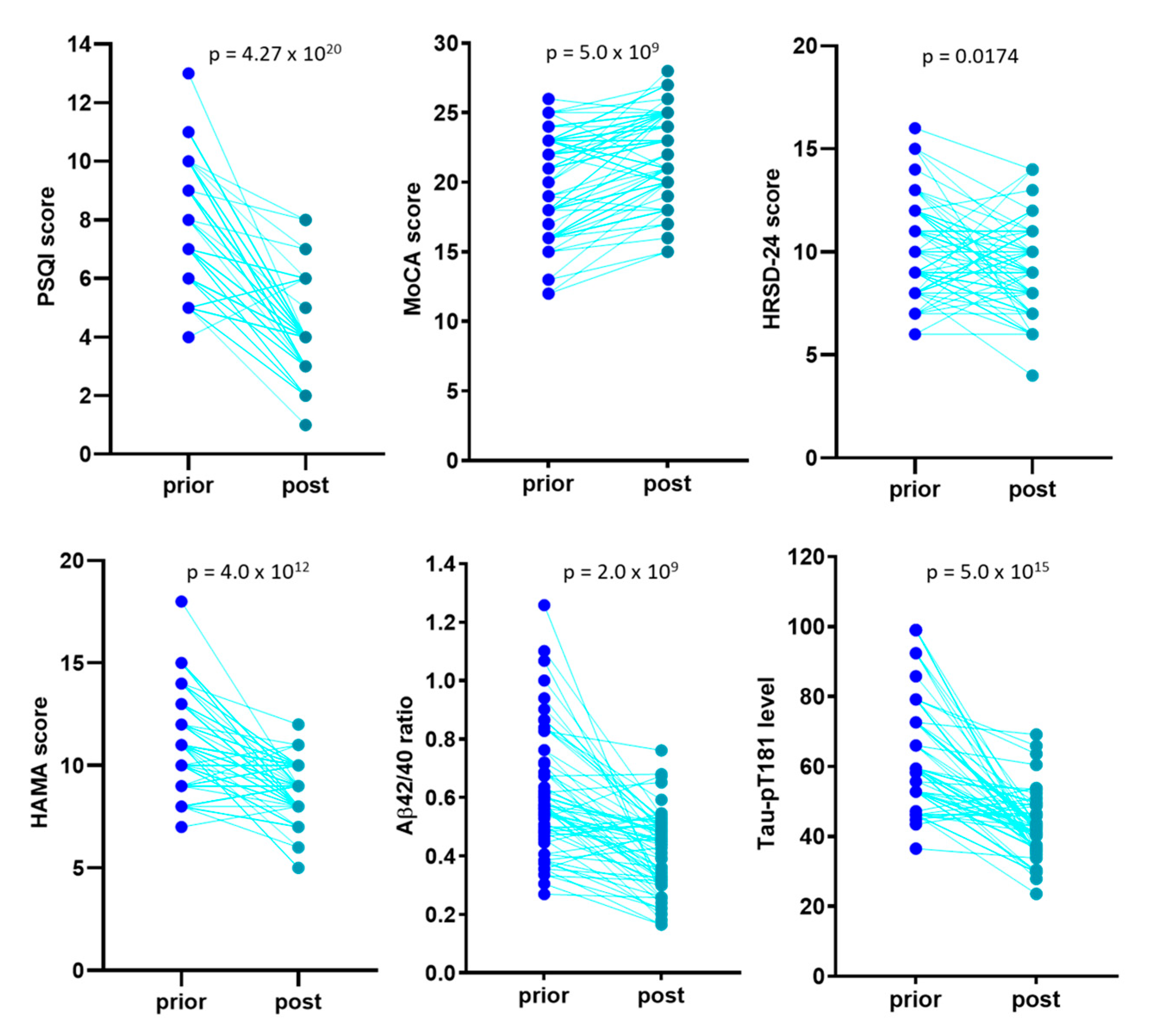
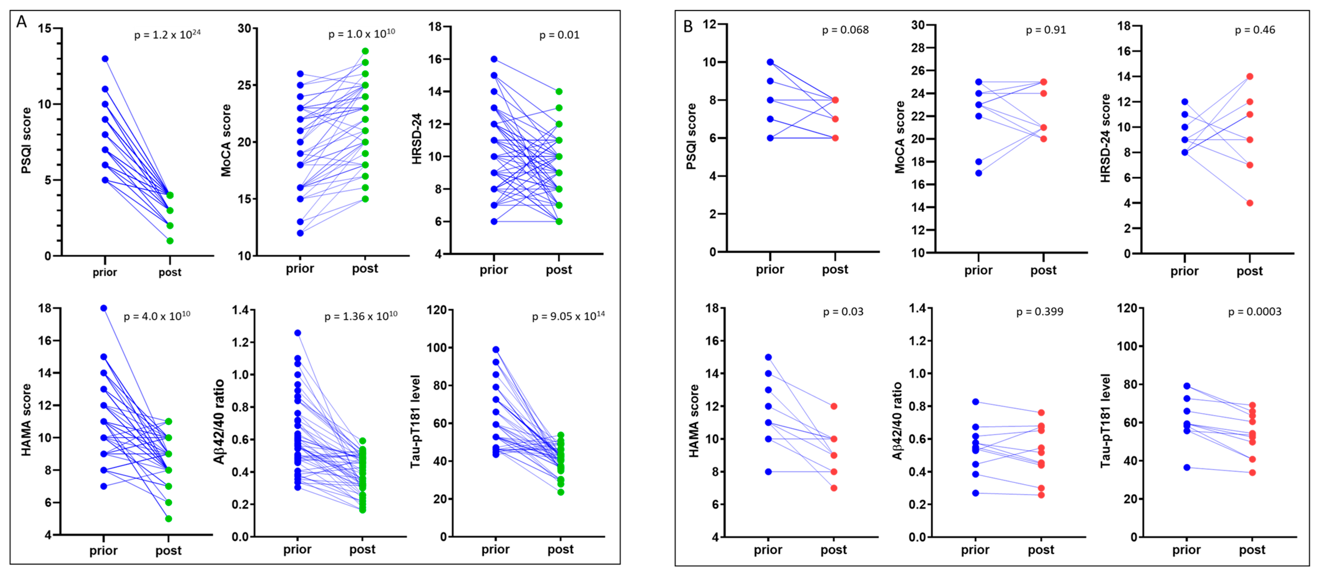
3.3. Sleep Treatment Improves COGNITION and Relieves Anxiety

Patients with sleep-disordered breathing (64 cases) were treated with continuous positive pressure ventilation for six months in addition to the standard care of anti-Alzheimer’s disease medicine donepezil. Pearson correlation analysis revealed a strong correlation among the PSQI, neuropsychological scores, blood Aβ42/40 ratio, and Tau-pT181 levels (Table 3), similar to the correlations for the entire cohort (Table 2).
After a 6-month sleep intervention, a significant improvement was achieved for all the neuropsychological scores, blood Aβ42/40 ratio, and Tau-pT181 levels (Figure 3). Among these patients, 53 cases (82.8%) showed a complete recovery in PSQI score (<5) but 10 cases were only showed a slight improvement (PSQI > 5 post-treatment). One patient had a PSQI increased from prior to treatment of 6 to post-treatment of 8. After separating these two subgroups, a substantial improvement was observed for the recovered cases in MoCA, HRSD-24, and HAMA scores after sleep treatment. blood Aβ42/40 ratio and Tau-pT181 levels were also significantly reduced after the 6-month sleep treatment in this subgroup (Figure 4A). In the unrecovered subgroup, although HAMA scores and blood Tau-pT181 levels reduced considerably, MoCA and HRSD-24 scores, as well as blood Aβ42/40 ratio, did not improve significantly (Figure 4B).
We then compared blood Aβ42/40 ratio and Tau-pT181 protein levels prior- or post-sleep treatment in unrecovered and recovered subgroups. As shown in Figure 5A, the Aβ42/40 ratio showed a significant reduction in the recovered subgroup but not in the unrecovered subgroup. In contrast, Tau-pT181 protein levels showed a significant reduction in both recovered and unrecovered subgroups (Figure 5B). However, the recovered subgroups showed a much lower Aβ42/40 ratio and Tau-pT181 protein than the unrecovered subgroups. These data suggest that a full recovery of sleep quality is necessary to reduce the blood Aβ42/40 ratio and improve recognition and depression status.

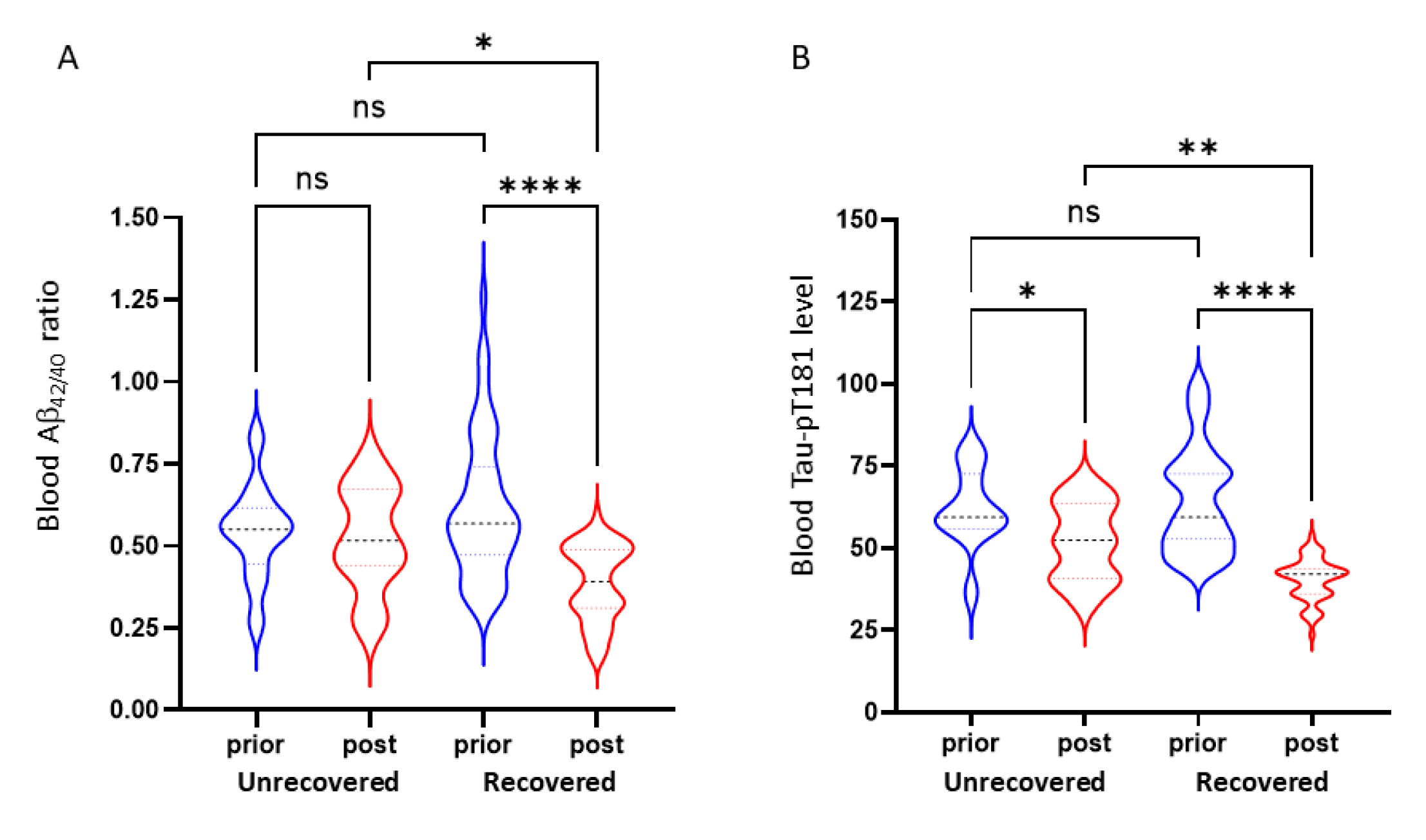
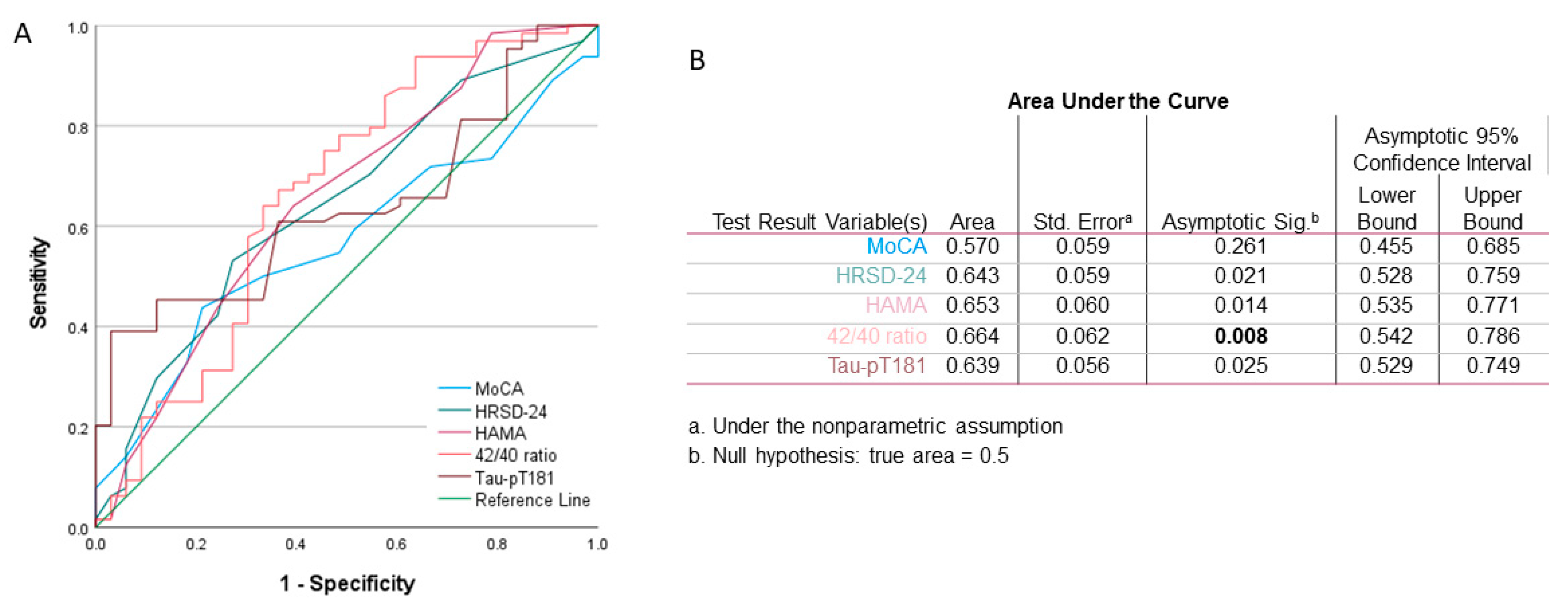
3.4. Blood Amyloid-β42/40 Ratio Is a Predictive Factor for Sleep Quality in MCI Patients

Finally, we determined if the neuropsychological score, blood Aβ42/40 ratio, and Tau-pT181 protein level had any predictive value as a risk factor for sleep quality. The ROC analysis included the prior-treatment values from all 97 patients. Sleep quality was set as the dependent variate (PSQI < 5). As shown in Figure 6, Aβ42/40 ratio had the highest significance as a predicting factor over HAMA and HRSD-24 scores and Tau-pT181 protein level. These data suggest that Aβ42/40 ratio is an important factor associated with sleep disorder in MCI patients due to Alzheimer’s disease.

4. Discussion

In this study, our main findings are listed below: (1) a very strong and negative correlation was discovered between education level and MCI onset age; (2) a strong correlation was identified among sleep quality (PSQI score), depression (HRSD-24 score), anxiety (HAMA score), blood Aβ42/40 ratio and Tau-pT181 levels; (3) patients with sleep disorder-breathing exhibited a worse HRSD-24 score and higher levels of blood Aβ42/40 ratio and Tau-pT181 levels; (4) sleep treatment for sleep-disordered breathing improved all neuropsychological scores, Aβ42/40 ratio, and Tau-pT181 levels; (5) a complete recovery of sleep quality is critical to fully improve the MoCA and HRSD-24 score and to reduce the Aβ42/40 ratio; and (6) blood Aβ42/40 ratio showed the highest significance in predicting sleep quality. These data suggest that sleep treatment improved neuropsychological symptoms and reduced blood Aβ42/40 ratio and Tau-pT181 protein levels in MCI patients due to Alzheimer’s disease.
Accumulating evidence has demonstrated sleep disordered-breathing as a clinical contributing factor in patients with MCI during Alzheimer’s disease development and progression [13,15]. However, very few studies reported the correlation of sleep quality (PSQI score) with neuropsychological scores that occur at any stage in Alzheimer’s disease [36]. This study assessed patient sleep quality with the PSQI and neuropsychological scores. Our results revealed that PSQI scores correlated significantly with HRSD-24 and HAMA scores, which were supported by a cross-sectional study showing an inverse correlation between sleep length and anxiety symptoms in Alzheimer’s disease patients [37] and by a recent study showing a close correlation between PSQI scores and depression in Alzheimer’s disease patients [38].
So far, there is a paucity of literature about the correlation of sleep quality (PSQI scores) with MoCA, GDS, and CDR scores. However, a linear correlation (coefficient of multiple correlations at 0.307–0.34) between sleep quality and GDS score was reported in Alzheimer’s disease patients with moderate to severe dementia (CDR 2–3) [39], indicating a possible connection of sleep disturbance with global deterioration scale at a late stage of AD patients. Also, lower MoCA scores were found in patients with obstructive sleep apnea-hypopnea syndrome (OSAHS) than non-insomnia patients [40], indicating a potential effect of sleep disturbance on cognitive impairment in patients without Alzheimer’s disease. Meanwhile, our results showed that treating SDB patients with continuous positive pressure ventilation significantly improved the MoCA, HRSD-24 and HAMA scores. Significantly, our results are supported by a previous report derived from a randomized clinical trial in patients with Alzheimer’s disease [22]. Therefore, sleep improvement might be able to slow down the process of Alzheimer’s disease (15), although further investigation is warranted to determine the clinical significance of sleep disordered-breathing in different phases of Alzheimer’s disease.
Aberrant aggregation of phosphorylated Tau protein is one of the major pathogenic factors in the development and progression of Alzheimer’s disease [3,8]. Also, excessive shedding and deposition of amyloid peptides have been considered a critical factor in Alzheimer’s disease [3,14]. Studies in mouse models of Alzheimer’s disease and humans have convincingly demonstrated a tight association between sleep disorders and Alzheimer’s disease in terms of Tau proteins [14,41]. Increased cerebrospinal fluid and blood levels of Amyloid-β peptide and phosphorylated Tau protein was reported in patients with sleep disturbances, including SDB [15], and have been considered biomarkers for disease progression [42,43,44]. A recent meta-analysis indicated that blood levels of Aβ42/40 ratio and Tau-pT181 protein strongly predicted the Aβ-PET status in patients [45] and that blood Aβ42/40 ratio was even recommended as a cost-effective marker for early AD pathological screening [46]. Our data also showed a strong correlation between PSQI scores and blood Aβ42/40 ratio and Tau-pT181 levels before and after sleep intervention in this study. Interestingly, we also found that Aβ42/40 ratio and Tau-pT181 levels correlated with HRSD-24 and HAMA scores. Although it is hard to postulate their causative relationship among these correlations, they are the first clue for further investigation to determine the correlation of Aβ42/40 ratio and Tau-pT181 level with cognition/dementia or depression/anxiety during Alzheimer’s disease progression.
For Alzheimer’s disease, a fundamental challenge is determining if sleep intervention improves neuropsychological status and reduces the Tau protein burden in patients [24]. Our data demonstrated that sleep intervention resulted in a significant reduction of blood Tau-pT181 levels. More specifically, patients with or without a full recovery achieved a substantial decrease in Tau-pT181 levels. In contrast to the Tau-pT181 decline, Aβ42/40 ratio was not significantly reduced in unrecovered patients, indicating that a complete recovery is critical in lowering the Aβ42/40 ratio.
Our data are supported by other clinical studies showing a 50% increase of Tau-pT181 proteins in human cerebrospinal fluid after sleep deprivation [18]. These results indicate a potential causative relationship between sleep disturbance and accumulation of Tau-pT181 in Alzheimer’s disease. Aberrant accumulation of Tau protein occurs due to an imbalanced production and clearance process reported recently [14,24]. A high-quality sleep-wake cycle is vital for successfully clearing these pathogenic molecules [21,23,47]. Therefore, it is plausible that sleep intervention at the preclinical stage of Alzheimer’s disease has a strong potential to prevent or slow down disease progression [48].
There were a few limitations in our study. First, we utilized the most economical but reliable blood biomarkers, Aβ42/40 ratio and Tau-pT181 level [42,45,46]. However, more precise and accurate methods are available in the field, such as HPLC measurement in CSF or blood specimens [49] and Florbetapir (18F)-positron emission tomography (PET) [50]. With advanced imaging technologies like functional MRI and 18F (or 11C-PIB)-PET, more precise brain tissue changes after sleep treatment will be clearly understood for mechanistic analysis. Second, we used the subjective assessments of sleep quality with PSQI and neuropsychological evaluation system of MoCA, GDS, CDR, HRSD-24, HAMA, which have been widely used in the field as standard tools in clinical practice. Although the self-assessment system by older patients might not reflect their actual sleep quality compared to a subjective measurement like polysomnography [51], the alterations of PSQI scores between prior- and post-treatment were analyzed using a paired t-test (longitudinal self-comparison) so that the derivations among individuals were minimized. It is plausible that objective measurements with advanced technology for sleep monitoring will significantly strengthen our conclusion. Third, 11 cases did not fully recover after a 6-month treatment. We are still monitoring these cases for a more extended intervention and follow-up until a possible complete recovery within two years.

5. Conclusions

This is a longitudinal study with 97 cases of mild–moderate cognitive impairment patients due to Alzheimer’s disease. Of these, 64 subjects were treated due to sleep-disordered breathing. The sleep quality scores were significantly correlated with depression and anxiety scores, blood Aβ42/40 ratio, and Tau-pT181 protein levels. Sleep treatment significantly improved sleep quality and enhanced cognition, relieving anxiety, and reduced blood Aβ42/40 ratio and Tau-pT181 protein levels. A full recovery of sleep quality was critical to improving cognition and depression status. Our data provided valuable evidence that sleep intervention might be feasible to improve neuropsychological symptoms and reduce disease progression when implicated in the early phase of Alzheimer’s disease.

Author Contributions

H.H. designed the study and acquired funding as the principal investigator of the project. H.H., M.L., M.Z., H.C., X.M., Q.C. and J.P. were involved in patient recruitment, clinical examination, the eligibility evaluation, the consent form, and the questionnaire assessment. J.Q., J.P. and M.Z. collected blood specimens and conducted ELISA analyses. H.H. and B.L. performed the statistical analysis and data interpretation. H.H., T.L. and B.L. drafted the manuscript, including data tables and graphs. All authors have read and agreed to the published version of the manuscript.

Funding

This clinical project was supported by a grant from Hubei Provincial Health Commission (Grant number WJ2018H182). The funder of this project had no role in the study design, data collection and analysis, result interpretation, and report preparation. All authors had full access to the data obtained from the study, and the corresponding authors had the final responsibility for the decision of submission and publication.

Institutional Review Board Statement

The study protocol was reviewed and approved by the Ethics Committee of the Jiangshan Oilfield General Hospital (study ethical code number 2017016, approval date 26 February 2017).

Informed Consent Statement

Informed consent was obtained from all subjects involved in the study.

Acknowledgments

We are very grateful for the technical support from the Hubei Clinical Research Center for Dementia & Cognitive Impairment. The staff members at the Department of Geriatrics in Jianghan Oilfield General Hospital provided technical assistance in blood sample collection and patient care. Jianghua He and Jinxiang Hu from the Department of Biostatistics & Data Science at the University of Kansas Medical Center provided valuable consultation on a statistical analysis of the clinical data.

Conflicts of Interest

The authors declare no conflict of interest. The funders had no role in the design of the study; in the collection, analyses, or interpretation of data; in the writing of the manuscript, or in the decision to publish the results.

References

  1. Kivipelto, M.; Mangialasche, F.; Snyder, H.M.; Allegri, R.; Andriey, S.; Arai, H.; Baker, L.; Belleville, S.; Brodaty, H.; Brucki, S.M.; et al. World-Wide FINGERS Network: A global approach to risk reduction and prevention of dementia. Alzheimers Dement 2020, 16, 1078–1094. [Google Scholar] [CrossRef]
  2. Alzheimer’s Association Report. 2020 Alzheimer’s disease facts and figures. Alzheimers Dement 2020, 16, 391–460. [Google Scholar] [CrossRef]
  3. Polanco, J.C.; Li, C.; Bodea, L.G.; Martinez-Marmol, R.; Meunier, F.A.; Gotz, J. Amyloid-beta and tau complexity—Towards improved biomarkers and targeted therapies. Nat. Rev. Neurol. 2018, 14, 22–39. [Google Scholar] [CrossRef] [PubMed]
  4. Gordon, B.A.; Blazey, T.M.; Su, Y.; Hari-Raj, A.; Dincer, A.; Flores, S.; Christensen, J.; McDade, E.; Wang, G.; Xiong, C.; et al. Spatial patterns of neuroimaging biomarker change in individuals from families with autosomal dominant Alzheimer’s disease: A longitudinal study. Lancet Neurol. 2018, 17, 241–250. [Google Scholar] [CrossRef] [Green Version]
  5. De Strooper, B.; Karran, E. The Cellular Phase of Alzheimer’s Disease. Cell 2016, 164, 603–615. [Google Scholar] [CrossRef] [Green Version]
  6. Selkoe, D.J.; Hardy, J. The amyloid hypothesis of Alzheimer’s disease at 25 years. EMBO Mol. Med. 2016, 8, 595–608. [Google Scholar] [CrossRef]
  7. D’Abramo, C.; D’Adamio, L.; Giliberto, L. Significance of Blood and Cerebrospinal Fluid Biomarkers for Alzheimer’s Disease: Sensitivity, Specificity and Potential for Clinical Use. J. Pers. Med. 2020, 10, 116. [Google Scholar] [CrossRef] [PubMed]
  8. Gallardo, G.; Holtzman, D.M. Amyloid-beta and Tau at the Crossroads of Alzheimer’s Disease. Adv. Exp. Med. Biol. 2019, 1184, 187–203. [Google Scholar] [CrossRef] [PubMed]
  9. Diaz-Roman, M.; Pulopulos, M.M.; Baquero, M.; Salvador, A.; Cuevas, A.; Ferrer, I.; Ciopat, O.; Gomez, E. Obstructive sleep apnea and Alzheimer’s disease-related cerebrospinal fluid biomarkers in mild cognitive impairment. Sleep 2020, 44, zsaa133. [Google Scholar] [CrossRef]
  10. Hefti, M.M.; Kim, S.; Bell, A.J.; Betters, R.K.; Fiock, K.L.; Iida, M.A.; Smalley, M.E.; Farrell, K.; Fowkes, M.E.; Crary, J.F. Tau Phosphorylation and Aggregation in the Developing Human Brain. J. Neuropathol. Exp. Neurol. 2019, 78, 930–938. [Google Scholar] [CrossRef]
  11. Olsson, B.; Lautner, R.; Andreasson, U.; Ohrfelt, A.; Portelius, E.; Bjerke, M.; Holtta, M.; Rosen, C.; Olsson, C.; Strobel, G.; et al. CSF and blood biomarkers for the diagnosis of Alzheimer’s disease: A systematic review and meta-analysis. Lancet Neurol. 2016, 15, 673–684. [Google Scholar] [CrossRef]
  12. Borges, C.R.; Poyares, D.; Piovezan, R.; Nitrini, R.; Brucki, S. Alzheimer’s disease and sleep disturbances: A review. Arq. Neuropsiquiatr. 2019, 77, 815–824. [Google Scholar] [CrossRef] [PubMed] [Green Version]
  13. Bubu, O.M.; Brannick, M.; Mortimer, J.; Umasabor-Bubu, O.; Sebastiao, Y.V.; Wen, Y.; Schwartz, S.; Borenstein, A.R.; Wu, Y.; Morgan, D.; et al. Sleep, Cognitive impairment, and Alzheimer’s disease: A Systematic Review and Meta-Analysis. Sleep 2017, 40, piizsw032. [Google Scholar] [CrossRef]
  14. Wang, C.; Holtzman, D.M. Bidirectional relationship between sleep and Alzheimer’s disease: Role of amyloid, tau, and other factors. Neuropsychopharmacology 2020, 45, 104–120. [Google Scholar] [CrossRef]
  15. Ulland, T.K.; Ewald, A.C.; Knutson, A.O.; Marino, K.M.; Smith, S.M.C.; Watters, J.J. Alzheimer’s Disease, Sleep-disordered breathing, and Microglia: Puzzling out a Common Link. Cells 2021, 10, 2907. [Google Scholar] [CrossRef] [PubMed]
  16. Bliwise, D.L. Sleep disorders in Alzheimer’s disease and other dementias. Clin. Cornerstone 2004, 6, S16–S28. [Google Scholar] [CrossRef]
  17. Bubu, O.M.; Umasabor-Bubu, O.Q.; Turner, A.D.; Parekh, A.; Mullins, A.E.; Kam, K.; Birckbichler, M.K.; Mukhtar, F.; Mbah, A.K.; Williams, N.J.; et al. Self-reported obstructive sleep apnea, amyloid and tau burden, and Alzheimer’s disease time-dependent progression. Alzheimers Dement 2020, 17, 226–245. [Google Scholar] [CrossRef] [PubMed]
  18. Barthelemy, N.R.; Liu, H.; Lu, W.; Kotzbauer, P.T.; Bateman, R.J.; Lucey, B.P. Sleep Deprivation Affects Tau Phosphorylation in Human Cerebrospinal Fluid. Ann. Neurol. 2020, 87, 700–709. [Google Scholar] [CrossRef]
  19. Blattner, M.S.; Panigrahi, S.K.; Toedebusch, C.D.; Hicks, T.J.; McLeland, J.S.; Banks, I.R.; Schaibley, C.; Ovod, V.; Mawuenyega, K.G.; Bateman, R.J.; et al. Increased Cerebrospinal Fluid Amyloid-beta During Sleep Deprivation in Healthy Middle-Aged Adults Is Not Due to Stress or Circadian Disruption. J. Alzheimers Dis. 2020, 75, 471–482. [Google Scholar] [CrossRef]
  20. Lucey, B.P.; Hicks, T.J.; McLeland, J.S.; Toedebusch, C.D.; Boyd, J.; Elbert, D.L.; Patterson, B.W.; Baty, J.; Morris, J.C.; Ovod, V.; et al. Effect of sleep on overnight cerebrospinal fluid amyloid beta kinetics. Ann. Neurol. 2018, 83, 197–204. [Google Scholar] [CrossRef]
  21. Holth, J.K.; Fritschi, S.K.; Wang, C.; Pedersen, N.P.; Cirrito, J.R.; Mahan, T.E.; Finn, M.B.; Manis, M.; Geerling, J.C.; Fuller, P.M.; et al. The sleep-wake cycle regulates brain interstitial fluid tau in mice and CSF tau in humans. Science 2019, 363, 880–884. [Google Scholar] [CrossRef]
  22. Ancoli-Israel, S.; Palmer, B.W.; Cooke, J.R.; Corey-Bloom, J.; Fiorentino, L.; Natarajan, L.; Liu, L.; Ayalon, L.; He, F.; Loredo, J.S. Cognitive effects of treating obstructive sleep apnea in Alzheimer’s disease: A randomized controlled study. J. Am. Geriatr. Soc. 2008, 56, 2076–2081. [Google Scholar] [CrossRef] [Green Version]
  23. Xie, L.; Kang, H.; Xu, Q.; Chen, M.J.; Liao, Y.; Thiyagarajan, M.; O’Donnell, J.; Christensen, D.J.; Nicholson, C.; Iliff, J.J.; et al. Sleep drives metabolite clearance from the adult brain. Science 2013, 342, 373–377. [Google Scholar] [CrossRef] [Green Version]
  24. Lucey, B.P. It’s complicated: The relationship between sleep and Alzheimer’s disease in humans. Neurobiol. Dis. 2020, 144, 105031. [Google Scholar] [CrossRef] [PubMed]
  25. McKhann, G.M.; Knopman, D.S.; Chertkow, H.; Hyman, B.T.; Jack, C.R., Jr.; Kawas, C.H.; Klunk, W.E.; Koroshetz, W.J.; Manly, J.J.; Mayeux, R.; et al. The diagnosis of dementia due to Alzheimer’s disease: Recommendations from the National Institute on Aging-Alzheimer’s Association workgroups on diagnostic guidelines for Alzheimer’s disease. Alzheimers Dement 2011, 7, 263–269. [Google Scholar] [CrossRef] [Green Version]
  26. Sateia, M.J. International classification of sleep disorders-third edition: Highlights and modifications. Chest 2014, 146, 1387–1394. [Google Scholar] [CrossRef]
  27. Yesavage, J.A.; Friedman, L.; Ancoli-Israel, S.; Bliwise, D.; Singer, C.; Vitiello, M.V.; Monjan, A.A.; Lebowitz, B. Development of diagnostic criteria for defining sleep disturbance in Alzheimer’s disease. J. Geriatr. Psychiatry Neurol. 2003, 16, 131–139. [Google Scholar] [CrossRef]
  28. General Assembly of the World Medical Association. World Medical Association Declaration of Helsinki: Ethical principles for medical research involving human subjects. J. Am. Coll. Dent. 2014, 81, 14–18. [Google Scholar]
  29. Buysse, D.J.; Reynolds, C.F., 3rd; Monk, T.H.; Berman, S.R.; Kupfer, D.J. The Pittsburgh Sleep Quality Index: A new instrument for psychiatric practice and research. Psychiatry Res. 1989, 28, 193–213. [Google Scholar] [CrossRef]
  30. Tsai, P.S.; Wang, S.Y.; Wang, M.Y.; Su, C.T.; Yang, T.T.; Huang, C.J.; Fang, S.C. Psychometric evaluation of the Chinese version of the Pittsburgh Sleep Quality Index (CPSQI) in primary insomnia and control subjects. Qual. Life Res. 2005, 14, 1943–1952. [Google Scholar] [CrossRef]
  31. Chen, X.; Zhang, R.; Xiao, Y.; Dong, J.; Niu, X.; Kong, W. Reliability and Validity of the Beijing Version of the Montreal Cognitive Assessment in the Evaluation of Cognitive Function of Adult Patients with OSAHS. PLoS ONE 2015, 10, e0132361. [Google Scholar] [CrossRef]
  32. Morris, J.C. The Clinical Dementia Rating (CDR): Current version and scoring rules. Neurology 1993, 43, 2412–2414. [Google Scholar] [CrossRef]
  33. Reisberg, B.; Ferris, S.H.; de Leon, M.J.; Crook, T. The Global Deterioration Scale for assessment of primary degenerative dementia. Am. J. Psychiatry 1982, 139, 1136–1139. [Google Scholar] [CrossRef] [Green Version]
  34. Hamilton, M. Rating depressive patients. J. Clin. Psychiatry 1980, 41, 21–24. [Google Scholar]
  35. Hamilton, M. Development of a rating scale for primary depressive illness. Br. J. Soc. Clin. Psychol. 1967, 6, 278–296. [Google Scholar] [CrossRef] [PubMed]
  36. Magierski, R.; Sobow, T.; Schwertner, E.; Religa, D. Pharmacotherapy of Behavioral and Psychological Symptoms of Dementia: State of the Art and Future Progress. Front. Pharmacol. 2020, 11, 1168. [Google Scholar] [CrossRef]
  37. De Oliveira, F.F.; Bertolucci, P.H.; Chen, E.S.; Smith Mde, A. Assessment of sleep satisfaction in patients with dementia due to Alzheimer’s disease. J. Clin. Neurosci. 2014, 21, 2112–2117. [Google Scholar] [CrossRef]
  38. Zhou, G.; Liu, S.; Yu, X.; Zhao, X.; Ma, L.; Shan, P. High prevalence of sleep disorders and behavioral and psychological symptoms of dementia in late-onset Alzheimer disease: A study in Eastern China. Medicine 2019, 98, e18405. [Google Scholar] [CrossRef] [PubMed]
  39. Stella, F.; Laks, J.; Govone, J.S.; de Medeiros, K.; Forlenza, O.V. Association of neuropsychiatric syndromes with global clinical deterioration in Alzheimer’s disease patients. Int. Psychogeriatr. 2016, 28, 779–786. [Google Scholar] [CrossRef] [Green Version]
  40. Guo, H.; Wei, M.; Ding, W. Changes in Cognitive Function in Patients with Primary Insomnia. Shanghai Arch. Psychiatry 2017, 29, 137–145. [Google Scholar] [CrossRef] [PubMed]
  41. Mullins, A.E.; Kam, K.; Parekh, A.; Bubu, O.M.; Osorio, R.S.; Varga, A.W. Obstructive Sleep Apnea and Its Treatment in Aging: Effects on Alzheimer’s disease Biomarkers, Cognition, Brain Structure and Neurophysiology. Neurobiol. Dis. 2020, 145, 105054. [Google Scholar] [CrossRef] [PubMed]
  42. Karikari, T.K.; Pascoal, T.A.; Ashton, N.J.; Janelidze, S.; Benedet, A.L.; Rodriguez, J.L.; Chamoun, M.; Savard, M.; Kang, M.S.; Therriault, J.; et al. Blood phosphorylated tau 181 as a biomarker for Alzheimer’s disease: A diagnostic performance and prediction modelling study using data from four prospective cohorts. Lancet Neurol. 2020, 19, 422–433. [Google Scholar] [CrossRef]
  43. Thijssen, E.H.; La Joie, R.; Wolf, A.; Strom, A.; Wang, P.; Iaccarino, L.; Bourakova, V.; Cobigo, Y.; Heuer, H.; Spina, S.; et al. Diagnostic value of plasma phosphorylated tau181 in Alzheimer’s disease and frontotemporal lobar degeneration. Nat. Med. 2020, 26, 387–397. [Google Scholar] [CrossRef]
  44. Ashton, N.J.; Leuzy, A.; Karikari, T.K.; Mattsson-Carlgren, N.; Dodich, A.; Boccardi, M.; Corre, J.; Drzezga, A.; Nordberg, A.; Ossenkoppele, R.; et al. The validation status of blood biomarkers of amyloid and phospho-tau assessed with the 5-phase development framework for AD biomarkers. Eur. J. Nucl. Med. Mol. Imaging 2021, 48, 2140–2156. [Google Scholar] [CrossRef] [PubMed]
  45. Cianflone, A.; Coppola, L.; Mirabelli, P.; Salvatore, M. Predictive Accuracy of Blood-Derived Biomarkers for Amyloid-beta Brain Deposition Along with the Alzheimer’s Disease Continuum: A Systematic Review. J. Alzheimers Dis. 2021, 84, 393–407. [Google Scholar] [CrossRef]
  46. Stockmann, J.; Verberk, I.M.W.; Timmesfeld, N.; Denz, R.; Budde, B.; Lange-Leifhelm, J.; Scheltens, P.; van der Flier, W.M.; Nabers, A.; Teunissen, C.E.; et al. Amyloid-beta misfolding as a plasma biomarker indicates risk for future clinical Alzheimer’s disease in individuals with subjective cognitive decline. Alzheimers Res. Ther. 2020, 12, 169. [Google Scholar] [CrossRef]
  47. Lucey, B.P.; McCullough, A.; Landsness, E.C.; Toedebusch, C.D.; McLeland, J.S.; Zaza, A.M.; Fagan, A.M.; McCue, L.; Xiong, C.; Morris, J.C.; et al. Reduced non-rapid eye movement sleep is associated with tau pathology in early Alzheimer’s disease. Sci. Transl. Med. 2019, 11, eaau6550. [Google Scholar] [CrossRef]
  48. Ju, Y.E.; McLeland, J.S.; Toedebusch, C.D.; Xiong, C.; Fagan, A.M.; Duntley, S.P.; Morris, J.C.; Holtzman, D.M. Sleep quality and preclinical Alzheimer disease. JAMA Neurol. 2013, 70, 587–593. [Google Scholar] [CrossRef]
  49. Nakamura, A.; Kaneko, N.; Villemagne, V.L.; Kato, T.; Doecke, J.; Dore, V.; Fowler, C.; Li, Q.X.; Martins, R.; Rowe, C.; et al. High performance plasma amyloid-beta biomarkers for Alzheimer’s disease. Nature 2018, 554, 249–254. [Google Scholar] [CrossRef]
  50. Clark, C.M.; Schneider, J.A.; Bedell, B.J.; Beach, T.G.; Bilker, W.B.; Mintun, M.A.; Pontecorvo, M.J.; Hefti, F.; Carpenter, A.P.; Flitter, M.L.; et al. Use of florbetapir-PET for imaging beta-amyloid pathology. JAMA 2011, 305, 275–283. [Google Scholar] [CrossRef] [PubMed] [Green Version]
  51. O’Donnell, D.; Silva, E.J.; Munch, M.; Ronda, J.M.; Wang, W.; Duffy, J.F. Comparison of subjective and objective assessments of sleep in healthy older subjects without sleep complaints. J. Sleep Res. 2009, 18, 254–263. [Google Scholar] [CrossRef] [PubMed] [Green Version]
Figure 1. A schematic illustration of the study design and protocol. A total of 97 patients participated; 64 patients were diagnosed with sleep disorders and were subjected to sleep interventions in addition to donepezil treatment. The other 33 cases without sleep disorders received donepezil only. After a 6-month treatment, patients were re-assessed and divided into recovered or unrecovered subgroups groups based on the Pittsburgh Sleep Quality Index (PSQI) scores. SD: sleep-disorder.
Medicina 57 01366 g001
Figure 2. The differences of neuropsychological scores and blood biomarkers between SD cases and non-SD cases, including MOCA score (A), HRSD-24 score (B), HAMA score (C), Aβ42/40 ratio (D), and Tau-pT181 level (E). The asterisk indicates a significant difference between the groups (* p < 0.05, ** p < 0.01, Student t-test).
Medicina 57 01366 g002
Figure 3. Sleep treatment improves neuropsychological scores, blood Aβ42/40 ratio, and Tau-pT181 protein level. Data were presented as case-controlled pairs of prior- and post-treatment values. The p values were derived from a pair-wise t-test.
Medicina 57 01366 g003
Figure 4. Different improvements in recovered (panel A) and unrecovered (panel B) subgroups. The neuropsychological scores, blood Aβ42/40 ratio, and Tau-pT181 protein level were presented as case-controlled pairs of prior- and post-treatment values. The p values were derived from a pair-wise t-test.
Medicina 57 01366 g004
Figure 5. Blood Aβ42/40 ratio (panel A) and Tau-pT181 protein levels (panel B) were reduced in recovered subgroups. Group comparison was conducted using Student t-test, and the asterisks indicate a significant difference between the subgroups as indicated. * p < 0.05; ** p < 0.01; **** p < 0.0001; ns, not significant.
Medicina 57 01366 g005
Figure 6. Receiver Operator Characteristic (ROC) analysis indicates that the Aβ42/40 ratio has the highest potential for predicting sleep quality. (A) The ROC curves for the variables of Aβ42/40 ratio, Tau-pT181, MoCA, HRSD-24, and HAMA scores. (B) Summary of the statistical values derived from the ROC curve analysis.
Medicina 57 01366 g006
Table 1. Patient Biometrics Parameters.
All CasesSD Cases with Treatment
Non-SD CasesSD Casesp-ValueUn-RecoveredRecoveredp-Value
Case number (%)33 (34%)64 (66%) 11 (17.2%)53 (82.8%)
Age (year)73 (66–81)73 (63–81)n.s.73 (67–78)73 (63–81)n.s.
Onset age (year)69 (63–79)71 (61–79)n.s.69 (63–79)71 (61–79)n.s.
Sex (Male/Female)M18/F15M30/F34n.s.M6/F5M24/F29n.s.
Disease length (month)32 (19–45)32 (16–59)n.s.32 (19–45)32 (16–59)n.s.
Secondary education4 (2–11)4 (0–11)n.s.4 (2–11)4 (0–11)n.s.
Body Mass Index23.7 (16.7–29.2)23.6 (15.48–33.02)n.s.23.7 (16.7–29.2)24.5 (15.48–33.02)n.s.
Note: The values for clinical assessments were shown as the Median (range). The p-value was derived from Student t-test analysis between non-recovered and recovered groups except Χ2 test for sex. n.s., not significant. SD, sleep disorder.
Table 2. Pearson Correlation Coefficient.
Correlation PairPearson rp-Value
Education vs. Onset age−0.5738.53 × 10−10
PSQI vs. HAMA0.4671.42 × 10−6
PSQI vs. HRSD-240.3530.0004
PSQI vs. Aβ42/40 ratio0.3480.0005
PSQI vs. Tau-pT1810.4241.52 × 10−5
HRSD-24 vs. HAMA0.4191.93 × 10−5
HRSD-24 vs. Aβ42/40 ratio0.5061.24 × 10−7
HRSD-24 vs. Tau-pT1810.6431.30 × 10−12
HAMA vs. Aβ42/40 ratio0.5061.28 × 10−7
HAMA vs. Tau-pT1810.5553.61 × 10−9
42/40 ratio vs. Tau-pT1810.5882.34 × 10−10
Note: 24-h Hamilton Rating Scale for Depression: HRSD-24 and Hamil-ton Anxiety Rating Scale: HAMA, Amyloid β peptide-42/40: Aβ42/40.
Table 3. Pearson Correlation Coefficient.
Correlation PairPearson rp-Value
Education vs. Onset age−0.7095.53 × 10−11
PSQI vs. HAMA0.4884.22 × 10−4
PSQI vs. HRSD-240.2680.032
PSQI vs. Aβ42/40 ratio0.3110.012
PSQI vs. Tau-pT1810.3280.008
MoCa vs. GDS−0.4090.0008
GDS vs. Aβ42/40 ratio−0.3160.011
HRSD-24 vs. HAMA0.4070.0008
HRSD-24 vs. Aβ42/40 ratio0.5804.96 × 10−7
HRSD-24 vs. Tau-pT1810.7095.13 × 10−11
HAMA vs. Aβ42/40 ratio0.5267.98 × 10−6
HAMA vs. Tau-pT1810.6632.43 × 10−9
42/40 ratio vs. Tau-pT1810.7727.84 × 10−14
Note: Montreal Cognitive Assessment: MoCA, Geriatric Depression Scale: GDS, Clinical De-mentia Rating: CDR.
Publisher’s Note: MDPI stays neutral with regard to jurisdictional claims in published maps and institutional affiliations.

