
Volumetric Modulated Arc Therapy Capabilities for Treating Lower-Extremity Skin Affected by Several Merkel Cell Carcinoma Nodules: When Technological Advances Effectively Achieve the Palliative Therapeutic Goal while Minimising the Risk of Potential Toxicities

 ,
, 
Abstract
:1. Introduction
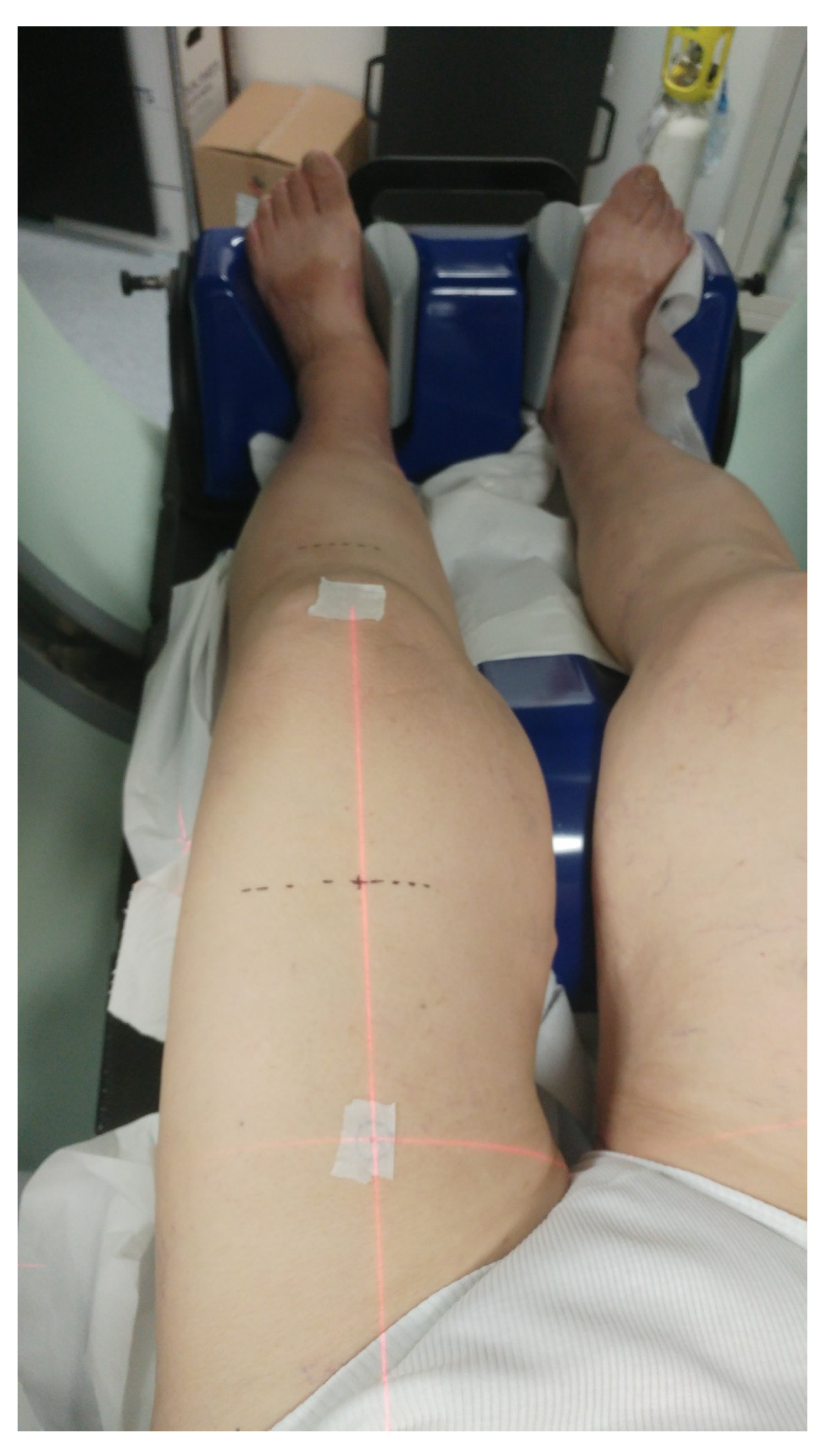
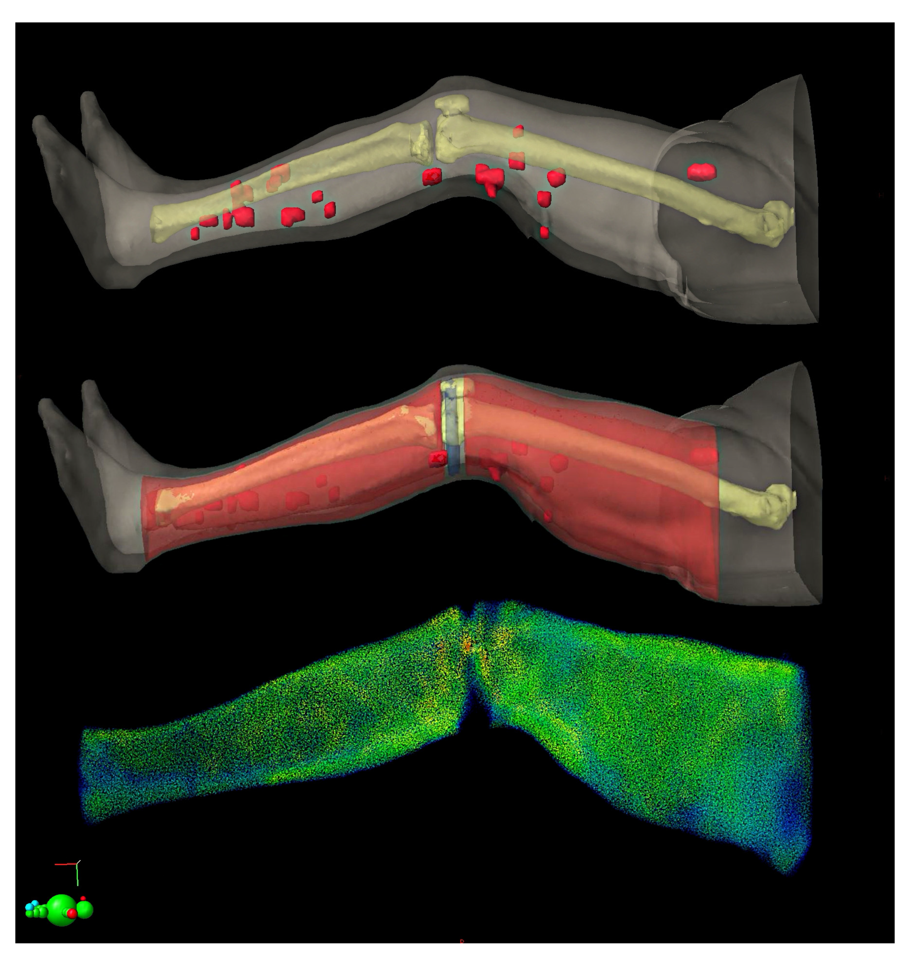
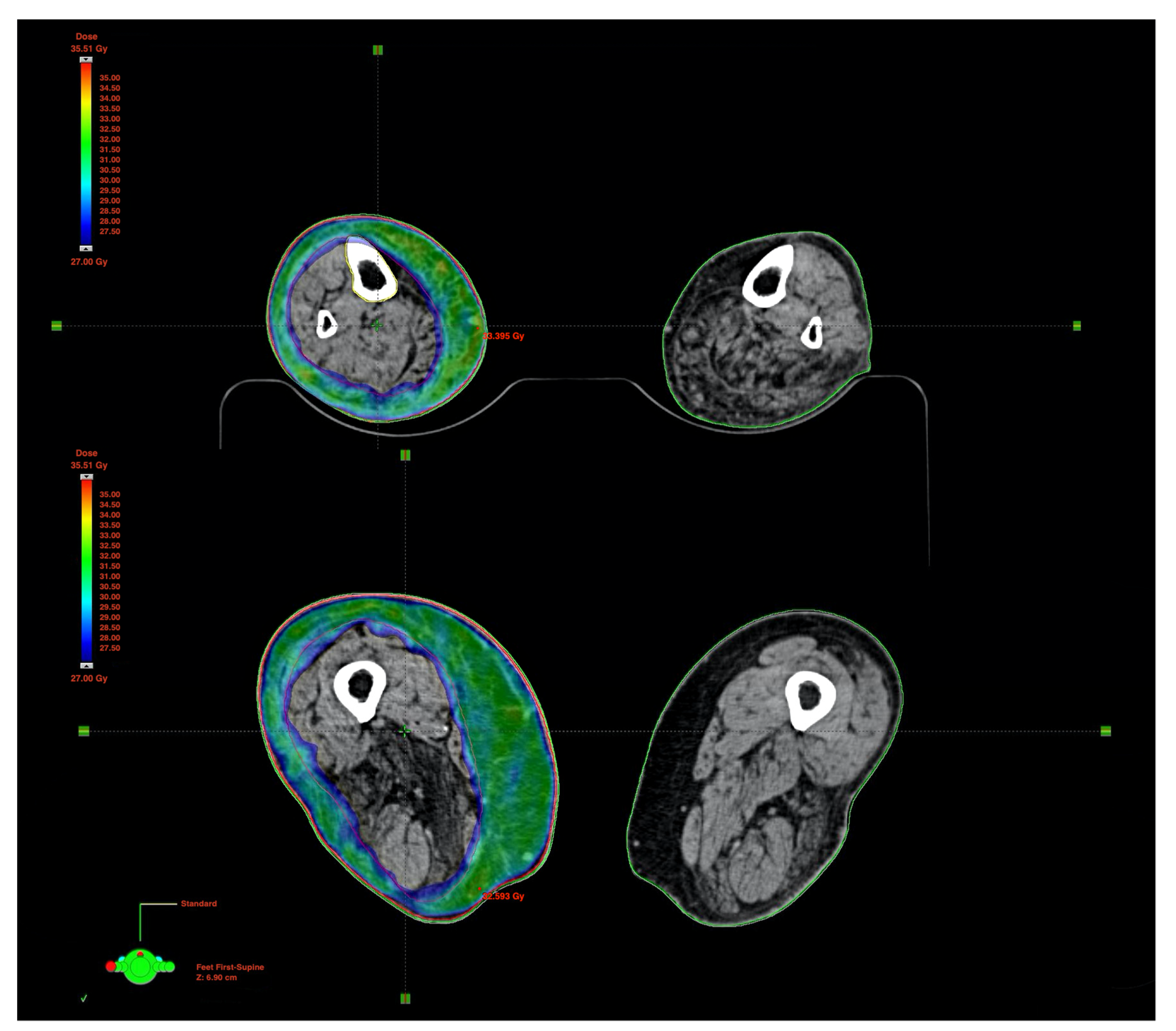
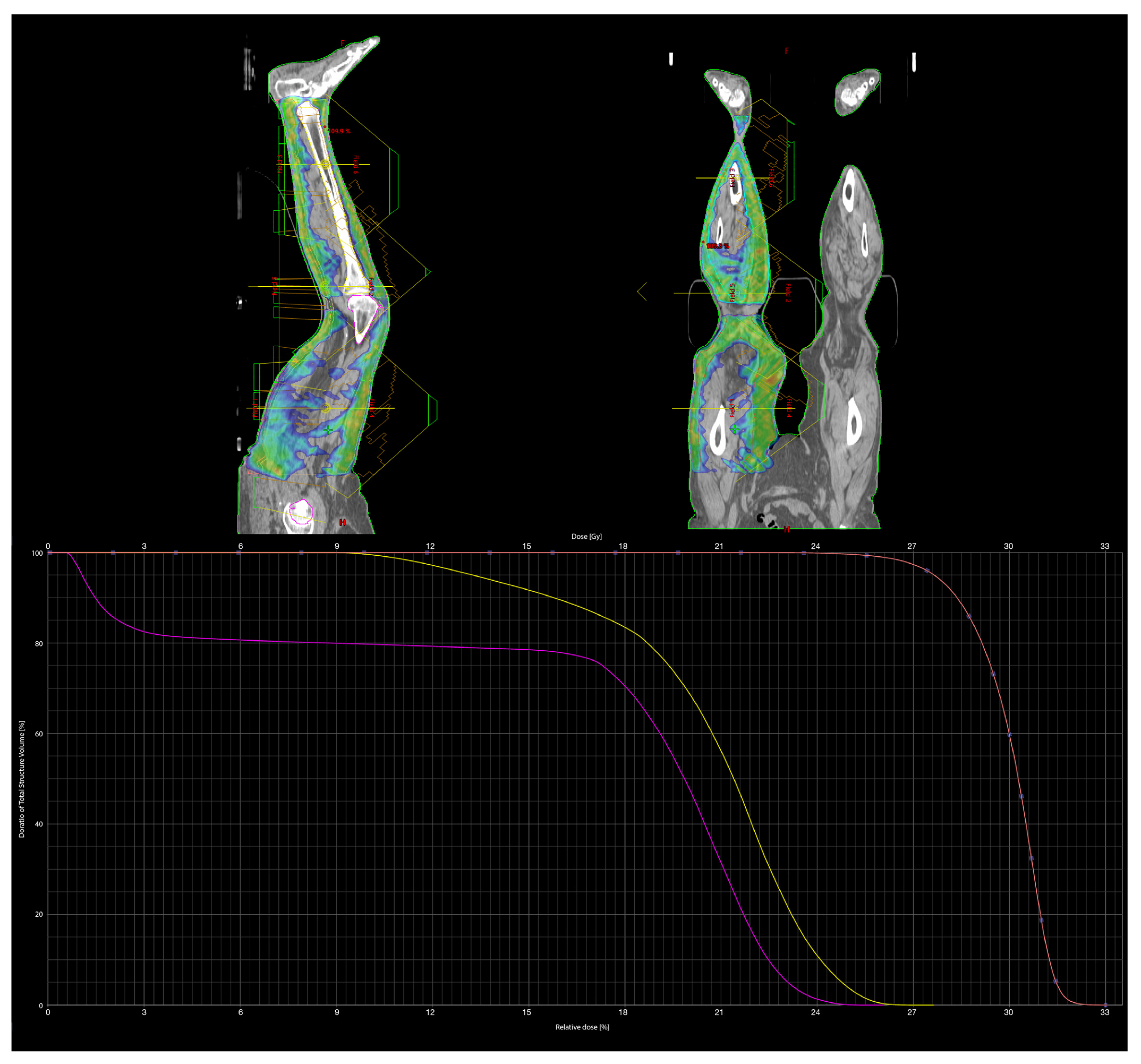
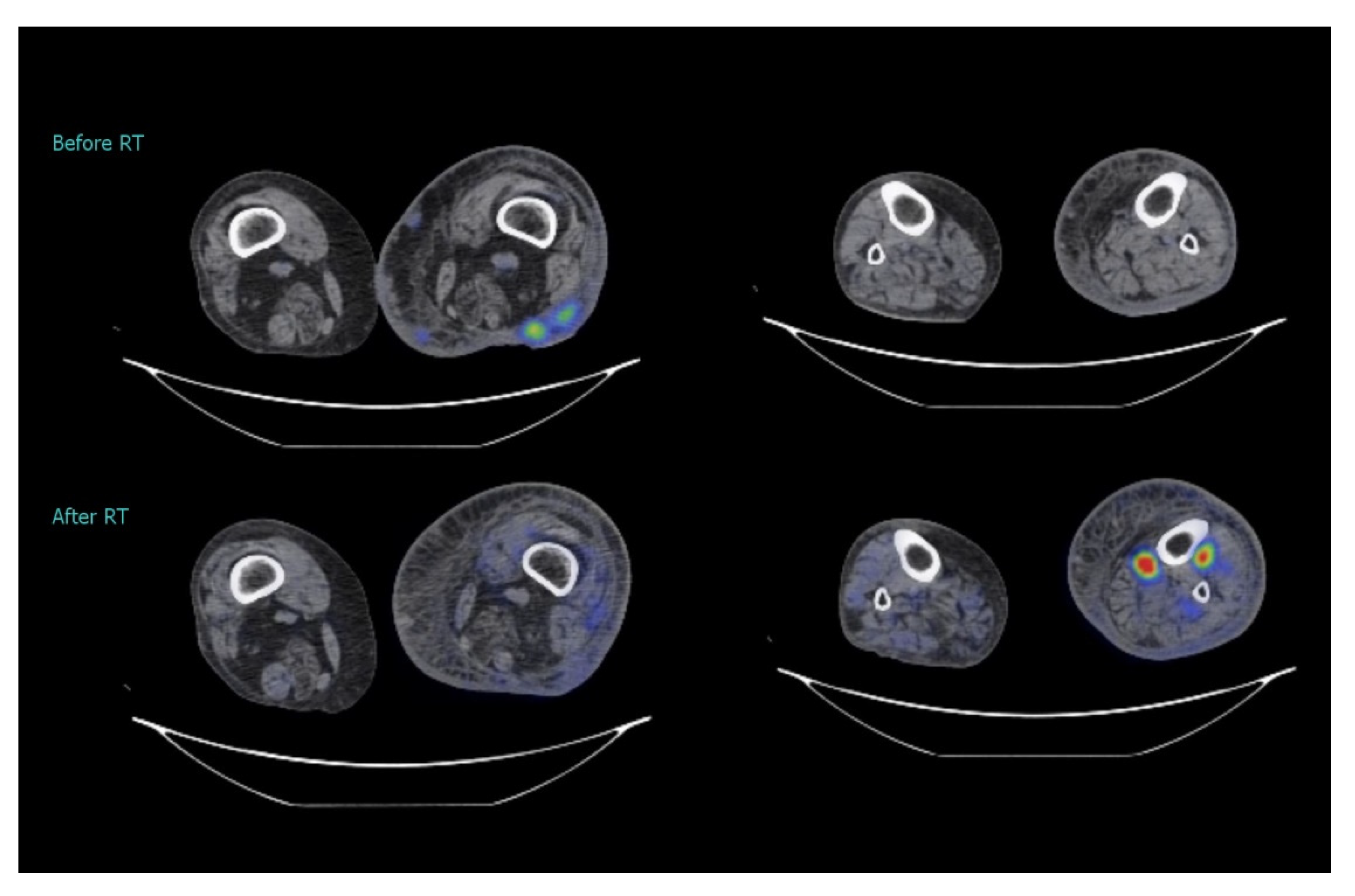
2. Case Study
3. Discussion
4. Conclusions
Additional Considerations: Weaknesses and Strengths
Author Contributions
Funding
Informed Consent Statement
Data Availability Statement
Conflicts of Interest
References
- Singh, G.K.; Sinha, A.; Mishra, P.S.; Jain, A.; Beniwal, N.S. Ulcerative Variant of Merkel Cell Carcinoma in an Immunocompetent Individual: An Unusual Presentation. Indian Dermatol. Online J. 2021, 12, 156–158. [Google Scholar] [CrossRef] [PubMed]
- Gaba, S.; Chopra, P.; Pankaj, P.; Belho, E.S.; Qadri, A.B.; Aggarwal, S. Merkel cell carcinoma—a rare cause of non-healing skin ulcer: A case report. J. Indian Med. Assoc. 2012, 110, 496–498. [Google Scholar] [PubMed]
- Shinogi, T.; Nagase, K.; Inoue, T.; Sato, K.; Onita, A.; Takamori, A.; Narisawa, Y. Merkel cell carcinoma: A systematic review of the demographic and clinical characteristics of 847 cases in Japan. J. Dermatol. 2021, 48, 1027–1034. [Google Scholar] [CrossRef] [PubMed]
- Carter, J.; Huang, H.Q.; Armer, J.; Carlson, J.W.; Lockwood, S.; Nolte, S.; Kauderer, J.; Hutson, A.; Walker, J.L.; Fleury, A.C.; et al. GOG 244—The Lymphedema and Gynecologic cancer (LeG) study: The impact of lower-extremity lymphedema on quality of life, psychological adjustment, physical disability, and function. Gynecol. Oncol. 2021, 160, 244–251. [Google Scholar] [CrossRef] [PubMed]
- Rodrigues, M.A.; Caetano, M.; Amorim, I.; Selores, M. Dermo-Hipodermites Bacterianas Agudas Não Necrotizantes: Erisipela e Celulite Infeciosa [Non-Necrotizing Acute Dermo-Hypodermal Infections: Erysipela and Infectious Cellulitis]. Acta Med. Port. 2021, 34, 217–228. [Google Scholar] [CrossRef] [PubMed]
- Barreira, J.V.; Valejo Coelho, M.M.; Ribeiro, C.; Semedo, M. Unknown primary Merkel cell carcinoma with cutaneous spread. BMJ Case Rep. 2019, 12, e224834. [Google Scholar] [CrossRef] [PubMed]
- Poulsen, M.; Round, C.; Keller, J.; Tripcony, L.; Veness, M. Factors influencing relapse-free survival in Merkel cell carcinoma of the lower limb—a review of 60 cases. Int. J. Radiat. Oncol. Biol. Phys. 2010, 76, 393–397. [Google Scholar] [CrossRef]
- Steven, N.; Lawton, P.; Poulsen, M. Merkel Cell Carcinoma—Current Controversies and Future Directions. Clin. Oncol. 2019, 31, 789–796. [Google Scholar] [CrossRef]
- Zerini, D.; Patti, F.; Spada, F.; Fazio, N.; Pisa, E.; Pennacchioli, E.; Prestianni, P.; Cambria, R.; Pepa, M.; Grana, C.M.; et al. Multidisciplinary team approach for Merkel cell carcinoma: The European Institute of Oncology experience with focus on radiotherapy. Tumori J. 2021, 107, 145–149. [Google Scholar] [CrossRef]
- Pacella, J.; Ashby, M.; Ainslie, J.; Minty, C. The role of radiotherapy in the management of primary cutaneous neuroendocrine tumors (Merkel cell or trabecular carcinoma): Experience at the Peter MacCallum Cancer Institute (Melbourne, Australia). Int. J. Radiat. Oncol. Biol. Phys. 1988, 14, 1077–1084. [Google Scholar] [CrossRef]
- Garibyan, L.; Cotter, S.E.; Hansen, J.L.; Noell, C.; Dorosario, A.; O’Farrell, D.A.; Devlin, P.M.; Wang, L.C. Palliative treatment for in-transit cutaneous metastases of Merkel cell carcinoma using surface-mold computer-optimized high-dose-rate brachytherapy. Cancer J. 2013, 19, 283–287. [Google Scholar] [CrossRef] [Green Version]
- Veness, M.; Howle, J. Patients with clinically node negative extremity Merkel cell carcinoma: The importance of identifying and treating patients with microscopic nodal metastases. Australas. J. Dermatol. 2010, 51, 274–278. [Google Scholar] [CrossRef]
- Semwal, M.K. Khan’s The Physics of Radiation Therapy. J. Med. Phys. 2020, 45, 134–135. [Google Scholar] [CrossRef]
- Fogliata, A.; Bergström, S.; Cafaro, I.; Clivio, A.; Cozzi, L.; Dipasquale, G.; Hållström, P.; Mancosu, P.; Navarria, P.; Nicolini, G.; et al. Cranio-spinal irradiation with volumetric modulated arc therapy: A multi-institutional treatment experience. Radiother. Oncol. 2011, 99, 79–85. [Google Scholar] [CrossRef]
- Maddalo, M.; Benecchi, G.; Altabella, L.; Ghetti, C.; D’Abbiero, N. Automatic feathering algorithm for VMAT craniospinal irradiation: A comprehensive comparison with other VMAT planning strategies. Med Dosim. 2021, 46, 103–110. [Google Scholar] [CrossRef]
- Executive Committee of the International Society of Lymphology. The diagnosis and treatment of peripheral lymphedema: 2020 Consensus Document of the International Society of Lymphology. Lymphology 2020, 53, 3–19. [Google Scholar] [PubMed]
- Ferini, G.; Valenti, V.; Tripoli, A.; Illari, S.I.; Molino, L.; Parisi, S.; Cacciola, A.; Lillo, S.; Giuffrida, D.; Pergolizzi, S. Lattice or Oxygen-Guided Radiotherapy: What If They Converge? Possible Future Directions in the Era of Immunotherapy. Cancers 2021, 13, 3290. [Google Scholar] [CrossRef]
- Allam, O.; Park, K.E.; Chandler, L.; Mozaffari, M.A.; Ahmad, M.; Lu, X.; Alperovich, M. The impact of radiation on lymphedema: A review of the literature. Gland. Surg. 2020, 9, 596–602. [Google Scholar] [CrossRef]
- Hayes, S.B.; Freedman, G.M.; Li, T.; Anderson, P.R.; Ross, E. Does axillary boost increase lymphedema compared with supraclavicular radiation alone after breast conservation? Int. J. Radiat. Oncol. Biol. Phys. 2008, 72, 1449–1455. [Google Scholar] [CrossRef]
- Wooden, K.K.; Hogstrom, K.R.; Blum, P.; Gastorf, R.J.; Cox, J.D. Whole-limb irradiation of the lower calf using a six-field electron technique. Med Dosim. Off. J. Am. Assoc. Med Dosim. 1996, 21, 211–218. [Google Scholar] [CrossRef]
- Soares, C.B.G.; De Araújo, I.D.; Pádua, B.J.; Vilela, J.C.S.; Souza, R.H.R.; Teixeira, L.E.M. Pathological fracture after radiotherapy: Systematic review of literature. Rev. Assoc. Med. Bras. 2019, 65, 902–908. [Google Scholar] [CrossRef]
- Ferini, G.; Tripoli, A.; Umina, V.; Borzì, G.R.; Marchese, V.A.; Illari, S.I.; Cacciola, A.; Lillo, S.; Parisi, S.; Valenti, V. Radiation Proctitis: The Potential Role of Hyaluronic Acid in the Prevention and Restoration of Any Damage to the Rectal Mucosa among Prostate Cancer Patients Submitted to Curative External Beam Radiotherapy. Gastroenterol. Insights 2021, 12, 446–455. [Google Scholar] [CrossRef]
- Mallory, M.; Pokhrel, D.; Badkul, R.; Jiang, H.; Lominska, C.; Wang, F. Volumetric modulated arc therapy treatment planning of thoracic vertebral metastases using stereotactic body radiotherapy. J. Appl. Clin. Med Phys. 2018, 19, 54–61. [Google Scholar] [CrossRef] [Green Version]
- Pontoriero, A.; Iatì, G.; Cacciola, A.; Conti, A.; Brogna, A.; Siragusa, C.; Ferini, G.; Davì, V.; Tamburella, C.; Molino, L.; et al. Stereotactic Body Radiation Therapy With Simultaneous Integrated Boost in Patients With Spinal Metastases. Technol. Cancer Res. Treat. 2020, 19, 1533033820904447. [Google Scholar] [CrossRef] [PubMed]
- Jaccard, M.; Zilli, T.; Dubouloz, A.; Escude, L.; Jorcano, S.; Linthout, N.; Bral, S.; Verbakel, W.; Bruynzeel, A.; Björkqvist, M.; et al. Urethra-Sparing Stereotactic Body Radiation Therapy for Prostate Cancer: Quality Assurance of a Randomized Phase 2 Trial. Int. J. Radiat. Oncol. Biol. Phys. 2020, 108, 1047–1054. [Google Scholar] [CrossRef]
- Vadalà, R.E.; Santacaterina, A.; Sindoni, A.; Platania, A.; Arcudi, A.; Ferini, G.; Mazzei, M.M.; Marletta, D.; Rifatto, C.; Risoleti, E.V.I.; et al. Stereotactic body radiotherapy in non-operable lung cancer patients. Clin. Transl. Oncol. Off. Publ. Fed. Span. Oncol. Soc. Natl. Cancer Inst. Mex. 2016, 18, 1158–1159. [Google Scholar] [CrossRef]
- Ferini, G.; Molino, L.; Bottalico, L.; De Lucia, P.; Garofalo, F. A small case series about safety and effectiveness of a hypofractionated electron beam radiotherapy schedule in five fractions for facial non melanoma skin cancer among frail and elderly patients. Rep. Pract. Oncol. Radiother. 2021, 26, 66–72. [Google Scholar] [CrossRef] [PubMed]
- Pontoriero, A.; Iatì, G.; Pergolizzi, S. A case report of a patient with squamous cell carcinoma of the face irradiated using a stereotactic technique. Radiat. Oncol. J. 2015, 33, 261–264. [Google Scholar] [CrossRef] [Green Version]
- Coles, A. A patient presenting with an advanced squamous cell carcinomas to the left thigh. Radiographer 2010, 57, 40–44. [Google Scholar] [CrossRef]
- Fitzgerald, E.; Miles, W.; Fenton, P.; Frantzis, J. Intensity-modulated radiation therapy to bilateral lower limb extremities concurrently: A planning case study. J. Med Radiat. Sci. 2014, 61, 210–215. [Google Scholar] [CrossRef] [PubMed]
- Servy, A.; Kramkimel, N.; Franck, N.; Park, S.; Kirova, Y. Helical tomotherapy in oncodermatology: Case report of circumferential cutaneous lymphoma treated by this optimized radiotherapy. Cancer Radiother. J. Soc. Fr. Radiother. Oncol. 2014, 18, 136–138. [Google Scholar] [CrossRef] [PubMed]
- Gunaratne, D.A.; Howle, J.R.; Veness, M.J. Merkel cell carcinoma: A case of palliative upper limb amputation in a patient with refractory in-transit metastases. Australas. J. Dermatol. 2016, 57, e53–e56. [Google Scholar] [CrossRef] [PubMed]
- Blumenthal, L.; VandenBoom, T.; Melian, E.; Peterson, A.; Hutchens, K.A. Multiple Primary Merkel Cell Carcinomas Presenting as Pruritic, Painful Lower Leg Tumors. Case Rep. Dermatol. 2015, 7, 316–321. [Google Scholar] [CrossRef] [PubMed]
- Pergolizzi, S.; Santacaterina, A.; Gaeta, M.; Blandino, A. Kaposi’s sarcoma in young patients treated with orthovoltage irradiation and having a minimum follow-up of forty-six years. Tumori 2009, 95, 325–328. [Google Scholar] [CrossRef] [PubMed]
- Narayan, J. Treatment options for Classic Kaposi’s. Radiographer 2012, 59, 104–107. [Google Scholar] [CrossRef]
- Sindoni, A.; Severo, C.; Vadala’, R.E.; Ferini, G.; Mazzei, M.M.; Vaccaro, M.; Iatì, G.; Pontoriero, A.; Pergolizzi, S. Levetiracetam-induced radiation recall dermatitis in a patient undergoing stereotactic radiotherapy. J. Dermatol. 2016, 43, 1440–1441. [Google Scholar] [CrossRef]
- Ferini, G.; Molino, L.; Tripoli, A.; Valenti, V.; Illari, S.I.; Marchese, V.A.; Cravagno, I.R.; Borzi, G.R. Anatomical Predictors of Dosimetric Advantages for Deep-inspiration-breath-hold 3D-conformal Radiotherapy Among Women With Left Breast Cancer. Anticancer Res. 2021, 41, 1529–1538. [Google Scholar] [CrossRef] [PubMed]
- Iatì, G.; Parisi, S.; Santacaterina, A.; Pontoriero, A.; Cacciola, A.; Brogna, A.; Platania, A.; Palazzolo, C.; Cambareri, D.; Davì, V.; et al. Simultaneous Integrated Boost Radiotherapy in Unresectable Stage IV (M0) Head and Neck Squamous Cell Cancer Patients: Daily Clinical Practice. Rep. Pract. Oncol. Radiother. 2020, 25, 399–404. [Google Scholar] [CrossRef]
- Tagliaferri, L.; Ciardo, F.G.; Fionda, B.; Casà, C.; DIStefani, A.; Lancellotta, V.; Placidi, E.; Macchia, G.; Capocchiano, N.D.; Morganti, A.G.; et al. Non-melanoma Skin Cancer Treated by Contact High-dose-rate Radiotherapy (Brachytherapy): A Mono-institutional Series and Literature Review. In Vivo 2021, 35, 2313–2319. [Google Scholar] [CrossRef]
- Stachyra, K.; Dudzisz-Śledź, M.; Bylina, E.; Szumera-Ciećkiewicz, A.; Spałek, M.J.; Bartnik, E.; Rutkowski, P.; Czarnecka, A.M. Merkel Cell Carcinoma from Molecular Pathology to Novel Therapies. Int. J. Mol. Sci. 2021, 22, 6305. [Google Scholar] [CrossRef]


Publisher’s Note: MDPI stays neutral with regard to jurisdictional claims in published maps and institutional affiliations. |
© 2021 by the authors. Licensee MDPI, Basel, Switzerland. This article is an open access article distributed under the terms and conditions of the Creative Commons Attribution (CC BY) license (https://creativecommons.org/licenses/by/4.0/).
Share and Cite
Ferini, G.; Valenti, V.; Puliafito, I.; Illari, S.I.; Marchese, V.A.; Borzì, G.R. Volumetric Modulated Arc Therapy Capabilities for Treating Lower-Extremity Skin Affected by Several Merkel Cell Carcinoma Nodules: When Technological Advances Effectively Achieve the Palliative Therapeutic Goal while Minimising the Risk of Potential Toxicities. Medicina 2021, 57, 1379. https://0-doi-org.brum.beds.ac.uk/10.3390/medicina57121379
Ferini G, Valenti V, Puliafito I, Illari SI, Marchese VA, Borzì GR. Volumetric Modulated Arc Therapy Capabilities for Treating Lower-Extremity Skin Affected by Several Merkel Cell Carcinoma Nodules: When Technological Advances Effectively Achieve the Palliative Therapeutic Goal while Minimising the Risk of Potential Toxicities. Medicina. 2021; 57(12):1379. https://0-doi-org.brum.beds.ac.uk/10.3390/medicina57121379
Chicago/Turabian StyleFerini, Gianluca, Vito Valenti, Ivana Puliafito, Salvatore Ivan Illari, Valentina Anna Marchese, and Giuseppina Rita Borzì. 2021. "Volumetric Modulated Arc Therapy Capabilities for Treating Lower-Extremity Skin Affected by Several Merkel Cell Carcinoma Nodules: When Technological Advances Effectively Achieve the Palliative Therapeutic Goal while Minimising the Risk of Potential Toxicities" Medicina 57, no. 12: 1379. https://0-doi-org.brum.beds.ac.uk/10.3390/medicina57121379