
Tubulocystic Renal Cell Carcinoma Is Not an Indolent Tumor: A Case Report of Recurrences in the Retroperitoneum and Contralateral Kidney


Abstract
:1. Introduction
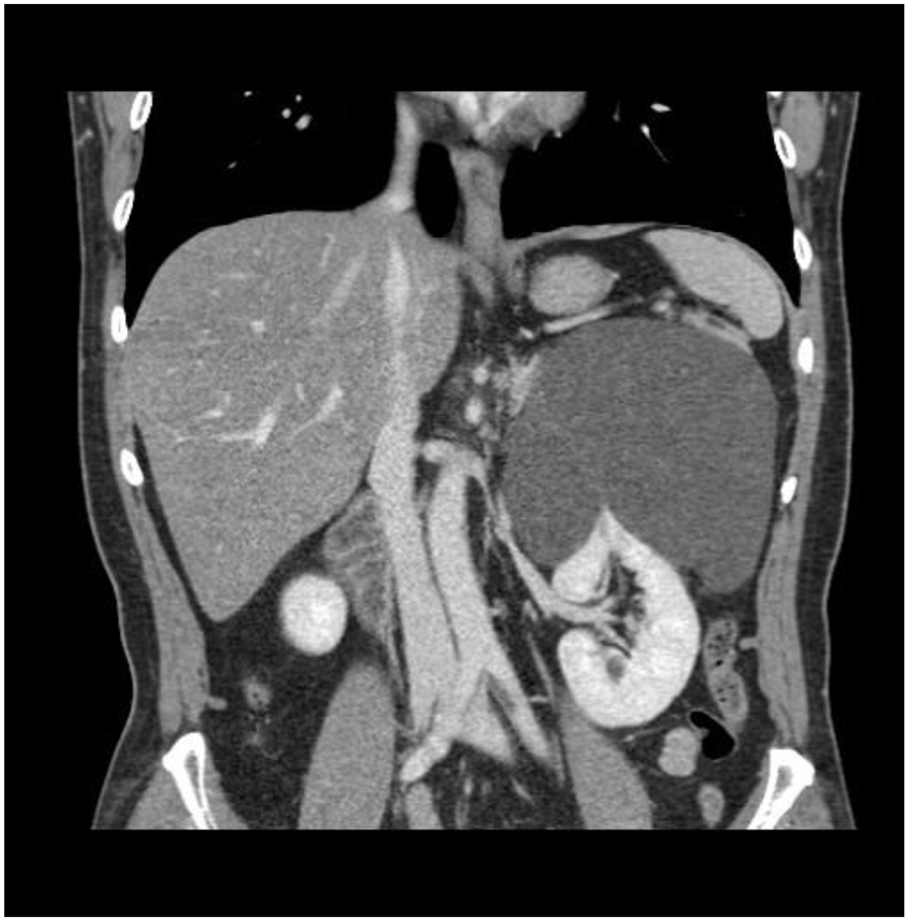
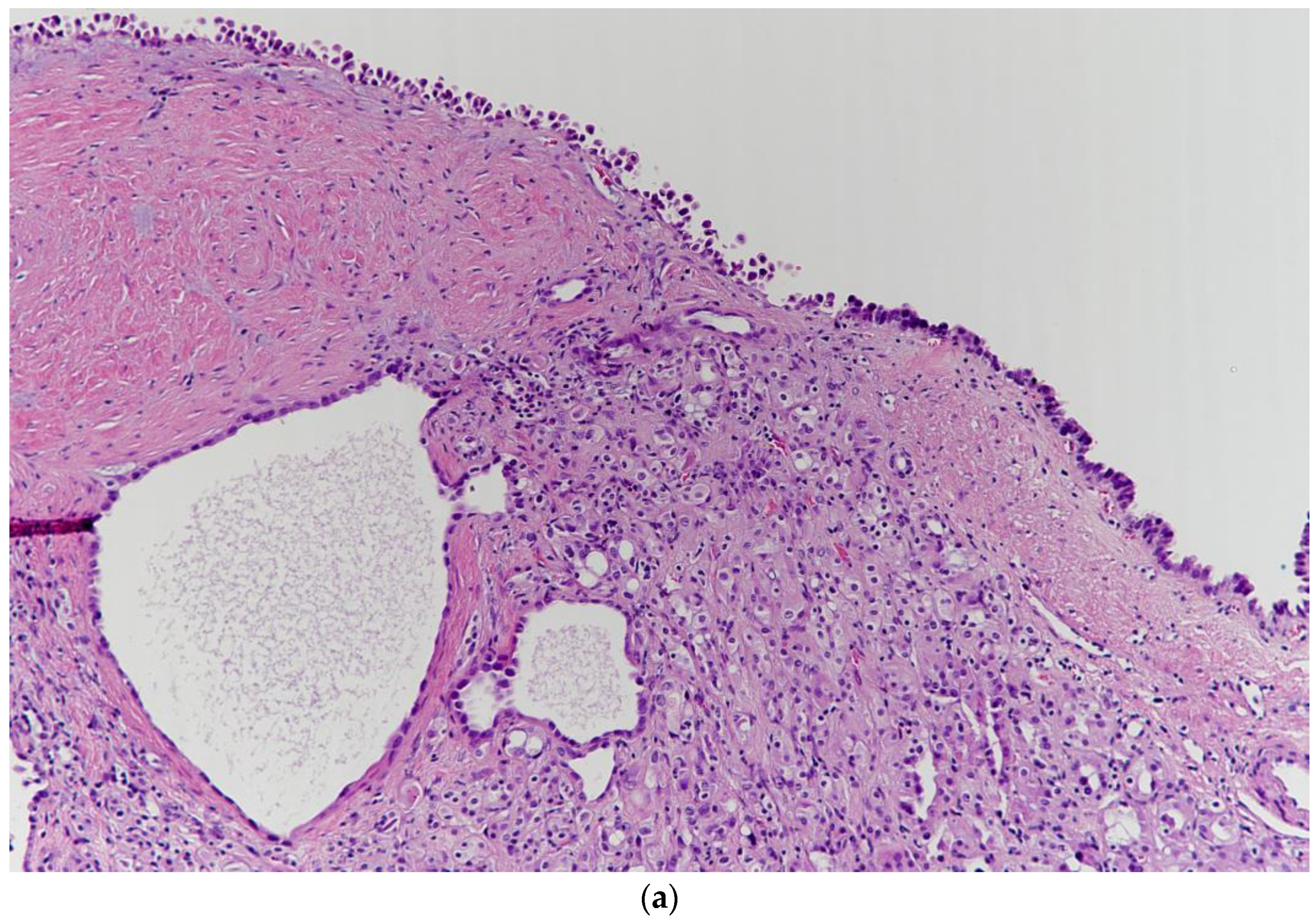
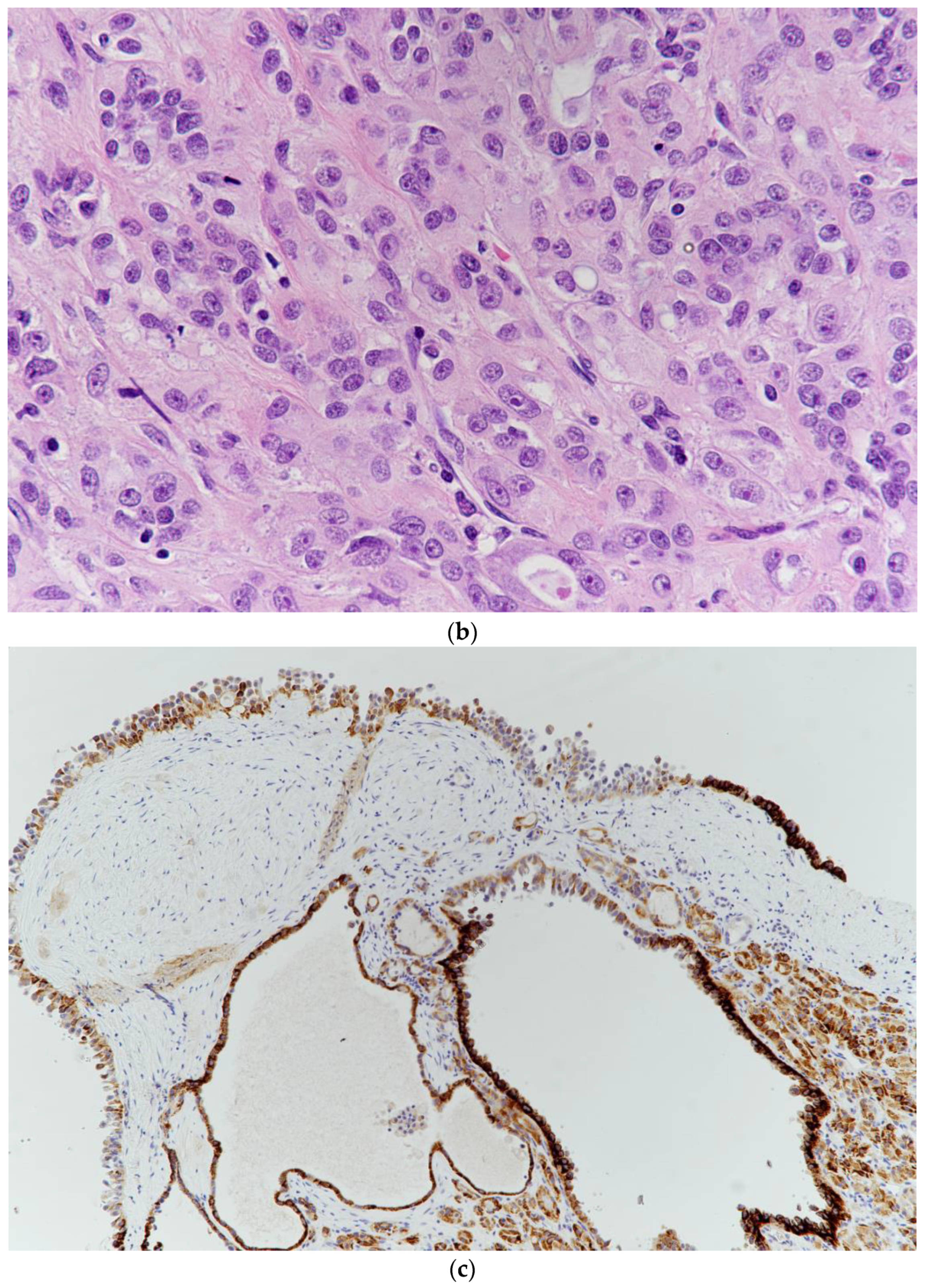
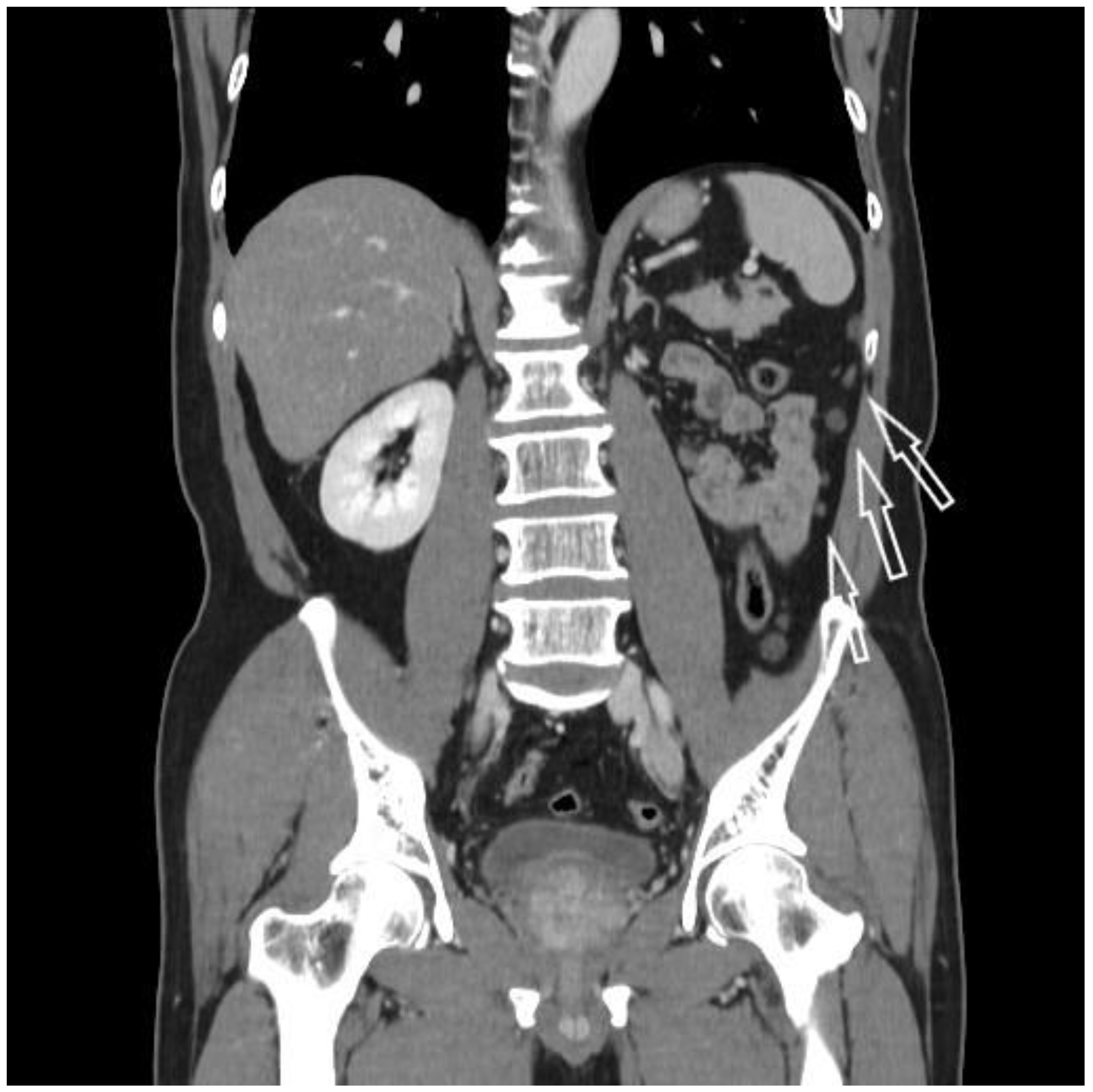
2. Case Report
3. Discussion
Author Contributions
Funding
Institutional Review Board Statement
Informed Consent Statement
Data Availability Statement
Acknowledgments
Conflicts of Interest
References
- Moch, H.; Cubilla, A.L.; Humphrey, P.A.; Reuter, V.E.; Ulbright, T.M. The 2016 WHO Classification of Tumours of the Urinary System and Male Genital Organs-Part A: Renal, Penile, and Testicular Tumours. Eur. Urol. 2016, 70, 93–105. [Google Scholar] [CrossRef] [PubMed]
- Banerjee, I.; Yadav, S.S.; Tomar, V.; Yadav, S.; Talreja, S. Tubulocystic Renal Cell Carcinoma: A Great Imitator. Rev. Urol. 2016, 18, 118–121. [Google Scholar] [CrossRef]
- Amin, M.B.; MacLennan, G.T.; Gupta, R.; Grignon, D.; Paraf, F.; Vieillefond, A.; Paner, G.P.; Stovsky, M.; Young, A.N.; Srigley, J.R.; et al. Tubulocystic carcinoma of the kidney: Clinicopathologic analysis of 31 cases of a distinctive rare subtype of renal cell carcinoma. Am. J. Surg. Pathol. 2009, 33, 384–392. [Google Scholar] [CrossRef] [PubMed]
- Yang, X.J.; Zhou, M.; Hes, O.; Shen, S.; Li, R.; Lopez, J.; Shah, R.B.; Yang, Y.; Chuang, S.T.; Lin, F.; et al. Tubulocystic carcinoma of the kidney: Clinicopathologic and molecular characterization. Am. J. Surg. Pathol. 2008, 32, 177–187. [Google Scholar] [CrossRef] [PubMed]
- Bhullar, J.S.; Thamboo, T.; Esuvaranathan, K. Unique case of tubulocystic carcinoma of the kidney with sarcomatoid features: A new entity. Urology 2011, 78, 1071–1072. [Google Scholar] [CrossRef]
- Cornelis, F.; Hélénon, O.; Correas, J.M.; Lemaitre, L.; André, M.; Meuwly, J.Y.; Sengel, C.; Derchi, L.; Yacoub, M.; Verkarre, V.; et al. Tubulocystic renal cell carcinoma: A new radiological entity. Eur. Radiol. 2016, 26, 1108–1115. [Google Scholar] [CrossRef] [PubMed]
- MacLennan, G.T.; Cheng, L. Tubulocystic carcinoma of the kidney. J. Urol. 2011, 185, 2348–2349. [Google Scholar] [CrossRef] [PubMed]
- Narayanasamy, S.; Krishna, S.; Prasad Shanbhogue, A.K.; Flood, T.A.; Sadoughi, N.; Sathiadoss, P.; Schieda, N. Contemporary update on imaging of cystic renal masses with histopathological correlation and emphasis on patient management. Clin. Radiol. 2019, 74, 83–94. [Google Scholar] [CrossRef] [PubMed]
- Oderda, M.; Maletta, F.; Palazzetti, A.; Faletti, R.; Falcone, M.; Marra, G.; Galliano, D.; Davico Bonino, L.; Gontero, P. Tubulocystic renal cell carcinoma disguised as a renal cyst. Minerva Urol. E Nefrol. Ital. J. Urol. Nephrol. 2016, 68, 451–455. [Google Scholar]
- Tran, T.; Jones, C.L.; Williamson, S.R.; Eble, J.N.; Grignon, D.J.; Zhang, S.; Wang, M.; Baldridge, L.A.; Wang, L.; Montironi, R.; et al. Tubulocystic renal cell carcinoma is an entity that is immunohistochemically and genetically distinct from papillary renal cell carcinoma. Histopathology 2016, 68, 850–857. [Google Scholar] [CrossRef] [PubMed]
- Osunkoya, A.O.; Young, A.N.; Wang, W.; Netto, G.J.; Epstein, J.I. Comparison of gene expression profiles in tubulocystic carcinoma and collecting duct carcinoma of the kidney. Am. J. Surg. Pathol. 2009, 33, 1103–1106. [Google Scholar] [CrossRef] [PubMed]
- Zhou, M.; Yang, X.J.; Lopez, J.I.; Shah, R.B.; Hes, O.; Shen, S.S.; Li, R.; Yang, Y.; Lin, F.; Elson, P.; et al. Renal tubulocystic carcinoma is closely related to papillary renal cell carcinoma: Implications for pathologic classification. Am. J. Surg. Pathol. 2009, 33, 1840–1849. [Google Scholar] [CrossRef] [PubMed]
- Al-Hussain, T.O.; Cheng, L.; Zhang, S.; Epstein, J.I. Tubulocystic carcinoma of the kidney with poorly differentiated foci: A series of 3 cases with fluorescence in situ hybridization analysis. Hum. Pathol. 2013, 44, 1406–1411. [Google Scholar] [CrossRef] [PubMed]
- Smith, S.C.; Trpkov, K.; Chen, Y.B.; Mehra, R.; Sirohi, D.; Ohe, C.; Cani, A.K.; Hovelson, D.H.; Omata, K.; McHugh, J.B.; et al. Tubulocystic Carcinoma of the Kidney with Poorly Differentiated Foci: A Frequent Morphologic Pattern of Fumarate Hydratase-deficient Renal Cell Carcinoma. Am. J. Surg. Pathol. 2016, 40, 1457–1472. [Google Scholar] [CrossRef]
- Zhao, M.; Teng, X.; Ru, G.; Zhao, Z.; Hu, Q.; Han, L.; He, X. Tubulocystic renal cell carcinoma with poorly differentiated foci is indicative of aggressive behavior: Clinicopathologic study of two cases and review of the literature. Int. J. Clin. Exp. Pathol. 2015, 8, 11124–11131. [Google Scholar]
- Al-Hussain, T.O.; Alahmadi, B.; Junejo, N.N.; Alshammari, K.; Bakshi, N.; Alzahrani, H.M. Tubulocystic renal cell carcinoma with poorly differentiated foci and loss of fumarate hydratase: A case report. Urol. Case Rep. 2020, 33, 101236. [Google Scholar] [CrossRef]
- Mego, M.; Sycova-Mila, Z.; Rejlekova, K.; Rychly, B.; Obertova, J.; Rajec, J.; Hes, O.; Mardiak, J. Sunitinib in the treatment of tubulocystic carcinoma of the kidney. A case report. Ann. Oncol. Off. J. Eur. Soc. Med. Oncol. 2008, 19, 1655–1656. [Google Scholar] [CrossRef]
- Teramoto, S.; Niwakawa, M.; Muraoka, K.; Ogawa, M.; Kunieda, F.; Matsuzaki, M.; Yamashita, R.; Matsui, T.; Yamaguchi, R.; Tobisu, K. Tubulocystic carcinoma of the kidney. Nihon Hinyokika Gakkai Zasshi Jpn. J. Urol. 2011, 102, 696–700. [Google Scholar] [CrossRef] [Green Version]
- Urabe, F.; Miki, J.; Yanagisawa, T.; Kimura, T.; Nakano, M.; Suzuki, M.; Kishimoto, K.; Egawa, S. A Case of Metastatic Tubulocystic Carcinoma of the Kidney Treated with Molecularly Targeted Therapy. Hinyokika Kiyo Acta Urol. Jpn. 2016, 62, 569–574. [Google Scholar] [CrossRef]
- Motzer, R.J.; Jonasch, E.; Michaelson, M.D.; Nandagopal, L.; Gore, J.L.; George, S.; Alva, A.; Haas, N.; Harrison, M.R.; Plimack, E.R.; et al. NCCN Guidelines Insights: Kidney Cancer, Version 2. 2020. J. Natl. Compr. Cancer Netw. JNCCN 2019, 17, 1278–1285. [Google Scholar] [CrossRef] [PubMed] [Green Version]
- Curry, D.; Pahuja, A.; Loan, W.; Thwaini, A. Radiofrequency Ablation of Small Renal Masses: Outcomes, Complications and Effects on Renal Function. Curr. Urol. 2017, 11, 196–200. [Google Scholar] [CrossRef] [PubMed]
- Kim, H.J.; Park, B.K.; Park, J.J.; Kim, C.K. CT-Guided Radiofrequency Ablation of T1a Renal Cell Carcinoma in Korea: Mid-Term Outcomes. Korean J. Radiol. 2016, 17, 763–770. [Google Scholar] [CrossRef] [PubMed] [Green Version]
- Gonnet, A.; Salabert, L.; Roubaud, G.; Catena, V.; Brouste, V.; Buy, X.; Gross Goupil, M.; Ravaud, A.; Palussière, J. Renal cell carcinoma lung metastases treated by radiofrequency ablation integrated with systemic treatments: Over 10 years of experience. BMC Cancer 2019, 19, 1182. [Google Scholar] [CrossRef] [PubMed] [Green Version]


Publisher’s Note: MDPI stays neutral with regard to jurisdictional claims in published maps and institutional affiliations. |
© 2021 by the authors. Licensee MDPI, Basel, Switzerland. This article is an open access article distributed under the terms and conditions of the Creative Commons Attribution (CC BY) license (https://creativecommons.org/licenses/by/4.0/).
Share and Cite
Choi, T.-S.; Lee, D.-G.; Won, K.-Y.; Min, G.-E. Tubulocystic Renal Cell Carcinoma Is Not an Indolent Tumor: A Case Report of Recurrences in the Retroperitoneum and Contralateral Kidney. Medicina 2021, 57, 851. https://0-doi-org.brum.beds.ac.uk/10.3390/medicina57080851
Choi T-S, Lee D-G, Won K-Y, Min G-E. Tubulocystic Renal Cell Carcinoma Is Not an Indolent Tumor: A Case Report of Recurrences in the Retroperitoneum and Contralateral Kidney. Medicina. 2021; 57(8):851. https://0-doi-org.brum.beds.ac.uk/10.3390/medicina57080851
Chicago/Turabian StyleChoi, Tae-Soo, Dong-Gi Lee, Kyu-Yeoun Won, and Gyeong-Eun Min. 2021. "Tubulocystic Renal Cell Carcinoma Is Not an Indolent Tumor: A Case Report of Recurrences in the Retroperitoneum and Contralateral Kidney" Medicina 57, no. 8: 851. https://0-doi-org.brum.beds.ac.uk/10.3390/medicina57080851