
Intracranial Hemorrhage in a Patient with TAFRO Syndrome Treated with Cyclosporine A and Rituximab


Abstract
:1. Introduction
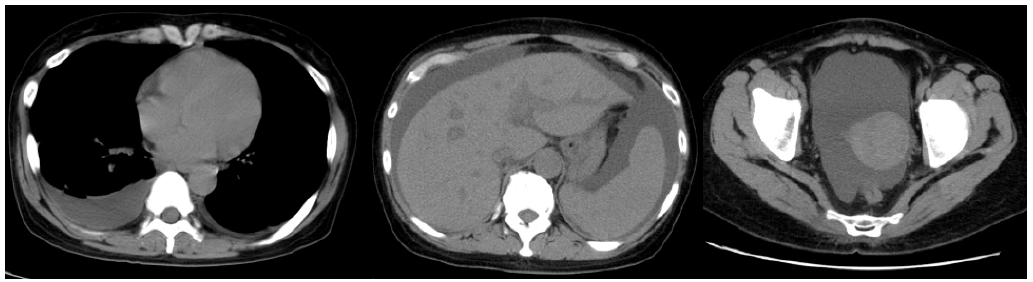
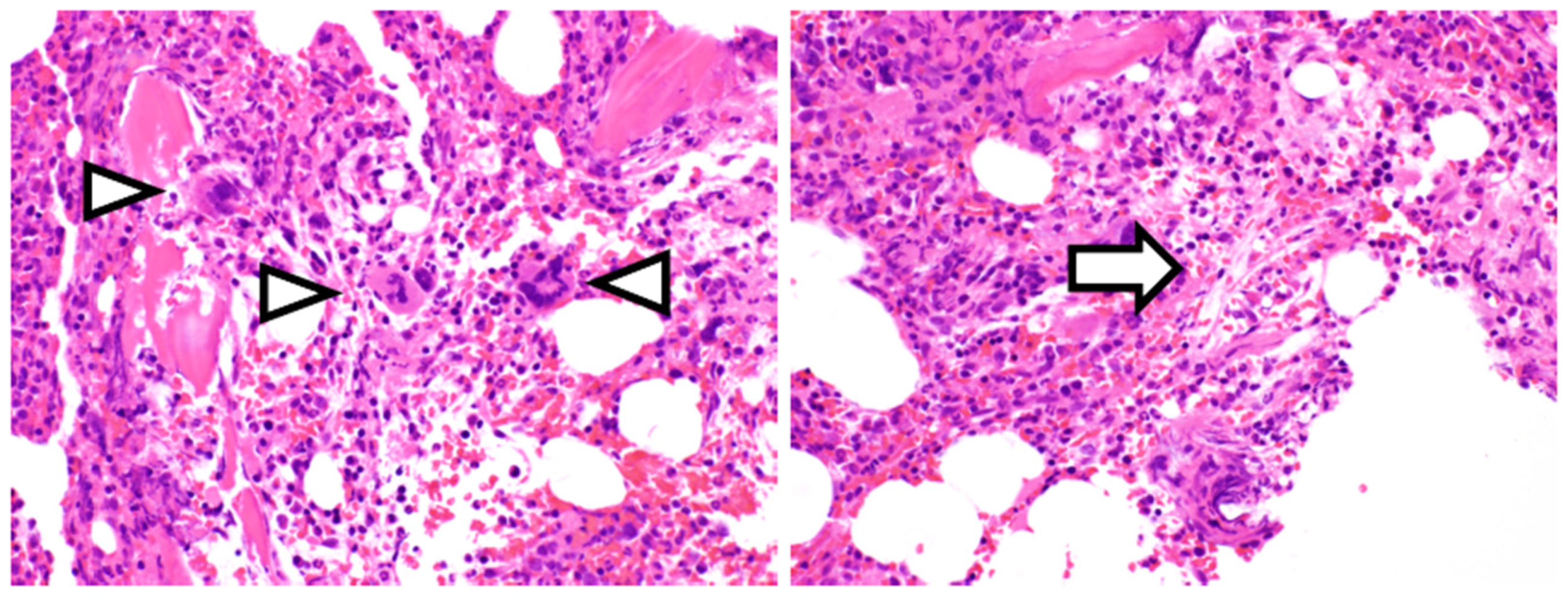
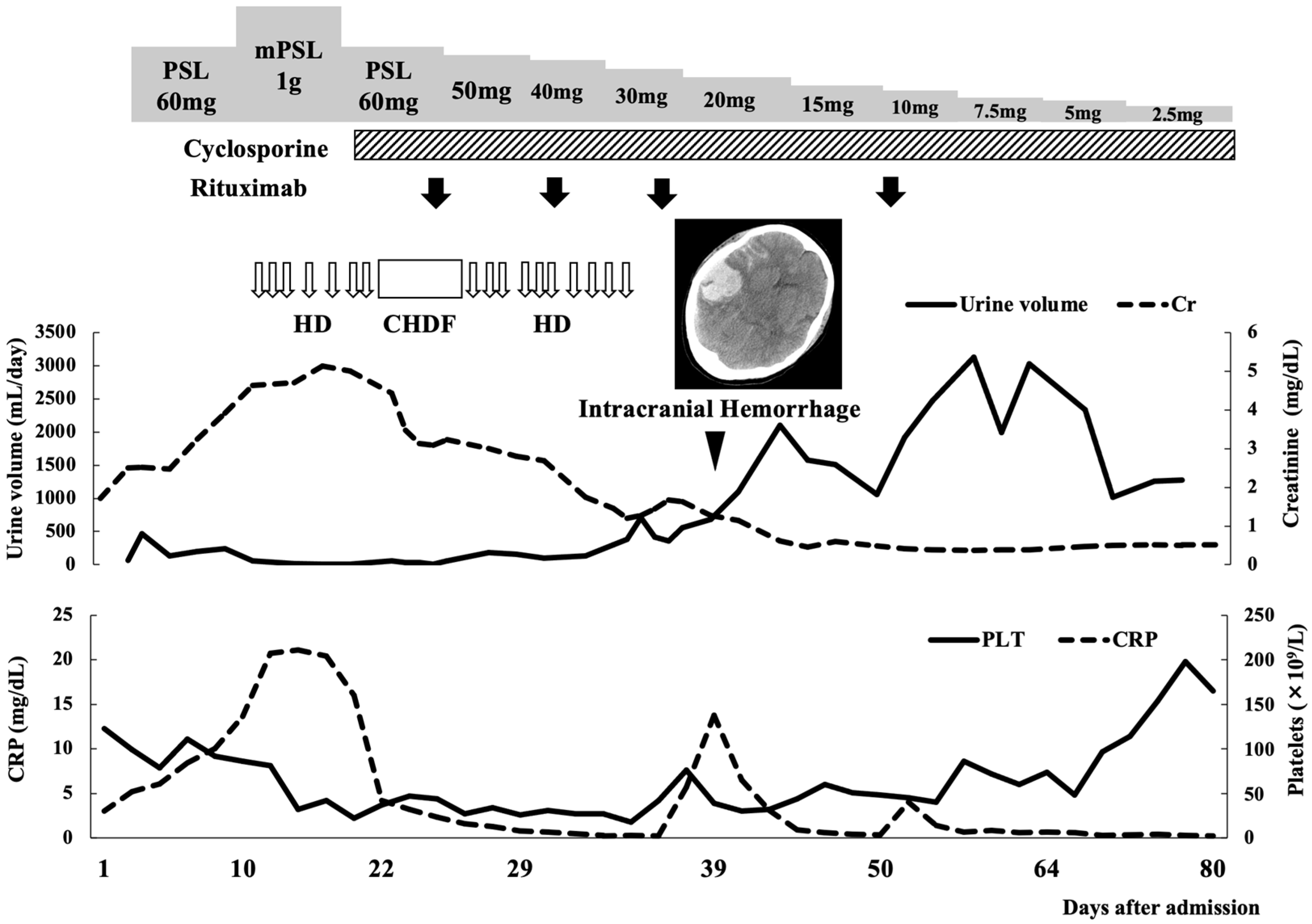
2. Case Report
3. Discussion
4. Conclusions
Author Contributions
Funding
Institutional Review Board Statement
Informed Consent Statement
Data Availability Statement
Acknowledgments
Conflicts of Interest
References
- Kawabata, H.; Takai, K.; Kojima, M.; Nakamura, N.; Aoki, S.; Nakamura, S.; Kinoshita, T.; Masaki, Y. Castleman-Kojima disease (TAFRO syndrome): A novel systemic inflammatory disease characterized by a constellation of symptoms, namely, thrombocytopenia, ascites (anasarca), microcytic anemia, myelofibrosis, renal dysfunction, and organomegaly: A status report and summary of Fukushima (6 June 2012) and Nagoya meetings (22 September 2012). J. Clin. Exp. Hematop. 2013, 53, 57–61. [Google Scholar] [CrossRef] [PubMed] [Green Version]
- Masaki, Y.; Kawabata, H.; Takai, K.; Kojima, M.; Tsukamoto, N.; Ishigaki, Y.; Kurose, N.; Ide, M.; Murakami, J.; Nara, K.; et al. Proposed diagnostic criteria, disease severity classification and treatment strategy for TAFRO Syndrome. Int. J. Hematol. 2016, 103, 686–692. [Google Scholar] [CrossRef] [PubMed]
- Takai, K.; Nikkuni, K.; Shibuya, H.; Hashidate, H. Thrombocytopenia with mild bone marrow fibrosis accompanied by fever, pleural effusion, ascites and hepatosplenomegaly. Rinsho Ketsueki 2010, 51, 320–325. [Google Scholar]
- Fajgenbaum, D.C.; van Rhee, F.; Nabel, C.S. HHV-8-negative, idiopathic multicentric Castleman disease: Novel insights into biology, pathogenesis, and therapy. Blood 2014, 123, 2924–2933. [Google Scholar] [CrossRef] [PubMed]
- Yamaga, Y.; Tokuyama, K.; Kato, T.; Yamada, R.; Murayama, M.; Ikeda, T.; Yamakita, N.; Kunieda, T. Successful treatment with cyclosporin A in tocilizumab-resistant TAFRO syndrome. Intern. Med. 2016, 55, 185–190. [Google Scholar] [CrossRef] [Green Version]
- Shirai, T.; Onishi, A.; Waki, D.; Saegusa, J.; Morinobu, A. Successful treatment with tacrolimus in TAFRO syndrome, two case reports and literature review. Medicine 2018, 97, e11045. [Google Scholar] [CrossRef]
- Kikuchi, T.; Shimizu, T.; Toyama, T.; Abe, R.; Okamoto, S. Successful treatment of TAFRO syndrome with tocilizumab, prednisone, and cyclophosphamide. Intern. Med. 2017, 56, 2205–2211. [Google Scholar] [CrossRef] [Green Version]
- José, F.F.; Kerbauy, L.N.; Perini, G.F.; Blumenschein, D.I.; Pasqualin, D.D.C.; Malheiros, D.M.A.C.; Campos Neto, G.C.; de Souza Santos, F.P.; Piovesan, R.; Hamerschlak, N. A life-threatening case of TAFRO syndrome with dramatic response to tocilizumab, rituximab, and pulse steroids: The first case report in Latin America. Medicine 2017, 96, e6271. [Google Scholar] [CrossRef]
- Tsurumi, H.; Fujigaki, Y.; Yamamoto, T.; Iino, R.; Taniguchi, K.; Nagura, M.; Arai, S.; Tamura, Y.; Ota, T.; Shibata, S.; et al. Remission of refractory ascites and discontinuation of hemodialysis after additional rituximab to long-term glucocorticoid therapy in a patient with TAFRO syndrome. Intern. Med. 2018, 57, 1433–1438. [Google Scholar] [CrossRef] [Green Version]
- Noda, Y.; Saka, Y.; Kato, A.; Mimura, T.; Naruse, T. Successful rituximab treatment of TAFRO syndrome with pathological findings of glomerular endothelial damage. Clin. Nephrol. Case Stud. 2018, 6, 16–20. [Google Scholar] [CrossRef]
- Nagayama, Y.; Yamano, M.; Yagame, M.; Nariyama, T.; Takahashi, M.; Kawamoto, M.; Matsui, K. TAFRO syndrome as a cause of glomerular microangiopathy: A case report and literature review. BMC Nephrol. 2019, 20, 375. [Google Scholar] [CrossRef] [PubMed]
- Zhou, Q.; Zhang, Y.; Zhou, G.; Zhu, J. Kidney biopsy findings in two patients with TAFRO syndrome: Case presentations and review of the literature. BMC Nephrol. 2020, 21, 499. [Google Scholar] [CrossRef]
- Mizuno, H.; Sekine, A.; Oguro, M.; Oshima, Y.; Kawada, M.; Sumida, K.; Yamanouchi, M.; Hayami, N.; Suwabe, T.; Hiramatsu, R.; et al. Renal histology in a patient with TAFRO syndrome: A case report. Hum. Pathol. 2018, 82, 258–263. [Google Scholar] [CrossRef] [PubMed]
- Imafuku, A.; Suwabe, T.; Hasegawa, E.; Mise, K.; Sumida, K.; Hiramatsu, R.; Yamanouchi, M.; Hayami, N.; Hoshino, J.; Sawa, N.; et al. Castleman’s disease accompanied by hypolipidemic cerebral hemorrhage and nephrosclerosis. Intern. Med. 2013, 52, 1611–1616. [Google Scholar] [CrossRef] [PubMed] [Green Version]
- Tanaka, J.; Fujita, A.; Hosoda, K.; Kohmura, E. Cerebral angiitis associated with subarachnoid hemorrhage in Castleman’s disease, report of two cases. BMC Neurol. 2016, 16, 60. [Google Scholar] [CrossRef] [PubMed] [Green Version]
- Kubokawa, I.; Yachie, A.; Hayakawa, A.; Hirase, S.; Yamamoto, N.; Mori, T.; Yanai, T.; Takeshima, Y.; Kyo, E.; Kageyama, G.; et al. The first report of adolescent TAFRO syndrome: A unique clinicopathologic variant of multicentric Castleman’s disease. BMC Pediatr. 2014, 14, 139. [Google Scholar] [CrossRef] [Green Version]
- Takayama, Y.; Kubota, T.; Ogino, Y.; Ohnishi, H.; Togitani, K.; Yokoyama, A. TAFRO syndrome with disseminated intravascular coagulation successfully treated with tocilizumab and recombinant thrombomodulin. Intern. Med. 2018, 57, 1291–1296. [Google Scholar] [CrossRef] [Green Version]
- Nara, M.; Komatsuda, A.; Itoh, F.; Kaga, H.; Saitoh, M.; Togashi, M.; Kameoka, Y.; Wakui, H.; Takahashi, N. Two cases of thrombocytopenia, anasarca, fever, reticulin fibrosis/renal failure, and organomegaly (TAFRO) syndrome with high serum procalcitonin levels, including the first case complicated with adrenal hemorrhaging. Intern. Med. 2017, 56, 1247–1252. [Google Scholar] [CrossRef] [PubMed] [Green Version]
- Iwaki, N.; Fajgenbaum, D.C.; Nabel, C.S.; Gion, Y.; Kondo, E.; Kawano, M.; Masunari, T.; Yoshida, I.; Moro, H.; Nikkuni, K.; et al. Clinicopathologic analysis of TAFRO syndrome demonstrates a distinct subtype of HHV-8-negative multicentric Castleman disease. Am. J. Hematol. 2016, 91, 220–226. [Google Scholar] [CrossRef] [PubMed]
- Fujimoto, S.; Kawabata, H.; Sakai, T.; Yanagisawa, H.; Nishikori, M.; Nara, K.; Ohara, S.; Tsukamoto, N.; Kurose, N.; Yamada, S.; et al. Optimal treatments for TAFRO syndrome, a retrospective surveillance study in Japan. Int. J. Hematol. 2021, 113, 73–80. [Google Scholar] [CrossRef]
- Coutier, F.; Meaux Ruault, N.; Crepin, T.; Bouiller, K.; Gil, H.; Humbert, S.; Bedgedjian, I.; Magy-Bertrand, N. A comparison of TAFRO syndrome between Japanese and non-Japanese cases: A case report and literature review. Ann. Hematol. 2018, 97, 401–407. [Google Scholar] [CrossRef]
- Fajgenbaum, D.C.; Langan, R.A.; Japp, A.S.; Partridge, H.L.; Pierson, S.K.; Singh, A.; Arenas, D.J.; Ruth, J.R.; Nabel, C.S.; Stone, K.; et al. Identifying and targeting pathogenic PI3K/AKT/mTOR signaling in IL-6-blockade-refractory idiopathic multicentric Castleman disease. J. Clin. Investig. 2019, 129, 4451–4463. [Google Scholar] [CrossRef] [PubMed]
- Arenas, D.J.; Floess, K.; Kobrin, D.; Pai, R.L.; Srkalovic, M.B.; Tamakloe, M.A.; Rasheed, R.; Ziglar, J.; Khor, J.; Parente, S.A.T.; et al. Increased mTOR activation in idiopathic multicentric Castleman disease. Blood 2020, 135, 1673–1684. [Google Scholar] [CrossRef] [PubMed]
- Pai, R.L.; Japp, A.S.; Gonzalez, M.; Rasheed, R.F.; Okumura, M.; Arenas, D.; Pierson, S.K.; Powers, V.; Layman, A.A.K.; Kao, C.; et al. Type I IFN response associated with mTOR activation in the TAFRO subtype of idiopathic multicentric Castleman disease. JCI Insight 2020, 5, e135031. [Google Scholar] [CrossRef] [PubMed]
- Ivashkiv, L.B.; Donlin, L.T. Regulation of type I interferon responses. Nat. Rev. Immunol. 2014, 14, 36–49. [Google Scholar] [CrossRef] [Green Version]


| Complete Blood Count | Chemistry | Calcium | 9.4 | mg/dL | ||||
|---|---|---|---|---|---|---|---|---|
| White Blood Cells | 11.1 | ×109/L | Total Protein | 5.9 | g/dL | Total Bilirubin | 0.5 | mg/dL |
| Neutrophil | 78 | % | Albumin | 3.0 | g/dL | C-Reactive Protein | 3.0 | mg/dL |
| Eosinophil | 0 | % | Creatine Kinase | 87 | IU/L | IgG | 1008 | mg/dL |
| Basophil | 0 | % | Aspartate Transaminase | 16 | IU/L | IgA | 60 | mg/dL |
| Monocyte | 7 | % | Alanine Transaminase | 7 | IU/L | IgM | 112 | mg/dL |
| Lymphocyte | 12 | % | Lactate Dehydrogenase | 187 | IU/L | IgG4 | 11 | mg/dL |
| Myelocytes | 1 | % | Alkaline Phosphatase | 325 | IU/L | TSH | 2.38 | µIU/mL |
| Metamyelocytes | 2 | % | γ-Glutamyl Transpeptidase | 32 | IU/L | Free T4 | 1.39 | ng/mL |
| Red Blood Cells | 4.14 | ×1012/L | Cholinesterase | 285 | IU/L | Soluble IL-2R | 981 | U/mL |
| Hemoglobin | 10.7 | g/dL | Amylase | 63 | IU/L | Ferritin | 118 | ng/mL |
| Reticulocytes | 77 | ×109/L | Creatinine | 1.71 | mg/dL | |||
| Platelets | 123 | ×109/L | Uric Acid | 8.4 | mg/dL | Coagulation system | ||
| Blood Urea Nitrogen | 32.7 | mg/dL | APTT | 32.3 | s | |||
| Serum Cytokine | Sodium | 139 | mEq/L | Prothrombin time | 1.04 | s | ||
| IL-6 * | 5.0 | pg/mL | Potassium | 4.3 | mEq/L | Fibrinogen | 423 | mg/dL |
| VEGF * | 256 | pg/mL | Chloride | 104 | mEq/L | D-dimer | 8.1 | μg/mL |
Publisher’s Note: MDPI stays neutral with regard to jurisdictional claims in published maps and institutional affiliations. |
© 2021 by the authors. Licensee MDPI, Basel, Switzerland. This article is an open access article distributed under the terms and conditions of the Creative Commons Attribution (CC BY) license (https://creativecommons.org/licenses/by/4.0/).
Share and Cite
Akagi, Y.; Kato, T.; Yamashita, Y.; Hosoi, H.; Murata, S.; Yamamoto, S.; Warigaya, K.; Nakao, T.; Murata, S.; Sonoki, T.; et al. Intracranial Hemorrhage in a Patient with TAFRO Syndrome Treated with Cyclosporine A and Rituximab. Medicina 2021, 57, 971. https://0-doi-org.brum.beds.ac.uk/10.3390/medicina57090971
Akagi Y, Kato T, Yamashita Y, Hosoi H, Murata S, Yamamoto S, Warigaya K, Nakao T, Murata S, Sonoki T, et al. Intracranial Hemorrhage in a Patient with TAFRO Syndrome Treated with Cyclosporine A and Rituximab. Medicina. 2021; 57(9):971. https://0-doi-org.brum.beds.ac.uk/10.3390/medicina57090971
Chicago/Turabian StyleAkagi, Yuina, Takashi Kato, Yusuke Yamashita, Hiroki Hosoi, Shogo Murata, Shuto Yamamoto, Kenji Warigaya, Taisei Nakao, Shinichi Murata, Takashi Sonoki, and et al. 2021. "Intracranial Hemorrhage in a Patient with TAFRO Syndrome Treated with Cyclosporine A and Rituximab" Medicina 57, no. 9: 971. https://0-doi-org.brum.beds.ac.uk/10.3390/medicina57090971