Article

An Anti-Inflammatory 2,4-Cyclized-3,4-Secospongian Diterpenoid and Furanoterpene-Related Metabolites of a Marine Sponge Spongia sp. from the Red Sea

1
Doctoral Degree Program in Marine Biotechnology, National Sun Yat-sen University, Kaohsiung 80424, Taiwan
2
Department of Marine Biotechnology and Resources, National Sun Yat-sen University, Kaohsiung 80424, Taiwan
3
Department of Pharmacognosy, College of Pharmacy, King Saud University, Riyadh 11451, Saudi Arabia
4
Department of Pharmacognosy, Faculty of Pharmacy, Mansoura University, Mansoura 35516, Egypt
5
Department of Marine Chemistry, Faculty of Marine Sciences, King Abdulaziz University, Jeddah 21589, Saudi Arabia
6
Department of Marine Recreation, National Penghu University of Science and Technology, Magong, Penghu 88046, Taiwan
7
Tropical Island Sustainable Development Research Center, National Penghu University of Science and Technology, Magong, Penghu 88046, Taiwan
8
Graduate Institute of Natural Products, College of Medicine, Chang Gung University, Taoyuan 33302, Taiwan
9
Research Center for Chinese Herbal Medicine, Research Center for Food and Cosmetic Safety, Graduate Institute of Health Industry Technology, College of Human Ecology, Chang Gung University of Science and Technology, Taoyuan 33303, Taiwan
10
Department of Anesthesiology, Chang Gung Memorial Hospital, Taoyuan 33305, Taiwan
11
Graduate Institute of Natural Products, Kaohsiung Medical University, Kaohsiung 80708, Taiwan
12
Department of Medical Research, China Medical University Hospital, China Medical University, Taichung 404333, Taiwan
*
Authors to whom correspondence should be addressed.
Submission received: 16 December 2020 / Revised: 6 January 2021 / Accepted: 14 January 2021 / Published: 16 January 2021
(This article belongs to the Special Issue Bioactive Compounds from Marine Sponges 2020)

Abstract

:
Chemical investigation of a Red Sea Spongia sp. led to the isolation of four new compounds, i.e., 17-dehydroxysponalactone (1), a carboxylic acid, spongiafuranic acid A (2), one hydroxamic acid, spongiafuranohydroxamic acid A (3), and a furanyl trinorsesterpenoid 16-epi-irciformonin G (4), along with three known metabolites (−)-sponalisolide B (5), 18-nor- 3,17-dihydroxy-spongia-3,13(16),14-trien-2-one (6), and cholesta-7-ene-3β,5α-diol-6-one (7). The biosynthetic pathway for the molecular skeleton of 1 and related compounds was postulated for the first time. Anti-inflammatory activity of these metabolites to inhibit superoxide anion generation and elastase release in N-formyl-methionyl-leucyl phenylalanine/cytochalasin B (fMLF/CB)-induced human neutrophil cells and cytotoxicity of these compounds toward three cancer cell lines and one human dermal fibroblast cell line were assayed. Compound 1 was found to significantly reduce the superoxide anion generation and elastase release at a concentration of 10 μM, and compound 5 was also found to display strong inhibitory activity against superoxide anion generation at the same concentration. Due to the noncytotoxic activity and the potent inhibitory effect toward the superoxide anion generation and elastase release, 1 and 5 can be considered to be promising anti-inflammatory agents.

1. Introduction

Marine sponges have been considered to be an important source for the discovery of structurally diverse bioactive secondary metabolites [1]. Many natural products from sponges have been shown to exhibit a variety of biological activities, such as antimicrobial [2,3,4,5], antiviral [6,7,8], antiprotozoal [8,9,10], cytotoxic [6,11,12,13], anti-inflammatory [14,15,16], antioxidant [4,17,18], immunosuppressive [1,19,20], and antifeedant [21,22,23]. The genus Spongia (Spongidae) has been chemically investigated since 1971 [24] and the studies have led to the discovery of a series of furanoterpenes [24,25,26], spongian diterpenoids [27,28,29,30,31,32], scalarane sesterterpenoids [33,34,35], sesquiterpene quinones [36,37], along with other kinds of metabolites, for example, sterols [38,39,40] and macrolides [41].
We report, herein, the chemical investigation of an unidentified Spongia species inhabiting along the eastern coast of the Red Sea. This study afforded four new natural products including a rare A-ring contracted diterpenoid, 17-dehydroxysponalactone (1), a C12 carboxylic acid, spongiafuranic acid A (2); a C12 hydroxamic acid, spongiafuranohydroxamic acid A (3); and a furanyl trinorsesterpenoid, 16-epi-irciformonin G (4); along with three known metabolites, (−)-sponalisolide B (5) [42], 18-nor-3,17-dihydroxyspongia-3,13(16),14-trien-2-one (6) [43], and cholesta-7-ene-3β,5α-diol-6-one (7) [40] (Figure 1 and Supplementary Materials Figures S1–S35 for 15). Furthermore, in order to discover bioactive lead compounds, assays for the anti-inflammatory activity of the isolated compounds by inhibition of the superoxide anion generation and elastase release in N-formyl-methionyl-leucyl phenylalanine/cytochalasin B (fMLF/CB)-induced human neutrophils, and the cytotoxicity of these compounds against three tumor cell lines, murine leukemia (P388), human bile duct carcinoma (HuCCT), and human colon adenocarcinoma (DLD-1), and a human dermal fibroblast (CCD-966SK) cell line were undertaken. Compounds 1 and 5 were shown to exhibit the promising anti-inflammatory activity.

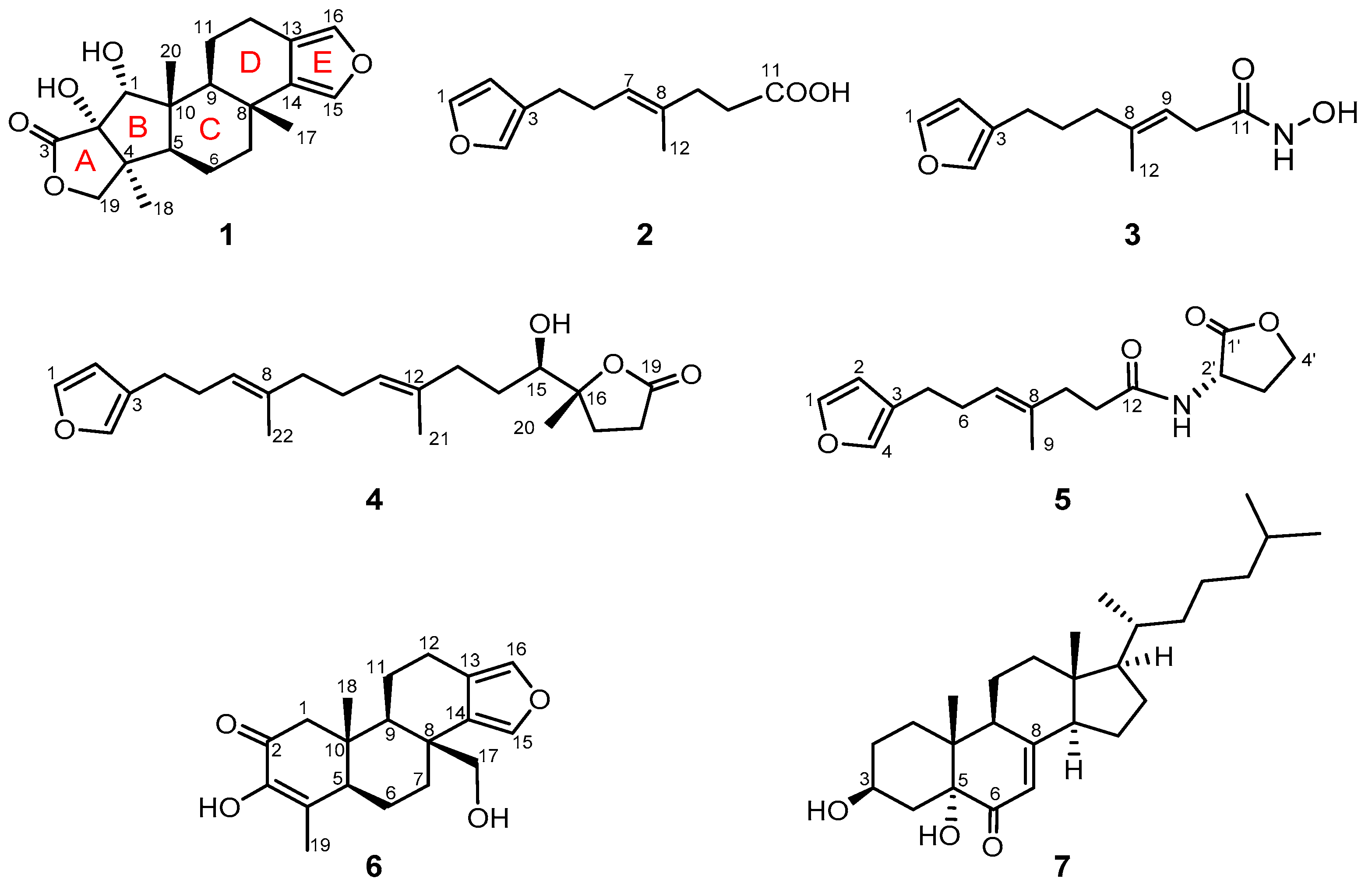
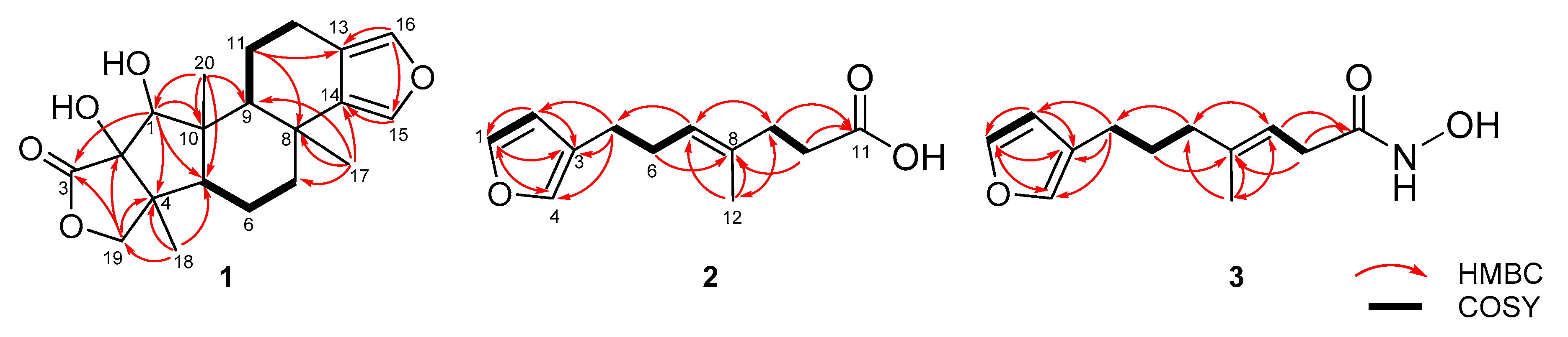
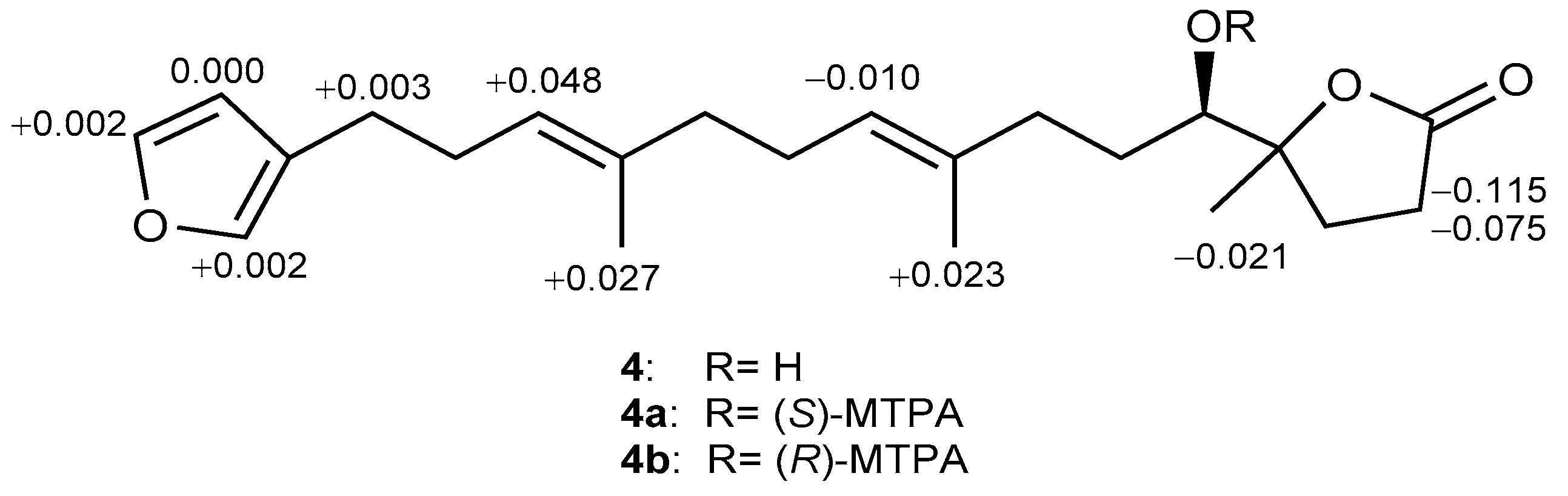
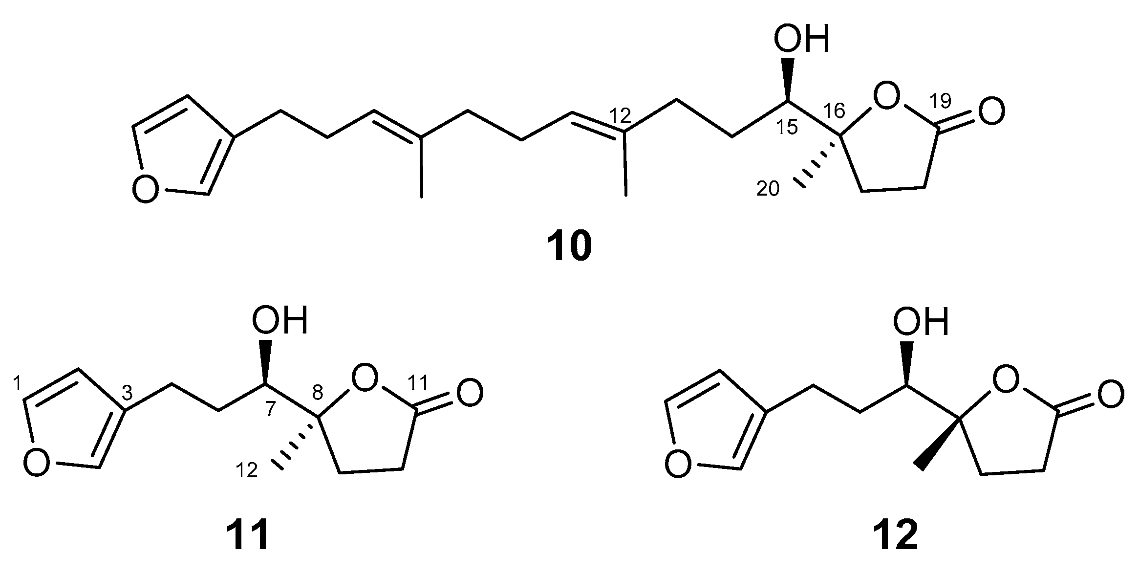
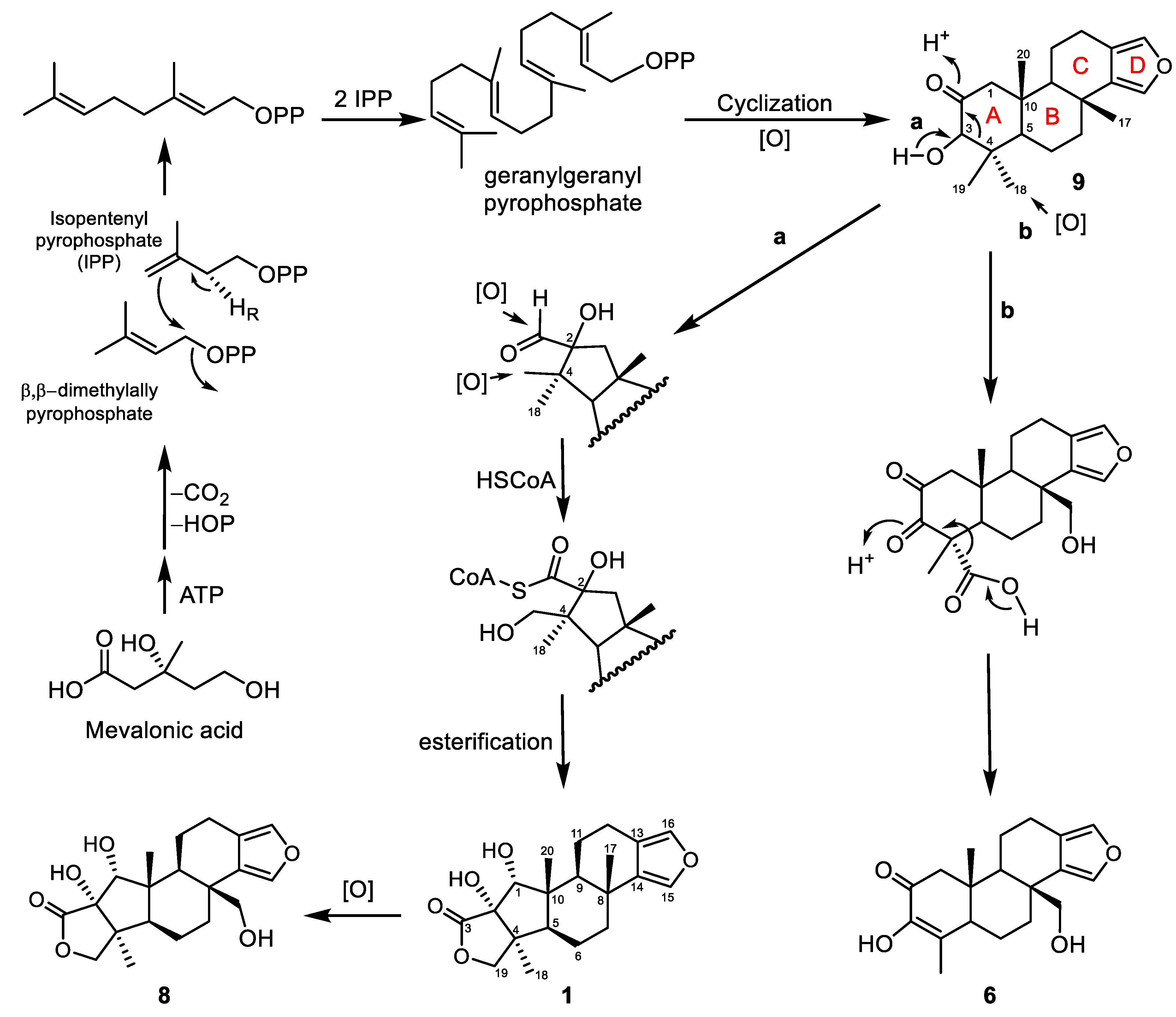
2. Results and Discussion

Compound 1 was obtained as a white powder. Its molecular formula C20H26O5 was established by the molecular ion peak at m/z 369.1672 [M + Na]+ in the HRESIMS, consistent with eight degrees of unsaturation. The IR spectrum showed absorptions of hydroxyl (3455 and 3401 cm−1) and lactone carbonyl (1752 cm−1) functionalities. The 13C NMR spectroscopic data of 1 exhibited 20 carbon signals (Table 1), which were assigned by the assistance of DEPT spectrum showing thirteen carbon signals of a diterpene, including three ring-juncture methyls (δC 26.9, 22.6, and 14.0; δH 1.24, 1.14, and 0.84) and a 3,4-disubstituted furan ring (δC 137.1, CH; 134.8, CH; 136.8, C; and 119.6, C and δH 7.06, 1H, br s and 7.09, 1H, br s) [30,31,35,44]. On the basis of the number of unsaturations, 1 was, thus, suggested to be a pentacyclic 3,4-disubstituted furan diterpenoid. The NMR spectroscopic data of 1 and 2D NMR corraltions (Figure 2) were similar to those of the previously described sponalactone (8) [30], except that a hydroxymethyl in 8 was replaced by a methyl at C-8 in 1. Compound 1 also possesses the same B, C, and D rings as 9 [32] (Scheme 1).
The relative and absolute configurations of 1 were established on the basis of nuclear Overhauser effect (NOE) correlation analysis (Figure 3) and by comparison of the observed NOE correlations with those of the related compounds [30,31], the observed pyridine-induced solvent shifts [45], and biogenetic consideration. The NOESY spectrum of 1 showed NOE correlations of H3-17/H3-20 and H-5/H-9, depicting the 5R*,8R*,9S*,10R*-configuration. H-1 displayed NOE interactions with the β-oriented H3-20 and H-11α (δH 1.68, m), indicating the α-orientation of the H-1. Furthermore, the NOE correlations of H-5/H3-18, H3-18/H-19αH 3.92) and H-19βH 4.37)/H3-20 disclosed the α- and β-orientations of H3-18 and the γ-lactone ring, respectively, and the α-orientation of the hydroxyl at C-2, accordingly. The analysis of the pyridine-induced deshielding effect of the axial hydroxy groups was also employed to support the configuration of 1. Therefore, the significant pyridine-induced downfield shifts (∆δ = δCDCl3 − δC6D5N) exerted on H-5 (∆δH = −0.24 ppm) could only be approached when 1-OH was axially oriented on the same α-face of the molecule. Also, H3-18 exhibited pyridine-induced downfield shift (∆δH = −0.14 ppm) due to the vicinal effect of 2-OH, which should be syn to H3-18 [45]. On the basis of the above findings, we propose that 1 can be derived from an intermediate spongian 9, which was biosynthesized from the mevalonic acid pathway, after oxygenation of the six-membered ring A and a subsequent ring contraction and formation of a five-membered carbocycle, as illustrated in Scheme 1.
Metabolite 2 was isolated as a colorless oil. Its molecular formula was determined to be C12H16O3 from the HREIMS (m/z 231.0992 [M + Na]+), indicating the four degrees of unsaturation. The IR spectrum displayed the absorptions of carboxylic acid (3105–2857 and 1708 cm–1) and olefin (1654 cm−1). The NMR data (Table 2) showed the presence of a monosubstituted furan ring (δC 142.5, CH; 138.8, CH; 111.0, CH; and 124.7, C; δH 7.34, 7.20, and 6.27, each 1H, s) [24,25,26,42], a trisubstituted olefin (δC 124.7, CH; δH 5.22, 1H, s), a methyl (δC 15.9; δH 1.61, 3H, s) and a carbonyl group (δC 180.0, C). Other 1H NMR signals in the shielded region (δH 2.25–2.47, 8H) were attributable to four methylene groups, as depicted from the COSY (Figure 2) correlations. The methylene protons H2-6 (δH 2.25, dt, J = 7.6, 7.2 Hz, 2H) was found to be further correlated with the olefinic proton (δH 5.22, dd, J = 7.2, 7.2 Hz, H-7) in 2. The detailed analysis of HMBC correlations (Figure 2) resolved the carbon positions of the furan ring, olefinic double bond, and the carboxyl group to be at C-1‒C-4, C-7/C-8, and C-11, respectively. Furthermore, the methyl group was positioned at C-8. The furanyl H-2 (δH 6.27, s), H-4 (δH 7.20, s), and the olefinic proton H-7 (δH 5.22, dd, J = 7.2, 7.2 Hz, 2H) displayed HMBC correlations with the sp3 carbon C-5 (δC 24.8, CH2), and H3-12 (δH 1.61, s) showed HMBC correlations with C-7 (δC 124.7, CH) and C-9 (δC 34.2, CH2), while the signal of H2-9 (δH 2.32, dd, J = 7.6, 7.6 Hz, 2H) was found to be correlated with the carboxyl carbon (C-11, δC 180.0). Moreover, the NOE correlations observed for H3-12 with H2-6 but not with H-5 and the chemical shift of C-12 (δC < 20 ppm) assigned the E-configuration of the 7,8-double bond [46]. Therefore, 2 was determined to be a furanotrinorsesquiterpenoid carboxylic acid with the structure of (E)-7-(furan-3-yl)-4-methylhept-4-enoic acid. The literature search showed that this compound had been prepared as a synthetic intermediate during the total syntheses of the furanosesquiterpenoids and dendrolasins [42,47], however, its NMR data had not been reported. Therefore, this is the first report of 2 as a natural product, with the NMR data assigned and reported for the first time.
Metabolite 3 exhibited almost the same NMR data as those of 2 (Table 2) from C-1 to C-6, with the carbon chemical shifts of the trisubstituted double bond (δC 139.7, C and 115.5, CH; δH 5.34, dd, J = 6.0, 6.0 Hz, 1H) and the carbonyl group (δC 176.1, C) in 3 showing significant differences of ∆δC −6.0, +9.2, and −3.9 ppm as compared with those of the corresponding carbons in 2, respectively. As illustrated by 1H-1H COSY correlations (Figure 2), the double bond has been isomerized from the C-7/C-8 position in 2 to the C-8/C-9 position in 3. However, the IR spectrum displayed the absorptions of the hydroxyl and NH groups (3407‒2858 cm–1), carbonyl group (1705 cm–1), and olefin (1634 cm–1) functionalities. Furthermore, the HREIMS m/z 246.1098 [M + Na]+ established the molecular formula of 3 to be C12H17NO3 and the chemical shift of the carbonyl group (176.1 ppm), showing that a hydroxamic acid moiety [48,49,50,51] replaced a carboxylic acid group at C-11 in 3.
Compound 4 was isolated as a colorless oil, [α] D 25 +4.4 (c 0.74, CHCl3). The ESIMS and NMR spectroscopic data (Table 3) established the molecular formula C22H32O4 for 4. The IR absorptions 3432, 1769, and 1647 cm–1 revealed the presence of hydroxyl, carbonyl, and olefin functionalities, respectively. Moreover, it was found that the NMR data of 4 was the same as those of irciformonin G (10) [52] in all aspects except for those at positions 17 and 18−20 (Table 4), proposing 4 as an isomer of 10. By using Mosher’s method [53,54], the 15R absolute configuration in 4 was established based on the calculated ∆δHS − δR) values of protons neighboring C-15 of (S)- and (R)-α-methoxy-α-(trifluorome- thyl)-phenylacetyl (MTPA) esters 4a and 4b, respectively (Figure 4). After the assignment of the 15R configuration, the 13C NMR data of C-15 to C-20 of 4 were further compared with the corresponding data of irciformonin G (10), (+)-sponalisolide A (11), and 8-epi-(+)-sponalisolide A (12) [42] of known absolute configurations (Table 4 and Figure 5). The 15R,16R-configuration of 4 was, thus, confirmed as those of the 7R, 8R configured 12, while 10 and 11 possessed the same configurations (R,S) at the corresponding asymmetric carbons. From the above findings, compound 4 was, thus, identified as 16-epi-irciformonin G.
(−)-Sponalisolide B (5) was isolated as a colorless oil, [α] D 25 −8.5 (c 0.34, CHCl3). Through detailed analysis of NMR spectroscopic data (Table 3), in particular two-dimensional (2D) NMR correlations, the structure of 5 was established to be identical to that of the known (‒)-sponalisolide B [42]. However, the coupling constants and spin-spin splitting patterns of the proton H2-6 (δH 2.25, dt, 2H, J = 7.5, 7.0 Hz at 500 MHz in CDCl3) were wrongly assigned. We, herein, reanalyzed the spectrum and provided the correct NMR data for 5.
With the aim of discovering bioactive compounds from these isolates, the cytotoxic activities of the isolated compounds 17 against the proliferation of three cancer cell lines including murine leukemia (P388), human bile duct carcinoma (HuCCT), and human colon adenocarcinoma (DLD-1), and a human dermal fibroblast cell line (CCD-966SK) were evaluated, using the Alamar Blue assay [55,56]. The results indicated that none of the tested metabolites exhibited cytotoxic activity (IC50 > 20 μg/mL).
The anti-inflammatory activities of compounds 17 on inhibition of superoxide anion (O2) generation and elastase release in the fMLF/CB-stimulated human neutrophils [57,58,59] were also evaluated. The results (Table 5) showed that 1 exhibited potent activity to inhibit the superoxide anion generation (91.38 ± 2.91%) and elastase release (90.29 ± 7.71%) at 10 μM, with the IC50 values of 3.37 ± 0.21 and 4.07 ± 0.60 μM, respectively. Compound 5 was also found to display significant inhibitory activity against the superoxide anion generation (IC50 = 5.31 ± 1.52 μM), and the percentage of inhibition was 67.12 ± 6.00% at 10 μM. Due to the noncytotoxic character and the potent activity toward the superoxide anion generation and elastase release, 1 and 5 can be considered to be the promising anti-inflammatory agents.

3. Materials and Methods

3.1. General Procedures

Measurements of optical rotations and IR spectra were carried out on a JASCO P-1020 polarimeter and FT/IR-4100 infrared spectrophotometer (JASCO Corporation, Tokyo, Japan), respectively. ESIMS and HRESIMS were performed on a Bruker APEX II (Bruker, Bremen, Germany) mass spectrometer. The NMR spectra were recorded on a Varian 400MR FT-NMR at 400 and 100 MHz for 1H and 13C, respectively or a Varian Unity INOVA500 FT-NMR at 500 and 125 MHz for 1H and 13C, respectively (Varian Inc., Palo Alto, CA, USA). Silica gel or reversed-phase (RP-18, 230–400 mesh) silica gel was used for column chromatography and analytical thin-layer chromatography (TLC) analysis (Kieselgel 60 F-254, 0.2 mm, Merck, Darmstadt, Germany), respectively. Isolation and purification of compounds by high-performance liquid chromatography (HPLC) were achieved using an Hitachi L-2455 HPLC apparatus (Hitachi, Tokyo, Japan) equipped with a Supelco C18 column (250 × 21.2 mm, 5 μm, Supelco, Bellefonte, PA, USA).

3.2. Animal Material

The sponge Spongia sp. was collected during March 2016, off the Red Sea Coast at Jeddah, Saudi Arabia (21o22′11.08″ N, 39 o06′56.62″ E). A voucher sample (RSS-1) has been deposited at the Department of Pharmacognosy, College of Pharmacy, King Saud University, Saudi Arabia.

3.3. Extraction and Separation

The Spongia sp. was collected and freeze-dried. The freeze-dried material (550 g dry wt) was minced and extracted exhaustively with EtOAc/MeOH/CH2Cl2 (1:1:0.5) (3 × 10 L). The solvent-free extract was suspended in water and partitioned with CH2Cl2, EtOAc, and then n-BuOH saturated with water to obtain CH2Cl2 (18.47 g), EtOAc (0.782 g), and n-BuOH (1.0 g) fractions. The CH2Cl2 fraction was chromatographed over silica gel column, using EtOAc in n-hexane (0% to 100%, stepwise), to yield 12 fractions (F1–F12). F6 (1.21 g), eluted with n-hexan/EtOAc (1:1), was re-chromatoraphed over a RP-18 column using MeOH in H2O (50% to 100%, stepwise) to give 15 subfractions (F6-1 to F6-15). F6-5 (83.0 mg), F6-8 (85.2 mg), F6-11 (21.1 mg), and F6-14 (23.5 mg) were purified on RP-18 HPLC separately, using MeOH/H2O (1.4:1), CH3CN/H2O (1:1.7), MeOH/H2O (1.5:1), and CH3CN/H2O (1.6:1), in order, to afford 2 (55.5 mg) from F6-8, 6 (6.2 mg) from F6-5, 1 (10.2 mg) from F6-11, and 4 (7.4 mg) from F6-14. F7 (1.1 g), eluted with n-hexane/EtOAc (1:3), was isolated using RP-18 silica gel column chromatography and MeOH in H2O (50% to 100%, stepwise) as a mobile phase to result in 20 subfractions (F7-1 to F7-20). F7-4 (16.1 mg) and F7-6 (25.7 mg) were further separated on RP-18 HPLC, using CH3CN/H2O (1:1.7) and (1:2.5), separately, to afford 3 (4.6 mg) from F7-4, 5 (9.1 mg) and 7 (4.3 mg) from F7-6.

3.3.1. 17-Dehydroxysponalactone (1)

White powder, [α] D 25 +27.7 (c = 0.71, CHCl3); IR (neat) νmax 3455, 3401, 2962, 2927, 2864, 1752, 1663, 1455, 1387, 1186, 1150, 1111, 1060, 1019, 890.0, and 757 cm–1; 1H NMR (500 MHz, CDCl3); and 13C (125 MHz, CDCl3) data, see Table 1. ESIMS m/z 369 [M + Na]+; 1H NMR (C5D5N, 400 MHz) δH 7.37 (1H, br s, H-16), 7.26 (1H, br s, H-15), 4.44 (1H, d, J = 9.6 Hz, H-19), 4.30 (1H, br s, H-1), 3.94 (1H, d, J = 9.6 Hz, H-19), 2.66 (1H, m, H-12), 2.60 (1H, m, H-12), 2.26 (1H, m, H-9), 2.14 (1H, d, J = 11.5 Hz, H-5), 2.12 (1H, m, H-7), 1.76 (1H, m, H-11), 1.67 (1H, m, H-6), 1.60 (1H, m, H-11), 1.57 (1H, m, H-6), 1.56 (1H, m, H-7), 1.28 (3H, s, H3-18), 1.24 (3H, s, H3-17), 0.94 (3H, s, H3-20); 13C NMR (C5D5N, 100 MHz) δC 181.0 (C, C-3), 138.0 (CH, C-15), 136.0 (C, C-14), 135.7 (CH, C-16), 120.5 (C, C-13), 84.3 (C, C-2), 82.7 (CH, C-1), 74.4 (CH2, C-19), 57.0 (CH, C-5), 47.8 (CH, C-9), 47.6 (C, C-4), 47.0 (C, C-10), 40.9 (CH2, C-7), 35.1 (C, C-8), 27.4 (CH3, C-17), 23.8 (CH3, C-18), 20.9 (CH2, C-11), 20.4 (CH2, C-12), 18.9 (CH2, C-6), 14.5 (CH3, C-20). HRESIMS m/z 369.1672 [M + Na]+ (calcd for C20H26O5Na, 369.1673).

3.3.2. Spongiafuranic Acid A (2)

Colorless oil, IR (neat) νmax 3105, 2920, 2918, 2857, 1708, 1654, 1500, 1446, 1386, 1298, 1210, 1163, 1024, and 874 cm–1; 1H NMR (400 MHz, CDCl3); and 13C (100 MHz, CDCl3) data, see Table 2. ESIMS m/z 231 [M + Na]+. HRESIMS m/z 231.0994 [M + Na]+ (calcd for C12H16O3Na, 231.0997).

3.3.3. Spongiafuranohydroxamic Acid A (3)

Colorless oil, IR (neat) max 3407, 3252, 2918, 2858, 1704, 1634, 1442, 1372, 1298, 1205, 1136, 1027, and 963 cm–1; 1H NMR (400 MHz, CDCl3); and 13C (100 MHz, CDCl3) data, see Table 2. ESIMS m/z 246 [M + Na]+. HRESIMS m/z 246.1098 [M + Na]+ (calcd for C12H17NO3Na, 246.1100).

3.3.4. 16-Epi-Irciformonin G (4)

Colorless oil, [α] D 25 +4.4 (c 0.74, CHCl3); IR (neat) νmax 3432, 2920, 2851, 1769, 1647, 1557, 1456, 1384, 1239, 1162, 1089, 944, 874, and 776 cm–1; 1H NMR (500 MHz, CDCl3); and 13C (125 MHz, CDCl3) data, see Table 3. ESIMS m/z 383 [M + Na]+. HRESIMS m/z 383.2195 [M + Na]+ (calcd for C22H32O4Na, 383.2198).

3.3.5. Preparation of (S)- and (R)-MTPA Esters of 4

To a solution of 4a (1 mg, 2.8 μM) in pyridine (100 μL), R-(−)-MTPA-Cl (5 μL) was added and left to react overnight at RT. The reaction was ended by addition of water (1.0 mL), and the mixture was further processed, as previously described [53,54], to afford (S)-MTPA ester (4a, 1.4 mg, 2.4 μM). The correspondent (R)-MTPA ester (4b, 0.9 mg, 1.6 μM) was similarly obtained from the reaction of S-(+)-MTPA-Cl with 4. 1H NMR (CDCl3, 400 MHz) of 4a: δH 7.340 (1H, br dd, J = 1.8, 1.8 Hz, H-1), 7.208 (1H, br s, H-4), 6.275 (1H, br s, H-2), 5.164 (1H, dd, J = 8.0, 8.0 Hz, H-11), 5.113 (1H, m, H-7), 2.489 (1H, m, H-18a), 2.450 (2H, dd, J = 7.6, 7.6 Hz, H2-5), 2.426 (1H, m, H-18a), 1.593 (3H, H3-21), 1.559 (3H, H3-22), and 1.355 (3H, H3-20). 1H NMR (CDCl3, 400 MHz) of 4b: δH 7.338 (1H, br s, H-1), 7.206 (1H, br s, H-4), 6.275 (1H, br s, H-2), 5.174 (1H, ddd, J = 9.2, 9.2, 3.2 Hz, H-11), 5.065 (1H, br dd, J = 7.8, 7.8 Hz, H-7), 2.563 (1H, m, H-18a), 2.541 (1H, m, H-18a), 2.447 (2H, dd, J = 8.0, 8.0 Hz, H2-5), 1.570 (3H, H3-21), 1.532 (3H, H3-22), and 1.376 (3H, H3-20).

3.4. In Vitro Bioassays

3.4.1. Anti-Inflammatory Activity

Human neutrophils were isolated from the blood of healthy adult volunteers and enriched by using dextran sedimentation, Ficoll–Hypaque gradient centrifugation, and hypotonic lysis, as described previously [59]. Then, neutrophils were incubated in Ca2+-free HBSS buffer (pH 7.4, ice-cold).

Superoxide Anion Generation

Neutrophils (6 × 10⁵ cells/mL) incubated (with 0.6 mg/mL ferricytochrome c and 1 mM Ca2+) in HBSS at 37 °C were treated with DMSO (as control) or tested compound for 5 min. Neutrophils were primed by 1 μg/mL cytochalasin B (CB) for 3 min before being activated by 100 nM fMLF for 10 min. The change of superoxide anion generation was spectrophotomically measured at 550 nm (U-3010, Hitachi, Tokyo, Japan) [57,58]. LY294002 [2-(4-morpholinyl)-8-phenyl-1(4H)-benzopyran- 4-one] was used as a positive control.

Elastase Release

Neutrophils (6 × 10⁵ cells/mL) incubated (with 100 μM MeO-Suc-Ala-Ala-Pro-Val-p- nitroanilide and 1 mM Ca2+) in HBSS at 37 °C were treated with DMSO or the tested compound for 5 min. Neutrophils were, then, activated with fMLF (100 nM)/CB (0.5 μg/mL) for 10 min. The change of elastase release was spectrophotomically measured at 405 nm (U-3010, Hitachi, Tokyo, Japan) [58].

3.4.2. Cytotoxic Activity

P388, HuCCT-1, DLD-1, and CCD-966SK cell lines were purchased from the American Type Culture Collection (ATCC). Cytotoxicities of compounds 17 were measured using Almar Blue assay [55,56], with doxorubicin hydrochloride used as a positive control.

3.4.3. Statistical Analysis

Data are displayed as the mean ± SEM and comparisons were performed by one-way ANOVA with Dunnett analysis. All results were obtained from more than 3 biological replicates. A p value of 0.05 or less was considered to be significant. The software Prism (GraphPad Software, San Diego, CA, USA) was used for the statistical analysis.

4. Conclusions

The chemical investigation of dichloromethane-soluble fraction of the organic extract of a Red Sea sponge Spongia sp. resulted in the isolation and identification of a rare A-ring contracted secospongian diterpenoid 17-dehydroxysponalactone (1) and three new furano-norterpenoids 24. Compound 1 was found to be noncytotoxic but was shown to exhibit potent inhibitory activity against the superoxide anion generation and elastase release in the fMLF/CB-induced neutrophils, and 5 was also found to display strong inhibitory activity against the superoxide anion generation. Therefore, 1 and 5 are the promising candidates for further development of anti-inflammatory agents.

Supplementary Materials

HRESIMS, 1H, 13C, DEPT, HMQC, COSY, HMBC ,and NOESY spectra of new compounds 14 are available online at https://0-www-mdpi-com.brum.beds.ac.uk/1660-3397/19/1/38/s1, Figure S1: HRESIMS spectrum of 1, Figure S2: 1H NMR spectrum of 1 in CDCl3 at 500 MHz, Figure S3: 13C NMR spectrum of 1 in CDCl3 at 125 MHz, Figure S4: HSQC spectrum of 1 in CDCl3, Figure S5: 1H -1H COSY spectrum of 1 in CDCl3, Figure S6: HMBC spectrum of 1 in CDCl3, Figure S7: NOESY spectrum of 1 in CDCl3, Figure S8: HRESIMS spectrum of 2, Figure S9: 1H NMR spectrum of 2 in CDCl3 at 400 MHz, Figure S10: 13C NMR spectrum of 2 in CDCl3 at 100 MHz, Figure S11: HSQC spectrum of 2 in CDCl3, Figure S12: 1H -1H COSY spectrum of 2 in CDCl3, Figure S13: HMBC spectrum of 2 in CDCl3, Figure S14: NOESY spectrum of 2 in CDCl3, Figure S15: HRESIMS spectrum of 3, Figure S16: 1H NMR spectrum of 3 in CDCl3 at 400 MHz, Figure S17: 13C NMR spectrum of 3 in CDCl3 at 100 MHz, Figure S18: HSQC spectrum of 3 in CDCl3, Figure S19: 1H -1H COSY spectrum of 3 in CDCl3, Figure S20: HMBC spectrum of 3 in CDCl3, Figure S21: NOESY spectrum of 3 in CDCl3, Figure S22: HRESIMS spectrum of 4, Figure S23: 1H NMR spectrum of 4 in CDCl3 at 500 MHz, Figure S24: 13C NMR spectrum of 4 in CDCl3 at 125 MHz, Figure S25: HSQC spectrum of 4 in CDCl3, Figure S26: 1H NMR spectrum of 4 in CD3OD at 400 MHz, figure S27: 13C NMR spectrum of 4 in CD3OD at 100 MHz, Figure S28: HSQC spectrum of 4 in CD3OD, Figure S29: 1H -1H COSY spectrum of 4 in CD3OD, Figure S30: HMBC spectrum of 4 in CD3OD, Figure S31: NOESY spectrum of 4 in CD3OD, Figure S32: HRESIMS spectrum of 5, Figure S33: 1H NMR spectrum of 5 in CDCl3 at 500 MHz, Figure S34: 13C NMR spectrum of 5 in CDCl3 at 125 MHz, Figure S35: HSQC spectrum of 5 in CDCl3.

Author Contributions

Conceptualization and guiding the experiment, J.-H.S.; investigation, C.-J.T. and A.F.A.; analysis, C.-J.T., C.-Y.H., A.F.A., W.M.A., and R.S.O.; writing—original draft, C.-J.T., A.F.A., and J.-H.S.; writing—review and editing, J.-H.S.; biological activity analyses, C.-J.T., Y.-H.W., and T.-L.H.; collection of the sponge, A.F.A.; species identification of the sponge, Y.M.H. All authors have read and agreed to the published version of the manuscript.

Funding

This study was mainly supported by grants from the Ministry of Science and Technology (MOST 104-2320-B-110-001-MY2, 105-2811-M-110-013-, 106-2113-M-110-002-, 107-2320-B-110-001-MY3, and 108-2320-B-110-003-MY2) awarded to J.-H.S. A.F.A would like to extend appreciation to the Deanship of Scientific Research at King Saud University for further funding this work through research group RG-1440-127.

Institutional Review Board Statement

The research protocol was granted approval by the institutional review board of Chang Gung Memorial Hospital (IRB No: 201601307A3, 20161124-20191123; 201902217A3, 20200501-20240630). The study was conducted in accordance with the Declaration of Helsinki.

Informed Consent Statement

All subjects gave their informed consent for inclusion before they participated in the study.

Data Availability Statement

Data available in a publicly accessible repository.

Conflicts of Interest

The authors declare no conflict of interest.

References

  1. Carroll, A.R.; Copp, B.R.; Davis, R.A.; Keyzers, R.A.; Prinsep, M.R. Marine natural products. Nat. Prod. Rep. 2019, 36, 122–173. [Google Scholar] [CrossRef] [PubMed] [Green Version]
  2. Keffer, J.L.; Plaza, A.; Bewley, C.A. Motualevic acids A-F, antimicrobial acids from the sponge Siliquariaspongia sp. Org. Lett. 2009, 11, 1087–1090. [Google Scholar] [CrossRef] [PubMed] [Green Version]
  3. Hagiwara, K.; Garcia Hernandez, J.E.; Harper, M.K.; Carroll, A.; Motti, C.A.; Awaya, J.; Nguyen, H.Y.; Wright, A.D. Puupehenol, a potent antioxidant antimicrobial meroterpenoid from a Hawaiian deep-water Dactylospongia sp. sponge. J. Nat. Prod. 2015, 78, 325–329. [Google Scholar] [CrossRef] [PubMed]
  4. Gotsbacher, M.P.; Karuso, P. New antimicrobial bromotyrosine analogues from the sponge Pseudoceratina purpurea and its predator Tylodina corticalis. Mar. Drugs 2015, 13, 1389–1409. [Google Scholar] [CrossRef] [PubMed] [Green Version]
  5. Cariello, L.; Zanetti, L.; Cuomo, V.; Vanzanella, F. Antimicrobial activity of avarol, a sesquiterpenoid hydroquinone from the marine sponge, Dysidea avara. Comp. Biochem. Physiol. B. 1982, 71, 281–283. [Google Scholar] [CrossRef]
  6. Gong, K.K.; Tang, X.L.; Liu, Y.S.; Li, P.L.; Li, G.Q. Imidazole alkaloids from the South China Sea sponge Pericharax heteroraphis and their cytotoxic and antiviral activities. Molecules 2016, 21, 150. [Google Scholar] [CrossRef] [Green Version]
  7. Bastos, J.C.; Kohn, L.K.; Fantinatti-Garboggini, F.; Padilla, M.A.; Flores, E.F.; da Silva, B.P.; de Menezes, C.B.; Arns, C.W. Antiviral activity of Bacillus sp. isolated from the marine sponge Petromica citrina against bovine viral diarrhea virus, a surrogate model of the hepatitis C virus. Viruses 2013, 5, 1219–1230. [Google Scholar] [CrossRef] [Green Version]
  8. El Sayed, K.A.; Hamann, M.T.; Hashish, N.E.; Shier, W.T.; Kelly, M.; Khan, A.A. Antimalarial, antiviral, and antitoxoplasmosis norsesterterpene peroxide acids from the Red Sea sponge Diacarnus erythraeanus. J. Nat. Prod. 2001, 64, 522–524. [Google Scholar] [CrossRef]
  9. Chianese, G.; Silber, J.; Luciano, P.; Merten, C.; Erpenbeck, D.; Topaloglu, B.; Kaiser, M.; Tasdemir, D. Antiprotozoal linear furanosesterterpenoids from the marine sponge Ircinia oros. J. Nat. Prod. 2017, 80, 2566–2571. [Google Scholar] [CrossRef]
  10. Regalado, E.L.; Tasdemir, D.; Kaiser, M.; Cachet, N.; Amade, P.; Thomas, O.P. Antiprotozoal steroidal saponins from the marine sponge Pandaros acanthifolium. J. Nat. Prod. 2010, 73, 1404–1410. [Google Scholar] [CrossRef]
  11. Qin, G.F.; Tang, X.L.; de Voogd, N.J.; Li, P.L.; Li, G.Q. Cytotoxic components from the Xisha sponge Fascaplysinopsis reticulata. Nat. Prod. Res. 2018, 1–7. [Google Scholar] [CrossRef] [PubMed]
  12. Urda, C.; Fernández, R.; Rodríguez, J.; Peérez, M.; Jiménez, C.; Cuevas, C. Daedophamide, a cytotoxic cyclodepsipeptide from a Daedalopelta sp. sponge collected in Indonesia. J. Nat. Prod. 2017, 80, 3054–3059. [Google Scholar] [CrossRef] [PubMed]
  13. Jiao, W.H.; Shi, G.H.; Xu, T.T.; Chen, G.D.; Gu, B.B.; Wang, Z.; Peng, S.; Wang, S.P.; Li, J.; Han, B.N.; et al. Dysiherbols A-C and dysideanone E, cytotoxic and NF-kB inhibitory tetracyclic meroterpenes from a Dysidea sp. marine sponge. J. Nat. Prod. 2016, 79, 406–411. [Google Scholar] [CrossRef] [PubMed]
  14. Gui, Y.H.; Jiao, W.H.; Zhou, M.; Zhang, Y.; Zeng, D.Q.; Zhu, H.R.; Liu, K.C.; Sun, F.; Chen, H.F.; Lin, H.W. Septosones A-C, in vivo anti-inflammatory meroterpenoids with rearranged carbon skeletons from the marine sponge Dysidea septosa. Org. Lett. 2019, 21, 767–770. [Google Scholar] [CrossRef] [PubMed]
  15. Randazzo, A.; Bifulco, G.; Giannini, C.; Bucci, M.; Debitus, C.; Cirino, G.; Gomez-Paloma, L. Halipeptins A and B: Two novel potent anti-inflammatory cyclic depsipeptides from the Vanuatu marine sponge Haliclona species. J. Am. Chem. Soc. 2001, 123, 10870–10876. [Google Scholar] [CrossRef]
  16. Costantino, V.; Fattorusso, E.; Mangoni, A.; Perinu, C.; Cirino, G.; De Gruttola, L.; Roviezzo, F. Tedanol: A potent anti-inflammatory ent-pimarane diterpene from the Caribbean sponge Tedania ignis. Bioorg. Med. Chem. 2009, 17, 7542–7547. [Google Scholar] [CrossRef]
  17. Liu, Y.; Ji, H.; Dong, J.; Zhang, S.; Lee, K.J.; Matthew, S. Antioxidant alkaloid from the South China Sea marine sponge lotrochota sp. Z. Naturforsch. C 2008, 63, 636–638. [Google Scholar] [CrossRef]
  18. Utkina, N.K. Antioxidant activity of zyzzyanones and makaluvamines from the marine sponge Zyzzya fuliginosa. Nat. Prod. Commun. 2013, 8, 1551–1552. [Google Scholar] [CrossRef] [Green Version]
  19. Costantino, V.; Fattorusso, E.; Mangoni, A.; Di Rosa, M.; Ianaro, A. Glycolipids from sponges. VII. Simplexides, novel immunosuppressive glycolipids from the Caribbean sponge Plakortis simplex. Bioorg. Med. Chem. Lett. 1999, 9, 271–276. [Google Scholar] [CrossRef]
  20. Gunasekera, S.P.; Cranick, S.; Longley, R.E. Immunosuppressive compounds from a deep water marine sponge, Agelas flabelliformis. J. Nat. Prod. 1989, 52, 757–761. [Google Scholar] [CrossRef]
  21. Kubanek, J.; Fenical, W.; Pawlik, J.R. New antifeedant triterpene glycosides from the Caribbean sponge Erylus formosus. Nat. Prod. Lett. 2001, 15, 275–285. [Google Scholar] [CrossRef] [PubMed]
  22. Assmann, M.; van Soest, R.W.; Kock, M. New antifeedant bromopyrrole alkaloid from the Caribbean sponge Stylissa caribica. J. Nat. Prod. 2001, 64, 1345–1347. [Google Scholar] [CrossRef] [PubMed] [Green Version]
  23. Albrizio, S.; Ciminiello, P.; Fattorusso, E.; Magno, S.; Pawlik, J.R. Amphitoxin, a new high molecular weight antifeedant pyridinium salt from the Caribbean sponge Amphimedon compressa. J. Nat. Prod. 1995, 58, 647–652. [Google Scholar] [CrossRef] [PubMed]
  24. Fattorusso, E.; Minale, L.; Sodano, G.; Trivellone, E. Isolation and structure of nitenin and dihydronitenin, new furanoterpenes from Spongia nitens. Tetrahedron 1971, 27, 3909–3917. [Google Scholar] [CrossRef]
  25. Abdjul, D.B.; Yamazaki, H.; Kanno, S.I.; Wewengkang, D.S.; Rotinsulu, H.; Sumilat, D.A.; Ukai, K.; Kapojos, M.M.; Namikoshi, M. Furanoterpenes, new types of protein tyrosine phosphatase 1B inhibitors, from two Indonesian marine sponges, Ircinia and Spongia spp. Bioorg. Med. Chem. Lett. 2017, 27, 1159–1161. [Google Scholar] [CrossRef]
  26. Bauvais, C.; Bonneau, N.; Blond, A.; Perez, T.; Bourguet-Kondracki, M.L.; Zirah, S. Furanoterpene diversity and variability in the marine sponge Spongia officinalis, from untargeted LC-MS/MS metabolomic profiling to furanolactam derivatives. Metabolites 2017, 7, 27. [Google Scholar] [CrossRef] [Green Version]
  27. Li, C.J.; Schmitz, F.J.; Kelly-Borges, M. Six new spongian diterpenes from the sponge Spongia matamata. J. Nat. Prod. 1999, 62, 287–290. [Google Scholar] [CrossRef]
  28. Gross, H.; Wright, A.D.; Reinscheid, U.; König, G.M. Three new spongian diterpenes from the Fijian marine sponge Spongia sp. Nat. Prod. Commun. 2009, 4, 315–322. [Google Scholar] [CrossRef] [Green Version]
  29. El-Desoky, A.H.; Kato, H.; Tsukamoto, S. Ceylonins G-I: Spongian diterpenes from the marine sponge Spongia ceylonensis. J. Nat. Prod. 2017, 71, 765–769. [Google Scholar] [CrossRef]
  30. Chen, Q.; Mao, Q.; Bao, M.; Mou, Y.; Fang, C.; Zhao, M.; Jiang, W.; Yu, X.; Wang, C.; Dai, L.; et al. Spongian diterpenes including one with a rearranged skeleton from the marine sponge Spongia officinalis. J. Nat. Prod. 2019, 82, 1714–1718. [Google Scholar] [CrossRef]
  31. Kazlauskas, R.; Murphy, P.T.; Wells, R.J.; Noack, K.; Oberhansli, W.E.; SchonhoIzer, P. A new series of diterpenes from Australian Spongia species. Aust. J. Chem. 1979, 32, 867–880. [Google Scholar] [CrossRef]
  32. Searle, P.A.; Molinzki, T.F. Scalemic 12-hydroxyambliofuran and 12-acetoxyambliofuran, five tetracyclic furanoditerpenes and a furanosesterterpene from Spongia sp. Tetrahedron 1994, 50, 9893–9908. [Google Scholar] [CrossRef]
  33. Yang, I.; Lee, J.; Lee, J.; Hahn, D.; Chin, J.; Won, D.H.; Ko, J.; Choi, H.; Hong, A.; Nam, S.J.; et al. Scalalactams A-D, scalarane sesterterpenes with a γ-Lactam moiety from a Korean Spongia sp. marine sponge. Molecules 2018, 23, 3187. [Google Scholar] [CrossRef] [PubMed] [Green Version]
  34. Nam, S.J.; Ko, H.; Ju, M.K.; Hwang, H.; Chin, J.; Ham, J.; Lee, B.; Lee, J.; Won, D.H.; Choi, H.; et al. Scalarane sesterterpenes from a marine sponge of the genus Spongia and their FXR antagonistic activity. J. Nat. Prod. 2007, 70, 1691–1695. [Google Scholar] [CrossRef] [PubMed]
  35. Tsukamoto, S.; Miura, S.; van Soest, R.W.M.; Ohta, T. Three new cytotoxic sesterterpenes from a marine sponge Spongia sp. J. Nat. Prod. 2003, 66, 438–440. [Google Scholar] [CrossRef]
  36. Li, J.; Gu, B.B.; Sun, F.; Xu, J.R.; Jiao, W.H.; Yu, H.B.; Han, B.N.; Yang, F.; Zhang, X.C.; Lin, H.W. Sesquiterpene quinones/hydroquinones from the marine sponge Spongia pertusa Esper. J. Nat. Prod. 2017, 80, 1436–1445. [Google Scholar] [CrossRef]
  37. Ito, T.; Nguyen, H.M.; Win, N.N.; Vo, H.Q.; Nguyen, H.T.; Morita, H. Three new sesquiterpene aminoquinones from a Vietnamese Spongia sp. and their biological activities. J. Nat. Prod. 2018, 72, 298–303. [Google Scholar] [CrossRef]
  38. Migliuolo, A.; Piccialli, V.; Sica, D. Two new 9,11-secosterols from the marine sponge Spongia officinalis. Synthesis of 9,11-seco-3β,6α,11-trihydroxy-5α-cholest-7-en-9-one. Steroids 1992, 57, 344–347. [Google Scholar] [CrossRef]
  39. Migliuolo, A.; Piccialli, V.; Sica, D.; Giordano, F. New D8- and D8(14)-5α,6α-epoxysterols from the marine sponge Spongia officinalis. Steroids 1993, 58, 134–140. [Google Scholar] [CrossRef]
  40. Aiello, A.; Fattorusso, E.; Magno, S.; Menna, M. Isolation of five new 5α-hydroxy-6-keto-D7 sterols from the marine sponge Oscarella lobularis. Steroids 1991, 56, 337–340. [Google Scholar] [CrossRef]
  41. Grassia, A.; Bruno, I.; Debitus, C.; Marzocco, S.; Pinto, A.; Gomez-Paloma, L.; Riccio, R. Spongidepsin, a new cytotoxic macrolide from Spongia sp. Tetrahedron 2001, 57, 6257–6260. [Google Scholar] [CrossRef]
  42. Sun, D.Y.; Han, G.Y.; Yang, N.N.; Lan, L.F.; Li, X.W.; Guo, Y.W. Racemic trinorsesquiterpenoids from the Beihai sponge Spongia officinalis: Structure and biomimetic total synthesis. Org. Chem. Front. 2018, 5, 1022–1027. [Google Scholar] [CrossRef]
  43. Parrish, S.M.; Yoshida, W.Y.; Kondratyuk, T.P.; Park, E.J.; Pezzuto, J.M.; Kelly, M.; Williams, P.G. Spongiapyridine and related spongians isolated from an Indonesian Spongia sp. J. Nat. Prod. 2014, 77, 1644–1649. [Google Scholar] [CrossRef] [PubMed] [Green Version]
  44. Pech-Puch, D.; Rodriguez, J.; Cautain, B.; Sandoval-Castro, C.A.; Jimenez, C. Cytotoxic furanoditerpenes from the sponge Spongia tubulifera collected in the Mexican Caribbean. Mar. Drugs 2019, 17, 416. [Google Scholar] [CrossRef] [PubMed] [Green Version]
  45. Demarco, P.V.; Farkas, E.; Doddrell, D.; Mylari, B.L.; Wenkert, E. Pyridine-induced solvent shifts in the nuclear magnetic resonance spectra of hydoxylic compounds. J. Am. Chem. Soc. 1968, 90, 5480–5486. [Google Scholar] [CrossRef]
  46. Kalinowski, H.O.; Berger, S.; Braun, S. Carbon13 NMR Spectroscopy; John Wiley & Sons: Chichester, UK, 1988. [Google Scholar]
  47. Parker, K.A.; Johnson, W.S. Synthesis of dendrolasin. Tetrahedron Lett. 1969, 17, 1329–1332. [Google Scholar] [CrossRef]
  48. Brown, D.A.; Glass, W.K.; Mageswaran, R.; Mohammed, S.A. 1H and 13C NMR studies of isomerism in hydroxamic acids. Magn. Reson. Chem. 1991, 29, 40–45. [Google Scholar] [CrossRef]
  49. Trabulsi, H.; Guillot, R.; Rousseau, G. Preparation of imino lactones by electrophilic cyclization of β, γ-unsaturated hydroxamates: Formation of 3-cyanoprop-2-en-1-ones through fragmentation reactions. Eur. J. Org. Chem. 2010, 2010, 5884–5896. [Google Scholar] [CrossRef]
  50. Tiecco, M.; Testaferri, L.; Marini, F.; Sternativo, S.; Bagnoli, L.; Santi, C.; Temperini, A. A sulfur-containing diselenide as an efficient chiral reagent in asymmetric selenocyclization reactions. Tetrahedron: Asymmetry 2001, 12, 1493–1502. [Google Scholar] [CrossRef]
  51. Pretsch, E.; Clerc, T.; Seibl, J.; Simon, W. Tables of Spectral Data for Structure Determination of Organic Compounds; Springer-Verlag: Berlin Heidelberg, Germany, 1983. [Google Scholar]
  52. Shen, Y.C.; Shih, P.S.; Lin, Y.S.; Lin, Y.C.; Kuo, Y.H.; Kuo, Y.C.; Khalil, A.T. Irciformonins E – K, C22-trinorsesterterpenoids from the sponge Ircinia formosana. Helv. Chim. Acta 2009, 92, 2101–2110. [Google Scholar] [CrossRef]
  53. Ohtani, I.; Kusumi, T.; Kashman, Y.; Kakisawa, H. High-field FT NMR application of Mosher's method. The absolute configurations of marine terpenoids. J. Am. Chem. Soc. 1991, 113, 4092–4096. [Google Scholar] [CrossRef]
  54. Huang, H.C.; Ahmed, A.F.; Su, J.H.; Chao, C.H.; Wu, Y.C.; Chiang, M.Y.; Sheu, J.H. Crassolides A-F, cembranoids with a trans-fused lactone from the soft coral Sarcophyton crassocaule. J. Nat. Prod. 2006, 69, 1554–1559. [Google Scholar] [CrossRef] [PubMed]
  55. O’Brien, J.; Wilson, I.; Orton, T.; Pognan, F. Investigation of the Alamar Blue (resazurin) fluorescent dye for the assessment of mammalian cell cytotoxicity. Eur. J. Biochem. 2000, 267, 5421–5426. [Google Scholar] [CrossRef]
  56. Nakayama, G.R.; Caton, M.C.; Nova, M.P.; Parandoosh, Z. Assessment of the Alamar Blue assay for cellular growth and viability in vitro. J. Immunol. Methods 1997, 204, 205–208. [Google Scholar] [CrossRef]
  57. Yu, H.P.; Hsieh, P.W.; Chang, Y.J.; Chung, P.J.; Kuo, L.M.; Hwang, T.L. 2-(2-Fluorobenzamido)benzoate ethyl ester (EFB-1) inhibits superoxide production by human neutrophils and attenuates hemorrhagic shock-induced organ dysfunction in rats. Free Radic. Biol. Med. 2011, 50, 1737–1748. [Google Scholar] [CrossRef] [PubMed]
  58. Yang, S.C.; Chung, P.J.; Ho, C.M.; Kuo, C.Y.; Hung, M.F.; Huang, Y.T.; Chang, W.Y.; Chang, Y.W.; Chan, K.H.; Hwang, T.L. Propofol inhibits superoxide production, elastase release, and chemotaxis in formyl peptide-activated human neutrophils by blocking formyl peptide receptor 1. J. Immunol. 2013, 190, 6511–6519. [Google Scholar] [CrossRef] [Green Version]
  59. Hwang, T.L.; Su, Y.C.; Chang, H.L.; Leu, Y.L.; Chung, P.J.; Kuo, L.M.; Chang, Y.J. Suppression of superoxide anion and elastase release by C18 unsaturated fatty acids in human neutrophils. J. Lipid Res. 2009, 50, 1395–1408. [Google Scholar] [CrossRef] [Green Version]
Figure 1. Structures of compounds 17 isolated from a Red Sea Spongia sp.
Marinedrugs 19 00038 g001
Figure 2. Selected 1H-1H COSY and HMBC correlations for 1, 2, and 3.
Marinedrugs 19 00038 g002
Figure 3. Selected nuclear Overhauser effect (NOE) correlations for 1.
Marinedrugs 19 00038 g003
Scheme 1. Plausible biosynthetic pathway of 1, 6, and related metabolites.
Marinedrugs 19 00038 sch001
Figure 4. 1H NMR chemical shift differences Δδ (δS − δR) in ppm for α-methoxy-α-(trifluorome- thyl)-phenylacetyl (MTPA) esters of 4.
Marinedrugs 19 00038 g004
Figure 5. Structures of known compounds 1012.
Marinedrugs 19 00038 g005
Table 1. 1H and 13C NMR data (500 and 125 MHz, CDCl3) for 1.
PositionδH, m (J in Hz)δC, Type
13.87, 1H, br s81.8, CH
2-83.3, C
3-180.6, C
4-47.6, C
51.90, 1H, d (11.5)56.0, CH
61.63, 1H, br dd (10.5, 10.5)18.3, CH2
1.66, 1H, m
71.64, 1H, m40.0, CH2
2.16, 1H, br d (10.5)
8-34.4, C
91.96, 1H, d (11.5)47.0, CH
10-46.4, C
111.68, 1H, m20.2, CH2
1.78, 1H, dq (12.5, 6.5)
122.59, 1H, ddd (16.0, 12.5, 6.5)19.7, CH2
2.76, 1H, dd (16.0, 6.0)
13-119.6, C
14-136.8, C
157.09, 1H, br s134.8, CH
167.06, 1H, br s137.1, CH
171.24, 3H, s26.9, CH3
181.14, 3H, s22.6, CH3
193.92, 1H, d (12.0)74.6, CH2
4.37, 1H, d (12.0)
200.84, 3H, s14.0, CH3
Table 2. 1H and 13C NMR data for compounds 2 and 3.
23
#δH, m (J in Hz) aδC bδH, m (J in Hz) aδC b
17.34, 1H, brs142.5, CH7.35, 1H, brs142.9, CH
26.27, 1H, brs111.0, CH6.27, 1H, brs111.2, CH
3-124.7, C-125.1, C
47.20, 1H, s138.8, CH7.21, 1H, s139.1, CH
52.45, 2H, dt
(7.6, 7.6)
24.8, CH22.40, 2H, dt
(7.6, 7.6)
24.4, CH2
62.25, 2H, dt
(7.6, 7.2)
28.3, CH22.25, 2H, dt
(7.6, 7.6)
28.1, CH2
75.22, 1H, dd
(7.2, 7.2)
124.7, CH2.08, 2H, dd
(7.2, 7.6)
39.1, CH2
8-133.7, C-139.7, C
92.32, 2H, dd
(7.6, 7.6)
34.2, CH25.34, 1H, dd
(6.0, 6.0)
115.5, CH
102.47, 2H, m32.9, CH23.10, 2H, d (6.8)33.2, CH2
11-180.0, C-176.1, C
121.61, 3H, s15.9, CH31.65, 3H, s16.5, CH3
a Spectrum recorded at 400 MHz in CDCl3.b Spectrum recorded at 100 MHz in CDCl3.
Table 3. 1H and 13C NMR data for compounds 4, 5, and (–)-sponalisolide B.
4 5(‒)-Sponalisolide B
#δH, m (J in Hz) aδC b#δH, m (J in Hz) aδC bδH, m (J in Hz) cδC d
17.34, 1H, brs142.5, CH17.34, 1H, brs142.6, CH7.33, 1H, t (1.6)142.7, CH
26.28, 1H, brs111.1, CH26.27, 1H, brs111.0, CH6.26, 1H, brs111.1, CH
3124.9, C3124.7, C124.9, C
47.21, 1H, s138.8, CH47.20, 1H, s138.8, CH7.20, 1H, brs139.0, CH
52.45, 2H, t (7.5)25.0, CH252.45, 2H, t (7.5)24.8, CH22.44, 2H, dd
(7.7, 7.3)
24.9, CH2
62.24, 2H, dt
(7.5, 7.0)
28.4, CH262.25, 2H, dt
(7.5, 7.0)
28.3, CH22.24, 2H, ddd
(14.6, 7.3, 7.0)
28.5, CH2
75.16, 1H, t, (6.0)123.9, CH75.23, 1H, t (7.0)125.1, CH5.22, 1H, t (7.0)125.2, CH
8135.5, C8134.1, C134.2, C
92.00, 2H, dd,
(7.5, 7.0)
39.5, CH291.61, 3H, s16.0, CH31.60, 3H, s16.1, CH3
102.08, 2H, m26.5, CH2102.35, 2H, m34.7, CH22.33, 2H, m35.1, CH2
115.17, 1H, t, (6.0)125.4, CH112.34, 2H, m34.9, CH22.33, 2H, m34.9, CH2
12134.3, C12173.3, C173.5, C
132.24, 1H, m;
2.07, 1H, m
36.2, CH21’175.4, C175.6, C
141.50, 1H, m;
1.58, 1H, m
28.9, CH22’4.50, 1H, ddd,
(11.5, 8.5, 5.5)
49.3, CH4.52, 1H, ddd,
(11.7, 8.6, 5.8)
49.4, CH
153.51, 1H, br d (10.5)76.7, CH3’2.86, 1H, ddd,
(12.0, 8.5, 6.0)
2.08, 1H, qd,
(11.5, 9.0)
30.7, CH22.82, 1H, ddd,
(12.2, 8.6, 5.8);
2.08, 1H, qd,
(11.7, 9.1)
30.7, CH2
1688.7, C4’4.47, t (9.5)
4.27, 1H, ddd,
(11.5, 9.5, 6.0)
66.1, CH24.45, 1H, t, (9.5);
4.27, 1H, ddd,
(11.3, 9.5, 5.8)
66.2, CH2
172.63, 2H, dd (9.0, 7.5)29.2, CH2NH6.00 brs6.16 brs
181.92, 1H, ddd (13.0, 8.0, 8.0);
2.20, 1H, m
30.6, CH2
19176.7, C
201.37, 3H, s21.3, CH3
211.61, 3H, s16.0, CH3
221.61, 3H, s15.9, CH3
a Spectrum recorded at 500 MHz in CDCl3.b Spectrum recorded at 125 MHz in CDCl3. c Spectrum recorded at 400 MHz in CDCl3 [42]. d Spectrum recorded at 125 MHz in CDCl3 [42].
Table 4. Selected 13C NMR data at C-15‒C-20 of 4 and 10 and the correspondent carbons C-7‒C-12 of the related compounds 11 and 12.
4 a10 (15R,16S) bC#11 (7R,8S) c12 (7R,8R) c
C-1576.775.5C-775.176.2
C-1688.788.9C-888.988.9
C-1729.227.8C-927.629.2
C-1830.629.5C-1029.530.7
C-19176.7177.3C-11177.1176.6
C-2021.323.0C-1223.121.4
a Spectrum recorded at 125 MHz in CDCl3.b Spectrum recorded at 75 MHz in CDCl3 [52].c Spectrum recorded at 125 MHz in CDCl3 [42].
Table 5. Effects of compounds 17 on superoxide anion generation and elastase release in N-formyl-methionyl-leucyl phenylalanine/cytochalasin B (fMLF/CB)-induced human neutrophils.
CompoundSuperoxide Anion Elastase
IC50 (μM) a Inh % IC50 (μM) a Inh %
13.37±0.21 91.38±2.91***4.07±0.60 90.29±7.71***
2 b 3.47±0.68** 14.03±3.28*
3 8.85±3.73 18.00±6.08*
4 2.61±1.26 -1.07±7.93
55.31±1.52 67.12±6.00*** 35.18±8.03**
6 9.44±5.04 19.24±3.86**
7 12.79±6.01 25.87±4.18**
LY294002 c1.88±0.77 90.27±3.87***2.58±0.67 77.59±2.34***
Percentage of inhibition (Inh %) at 10 μM. Results are presented as mean ± SEM (n ≥ 3). * p < 0.05, ** p < 0.01, *** p < 0.001 as compared with the control (DMSO). a Concentration necessary for 50% inhibition (IC50). b The compound is not considered to be anti-inflammatory when IC50 value is >10 μM. c A phosphatidylinositol-3-kinase inhibitor was used as a positive control.
Publisher’s Note: MDPI stays neutral with regard to jurisdictional claims in published maps and institutional affiliations.

