Article

Bioactive Bromotyrosine Derivatives from the Pacific Marine Sponge Suberea clavata (Pulitzer-Finali, 1982)

1
CNRS, Institut de Chimie des Substances Naturelles, Université Paris-Saclay, F-91190 Gif-sur-Yvette, France
2
IRD, CNRS, Ifremer, LEMAR, Univ Brest, F-29280 Plouzane, France
3
IRD, Ifremer, ILM, EIO, Univ de la Polynésie française, F-98713 Papeete, French Polynesia
4
Laboratoire de Biologie Moléculaire et Cellulaire du Cancer, Hôpital Kirchberg, 9, rue Edward Steichen, L-2540 Luxembourg, Luxembourg
5
Department of Pharmacy, Research Institute of Pharmaceutical Sciences, College of Pharmacy, Seoul National University, 1 Gwanak-ro, Gwanak-gu, Seoul 08826, Korea
*
Authors to whom correspondence should be addressed.
Submission received: 14 February 2021 / Revised: 25 February 2021 / Accepted: 2 March 2021 / Published: 6 March 2021
(This article belongs to the Special Issue Marine Natural Product of the South Pacific Area)

Abstract

:
Chemical investigation of the South-Pacific marine sponge Suberea clavata led to the isolation of eight new bromotyrosine metabolites named subereins 1–8 (29) along with twelve known co-isolated congeners. The detailed configuration determination of the first representative major compound of this family 11-epi-fistularin-3 (11R,17S) (1) is described. Their chemical characterization was achieved by HRMS and integrated 1D and 2D NMR (nuclear magnetic resonance) spectroscopic studies and extensive comparison with literature data. For the first time, a complete assignment of the absolute configurations for stereogenic centers C-11/17 of the known members (11R,17S) 11-epi-fistularin-3 (1) and 17-deoxyfistularin-3 (10) was determined by a combination of chemical modifications, Mosher’s technology, and ECD spectroscopy. Consequently, the absolute configurations of all our new isolated compounds 29 were determined by the combination of NMR, Mosher’s method, ECD comparison, and chemical modifications. Interestingly, compounds 27 were obtained by chemical transformation of the major compound 11-epi-fistularin-3 (1). Evaluation for acetylcholinesterase inhibition (AChE), DNA methyltransferase 1 (DNMT1) modulating activity and antifouling activities using marine bacterial strains are also presented.

Graphical Abstract

1. Introduction

The Verongiida order is well known for producing highly diversified brominated metabolites, which are biogenetically linked to tyrosine. A large number of bromotyrosine derivatives, including “dimeric” spiro-isoxazoline alkaloids, phenolic oximino amides, and tetrameric macrocyclic bastadins have been reported [1]. Many of these metabolites possess diverse biological activities and also ecological significance. These specialized metabolites play a crucial role in the interactions between organisms and with their environment in their ecosystem. Bromotyrosines compounds are chemical markers of the Verongiida order [2]. They are of great interest for chemical exploration: although bromotyrosine metabolites were mainly isolated from sponge species belonging to Verongiida order, it is worth to note that fistularin-3 was very recently identified by Nicacion et al. in cultures of the marine bacterium Pseudovibrio denitrificans Ab134 isolated from the sponge Arenosclera brasiliensis [3]. The very special interest of this result lies in the sustainable supply potential of fistularin for further use. Nevertheless, the association of the bacterium Pseudovibrio denitrificans with a sponge from the order Haplosclerida deflects the presence of bromotyrosine specifically in the sponges of the order Verongiida. The identification of the bromotyrosine-derived alkaloids agelorins A-B and 11-epi-fistularin-3 from an Australian Agelas oroides (order Agelasida) by König et al. [4] is another exception to the idea that these compounds are specifically associated with Verongiida sponges.
From a chemical ecology point of view, some of these polybrominated compounds have been suggested to undergo enzyme-mediated transformation into aeroplysinin-1 and dibromocyclohexadienone which could play an important role in the sponge’s defense mechanism as antifeedant, antifouling, and also in response to tissue damage [5,6]. Regarding pharmacology, bromotyrosine derivatives have been shown to possess a myriad of biological potentialities including antimalarial [7], biocidal antifouling [8,9], antimicrobial [10], antimycobacterial [11], antiviral [12,13], and antifungal activities [14], cytotoxicity [15], specific histamine H-3 receptor antagonist [16], or enzyme inhibitors [17]. Several bromotyrosine derivatives analogs have been chemically synthesized and were able to inhibit human prostate cancer proliferation, invasion, and migration [18].
We previously discovered several brominated tyrosine congeners from the pacific marine sponge Suberea ianthelliformis with both chemical and ecological aspects [19]. As a part of our running program on the chemical exploration of pharmacologically active bromotyrosine products from Suberea marine sponges genus, we describe herein the isolation and structural assignment of eight new brominated tyrosine-containing metabolites (29), the absolute configuration determination of the known 11-epi-fistularin-3 (1) and 17-deoxyfistularin-3 (10) along with 11 other known derivatives from the sponge Suberea clavata. Biological evaluations for antibacterial and acetylcholinesterase (AChE) inhibitory activities and DNA methyltransferase 1 (DNMT1) modulating activity are described as well.

2. Results and Discussion

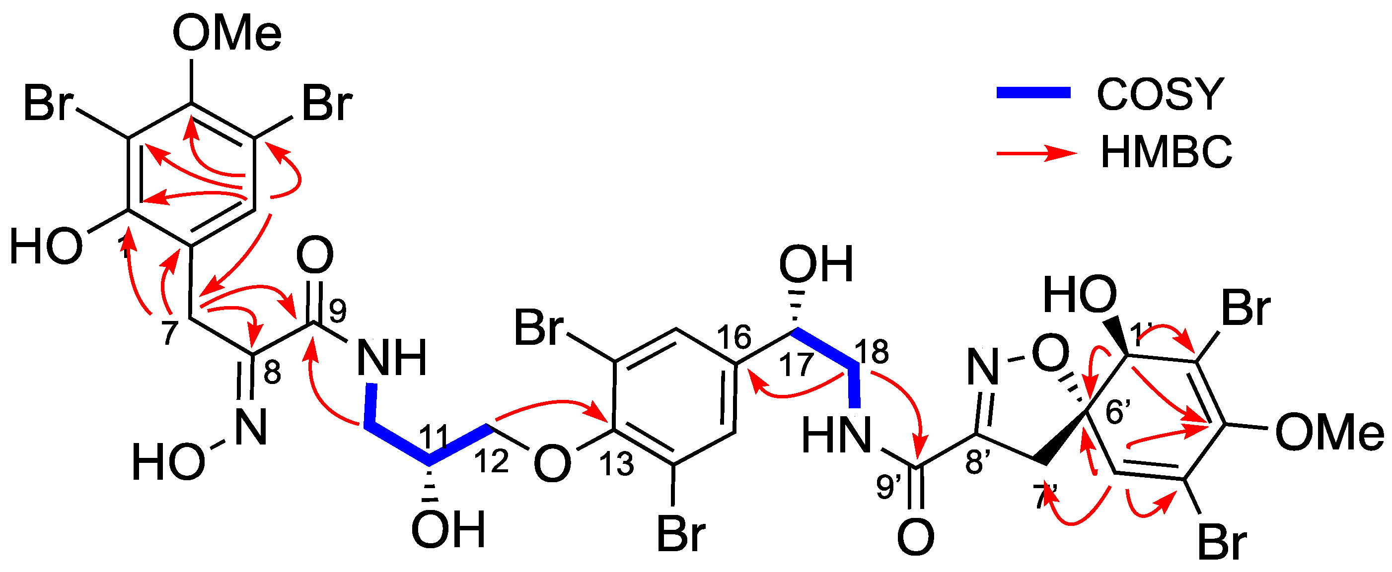
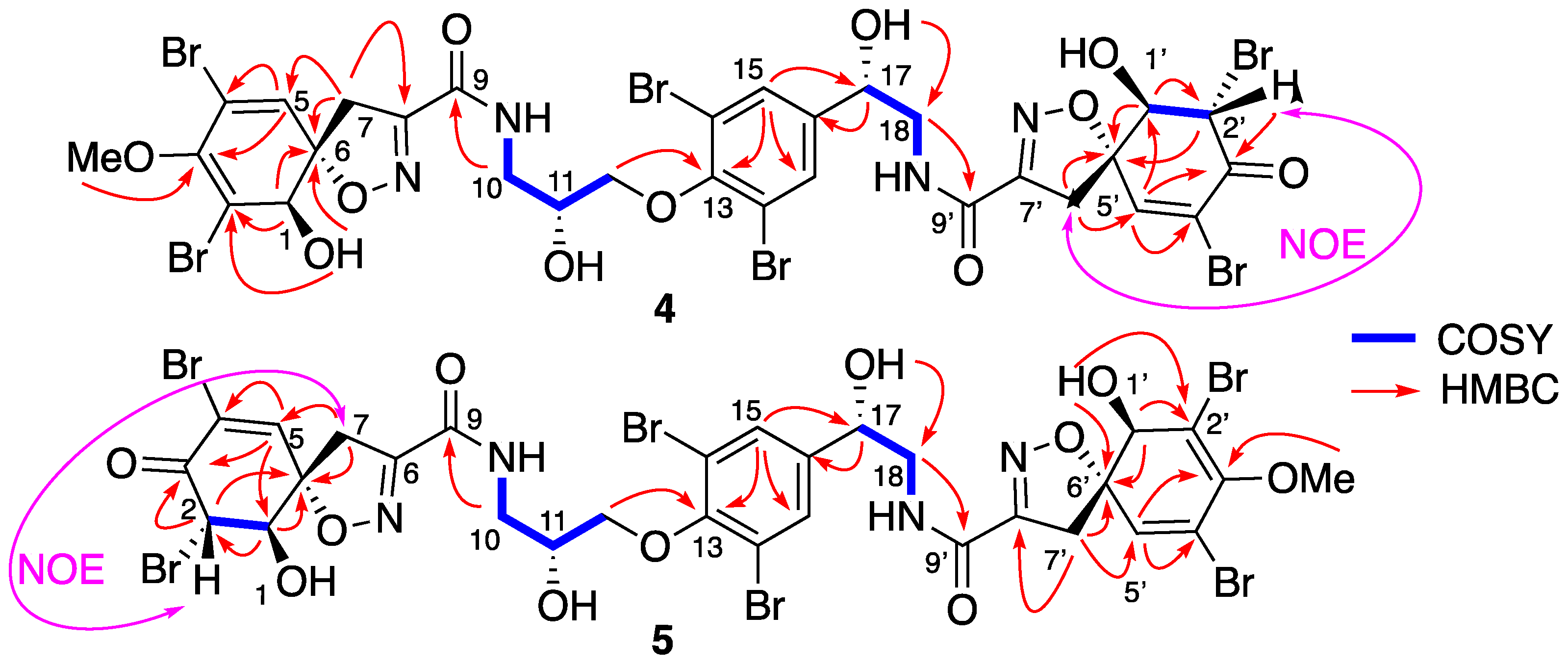
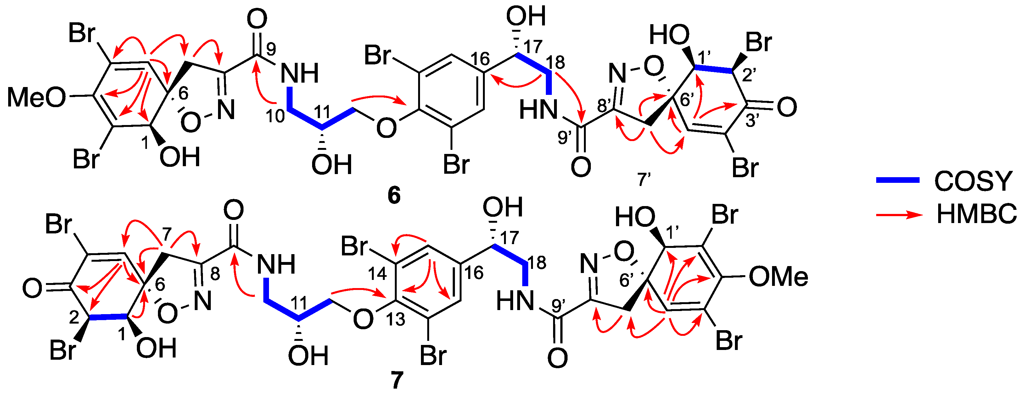
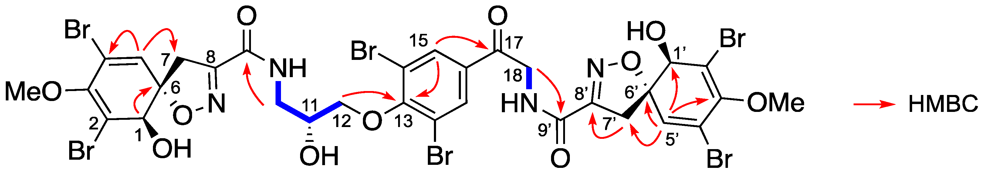
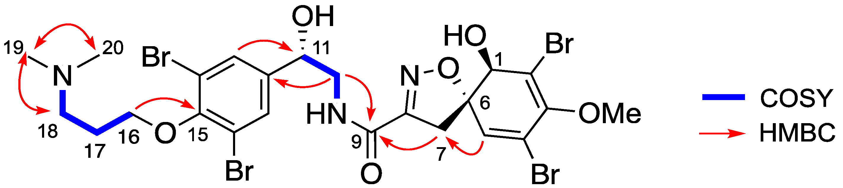
Isolation and Structure Elucidation

The sponge Suberea clavata [20,21] was collected in the Solomon Islands, extracted with a mixture of CH2Cl2/MeOH (1/1), and partitioned between n-BuOH and H2O. A part of the butanolic extract (5 g) was submitted to reversed-phase C18 SPE and eluted with successive solvents (H2O; MeOH; CH2Cl2) to afford six fractions. Step-wise repetitive purifications using HPLC (high performance liquid chromatography) and SFC (supercritical fluid chromatography) separation methods gave eight new bromotyrosine compounds 29 (Figure 1) named subereins 18, as well as eleven known metabolites (Figure S2, supporting information) such as the major compound of the extract (3 g, 14.3%) 11-epi-fistularin-3 (1) [4], 17-deoxy-epi-fistularin-3 (10) [22], agelorins A and B (1112) [4], 11-deoxyfistularin-3 (13) [23], 11,17-dideoxyfistularin-3 (14), 11-hydroxy-aerothionin (15) [14], aerophobine 2 (16), aplysinamisin-1 (17) [24], 7R,11S [3,5-dibromo-4-[(2-oxo-5-oxazolidinyl)]methoxyphenyl]-2-oxazolidinone (18) [25], subereaphenol K (19) [26], and pseudoceralidinone A (20) [27], whose structures were confirmed by comparison of their spectroscopic data with those reported in the literature. We have recently reported the in vitro inhibition of DNMT1 by our 11-epi-fistularin-3 (1) and described docking studies of the determined configuration [28].
The structure of the known epi-fistularin-3 (1) was confirmed by 1H and 13C NMR (nuclear magnetic resonance) data comparison with the literature [4,29]. In fact, similarities of 1H and 13C NMR spectra of fistularin-3 stereoisomers allow the room for confusion that exists regarding the absolute configurations of C-11 an C-17 carbons. The latter problem was pointed by Molinsky by studying samples from various sponges [29]. In the latter study, the configuration of C-11 was determined for different epimers. Finally, the C-17 configuration of all fistularin-3 isomers remains still undefined and the term isofistularin-3 is mainly used as generic name for undetermined fistularin-3 isomers so far. While the configuration of the verongidoic acid part (Figure 2) was established as 1(R), 6(S) for C-1 and C-6 by NMR spectroscopy and CD analysis [4,29,30], the absolute configurations at the two secondary carbinols C-11 and C-17 require precise determination. In the literature, this task was hampered by the presence of four secondary alcohols that significantly complicated the co-determination of their absolute configuration by chemical modification and NMR studies on the native molecule without degradation. Suitable crystals for X-ray studies have never been obtained for any one of the stereoisomers of fistularin-3. Moreover, the carbon configuration also seems to vary depending on sponge species. The name 11-epi-fistularin-3 has been used to identify a molecule isolated from Agelas oroides, which was recognized as a 11(R) [29]. Fistularin-3 extracted from Aplysina cauliformis has an 11S configuration, leaving the configuration C-17 undetermined. Our contribution starts with the determination of the absolute configuration of both C-11 and C-17. The question that arose for us is which of the two epimers have we isolated by determining the configuration of C-11 on the one hand, and on the other hand if we can determine the configuration of C-17 for the first time.
The isomer that we have isolated 1 (Figure 2) showed similar NMR spectra and an optical rotation of +148.0° (c 1.0, MeOH) or +169.0 (0.2, acetone) compared to +147° (c 0.275, acetone) for fistularin-3. The first point was to make sure that our product is not a mixture of two C-11 epimers, as Molinsky demonstrated with epi-fistularin-3 [29]. Indeed, the zoom on C-11 and C-17 (see SI, Figure S3) of our C-13 NMR spectrum shows that our sample is indeed diastereomerically pure. As already noted in the literature, these bromotyrosine metabolites crystallize poorly and do not allow X-ray analysis, and indeed we tried the derivatization of the OH functionalities of 1 with crystallogenic groups such 3,5 dinitrobenzoic acid and camphanic acid without success. For both 1, 1′, 11, 17-tetradinitrobenzoic ester and 1, 1′, 11, 17-tetracamphanic derivatives, we were not able to obtain suitable crystals. We then decided to analyze the stereochemistry by alternative methods.
The known absolute configurations of the asymmetric centers (R)C-1, (R)C-1′, (S)C-6 and (S)C-6′ on the spiroisoxazole moiety was confirmed by comparison with NMR data and the ECD spectrum that showed characteristic positive Cotton effects at 253 nm (Δє + 7.5) and 285 nm (Δє + 8.0) [31,32].
Following the method described by Molinsky [29], we hydrolyzed the epi-fistularin-3 (1) with aqueous trifluoracetic acid at 120 °C overnight to obtain the 1,2-aminopropanediol bearing the C-2 carbinol corresponding to the C-11 of compound 1. After derivatization with L-FDAA (2,4-dinitrophenyl-1-fluoro-Lalaninamide) and comparison with the C-2 configurations of the derivatized commercial standard aminodiol enantiomers, our sample was found to have the same retention time as the (+)-R-aminodiol standard, indicating that the C-11 of compound 1 is R.
At this stage, the configuration of the C-17 is still undetermined. To obtain the diastereomeric esters, 20 mg of 1 was reacted with (R) and (S)-methoxytrifluoromethylphenylacetyl chloride (MTPA-Cl) [33]. The reactions were performed in dichloromethane (DCM) in the presence of triethylamine or pyridine (10 equiv.) and a catalytic amount of 4-dimethylaminopyridine (DMAP). The monoester at position 17 was formed in minute quantity together with other polyesters at positions 1, 11, 17, and 1′. Reversed-phase HPLC (Sunfire C18, 10 × 150 mm, 5 µm; eluting with H2O/Acetonitrile (98/2 to 0/100 in 35 min) led to the pure desired monoesters. It should be noted that upon ester formation, the stereochemical descriptor of the MTPA-Cl stereocenter changes, because the COCl group has a Cahn–Ingold–Prelog (CIP) priority different from COOR. The Mosher esters obtained by reacting 1 with (R)- and (S)-MTPA chloride to yield the (S)- and (R)-MTPA esters respectively were analyzed using 1D and 2D NMR spectroscopy [33]. The difference in the chemical shift (δSR) was calculated for each of the analogous pairs of protons for both the (S)- and (R)-MTPA esters (Table 1). The positive values were assigned to the left side of the model (R1) and the negative values were placed on the right side (R2). By applying the Cahn–Ingold–Prelog system, the groups’ priorities were assigned as 1 (OH), 2 (C-18), 3 (C-15/15′), and 4 (H), and the absolute configuration at C-17 was determined as (S) for the first time.
In a slightly less polar fraction, we identified a mixture of two compounds 2 and 3 with the same HRESIMS observed for compound 1. Pure compounds 2 and 3 were obtained by SFC, using a Cyano column. Compound 2 (Figure 2) was isolated as an amorphous white solid, [α]D25 + 61.0° (c 1.0, MeOH). Its HRESIMS spectrum showed a predominant peak at m/z 1136.6924 [M+Na]+ corresponding to the molecular formula C31H3079Br381Br3N4O11 (calc. for C31H30Br6N4O11Na: 1136.6849). This isomer of fistularin-3 presented some differences on the 1H NMR spectrum. The singlet H-5 expected at δH 6.55 ppm was not observed, but another one appeared at δH 7.56 ppm, a chemical shift in accordance with an aromatic proton. An interesting change was observed for the CH2-7′ observed at δH 3.81 ppm as a singlet associated by HSQC with a carbon observed at the low chemical shift δC 25.5 ppm. The COSY spectrum (Figure 2) displayed cross-peak correlations between the signals at δH 7.56 and 3.81 corresponding to a 4J coupling suggesting an aromatization together with one of the spiroisoxazoline opening. The second spiroisoxazoline with its diastereotopic protons at δH 3.87–3.84/3.22–3.18 was observed. The 13C NMR spectrum showed more signals similar to those observed for compound 1, suggesting its partial modification. The HMBC spectrum showed a key correlation between the protons of the methylene group at δH 3.81 and the carbons at δC 155.1(C1′) and 122.1(C6′). Furthermore, the proton H-5′ at δH 7.56 was also correlated to C-1′, C-6′, and to those at δC 106.3 (C-4′) and 108.9 (C-2′). The COSY spectrum (recorded in acetone-d6) revealed a spin system including a methylene group CH2-10 at δH 3.78/3.52, followed by an -NH proton at δH 7.62 and oxymethine CH-11 at δH 4.24, and terminated with two diastereotopic protons CH2-12 at δH 4.02/4.05. The HMBC spectrum displayed key correlations between H-10 and H-7 with the amidic carbonyl C-9 at δC 160.4 (Figure 3), which confirmed that the left-hand side of the molecule was retained is an oxazolinic motif like in compound 1. Further significant HMBC correlations were observed between CH2-12 δH 4.02/4.05 and C-13 at δC 152.7. Additionally, the COSY spectrum allowed us to identify a second partial structure including the oxymethine H-17 at δH 4.92, two diastereotopic protons at δH 3.64/3.49, and -NH at δH 8.05 ppm. The proton CH-17 at δH 4.92 displayed key HMBC corrections with C-15/15′ and C-16 at δC 131.5 and 143.1, respectively. Furthermore, the methylene groups CH2-18 at δH 3.64/3.49 and CH2-7′ at δH 3.85/3.19 exhibited additional HMBC correlations with the quaternary carbon C-9′ at δC 166.7, which confirmed the planar structure of the compound 2. The stereochemistry of the spiroisoxazoline moiety was determined as 1R, 6S by its ECD spectrum, which showed characteristic positive Cotton effects at 256 nm (Δє + 2.55) and 290 nm (Δє + 2.17). Compound 2 was named suberein-1.
Compound 3 (Figure 4) was a regioisomer of compound 2 ([α]D25 + 70.7° (c 1.0, MeOH)). The HRESIMS spectrum showed the predominant peak at m/z 1136.6864 [M+Na]+, corresponding to a molecular formula C31H30Br6N4O11 (calc. for C31H30Br6N4O11Na, 1136.6849). The 1H and 13C NMR spectral data (Table 2) were similar to those of compound 2 and suggested the opening of the other spiroisoxazoline moiety. This was confirmed by the key HMBC correlations between the methylene groups CH2-7 at δH 3.84 and CH2-10 at δH 3.78/3.58 with the amidic carbonyl C-9 at δC 166.6 (Figure 4). The absolute configuration of the spiroisoxazoline moiety was determined as 1R, 6S based on positive Cotton effects at 255 nm (Δє + 2.33) and 286 nm (Δє + 2.19), in its ECD spectrum. Compound 3 was named suberein-2.
Compounds 4 + 5 were isolated together as an inseparable 1/1 mixture. Their HRESIMS spectrum exhibited a predominant peak at m/z 1100.6935 [M+H]+ corresponding to a molecular formula C30H28Br6N4O11 (calc. for C30H29Br6N4O11, 1100.6872), which indicated 14 a.m.u less than 11-epi-fistularin-3 (1). The 1H NMR spectrum (Table 3) showed only one methyl group as a singlet at δH 3.73 instead of two in 11-epi-fistularin-3 (1). Indeed, the 13C NMR spectrum disclosed two characteristic resonances at δC 183.6 (C-3′) assigned to a carbonyl group, and δC 75.3 attributed to C-1′, which were not recorded for 11-epi-fistularin-3 (1). The COSY spectrum showed key correlations between the proton at δH 5.08 (H-2′) and the oxymethine at δH 4.41 (H-1′), with a coupling constant J = 11.4 Hz. Significant HMBC correlations were observed between the proton H-5′ and the carbons at δC 75.3 (C-1′), 57.4 (C-2′), 183.6 (C-3′), 122.6 (C-4′), and 91.8 (C-6′). Additionally, the oxymethine H-1′ was correlated with C-2′ and meanwhile, H-2′ was correlated with C-3′. Thus, the structure of 4 was determined as described in Figure 5. The same demethoxylated substructure was observed on its isomer 5 in the left side of the molecule (C-1 to C-6 instead of C-1′ to C-6′). The observed NOE correlation between H-2′ and one of the H-5′ together with the absence of NOE correlations between H-1′ and H2′ suggested a trans configuration for 4 + 5. Compounds 4 and 5 are named suberein-3 and suberein-4, respectively.
As for 4 + 5, the following compounds 6 + 7 were also isolated together as an inseparable in 1/1 mixture. Their molecular formulas were established as C30H28Br6N4O11 from their positive HRESIMS spectrum with a predominant peak at m/z 1100.6935 [M+H]+, (calc. for C30H2979Br381Br6N4O11, 1100.6872). Their 1H NMR spectrum (Table 4) showed strong similarities with their stereoisomers 4 + 5, but the main difference appeared in the chemical shift of H-5/H-5′, H-1/H-1′ and H-2/H-2′. The coupling constant between H-1/H-1′ and H-2/H-2′ was close very weak (br s) suggesting a cis configuration, which was further confirmed by the NOE correlations. Therefore, the structure of 6 and 7 were assigned as described in Figure 6 and named suberein-6 and suberein-7, respectively.
It should be noted that due to the keto-enolic equilibrium, carbons C-2 and C-2′ in both mixtures 4 + 5 and 6 + 7 epimerize within two days in solution into a mixture of 4/5/6/7: 1/1/1/1. Their purification just allowed their characterisation as mixtures of the structures of the cis stereoisomers on the one hand and of the trans stereoisomers on the other hand.
Compound 8 was isolated as an amorphous powder ([α]D25 + 149.5° (c 0.5, MeOH)). Its HRESIMS showed an isotopic cluster indicating six bromines with a principal peak at m/z 1110.6661 [M−H] corresponding to a molecular formula C31H28Br6N4O11 (calc. for C31H27Br6N4O11, 1110.6715). Its 1H NMR spectrum (Table 2) showed the characteristic signals of the fistularin-3 with some modifications, including the presence of a doublet at δH 4.87 (H-18) together with the absence of the H-17 signal observed for fistularin-3 at δH 4.90. The chemical shift of the aromatic protons (H-15/15′) was observed at δH 8.28. The 13C NMR spectrum (Table 2) showed a quaternary carbon at δC 192.2, characteristic for a carbonyl group. The HMBC spectrum displayed significant correlations between H2-18 (δH 4.87), H-15/15′ (δH 8.28), and the non-protonated carbon at δC 192.2, thus assigning the carbonyl group to C-17. Further key HMBC correlations were consistent with the backbone of fistularin-3. Thus, the structure was depicted as 17-oxo-11-epi-fistularin-3 for compound 8 and named suberein-7 (Figure 7), a new member of the bromotyrosine-derived compounds.
Compound 9 ([α]D25 + 84.0° (c 0.2, MeOH)) showed in its HRESIMS an isotopic cluster of peaks indicating a tetrabrominated compound with a principal peak at m/z 761.8695 [M+H]+ corresponding to the molecular formula C23H26Br4N3O6 (calc. for C23H28Br4N3O6, 761.8671). The 1H NMR spectrum (Table 5) disclosed characteristic signals with similar chemical shifts to those reported for the right-hand side of epi-fistularin-3 (1). The COSY spectrum allowed the identification of additional spin system including an oxymethylene group (CH2-16) at δH 4.07 based on its chemical shift and CH2 group at δH 2.11 (CH2-17), and terminated with an azomethylene group (CH2-18) at δH 2.87. A singlet at δH 2.47 integrated for six protons was assigned to the N,N-dimethyl terminus (CH3-19/20). The HMBC spectrum displayed key correlations between the oxymethylene group at δH 4.07 (CH2-16) and the quaternary carbon C-15 at δC 152.1, linking this partial structure to the tetrasubstituted phenyl ring. Furthermore, the signal corresponding to the N,N-dimethyl functionality (δH 2.47, CH3-19/20) displayed HMBC correlation with the carbon C-18 at δC 56.0. The other HMBC correlations were consistent with the proposed structure for 9 and named suberein-8 (Figure 8).
Compound 10 ([α]D25 + 71.0° (c 0.3, MeOH)) showed in its negative HRESIMS a characteristic isotopic cluster of a hexabrominated compound with a predominant peak at m/z 1096.6923 [M−H] corresponding to the molecular formula C31H30Br6N4O10 (calc. for C31H29Br6N4O10, 1096.6923). Its 1H NMR spectral pattern was identical to the one of 17-deoxyfistularin-3 [21]. However, the absolute configuration of C-11 was not determined. As for compound epi-fistularin (1) described above, placing 17-deoxyfistularin-3 (10) after hydrolysis in aqueous trifluoroacetic acid to obtain the 1,2-aminopropanediol and further Marfey’s derivatization and comparison with R and S 1,2-aminopropanediol standards, we found that the absolute configuration of C-11 was R. Thus, compound 10 was assigned as a 17-deoxy-11(R)-epi-fistularin-3 (Figure 9).
The presence of agelorins and compounds 28 with 11-epi-fistularin (1) in our sponge brings us to the question of which one the precursor is, and if degradation processes can take place during purification. This is interesting because all the reactions involved are simple and can be imagined without any particular catalysis. Hydrolysis and simple oxidation of the benzylic alcohol into ketone can take place spontaneously. The presence of water, oxygen, and the variation of the acid–base conditions are sufficient. As noted above, such transformations were previously observed for agelorins A-B (1112) which were isolated separately with a smaller retention time [4,29]. Rogers et al. identified a mixture of these two products in a sample of epi-fistularin-3 (1) after storing for several years at −20 °C [29]. When epi-fistularin-3 is heated in acidic conditions, several degradation compounds appeared. We then decided to manage the degradation process of epi-fistularin-3 (1) in a less drastic conditions by dissolving it in a mixture of acetonitrile/water (+0.1% HCOOH) 1/1 and stirring at room temperature for two weeks. The reaction mixture was then dried under vacuum and chromatographed. In addition to the formation of agelorin A and agelorin B, we identified compounds 27 as well. This result raises the interesting question as to whether these compounds are natural or simply degradation derivatives of 11-epi-fistularin-3 (1).

3. Biological Activities

3.1. Antibacterial Activity Inhibition

Minimum inhibitory concentration (MIC values) results are presented in Table 6. Compounds were tested against a panel of bacterial strains commonly used for assessment of their anti-biofilm properties [34]. None of the compounds tested displayed antibacterial activity (at the concentrations tested) against all the panel of bacteria tested (four marine bacteria): Vibrio natriegens, Shewanella putrefaciens, Pseudoalteromonas elyakovii, Polaribacter irgensii; and two terrestrial bacteria: Staphylococcus aureus and Pseudomonas aeruginosa. Moreover, among all the tested compounds, no activity was observed (at the tested concentrations) for 11-epi-fistularin-3 (1), compounds 4 and 5, 11-hydroxyaerothionin (15), and aplysinamin-1 (17).
The most active molecule was compound 3, which displayed an MIC value of 0.01 µg/mL (the lowest concentration tested) against V. aesturianus and E. coli. These results are of great importance as new inhibitors against these strains are sought-after actively by scientists, and indeed not only for antifouling purposes. E. coli is a common inhabitant of the human gut microbiota and commonly causes nosocomial infection, urinary tract infections, neonate meningitis, and bacteria-related diarrhea. Resistance to antimicrobial agents (e.g., broad-spectrum penicillin and trimethoprim or third generation cephalosporin and nitrofurantoin) in E. coli has been reported worldwide and has an increasing impact on available therapeutic options [35]. There is an urgent need for new compounds inhibiting the growth of E. coli. The inhibition of V. aesturianus is of high importance as well as this bacterial strain is involved in surface colonization, but it is as well a known pathogen of the commercial oyster Crassostrea gigas [36,37,38]. Compounds 24 could possibly be used in aquaculture for the prevention of diseases. The oyster industry has grown to be very important in many regions of the world contributing substantially to social and economic activity in coastal zones. Abnormal mortality events due to outbreaks of V. aesturianus have been reported increasingly since 2008, leading to very high levels of mortality which were sudden and severe (up to 100%), and affected essentially spats (oysters less than one year old) and juveniles (12- to 18-month-old oysters). Massive mortality outbreaks resulted in a shortage in supplies of the shellfish over the next years [39].
It is worth mentioning that the mono- or di-deoxyfistularins (11-deoxyfistularin-3 (13), 11,17-dideoxyfistularin-3 (15)) displayed MIC values of 0.01 µg/mL (the lowest concentration tested) specifically toward Halomonas aquamarina. The discovery of H. aquamarina inhibitors is also of great interest since this strain is involved in both marine biofilm formation [40,41] and pathogenicity towards lobster Homarus americanus, an important species in aquaculture [42]. Halomonas aquamarina also poses a threat as a potential invasive species because it can lead to potential invasion when transported on biofilms inside ballast water tanks [43].
Molecules with targeted activities are of major interest because they make it possible to fight specifically against targeted bacteria without altering non-targeted organisms. This significantly slows down the development of resistance.

3.2. Acetylcholinesterase Activity Inhibition

The activity of these compounds was also evaluated on acetylcholinesterase, which is responsible in terrestrial invertebrates for various primordial biosynthetic pathways, the inhibition of which can be lethal for insect species and thus represents a great phytosanitary interest [44,45], and also might be a toxicity indicator in the marine environment, especially in bivalves [46].
The involvement of neurotransmission inhibition through acetylcholinesterase inhibition has been shown in a settlement of cyprid larvae of barnacles [47,48]. Agelorin A (12) displayed a better activity than the reference compound Galantamine used as a control for the bioassay (IC50 = 0.7 ± 0.1 µM). The acetylcholinesterase inhibition of bromotyrosine derivatives enhances their interest as efficient antifouling natural products especially in their natural environment as a mode of chemical defense.

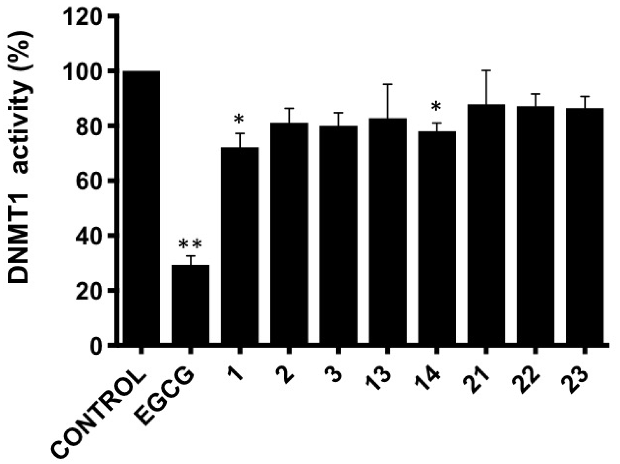
3.3. DNMT1 Activity Inhibition

Aberrant DNA methylation is a hallmark of cancer cells, and the enzyme DNA methyltransferase 1 (DNMT1) represents a recognized target in cancer treatment. As a continuation of our previously demonstrated potential of 11-epi-fistularin (1) to bind DNMT1 and reduce its activity [28], and that of isofistularin-3 extracted from the sponge Aplysina aerophoba, and its ability to inhibit DNMT1 leading to DNA demethylation in leukemia cells [49], we also investigated the capacity of our compounds 2, 3, 13, and 14 to modulate the activity of DNMT1 (Figure 10). Results were compared to bromotyrosine compounds psammaplysene D (21), F (22), and G (23) (SI Figure S0), previously isolated from another sponge species in our group, Suberea ianthelliformis [19].
In line with our previous results, compound 1 reduced DNMT1 activity to around 70% of control values and compound 14 also provided a statistically significant decrease.
In summary of this manuscript, the chemical examination of the Pacific marine sponge Suberea clavata led to the isolation and identification of twenty structurally diverse brominated tryrosine metabolites, of which eight are new congeners, subereins-1–8 (29), along with two major co-isolated known products 11-epi-fistularin-3 (1) and 17-deoxyfistularin-3 (10). A combination of chemical modifications, Mosher’s technology, and ECD spectroscopy allowed the complete assignment of the absolute configurations of the new compounds on one hand, and the assignment of the stereogenic centers C-11/17 of the known congeners (11R,17S) 11-epi-fistularin-3 (1) and 17-deoxyfistularin-3 (10) for the first time on the other hand.
This solves the problem of the absolute configuration of the C-17 of 11-epi-fistularin-3 (1), which has remained undetermined since 1993 [4]. Whereas this metabolite is predominant in several sponges and presents interesting biological activities, the easily observed transformation of the major compound 11-epi-fistularin-3 (1) into subereins 1–6 (27) rise the question of artifacts of extraction. We believe that all compounds are also present in the sponge since they are present in the crud extract of the sponge. This does not exclude their partial formation during chromatographic purifications. The isolated compounds 3, 13, and 15 displayed antimicrobial inhibition; 12 displayed acetylcholinesterase inhibition; and 1 and 14 inhibited DNMT1.

4. Materials and Methods

4.1. General Experimental Procedure

Optical rotations [α]D were measured at 25 °C on a Jasco P-1010 polarimeter (JASCO, Lisses, France). Electronic circular dichroism (ECD) experiments were performed at 25 °C on a JASCO J-810 spectropolarimeter (JASCO, Lisses, France). IR spectra were recorded on a Perkin Elmer BX FT-IR spectrometer (Perkin Elmer, Villebon-sur-Yvette, France). The NMR spectra were recorded on a Bruker 300 MHz instrument (Avance 300), a Bruker 500 MHz instrument (Avance 500,) and a Bruker DRX 600 MHz with a 5 mm or 1.7 mm triple resonance (HCN) probe (Bruker, Wissembourg, France). The chemical shifts are reported in ppm relative to the residual signal solvent (MeOH-d4: δH 3.31; δC 49.15; DMF-d7: δH 8.03, 2.92, 2.75; δC 163.15, 34.89, 29.76; acetone-d6: δH 2.05; δC 206.68, 29.92). High-resolution mass spectra were obtained with a hybrid linear trap/orbitrap mass spectrometer (LTQ-orbitrap, Thermofisher, Illkirch, France) in electrospray ionization mode by direct infusion of the purified compounds. MPLC was performed using a Combiflash-Companion apparatus (Serlabo Technologies, Entraigues-sur-la-Sorgue, France) and a prepacked C18 Versapak cartridge. Preparative HPLC purifications were performed on an autoprep system (Waters 600 controller and Waters 600 pump with a Waters 996 photodiode array detector (Waters France, Guyancourt, France)), equipped with a Waters Atlantis T3 (19 × 150 mm, 5 μm) column, and a Waters Sunfire C18 (19 × 150 mm, 5 μm) column or a X-bridge RP-18 (19 × 150 mm, 5 μm) column. SFC purifications were performed on a Thar Waters SFC Investigator II with a Waters 2998 photodiode array detector, equipped with a SFC 2-Ethyl-pyridine (10 × 250 mm, 6 μm) column or a Thar Cyano (10 × 250, 6 μm) column. Analytical UHPLC was performed on UPLC Waters Acquity with PDA, DEDL, and TQD, using a HSS C18 (2.1 × 50 mm, 1.8 μm) column. All other chemicals and solvents were purchased from SDS (France).

4.2. Biological Material

The sponge was collected by hand using SCUBA in the Solomon Islands, Russell Group, during the sampling cruise BSMS-1 [50] aboard the R/V Alis, off the coast of Lologhan Island (30 June 2004, 9°06.658′ S; 159°21.664′ E) between 6 and 12 m deep. A voucher sample is deposited at the Queensland Museum (Brisbane, Australia) under the access number G322641 and was identified by Dr. J. N. A. Hooper as Suberea clavata. The sponge was deep frozen on board until work up. It was then grounded, freeze-dried, and extracted.

4.3. Isolation and Spectroscopic Data

4.3.1. Isolation

The lyophilized sponge Suberea clavata (200 g) was extracted at room temperature with MeOH/DCM (1/1) to give 57.8 g of dried extract. The crude extract was submitted to n-butanol/H2O partition to obtain a desalted butanol extract (30 g). A portion (25 g) of the later extract was submitted to reversed-phase C18 SPE (50 μm, 65 A, Phenomenex Sepra) and eluted with a gradient H2O/MeOH (100/0 to 0/100) and MeOH/DCM (100/0 to 0/100) to give six fractions f1 to f6. The fraction f4 (10 g) was submitted to reversed-phase HPLC (Waters Sunfire C18 column, H2O + 0.1% HCOOH/CH3CN + 0.1% HCOOH: 85/15 to 0/100 in 40 min) yielding 13 subfractions, F1 to F13. The sub-fraction F1 afforded pure aerophobin-2 (16) (140 mg, RT = 10 min).
Fraction F6 only contained 11-epi-fistularin-3 (1) (2.7 g, RT = 21.8 min) and F9 pure 11,17-dideoxyfistularin-3 (14) (220 mg, RT = 25.6 min).
Fraction F7 was purified by SFC (ethylpyridine column, CO2/MeOH (75/25), isocratic condition) and yielded 17-deoxy-epi-fistularin-3 (10) (8 mg, RT = 12.0 min), 11-deoxyfistularin-3 (14) (36 mg, RT = 11.5 min), and 17-oxo-11-epi-fistularin-3 (8) (9 mg, RT = 14.1 min).
Fraction F8 was purified by SFC (cyano column, CO2/iPrOH (75/25), isocratic condition) to yield 11-epi-fistularin-3 (1), suberein-1 (2) (10 mg, RT = 11.8 min), and suberein-1 (3) (10 mg, RT = 13.0 min).
Fraction F5 was submitted to reversed-phase HPLC (Waters Sunfire C18, H2O + 0.1% HCOOH/CH3CN + 0.1% HCOOH, 85/15 to 25/75 in 25 min). Agelorin A (11) (3 mg, RT = 19.6) and B (12) (3 mg, RT = 20.1), 11-hydroxyaerothionin (15) (2 mg, RT = 22.2 min), as well as a mixture of 4 + 5 (5 mg, RT = 20.9 min) and a mixture of 6 + 7 (5 mg, RT = 21.2 min), were obtained.
Fraction F1 (1.5 g) was purified by MPLC (Versapak C18 silica-gel cartridge (23 × 110 mm); H2O + 0.1% HCOOH/MeOH+0.1% HCOOH, 100/0 to 0/100, in 35 min). It yielded eight subfractions F’1 to F’8. Pseudoceralidinone A (20) (5 mg, RT = 3.5 min) was obtained from F’1. Fraction F’7 was submitted to reversed-phase HPLC (Atlantis T3 C18, H2O + 0.1% HCOOH/CH3CN + 0.1% HCOOH, 100/0 to 75/25 in 7 min, 75/25 to 50/50 in 18 min) to afford aplysinamisin-1 (17) (15 mg, RT = 10.5 min), subereaphenol K (19) (4 mg, RT = 11.1 min), and 7R, 11S [3,5-dibromo-4-[(2-oxo-5-oxozolidinyl)]methoxyphenyl]-2-oxazolidinone (18) (17 mg, RT = 12.5 min). Fraction F’8 afforded suberein-8 (9) (3 mg, RT = 24.0 min) by reversed-phase HPLC purification using an Xbridge C18 column (pH 10.5, H2O + 0.15%NH4OH)/ CH3CN + 0.15% NH4OH, 90/10 to 0/100 in 35 min).

4.3.2. Spectroscopic Data and Absolute Configurations Determination

17S-epi-fistularin-3 (1): yellowish amorphous solid (3 g); [α]D25 + 148.0° (c 1.0, MeOH); [α]D25 + 169.0 (c 0.2, acetone); ECD (0.15 mM, MeOH) λmax (Δє) 253 (+7.5), 285 (+8.0); 1H NMR (500 MHz, acetone-d6): δH ppm 7.67 (m, 1H, NH’), 7.66 (s, 2H, H-15, H-15′), 7.62 (m, 1H, NH), 6.53 (d, 2H, H-5, H-5′), 5.41 (d, J = 8.0 Hz, 2H, OH-1, OH-1′), 5.00 (d, J = 4.3 Hz, 1H, OH-17), 4.90 (dd, J = 7.7, 5.5, 4.3 Hz, 1H, H-17), 4.44 (d, J = 5.3 Hz, 1H, OH-11), 4.25 (m, 1H, H-11), 4.18 (dd, J = 8.0 Hz, 2H, H-1, H-1′), 4.04 (m, 2H, H-12), 3.85 (d, J = 18.0 Hz, 1H, H-7a), 3.82 (d, J = 18.0 Hz, 1H, H-7a’), 3.80 (m, 1H, H-10a), 3.73 (s, 6H, OCH3, OCH3′), 3.63 (m, 1H, H-18a), 3.54 (m, 1H, H-10b), 3.49 (m, 1H, H-18b), 3.19 (d, J = 18.0 Hz, 1H, H-7b), 3.16 (d, J = 18.0 Hz, 1H, H-7b’); 13C NMR (125 MHz, acetone-d6): δC ppm 160.5 (C-9, C-9′), 155.2 (C-8′), 155.1 (C-8), 152.7 (C-13), 148.8 (C-3, C-3′), 143.3 (C-16), 132.4 (C-5′), 132.3 (C-5), 131.5 (C-15, C-15′), 122.1 (C-4, C-4′), 118.4 (C-14, C14′), 113.9 (C-2′), 113.8 (C-2), 91.8 (C-6, C-6′), 75.9 (C-12), 75.2 (C-1, C-1′), 71.0 (C-17), 69.9 (C-11), 60.2 (OCH3, OCH3′), 47.7 (C-18), 43.6 (C-10), 40.0 (C-7, C-7′); HRESIMS m/z 1136.6934 [M+Na]+ (calc. for C31H29Br6N4O11Na, 1136.6847).

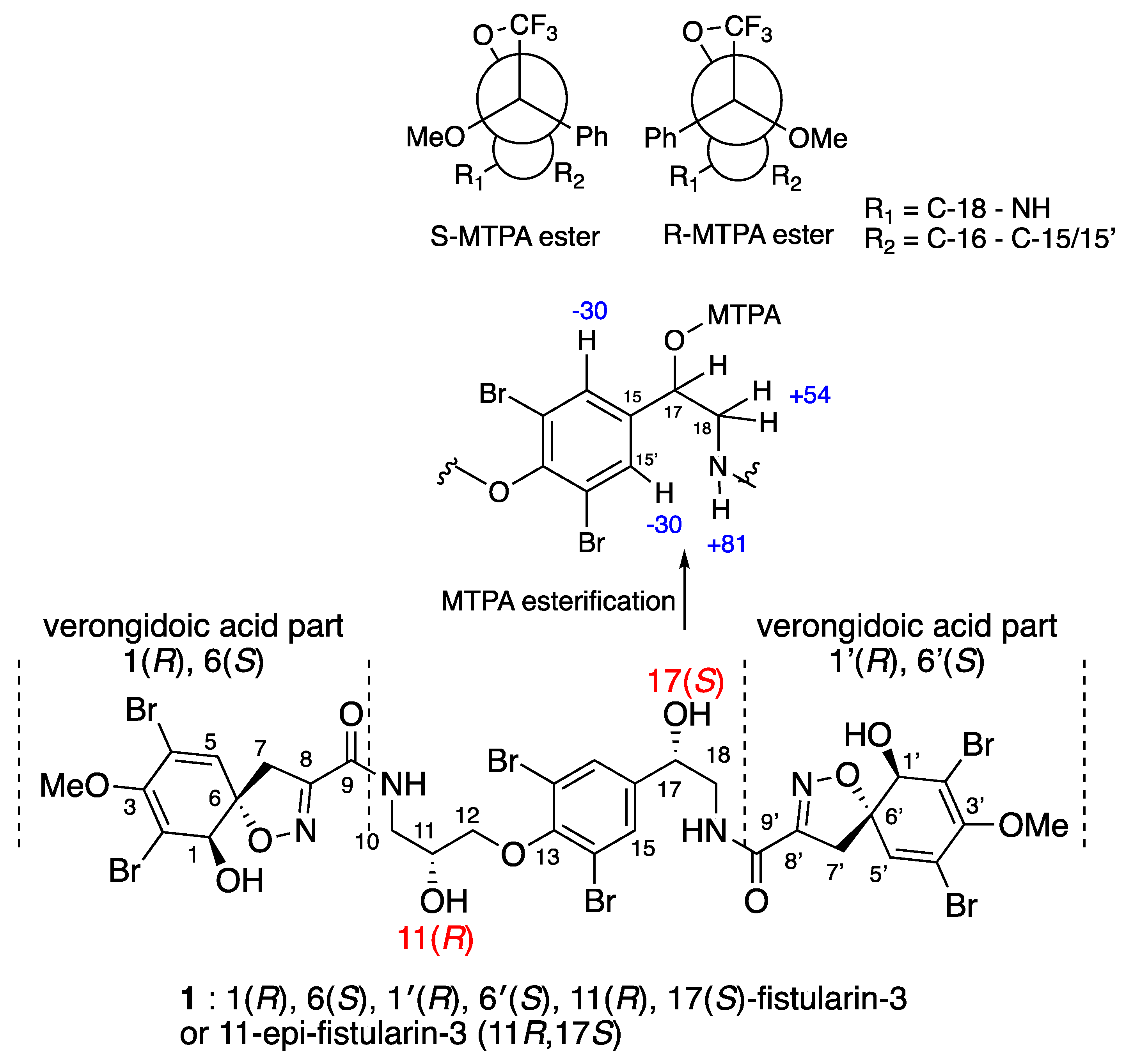
Determination of the C-17 Configuration for 11-epi-fistularin-3 (1)

To a stirred solution of compound 1 (20 mg, 0.018 mmol) and anhydrous Pyridine (15 μL, 0.18 mmol, 10 equiv.) in anhydrous DCM (270 μL, [1] = 0.066 M) at room temperature, R-(-)-MTPA-Cl (34 μL, 0.18 mmol, 10 equiv.) and DMAP (0.5 mg) were added. The reaction progress was monitored by thin-layer chromatography (TLC) on silica gel (95:5 DCM/MeOH). After 3 h, compound 1 was totally consumed. The reaction mixture was quenched by the addition of water and EtOAc. The aqueous layer was extracted with two additional portions of EtOAc, and the combined organic layers were dried on MgSO4, filtered, and concentrated in vacuo. The crude product mixture was purified by reversed-phase HPLC (Sunfire C18, 10 × 150 mm, 5 μm; eluting with H2O/acetonitrile (98/2 to 0/100 in 25 min, RT = 22.3 min.)) to give the S-MTPA-epi-fistularin-3 ester (2.6 mg, 11% yield) as a white solid: 1H NMR (300 MHz, acetone-d6): δH 7.95 (m, 1H, NH’), 7.63 (m, 3H, NH’, H-15, H-15′), 7.46 (m, 4H, MTPA), 7.40 (m, 1H, MTPA), 6.52 (dd, J = 9, 0.7 Hz, 2H, H-5, H5′), 6.13 (dd, J = 8.5, 4.0 Hz, 1H, H-17), 5.42 (m, 2H, OH-1, OH-1′), 4.48 (m, 1H, OH-11), 4.25 (m, 1H, H-11), 4.18 (m, 2H, H-1, H-1′), 4.08 (m, 2H, H-12), 3.98 (m, 2H, H-18), 3.88–3.82 (d, J = 18 Hz, 1H, H-7), 3.86–3.80 (d, J = 18 Hz, 1H, H-7′), 3.82 (m, 1H, H-10a), 3.73 (d, 6H, 3-OCH3, 3′-OCH3), 3.62 (m, 3H, MTPA-OCH3), 3.55 (m, 1H, H-10b), 3.23–3.17 (d, J = 18 Hz, 1H, H-7), 3.19–3.13 (d, J = 18 Hz, 1H, H-7′); HRESIMS m/z 1352.7357 [M+Na]+, (calc. for C41H3779Br381Br3F3N4O13Na, 1352.7246).
To a stirred solution of compound 1 (20 mg, 0,018 mmol) and anhydrous Pyridine (15 mL, 0.18 mmol, 10 equiv.) in anhydrous DCM (270 μL, (1) = 0.066 M) at room temperature, S-(+)-MTPA-Cl (34 μL, 0.18 mmol, 10 equiv.) and DMAP (0.5 mg) were added. The reaction progress was monitored by thin-layer chromatography (TLC) on silica gel (95:5 DCM/MeOH). After 4 h, compound 1 was totally consumed. The reaction mixture was quenched by the addition of water and EtOAc. The aqueous layer was extracted with two additional portions of EtOAc, and the combined organic layers were dried on MgSO4, filtered, and concentrated in vacuo. The crude product mixture was purified by reversed-phase HPLC (Sunfire C18, 10 × 150 mm, 5 μm; eluting with H2O/acetonitrile (98/2 to 0/100 in 25 min, RT = 21.9 min.)) to give the R-MTPA-epi-fistularin-3 ester (3.2 mg, 13% yield) as a white solid: 1H NMR (300 MHz, acetone-d6): δH 7.73 (s, 2H, H-15, H15′), 7.68 (m, 2H, NH, NH’), 7.48 (m, 4H, MTPA,), 7.30 (m, 1H, MTPA), 6.52 (dd, J = 11, 0.9 Hz, 2H, H-5, H5′), 6.21 (dd, J = 8, 5 Hz, 1H, H-17), 5.43 (dd, J = 8.5, 3.0 Hz, 2H, OH-1, OH-1′), 4.49 (m, 1H, OH-11), 4.26 (m, 1H, H-11), 4.17 (m, 2H, H-1, H-1′), 4.09 (m, 2H, H-12), 3.88–3.83 (d, J = 18.3 Hz, 1H, H-7), 3.82–3.77 (d, J = 18.3 Hz, 1H, H-7′), 3.80 (m, 1H, H-18), 3.78 (m, 1H, H-10a), 3.73 (d, 6H, 3-OCH3, 3′-OCH3), 3.60 (m, 1H, H-10b), 3.50 (s, 3H, MTPA-OCH3), 3.23–3.16 (d, J = 18.5 Hz, 1H, H-7), 3.12–3.06 (d, J = 18.5 Hz, 1H, H-7′); HRESIMS m/z 1352.7282 [M+Na]+ (calc. for C41H3779Br381Br3F3N4O13Na, 1352.7246).
Suberein-1 (2): colourless amorphous solid (10 mg); [α]D25 + 61.0 (c 1.0, MeOH); [α]D25 + 32.5 (c 0.2, acetone); UV (MeOH) λmax (є) 208 (38200), 286 (10100) nm; ECD (0.15 mM, MeOH) λmax (Δє) 214 (−2.79), 256 (2.55), 290 (2.17) nm; IR νmax 3380, 2990, 2900, 1655, 1630, 1545, 1460, 1400, 1255 cm-1; 1H and 13C NMR data, Table 2; HRESIMS m/z 1136.6924 [M+Na]+ (calc. for C31H30Br6N4O11Na, 1136.6849).
Suberein-2 (3): colourless amorphous solid (10 mg); [α]D25 +70.7 (c 1.0, MeOH); [α]D25 + 32.0 (c 0.2, acetone); UV (MeOH) λmax (є) 208 (38200), 286 (10100) nm; ECD (0.15 mM, MeOH) λmax (Δє) 215 (–1.73), 255 (2.33), 286 (2.19) nm; IR νmax 3360, 2985, 2900, 1655, 1545, 1460, 1415, 1260 cm-1; 1H and 13C NMR data, Table 2; HRESIMS m/z 1136.6864 [M+Na]+ (calc. for C31H30Br6N4O11Na, 1136.6849).
Suberein-3 and suberein-4 (4 and 5): amorphous off-white powder (5 mg); UV (MeOH) λmax (є) 210 (22500), 232 (7800), 260 (5100) nm; 1H and 13C NMR data, Table 3; HRESIMS m/z 1100.6935 [M+H]+ (calc. for C30H29Br6N4O11, 1100.6872)
Suberein-5 and Suberein-6 (6 and 7): amorphous off-white powder (5 mg); UV (MeOH) λmax (є) 210 (22500), 232 (7800), 260 (5000) nm; 1H and 13C NMR data, Table 4; HRESIMS m/z 1100.6935 [M+H]+ (calc. for C30H29Br6N4O11, 1100.6872).
Suberein-7 (8): yellow amorphous solid (9 mg); [α]D25 +149.5 (c 0.5, MeOH); [α]D25 + 14.5 (c 0.2, acetone); UV (MeOH) λmax (є) 210 (29500), 235 (15800), 283 (8800) nm; ECD (0.15 mM, MeOH) λmax (Δє) 253 (7.5), 285 (8.0) nm; IR νmax 3380, 2930, 1670, 1580, 1540, 1270 cm-1; 1H and 13C NMR data, Table 2; HRESIMS m/z 1110.6661 [M−H] (calc. for C31H27Br6N4O11, 1110.6715).
Suberein-8 (9): yellow amorphous solid (3 mg); [α]D25 +84.0 (c 0.2, MeOH); UV (MeOH) λmax (є) 208 (27800), 283 (6700) nm; ECD (0.15 mM, MeOH) λmax (Δє) 245 (7.2), 285 (7.8); IR νmax 3350, 2930, 2855, 1665, 1600, 1540, 1460, 1400, 1380, 1250 cm−1; 1H and 13C NMR data, Table 5; 1H NMR (CD3OD, 500 MHz, 298 K): δ 4.09 (s, H-1), 6.41 (s, H-5), 3.75 (d, J = 18.0, H-7), 3.06 (d, J = 18.0, H-7), 3.74 (m, H-10), 3.40 (m, H-10), 4.76 (dd, J = 4.7; 7.2, H-11), 7.62 (s, H-13, H-13′), 4.07 (t, J = 6.5; 6.5, H-16), 2.11 (m, H-17), 2.87 (m, H-18), 2.47 (s, H-19), 2.47 (s, H-20), 3.73 (s, OMe); 13C NMR (CD3OD, 125 MHz, 298 K): δ 74.2 (C-1), 112.6 (C-2), 147.9 (C-3), 121.5 (C-4), 130.7 (C-5), 91.1 (C-6), 38.3 (C-7), 153.0 (C-8), 160.8 (C-9), 47.3 (C-10), 70.1 (C-11), 142.5 (C-12), 130.2 (C-13, C-13′), 117.6 (C-14, C14′), 152.1 (C-15), 70.8 (C-16), 26.5 (C-17), 56.0 (C-18), 43.8 (C-19), 43.8 (C-20), 59.0 (OMe); HRESIMS m/z 761.8695 [M+H]+ (calc. for C23H28Br4N3O6, 761.8671).
17-deoxy-11-epi-fistularin-3 (10): yellow amorphous solid (8 mg); [α]D25 +71.0 (c 0.3, MeOH); [α]D25 +20.0 (c 0.2, acetone); ECD (0,15 mM, MeOH)) λmax (Δє) 251 (7.7), 286 (7.6); 1H NMR (acetone-d6, 500 MHz, 298 K): δ 7.74 (s, 1H, NH), 7.63 (s, 1H, NH’), 7.52 (s, 2H, H-15, H-15′), 6.52 (d, J = 11.0 Hz, 2H, H-5, H-5′), 5.45 (br s, 2H, OH-1, OH-1′), 4.47 (br s, 1H, OH-11), 4.24 (m, 1H, H-11), 4.18 (d, J = 13 Hz, 2H, H-1, H-1′), 4.03 (m, 2H, H-12), 3.86 (d, J = 18.6 Hz, 1H, H-7), 3.82 (d, J = 18.6 Hz, 1H, H-7′), 3.78 (m, 1H, H-10a), 3.73 (s, 6H, OCH3), 3.57 (dd, J = 7.0, 13 Hz, 2H, H-18), 3.52 (m, 1H, H-10b), 3.20 (d, J = 18.6 Hz, 1H, H-7), 3.15 (d, J = 18.6 Hz, 1H, H-7′), 2.88 (t, dd, J = 7.0, 13 Hz, H-17); HRESIMS m/z 1096.6923 [M−H] (calc. for C31H29Br6N4O10, 1096.6923).
General acid hydrolysis of compounds 1, 2, 3, 8, and 10 for the preparation of the 1-aminodiol for Marfey derivatization: the compounds (2 mg of each) were dissolved in a mixture of TFA/H2O (2/1, 2 mL) and heated in a closed tube at 120 °C for 36 h. Each solution was evaporated to dryness to obtain the corresponding crude hydrolysate.

Determination of C-11 Configuration for epi-fistularin-3 (1):

(a) Marfey derivatization: the hydrolysate was dissolved in water (100 μL) and combined with 1-fluoro-2,4-dinitrophenyl-5-L-alaninamide (L-FDAA) (1% w/v in acetone, 100 μL) and aqueous NaHCO3 (1.0 M, 40 μL). The solution was heated at 60 °C for 5 min, then cooled. The mixture was filtered on 0.2 μm Millex-LG. Standards (R) and (S)-3-amino-1,2-propanediol were converted to their L-FDAA derivatives following the same procedure.
(b) LC analysis: the solutions of DAA derivatives were analyzed directly by UPLC/MS using HSS C18 column and eluted with H2O + 0.1% HCOOH/CH3CN+ 0.1% HCOOH (98/2 to 70/30 in 10 min). The (R)- and (S)-DAA derivatives of 3-amino-1,2-propanediol were eluted at RT = 6.59 min and RT = 6.68 min, respectively. Analysis of 1-L-DAA derivative gave a retention time of RT = 6.58 min, corresponding to an 11R configuration for epi-fistularin-3 (1).

Determination of C-11 Configuration for Compounds 2 and 3.

(a) Marfey derivatization: the hydrolysate was dissolved in water (100 μL) and combined with 1-fluoro-2,4-dinitrophenyl-5-L-alaninamide (L-FDAA) (1% w/v in acetone, 100 μL) and aqueous NaHCO3 (1.0 M, 40 μL). The solution was heated at 60 °C for 5 min, then cooled. The mixture was filtered on 0.2 μm Millex-LG. Standards (R) and (S)-3-amino-1,2-propanediol were converted to their L-FDAA derivatives following the same procedure.
(b) LC analysis: the solutions of DAA derivatives were analyzed directly by UPLC/MS using HSS C18 column and eluted with H2O + 0.1% HCOOH/CH3CN + 0.1% HCOOH (98/2 to 70/30 in 10 min). The (R)- and (S)-DAA derivatives of 3-amino-1,2-propanediol were eluted at RT = 7.01 min and RT = 7.10 min, respectively. Analysis of the compound L-DAA derivative gave a retention time of RT = 7.01 min, corresponding to an 11R configuration for compounds 2 and 3.

Determination of C-11 Configuration for Compound 8

(a) Marfey derivatization: the hydrolysate was dissolved in water (100 μL) and combined with 1-fluoro-2,4-dinitrophenyl-5-L-alaninamide (L-FDAA) (1% w/v in acetone, 100 μL) and aqueous NaHCO3 (1.0 M, 40 μL). The solution was heated at 60 °C for 5 min, then cooled. The mixture was filtered on 0.2 μm Millex-LG. Standards (R) and (S)-3-amino-1,2-propanediol were converted to their L-FDAA derivatives following the same procedure.
(b) LC analysis: the solutions of DAA derivatives were analyzed directly by UPLC/MS using HSS C18 column and eluted with H2O + 0.1% HCOOH/CH3CN+ 0.1% HCOOH (98/2 to 70/30 in 10 min). The (R)- and (S)-DAA derivatives of 3-amino-1,2-propanediol were eluted at RT = 10.14 min and RT = 10.30 min, respectively. Analysis of 8-L-DAA derivative gave a retention time of RT = 10.13 min, corresponding to an 11R configuration for compound 8.

Determination of C-11 Configuration for 17-deoxy-epi-fistularin-3 (10)

(a) Marfey derivatization: the hydrolysate was dissolved in water (100 μL) and combined with 1-fluoro-2,4-dinitrophenyl-5-L-alaninamide (L-FDAA) (1% w/v in acetone, 100 μL) and aqueous NaHCO3 (1.0 M, 40 μL). The solution was heated at 60 °C for 5 min, then cooled. The mixture was filtered on 0.2 μm Millex-LG. Standards (R) and (S)-3-amino-1,2-propanediol were converted to their L-FDAA derivatives following the same procedure.
(b) LC analysis: the solutions of DAA derivatives were analyzed directly by UPLC/MS using HSS C18 column and eluted with H2O + 0.1% HCOOH/CH3CN + 0.1% HCOOH (98/2 to 70/30 in 10 min). The (R)- and (S)-DAA derivatives of 3-amino-1,2-propanediol were eluted at RT = 6.78 min and RT = 6.88 min, respectively. Analysis of 10-L-DAA derivative gave a retention time of RT = 6.80 min, corresponding to an 11R configuration for 17-deoxy-epi-fistularin-3 (10).
Conversion of 11-epi-fistularin-3 (1) to compounds 27 and agelorins A/B: compound 1 was dissolved in a mixture 1/1 of acetonitrile (+0.1% HCOOH)/water (+0.1% HCOOH) and stored at room temperature for two weeks. The reaction mixture was then dried under vacuum and submitted for HPLC analysis. Compound 1 gave agelorin A, agelorin B, and the compounds 2–7 by comparison with the retention time of each product.

4.4. Antibacterial Bioassay

Preparation of the compounds: antifouling efficiency of the compounds was assessed towards the inhibition of growth of key bacteria involved in marine and terrestrial surface colonisation. All bioassays were run in six replicates and using two batches of organisms. Compounds were tested at 0.01, 0.1, and 1 µg/mL. 96 well-plates were used and were coated with the compounds. Methanol was used as a carrier solvent and after its evaporation plates were sterilized under UV exposure for 30 min [41].

4.4.1. Biological Material

Seven strains of marine bacteria i.e., Halomonas aquamarina (ATCC 14400), Polaribacter irgensii (ATCC 700398), Shewanella putrefaciens (ATCC 8071), Roseobacter littoralis (ATCC 495666), Pseudoalteromonas elyakovii (ATCC 700519), Vibrio aestuarianus (ATCC 35048), Vibrio natriegens (ATCC 14058), and three strains of terrestrial bacteria, i.e., Escherichia coli (ATCC 11775), Staphylococcus aureus (ATCC 126000), and Pseudomonas aeruginosa (ATCC 16145) were used to assess the antibacterial potency of the compounds.
For maintenance, bacterial strains were kept on solid media: autoclaved seawater, 0.5% peptone (oxoid), and 1% agar for marine strains [51].

4.4.2. Bioassay

Inhibition of bacterial growth: experiments were run as previously described by Maréchal et al. [52]. 96-well plates containing the compounds were inoculated with the bacteria (2 × 10⁸ cells/mL) in LB medium (Luria Hinton Broth, Sigma, Andover, UK), (supplemented with NaCl (35 g/L) for marine strains), at 30 °C for 48 h. After incubation, the intensity of growth in the presence of the tested compounds and control (media only) was assessed by measurement of the OD 620 nm using a spectrophotometer (Apollo LB912, Berthold Technologies). Results are expressed as minimum inhibitory concentrations (MIC values).

4.5. Acetylcholinesterase Inhibition Bioassay

This bioassay was set up after Ellman [53]. This method is the most commonly used to screen compounds against this target. All reagents, buffers, and salts were purchased from Sigma-Aldrich (Saint-Louis, MO, USA). Two buffer solutions were prepared extemporaneously: buffer A (50 mM Tris-HCl pH 8 + 0.1% BSA); and buffer B (50 mM Tris-HCl pH 8 + 0.1 M NaCl + 0.02 M MgCl2.6H2O). Acetylcholinesterase from Electrophorus electricus (EC 3.1.1.7, AChE−0.2 U/mL) was dissolved in buffer A, 5,5′-dithiobis-2-nitrobenzoic acid (DTNB-Ellman’s reagent−3 mM) in buffer B, and acetylthiocholine iodide (ACTI−6 mM) in H2O. Tested compounds were dissolved in DMSO at 10 mg/mL, stored at −20 °C, and subsequently 100 times diluted in H2O before the assay. 10 μL AChE was preincubated for 10 min with 70 μL DTNB and 10 μL compounds solution in 96-well microplates. The enzymatic reaction was initiated with the addition of 10 μL ACTI, a synthetic substrate of AChE. After 30 min at 25 °C, absorbance of the plate was measured at 405 nm using a microplate reader (Tecan Infinite M200). Percentage of inhibition was calculated using the following equation: 100 × (Acontrol − Asample)/(Acontrol − A0), where Acontrol is the average absorbance of the wells in which tested compounds were replaced by water, Asample is the absorbance of the well with tested compound, and A0 the average absorbance of blank wells where AChE was omitted. Products leading to 50% inhibition of AChE were then submitted to dose-response experiments. Serial dilutions of the products were prepared (100, 50, 25, 12.5, 6.25, 3.125, 1.5625, and 0.78125 µg/mL). Enzymatic assay was performed according to the same procedure with 10 µL of each concentration added. IC50 values (concentration leading to 50% inhibition of enzymatic activity) were graphically determined by plotting the percentage of inhibition against the compound concentrations.
Each assay was performed in triplicate and results are expressed as mean values ± SD.

4.6. DNMT1 Activity Inhibition Bioassay

DNMT1 activity was measured using in vitro DNMT activity/inhibition assay (Active Motif, Rixensart, Belgium) according to manufacturer’s instructions. All compounds were used at a final 50 µM concentration. The methylation reaction was performed by incubating 25 ng of purified DNMT1 with compounds for 2 h at 37 °C in the presence of 0.01% Triton X-100. The methylated DNA was then recognized by the His-tagged methyl-CpG binding domain protein 2b. The addition of a poly-histidine antibody conjugated to horseradish peroxidase provided a colorimetric readout quantified with a spectrophotometer (SpectraCount, Packard) at the wavelength of 450 nm. Data of DNMT1 activity are reported as percentage of control.

Supplementary Materials

The following are available online at https://0-www-mdpi-com.brum.beds.ac.uk/1660-3397/19/3/143/s1: Spectroscopic data (HRMS, 1H NMR, 13C NMR, COSY, HSQC, ECD, and HMBC spectra) for compounds 110.

Author Contributions

A.A.-M. and C.D. conceived and directed the project. A.A.-M. supervised the findings of this work and prepared the manuscript. C.D. and S.P. organized the field work and sponge collection, the conditioning and the realization of the library of extracts in preliminary to this thorough study, and participated in the correction of the final version of the manuscript. C.M. and D.L. did the major chemical experimental part including isolation and structural identification of the compounds and wrote the first experimental draft of the manuscript. C.M. solved several determining points for this paper. A.E.-D. participated in the project occasionally and read the manuscript. The biological assays were designed and performed by R.T. and C.H. (antibacterial bioassay), T.M.L. (Acetylcholinesterase inhibition), and C.F. and M.D. (DNMT1 activity inhibition). All authors have read and agreed to the published version of the manuscript.

Funding

This research was funded by CNRS, IRD and AFD (grant CZZ 3012.01 T).

Acknowledgments

This work is part of the C2 component of the CRISP (Coral Reef Initiative in the South Pacific) project and granted by the Agence Française de Développement (grant CZZ 3012.01 T, AFD). We thank John N.A. Hooper for the identification of the sponges. We thank the Solomon Islands government for allowing us to collect in their country, the Fisheries Department and R. Sulu (University of the South Pacific in Honiara) for their help and assistance. We acknowledge C. Payri (IRD-Université de Polynésie Française), the crew of the R/V ALIS, and IRD’s diving team (SEOH IRD Noumea, New Caledonia) for their essential contribution to the field trip. We thank John N.A. Hooper for the identification of the sponges.

Conflicts of Interest

The authors declare no conflict of interest.

References

  1. Kornprobst, J.M. Encyclopedia of Marine Natural Products; Wiley-Blackwell: Weinheim, Germany, 2010; ISBN 978-3-527-32703-4. [Google Scholar]
  2. Nuñez, C.V.; de Almelda, E.V.R.; Granato, A.C.; Marques, S.O.; Santos, K.O.; Pereira, F.R.; Macedo, M.L.; Ferreira, A.G.; Hajdu, E.; Pinheiro, U.S.; et al. Chemical variability within the marine sponge Aplysina fulva. Biochem. Syst. Ecol. 2008, 36, 283–296. [Google Scholar]
  3. Nicacio, K.J.; Ioca, L.P.; Froes, A.M.; Leomil, L.; Appolinario, L.R.; Thompson, C.C.; Thompson, F.L.; Ferreira, A.G.; Williams, D.E.; Andersen, R.J.; et al. Cultures of the Marine Bacterium Pseudovibrio denitrificans Ab134 Produce Bromotyrosine-Derived Alkaloids Previously Only Isolated from Marine Sponges. J. Nat. Prod. 2017, 80, 235–240. [Google Scholar] [CrossRef] [PubMed]
  4. König, G.M.; Wright, A.D. Agelorins A and B, and 11-epi-fistularin-3, three new antibacterial fistularin-3 derivatives from the tropical marine sponge Agelas oroides. Heterocycles 1993, 36, 1351–1358. [Google Scholar]
  5. Ebel, R.; Brenzinger, M.; Kunze, A.; Gross, H.J.; Proksch, P. Wound Activation of Protoxins in Marine Sponge Aplysina Aerophobaj. Chem. Ecol. 1997, 23, 1451–1462. [Google Scholar] [CrossRef]
  6. Thoms, C.; Wolff, M.; Padmakumar, K.; Ebel, R.; Proksch, P. Chemical Defense of Mediterranean Sponges Aplysina cavernicola and Aplysina aerophoba. Z. Für Nat. C 2004, 59, 113–122. [Google Scholar] [CrossRef] [PubMed]
  7. Yang, X.; Davis, R.A.; Buchanan, M.S.; Duffy, S.; Avery, V.M.; Camp, D.; Quinn, R.J. Antimalarial Bromotyrosine Derivatives from the Australian Marine Sponge Hyattella sp. J. Nat. Prod. 2010, 73, 985–987. [Google Scholar] [CrossRef]
  8. Diers, J.A.; Pennaka, H.K.; Peng, J.; Bowling, J.J.; Duke, S.O.; Hamann, M.T. Structural Activity Relationship Studies of Zebra Mussel Antifouling and Antimicrobial Agents from Verongid Sponges. J. Nat. Prod. 2004, 67, 2117–2120. [Google Scholar] [CrossRef] [PubMed] [Green Version]
  9. Tsukamoto, S.; Kato, H.; Hirota, H.; Fusetani, N. Ceratinamides A and B: New antifouling dibromotyrosine derivatives from the marine sponge Pseudoceratina purpurea. Tetrahedron 1996, 52, 8181–8186. [Google Scholar] [CrossRef]
  10. Lebouvier, N.; Jullian, V.; Desvignes, I.; Maurel, S.; Parenty, A.; Dorin-Semblat, D.; Doerig, C.; Sauvain, M.; Laurent, D. Antiplasmodial Activities of Homogentisic Acid Derivative Protein Kinase Inhibitors Isolated from a Vanuatu Marine Sponge Pseudoceratina sp. Mar. Drugs 2009, 7, 640–653. [Google Scholar] [CrossRef] [PubMed]
  11. Oliveira, M.F.; de Oliveira, J.H.H.L.; de Galetti, F.C.S.; Souza, A.O.; de Silva, C.L.; Hajdu, E.; Peixinho, S.; Berlinck, R.G.S. Antimycobacterial Brominated Metabolites from Two Species of Marine Sponges. Planta Med. 2006, 72, 437–441. [Google Scholar] [CrossRef]
  12. Gunasekera, S.P.; Cross, S.S. Fistularin 3 and 11-Ketofistularin 3. Feline Leukemia Virus Active Bromotyrosine Metabolites from the Marine Sponge Aplysina archeri. J. Nat. Prod. 1992, 55, 509–512. [Google Scholar] [CrossRef]
  13. Ross, S.A.; Weete, J.D.; Schinazi, R.F.; Wirtz, S.S.; Tharnish, P.; Scheuer, P.J.; Hamann, M.T.; Mololipids, A. New Series of Anti-HIV Bromotyramine-Derived Compounds from a Sponge of the Order Verongiida. J. Nat. Prod. 2000, 63, 501–503. [Google Scholar] [CrossRef] [PubMed]
  14. Kernan, M.R.; Cambie, R.C.; Bergquist, P.R. Chemistry of Sponges, VII. 11,19-Dideoxyfistularin 3 and 11-Hydroxyaerothionin, Bromotyrosine Derivatives from Pseudoceratina durissima. J. Nat. Prod. 1990, 53, 615–622. [Google Scholar] [CrossRef]
  15. Koulman, A.; Proksch, P.; Ebel, R.; Beekman, A.C.; van Uden, W.; Konings, A.W.T.; Pedersen, J.A.; Pras, N.; Woerdenbag, H.J. Cytoxicity and Mode of Action of Aeroplysinin-1 and a Related Dienone from the Sponge Aplysina Aerophoba. J. Nat. Prod. 1996, 59, 591–594. [Google Scholar] [CrossRef] [PubMed]
  16. Mierzwa, R.; King, A.; Conover, M.A.; Tozzi, S.; Puar, M.S.; Patel, M.; Coval, S.J.; Pomponi, S.A. Verongamine, a Novel Bromotyrosine-Derived Histamine H3-Antagonist from the Marine Sponge Verongula gigantean. J. Nat. Prod. 1994, 57, 175–177. [Google Scholar] [CrossRef]
  17. Kobayashi, J.; Honma, K.; Sasaki, T.; Tsuda, M. Purealidins J-R, New Bromotyrosine Alkaloids from the Okinawan Marine Sponge Psammaplysilla purea. Chem. Pharm. Bull. 1995, 43, 403–407. [Google Scholar] [CrossRef]
  18. Sallam, A.A.; Ramasahayam, S.; Meyer, S.A.; Sayed, K.A.E. Design, synthesis, and biological evaluation of dibromotyrosine analogues inspired by marine natural products as inhibitors of human prostate cancer proliferation, invasion, and migration. Bioorganic Med. Chem. 2010, 18, 7446–7457. [Google Scholar] [CrossRef] [PubMed]
  19. El-Demerdash, A.; Moriou, C.; Toullec, J.; Besson, M.; Soulet, S.; Schmitt, N.; Petek, S.; Lecchini, D.; Debitus, C.; Al-Mourabit, A. Bioactive Bromotyrosine-Derived Alkaloids from the Polynesian Sponge Suberea ianthelliformis. Mar. Drugs 2018, 16, 146. [Google Scholar] [CrossRef] [PubMed] [Green Version]
  20. Quinn, R.J.; Buchanan, M.S.; Carroll, A.R.; Wessling, D.; Jobling, M.; Avery, V.M.; Davis, R.A.; Feng, Y.; Xue, Y.; Öster, L.; et al. A sponge with the same name Suberea clavata was chemically studied. Med. Chem. 2008, 51, 3583–3587. [Google Scholar]
  21. Buchanan, M.S.; Carroll, A.R.; Wessling, D.; Jobling, M.; Avery, V.M.; Davis, R.A.; Feng, Y.; Hooper, J.N.A.; Quinn, R.J.J. Clavatadines C−E, Guanidine Alkaloids from the Australian Sponge Suberea clavata. Nat. Prod. 2009, 72, 973–975. [Google Scholar] [CrossRef] [PubMed]
  22. Mancini, I.; Guella, G.; Laboute, P.; Debitus, C.; Pietra, F. Hemifistularin 3: A degraded peptide or biogenetic precursor? Isolation from a sponge of the order verongiida from the coral sea or generation from base treatment of 11-oxofistularin 3. J. Chem. Soc. Perkin Trans. 1993, 1, 3121–3125. [Google Scholar] [CrossRef]
  23. Compagnone, R.S.; Avila, R.; Suárez, A.I.; Abrams, O.V.; Rangel, H.R.; Arvelo, F.; Piña, I.C.; Merentes, E. 11-Deoxyfistularin-3, a New Cytotoxic Metabolite from the Caribbean Sponge Aplysina fistularis insularis. J. Nat. Prod. 1999, 62, 1443–1444. [Google Scholar] [CrossRef] [PubMed]
  24. Gunasekera, M.; Gunasekera, S.P. Dihydroxyaerothionin and Aerophobin 1. Two Brominated Tyrosine Metabolites from the Deep Water Marine Sponge Verongula rigida. J. Nat. Prod. 1989, 52, 753–756. [Google Scholar] [CrossRef]
  25. Kossuga, M.H.; MacMillan, J.B.; Rogers, E.W.; Molinski, T.F.; Nascimento, G.G.F.; Rocha, R.M.; Berlinck, R.G.S. (2S,3R)-2-Aminododecan-3-ol, a New Antifungal Agent from the Ascidian Clavelina oblonga. J. Nat. Prod. 2004, 67, 1879–1881. [Google Scholar] [CrossRef]
  26. Shaker, K.H.; Zinecker, H.; Ghani, M.A.; Imhoff, J.F.; Schneider, B. Bioactive Metabolites from the Sponge Suberea sp. Chem. Biodivers. 2010, 7, 2880–2887. [Google Scholar] [CrossRef] [PubMed]
  27. Tran, T.D.; Pham, N.B.; Fechner, G.; Hooper, J.N.A.; Quinn, R.J. Bromotyrosine Alkaloids from the Australian Marine Sponge Pseudoceratina verrucosa. J. Nat. Prod. 2013, 76, 516–523. [Google Scholar] [CrossRef]
  28. Florean, C.; Kim, K.R.; Schnekenburger, M.; Kim, H.-J.; Moriou, C.; Debitus, C.; Dicato, M.; Al-Mourabit, A.; Han, B.W.; Diederich, M. Synergistic AML Cell Death Induction by Marine Cytotoxin (+)-1(R), 6(S), 1′(R), 6′(S), 11(R), 17(S)-Fistularin-3 and Bcl-2 Inhibitor Venetoclax. Mar. Drugs 2018, 16, 518. [Google Scholar] [CrossRef] [PubMed] [Green Version]
  29. Rogers, E.W.; de Oliveira, M.F.; Berlinck, R.G.; Konig, G.M.; Molinski, T.F. Stereochemical heterogeneity in Verongid sponge metabolites. Absolute stereochemistry of (+)-fistularin-3 and (+)-11-epi-fistularin-3 by microscale LCMS-Marfey’s analysis. J. Nat. Prod. 2005, 68, 891–896. [Google Scholar] [CrossRef]
  30. Peng, J.; Li, J.; Hamann, M.T. The Marine Bromotyrosine Derivatives. Alkaloids Chem. Biol. 2005, 61, 59–262. [Google Scholar]
  31. Me Millan, J.A.; Paul, I.C.; Goo, Y.M.; Rinehart, K.L.; Krueger, W.C.; Pschigoda, L.M. An x-raystudy of aerothionin from Aplysina fistularis (PAL­LAS). Tetrahedron Lett. 1981, 22, 39–42. [Google Scholar] [CrossRef]
  32. Wang, Q.; Tang, X.L.; Luo, X.C.; de Voog, N.J.; Li, P.L.; Li, G.Q. Aplysinopsin-type and Bromotyrosine-derivedAlkaloids from the South China Sea Sponge Fascaplysinopsis reticulate. Sci. Rep. 2019, 9, 2248. [Google Scholar] [CrossRef] [Green Version]
  33. Hoye, T.R.; Jeffrey, J.; Shao, F. Mosher ester analysis for the determination of absolute configuration of stereogenic (chiral) carbinol carbons. Nat. Protoc. 2007, 2, 2451–2458. [Google Scholar] [CrossRef] [PubMed]
  34. Zhou, Y.; Debbab, A.; Wray, V.; Lin, W.; Schulz, B.; Trepos, R.; Pile, C.; Hellio, C.; Proksch, P.; Aly, A.H. Marine bacterial inhibitors from the sponge-derived fungus Aspergillus sp. Tetrahedron Lett. 2014, 55, 2789–2792. [Google Scholar] [CrossRef] [Green Version]
  35. Von Baum, H.; Marre, R. Antimicrobial resistance of Escherichia coli and therapeutic implications. Int. J. Med Microbiol. 2005, 295, 503–511. [Google Scholar] [CrossRef]
  36. Labreuche, Y.; Soudant, P.; Gonçalves, M.; Lambert, C.; Nicolas, J.-L. Effects of extracellular products from the pathogenic Vibrio aestuarianus strain 01/32 on lethality and cellular immune responses of the oyster Crassostrea gigas. Dev. Comp. Immunol. 2006, 30, 367–379. [Google Scholar] [CrossRef] [Green Version]
  37. Labreuche, Y.; Lambert, C.; Soudant, P.; Boulo, V.; Huvet, A.; Nicolas, J.-L. Cellular and molecular hemocyte responses of the Pacific oyster, Crassostrea gigas, following bacterial infection with Vibrio aestuarianus strain 01/32. Microbes Infect. 2006, 8, 2715–2724. [Google Scholar] [CrossRef] [PubMed] [Green Version]
  38. Labreuche, Y.; Le Roux, F.; Henry, J.; Zatylny, C.; Huvet, A.; Lambert, C.; Soudant, P.; Mazel, D.; Nicolas, J.-L. Vibrio aestuarianus zinc metalloprotease causes lethality in the Pacific oyster Crassostrea gigas and impairs the host cellular immune defenses. Fish Shellfish. 2010, 29, 753–758. [Google Scholar] [CrossRef] [Green Version]
  39. Cochennec-Laureau, N.; Baud, J.-P.; Pepin, J.-F.; Benabdelmouna, A.; Soletchnik, P.; Lupo, C.; Garcia, C.; Arzul, I.; Boudry, P.; Huvet, A.; et al. Les Surmortalités des Naissains D’huîtres Creuses, Crassostrea Gigas: Acquis des Recherches en 2010. RST/LER/MPL/11.07. 2011. Available online: https://archimer.ifremer.fr/doc/00033/14423/ (accessed on 25 February 2021).
  40. Qurashi, A.W.; Sabri, A.N. Biofilm formation in moderately halophilic bacteria is influenced by varying salinity levels. J. Basic Microbiol. 2012, 52, 566–572. [Google Scholar] [CrossRef]
  41. Chambers, L.D.; Hellio, C.; Stokes, K.R.; Dennington, S.P.; Goodes, L.R.; Wood, R.J.K.; Walsh, F.C. Investigation of Chondrus crispus as a potential source of new antifouling agents. Int. Biodeterior. Biodegrad. 2011, 65, 939–946. [Google Scholar] [CrossRef]
  42. Austin, B.; Rodgers, C.J.; Forns, J.M.; Colwell, R.R. Alcaligenes faecalis subsp. homari subsp. nov., a New Group of Bacteria Isolated from Moribund Lobsters. Int. J. Syst. Bacteriol. 1981, 31, 72–76. [Google Scholar] [CrossRef]
  43. Drake, L.A.; Meyer, A.E.; Forsberg, R.L.; Baier, R.E.; Doblin, M.A.; Heinemann, S.; Johnson, W.P.; Koch, M.; Rublee, P.A.; Dobbs, F.C. Potential Invasion of Microorganisms and Pathogens via ‘Interior Hull Fouling’: Biofilms Inside Ballast Water Tanks. Biol. Invasions 2005, 7, 969–982. [Google Scholar] [CrossRef]
  44. Huchard, E.; Martinez, M.; Alout, H.; Douzery, E.J.P.; Lutfalla, G.; Berthomieu, A.; Berticat, C.; Raymond, M.; Weill, M. Acetylcholinesterase genes within the Diptera: Takeover and loss in true flies. Proc. R. Soc. B Biol. Sci. 2006, 273, 2595–2604. [Google Scholar] [CrossRef] [PubMed] [Green Version]
  45. Ilg, T.; Cramer, J.; Lutz, J.; Noack, S.; Schmitt, H.; Williams, H.; Newton, T. The characterization of Lucilia cuprina acetylcholinesterase as a drug target, and the identification of novel inhibitors by high throughput screening. Insect Biochem. Mol. Biol. 2011, 41, 470–483. [Google Scholar] [CrossRef] [PubMed]
  46. Rickwood, C.J.; Galloway, T.S. Acetylcholinesterase inhibition as a biomarker of adverse effect: A study of Mytilus edulis exposed to the priority pollutant chlorfenvinphos. Aquat. Toxicol. 2004, 67, 45–56. [Google Scholar] [CrossRef]
  47. Faimali, M.; Falugi, C.; Gallus, L.; Piazza, V.; Tagliafierro, G. Involvement of Acetyl Choline in Settlement of Balanus Amphitrite. Biofouling 2003, 19, 213–220. [Google Scholar] [CrossRef]
  48. Piazza, V.; Dragić, I.; Sepčić, K.; Faimali, M.; Garaventa, F.; Turk, T.; Berne, S. Antifouling Activity of Synthetic Alkylpyridinium Polymers Using the Barnacle Model. Mar. Drugs 2014, 12, 1959–1976. [Google Scholar] [CrossRef] [Green Version]
  49. Florean, C.; Schnekenburger, M.; Lee, J.; Kim, K.R.; Mazumder, A.; Song, S.; Kim, J.-M.; Grandjenette, C.; Kim, J.-G.; Yoon, A.-Y.; et al. Discovery and characterization of Isofistularin-3, a marine brominated alkaloid, as a new DNA demethylating agent inducing cell cycle arrest and sensitization to TRAIL in cancer cells. Oncotarget 2016, 7, 24027–24049. [Google Scholar] [CrossRef]
  50. Payri, C. BSMS-1 cruise. RV Alis. 2004. [Google Scholar] [CrossRef]
  51. Plouguerne, E.; Ioannou, E.; Georgantea, P.; Vagias, C.; Roussis, V.; Hellio, C.; Kraffe, E.; Stiger-Pouvreau, V. Anti-microfouling Activity of Lipidic Metabolites from the Invasive Brown Alga Sargassum muticum (Yendo) Fensholt. Mar. Biotechnol. 2010, 12, 52–61. [Google Scholar] [CrossRef]
  52. Maréchal, J.-P.; Culioli, G.; Hellio, C.; Thomas-Guyon, H.; Callow, M.E.; Clare, A.S.; Ortalo-Magné, A. Seasonal variation in antifouling activity of crude extracts of the brown alga Bifurcaria bifurcata (Cystoseiraceae) against cyprids of Balanus amphitrite and the marine bacteria Cobetia marina and Pseudoalteromonas haloplanktis. J. Exp. Mar. Biol. Ecol. 2004, 313, 47–62. [Google Scholar] [CrossRef]
  53. Ellman, G.L.; Courtney, K.D.; Andres, V.; Featherstone, R.M. A new and rapid colorimetric determination of acetylcholinesterase activity. Biochem. Pharmacol. 1961, 7, 88–95. [Google Scholar] [CrossRef]
Figure 1. Structures and absolute configurations of the isolated compounds 110. The dotted rectangle groups the regioisomers two by two. The absolute configurations of all compounds were confirmed by the combination of NMR, Mosher’s method, and ECD comparison.
Marinedrugs 19 00143 g001
Figure 2. (+)-1(R), 6(S), 1′(R), 6′(S), 11(R), 17(S)-fistularin-3 and Mosher AC (representation of the conformation of each MTPA esters and the resulting ∆SR values (in Hz) for each of the protons).
Marinedrugs 19 00143 g002
Figure 3. Selected COSY and HMBC correlations for suberein-1 (2).
Marinedrugs 19 00143 g003
Figure 4. Selected COSY and HMBC correlations for suberein-2 (3).
Marinedrugs 19 00143 g004
Figure 5. Key COSY, HMBC, and NOE correlations differentiating suberein-3 (4) and suberein-4 (5).
Marinedrugs 19 00143 g005
Figure 6. Key COSY and HMBC correlations of suberein-5 (6) and suberein-6 (7).
Marinedrugs 19 00143 g006
Figure 7. Key HMBC correlations for suberein-7 (8).
Marinedrugs 19 00143 g007
Figure 8. Key COSY and HMBC correlations for suberein-8 (9).
Marinedrugs 19 00143 g008
Figure 9. Key HMBC correlations of 17-deoxy-11(R)-epi-fistularin-3 (10).
Marinedrugs 19 00143 g009
Figure 10. In vitro DNMT1 activity inhibition assays with 11-epi-fistularin (1), compounds (2, 3, 13, and 14), and psammaplysene D (21), F (22), and G (23) (all compounds are used at 50µM). The graph corresponds to the mean ± SD of three independent experiments. * p < 0.05; ** p < 0.01 (paired repeated measure one-way Anova with Dunnet’s multiple comparison test).
Marinedrugs 19 00143 g010
Table 1. Δδ (= δSδR) (at 300 MHz) data for the S- and R-MTPA-epi-fistularin-3 esters.
PositionδS-Ester (ppm)δR-Ester (ppm)ΔδSR (ppm)ΔδSR (Hz)
15, 15′7.637.73−0.10−30
176.136.21−0.08−24
183.983.80+0.18+54
NH’7.957.68+0.27+81
Table 2. NMR spectroscopic data of suberein-1 (2) and suberein-2 (3) in acetone-d6.
Suberein-1 (2) (500 MHz)Suberein-2 (3) (600 MHz)
PositionδC, TypeδH Mult, (J in Hz)δC, TypeδH Mult, (J in Hz)
175.2, CH4.20, s155.1, C-
2113.9, C-108.9, C-
3148.8, C-154.7, C-
4122.1, C-106.1, C-
5132.1, CH6.53, s134.7, CH7.57, s
691.9, C-122.2, C-
740.1, CH23.85, d (18.4)
3.19, d (18.4)
25.6, CH23.84, s
8155.2, C-151.1, C-
9160.4, C-166.6, C-
1043.5, CH23.78, m
3.52, m
43.6, CH23.78, m
3.58, m
1169.8, CH4.24, m69.2, CH4.25, m
1275.9, CH24.05, dd (9.0, 5.2)
4.02, dd (9.0, 5.2)
75.6, CH24.04, dd (9.1, 5.4)
3.99, dd (9.1, 5.4)
13152.7, C-152.5, C-
14, 14′118.4, C-118.3, C-
15, 15′131.5, CH7.65, s131.4, CH7.63, s
16143.1, C-143.2, C-
1771.0, CH4.92, dd (7.7, 4.5)71.3, CH4.89, dd (7.7, 4.5)
1847.7, CH23.64, m
3.49, m
47.4, CH23.61, m
3.45, m
1′155.1, C *-75.1, CH4.18, s
2′108.9, C-113.9, C-
3′154.8, C-148.7, C-
4′106.3, C-122.1, C-
5′134.9, CH7.56, s132.1, CH6.52, s
6′122.1, C-91.8, C-
7′25.7, CH23.81, s39.8, CH23.83, d (18.4)
3.17, d (18.4)
8′151.2, C-155.3, C-
9′166.7, C-160.4, C-
OMe60.2, CH33.73, s60.5, CH33.80, s
OMe’60.6, CH33.81, s60.5, CH33.72, s
NH-7.62, bt (5.7)-8.10, bt (6.8)
NH’-8.05, bt (6.8)-7.74, bt (5.7)
OH-1, OH-1′----
OH-11----
OH-17----
Table 3. NMR spectroscopic data of suberein-3 (4) and suberein-4 (5) (500, 125 MHz, acetone-d6).
Suberein-3 (4)Suberein-4 (5)
PositionδC, TypeδH Mult, (J in Hz)δC, TypeδH Mult, (J in Hz)
175.0, CH4.18, d (6.0)75.2, CH4.40, dd (11.4, 6.0)
2113.9, C-57.4, CH5.09, d (11.4)
3148.8, C-183.6, C-
4122.2, C-122.6, C-
5132.3, CH6.53, s149.3, CH7.62, s
691.8, C-91.8, C-
740.0, CH23.82, d (18.4)
3.16, d (18.4)
38.4, CH23.86, d (18.4)
3.25, d (18.4)
8155.2, C-155.1, C-
9160.3, C-160.4, C-
1043.7, CH23.78, m
3.52, m
43.7, CH23.78, m
3.52, m
1169.9, CH4.25, m69.9, CH4.25, m
1276.0, CH24.06, m
4.04, m
76.0, CH24.06, m
4.04, m
13152.7, C-152.2, C-
14, 14′118.4, C-118.3, C-
15, 15′131.5, CH7.66, s131.5, CH7.67, s
16143.3, C-143.3, C-
1771.4, CH4.91, m71.4, CH4.91, m
1847.6, CH23.62, m
3.48, m
47.6, CH23.62, m
3.48, m
1′75.3 CH4.41, dd (11.4, 6.0)75.1, CH4.20, d (6.0)
2′57.4, CH5.08, d (11.4)113.9, C-
3′183.6, C-148.7, C-
4′122.6, C-122.2, C-
5′149.4, CH7.64, s132.1, CH6.52, s
6′91.8, C-91.8, C-
7′38.5, CH23.88, d (18.4)
3.29, d (18.4)
40.1, CH23.86, d (18.4)
3.20, d (18.4)
8′155.1, C-155.2, C-
9′160.4, C-160.3, C-
OMe60.2, CH33.73, s--
OMe’--60.2, CH33.73, s
NH-7.63, m-7.63, m
NH’-7.67, m-7.67, m
OH-1-5.41, d (6.0)-5.96, d (6.0)
OH-1′-5.97, d (6.0)-5.41, d (6.0)
OH-11-4.44, m-4.44, m
OH-17-5.01, m-5.01, m
Table 4. NMR spectroscopic data of suberein-5 (6) and suberein-6 (7) (500, 125 MHz, acetone-d6).
Suberein-5 (6)Suberein-6 (7)
PositionδC, TypeδH Mult, (J in Hz)δC, TypeδH Mult, (J in Hz)
175.2, CH4.18, br s73.1, CH4.57, br s
2113.9, C-54.8, CH5.27, br s
3148.8, C-183.6, C-
4122.2, C-122.1, C-
5132.4, CH6.53, s146.4, CH7.47, s
691.8, C-90.8, C-
740.1, CH23.82, d (18.4)
3.16, d (18.4)
41.5, CH23.88, d (18.4)
3.35, d (18.4)
8155.2, C-155.1, C-
9160.4, C-160.5, C-
1043.6, CH23.78, m
3.52, m
43.6, CH23.78, m
3.52, m
1169.9, CH4.25, m69.9, CH4.25, m
1276.0, CH24.06, m
4.04, m
76.0, CH24.06, m
4.04, m
13152.7, C-152.7, C-
14, 14′118.4, C-118.4, C-
15, 15′131.4, CH7.67, s131.3, CH7.66, s
16143.3, C-143.3, C-
1771.4, CH4.91, m71.4, CH4.91, m
1847.6, CH23.62, m
3.48, m
47.6, CH23.62, m
3.48, m
1′73.2 CH4.57, br s75.3, CH4.19, br s
2′54.8, CH5.27, br s113.9, C-
3′183.6, C-148.8, C-
4′122.1, C-122.2, C-
5′146.4, CH7.49, s132.3, CH6.52, s
6′90.8, C-91.8, C-
7′41.5, CH23.91, d (18.4)
3.39, d (18.4)
40.1, CH23.86, d (18.4)
3.20, d (18.4)
8′155.1, C-155.2, C-
9′160.5, C-160.4, C-
OMe60.2, CH33.73, s--
OMe’--60.2, CH33.73, s
NH-7.63, br t (5.0)-7.66, br t (5.0)
NH’-7.66, br t (5.0)-7.73, br t (5.0)
OH-1-5.45, br s-5.95, br s
OH-1′-5.95, br s-5.45, br s
OH-11-4.47, br s-4.47, br s
OH-17-5.04, br s-5.04, br s
Table 5. Spectroscopic data of compounds 8 (600 MHz, acetone-d6) and 9 (500, MeOH-d4).
Suberein-7 (8) (600 MHz, Acetone-d6)Suberein-8 (9) (500, MHz, MeOH-d4)
PositionδC, TypeδH Mult, (J in Hz)PositionδC, TypeδH Mult, (J in Hz)
175.2, CH4.19, s174.2, CH4.09, s
2113.9, C-2112.6, C-
3148.7, C-3147.9, C-
4122.1, C-4121.5, C-
5132.3, CH6.53, s5130.7, CH6.41, s
691.8, C-691.1, C-
740.1, CH23.86, d (18.4)
3.22, d (18.4)
738.3, CH23.75, d (18.0)
3.06, d (18.0)
8154.8, C-8153.0, C-
9160.6, C-9160.8, C-
1043.5, CH23.78, m
3.53, m
1047.3, CH23.74, m
3.40, m
1169.9, CH4.29, m1170.1, CH24.76, dd (7.2, 4.7)
1276.3, CH24.16, m12142.5, C-
13158.0, C-13, 13′130.2, CH7.62, s
14, 14′119.2, C-14, 14′117.6, C-
15, 15′133.6, CH8.28, s15152.1, C-
16134.3, C-1671.8, CH24.07, t (6.5)
17192.2, C-1726.5, CH22.11, m
1845.9, CH24.87, d (5.2)1856.0, CH22.87, m
1′75.2, CH4.23, s1943.8 CH32.47, s
2′113.9, C-2043.8, CH2.47, s
3′148.7, C-OMe59.0, CH33.73, s
4′122.1, C-
5′132.3, CH6.57, s
6′92.0, C-
7′39.9, CH23.86, d (18.4)
3.19, d (18.4)
8′155.2, C-
9′160.2, C-
OMe60.2, CH33.73, s
OMe’60.2, CH33.73, s
NH-7.67, bt (6.2)
NH’-7.91, bt (5.2)
OH-1, OH-1′-5.47, br s
OH-11-4.58, br s
OH-17--
Table 6. Biological activity of epi-fistularin-3 (1) and related compounds.
Vibrio
aesturianus
Roseobacter littoralisHalomonas
aquamarina
Escherichia coliAcetylcholinesterase
CompoundsMIC µMMIC µMMIC µMMIC µMIC50 µM
epi-fistularin-3 (1)>1>1>1>1>10
suberein-1 (2)0.011>1>1>10
suberein-2 (3) 0.01>1>10.01>10
17-oxo-11-epi-fistularin-3 (8) 0.01>1>1>1>10
17-deoxy-11-epi-fistularin-3 (10)>1>10,01>1>10
agelorin A (11) 0.10.10.1nt0.19 ± 0.2
agelorin B (12) >1>1>1>1nt
11-deoxyfistularin-3 (13) >1>10,01>1>10
11,17-dideoxyfistularin (14)>1>10,01>110 ± 0.3
11-hydroxyaerothionin (15)>1>10,01>110 ± 0.3
Publisher’s Note: MDPI stays neutral with regard to jurisdictional claims in published maps and institutional affiliations.

