
Marine Pyrrole Alkaloids


Department of Chemistry, Organic Chemistry Section, Johannes Gutenberg University, Duesbergweg 10–14, 55128 Mainz, Germany
*
Author to whom correspondence should be addressed.
†
Both authors contributed equally to this work.
Mar. Drugs 2021, 19(9), 514; https://0-doi-org.brum.beds.ac.uk/10.3390/md19090514
Submission received: 20 August 2021
/
Revised: 5 September 2021
/
Accepted: 7 September 2021
/
Published: 10 September 2021
(This article belongs to the Special Issue Heterocyclic Compounds from Marine Organisms)
Abstract
:Nitrogen heterocycles are essential parts of the chemical machinery of life and often reveal intriguing structures. They are not only widespread in terrestrial habitats but can also frequently be found as natural products in the marine environment. This review highlights the important class of marine pyrrole alkaloids, well-known for their diverse biological activities. A broad overview of the marine pyrrole alkaloids with a focus on their isolation, biological activities, chemical synthesis, and derivatization covering the decade from 2010 to 2020 is provided. With relevant structural subclasses categorized, this review shall provide a clear and timely synopsis of this area.
1. Introduction
The oceans cover more than 70% of the earth’s surface and comprise around 95% of the volume of the biosphere. This impressive size of the marine habitat and its biological diversity known to date lead to the assumption of an enormous, yet still largely unexplored world, carrying an unused potential for research areas such as pharmacology, medicine, crop protection, or food technology. Furthermore, the uniqueness of marine life is reflected by the fact that only a small fraction of the 30,000 marine natural products (MNPs) known at present can also be found in terrestrial sources [1]. Additionally, the isolation and investigation of MNPs is a rapidly expanding field of research at the interface of biology and chemistry [2,3,4,5,6,7,8,9,10]. Looking back to 2009, when only 20,000 MNPs were known, an impressive increase of 50% has been achieved in the past 11 years, which highlights the importance of the marine habitat in this context [11].
Among the marine alkaloids, which are largely composed of nitrogen-containing heterocycles, the pyrroles form a large group of intriguing natural products which occur in marine organisms ranging from microbes over algae and sponges to animals. Their structural diversity including terpenoid-, polyketide-, carbohydrate-, lipid-, and peptide-frameworks [7,12] accompanied by attractive biological properties, has spurred a considerable interest of chemists [6,13,14,15,16,17,18,19].
This review focuses on marine pyrrole alkaloids containing at least one pyrrole moiety, which were discovered during the decade of 2010 to 2020. The number of newly discovered pyrrole MNPs surged in this decade and many structural revisions resulted in a deeper knowledge of their biogenetic origin and structural relations.
In addition to the reported structures and their biological sources, known biological activities and, where applicable, the first total syntheses of these compounds will be shown. Furthermore, this review is subdivided by structural subclasses based on the substitution pattern of the pyrrole core. As a delineation, only MNPs with intact pyrrole functionality are described, whereas indole alkaloids [20], the saturated heterocycles pyrroline and pyrrolidine [21], as well as other fused systems (e.g., carbazoles) and pyrrole derivatives lacking a genuine pyrrole core [22,23,24,25], will not be covered. Several other specific overviews focusing on subclasses such as bromopyrroles [26,27] and pyrrole-imidazole alkaloids (PIA) [13,14,28] or with the focus on the isolation source [14,25,27], have been published. In contrast, we intend to provide the reader with an impression of the multiple facets of pyrrole alkaloids in the marine environment.
The five-membered planar 6π heteroaromatic pyrrole core with its high electron density is a reactive and privileged structural motif found in many biomolecules. It can provide stacking interactions, coordinate metal ions, or form hydrogen bonds when devoid of a substituent in the 1-position. Probably, the most well-known pyrrole derivatives in nature possess a tetrapyrrole skeleton, which can, e.g., be found in heme, chlorophyll, and several other porphyrinoid cofactors [29,30]. However, pyrroles possessing much simpler architectures have also attracted considerable interest, e.g., as promising lead structures in medicinal chemistry [15]. The biggest-selling drug of all time, the blood cholesterol lowering HMG-CoA reductase inhibitor atorvastatin (Lipitor®), is a pyrrole derivative. Not surprisingly, many pyrrole MNPs have also been associated with various pharmacological activities, such as cytotoxic [31,32], anti-bacterial [33,34], anti-fungal [35], and anti-cancer properties [6,36,37].
2. Non-Halogenated Marine Pyrrole Alkaloids
The alkaloids presented in this chapter are identified by a non-halogenated pyrrole core. Despite their structural diversity, the biosynthetic origin of these alkaloids can be traced back to a small number of possible biosynthetic pathways. According to the stunning logic of nature, only a few building blocks such as the amino acids glycine, serine, tryptophan, and proline are necessary to construct their pyrrole units.
A well-known pathway involves δ-aminolevulinic acid (ALA) as a key intermediate, which is produced from glycine and succinyl-CoA. An enzyme-catalyzed Knorr-type condensation–cyclization reaction of two molecules of δ-aminolevulinate yields porphobilinogen as a central intermediate, from which the trialkyl-substituted pyrroles are derived. Porphobilinogen is prone to self-condensation under acidic conditions and can further react to polypyrrolic systems, most notably the tetrapyrroles. Another major biosynthetic pathway is the dehydrogenation of proline to the common pyrrole-2-carboxylate unit. The activation of proline is suggested to involve a peptidyl carrier protein (PCP) forming a thioester linkage. In the next step, a controlled four-electron oxidation process with a flavoprotein desaturase occurs. These two C−N desaturation steps of the prolyl-S-PCP and subsequent tautomerization lead to the desired pyrrolyl-2-carboxyl-S-PCP product. Starting from this activated intermediate, a broad spectrum of reactions such as enzymatic transfer to nucleophiles or enzymatic halogenations can occur to create the world of marine pyrrole alkaloids [25,30,38,39].
2.1. Simple Pyrroles
The pyrrole derivative 1-(4-benzyl-1H-pyrrol-3-yl)ethanone (1) was found in a co-culture of the marine-derived fungi Aspergillus sclerotiorum and Penicillium citrinum in 2017 (Figure 1). The acylated pyrrole 1 shows only medium toxicity against brine shrimp (LC50 values of 46.2 µM) and oppositely increases the growth of Staphylococcus aureus at 100 µg/mL [40].
Investigation of an endophytic strain of Fusarium incarnatum yielded another acylated pyrrole, fusarine (2), isolated from the marine mangrove fruit Aegiceras corniculatum in 2012 (Figure 1). Alkaloid 2 is expected to be formed biosynthetically via a Paal–Knorr cyclization of a primary amine and a 1,3-dicarbonyl, but showed neither antiproliferative nor cytotoxic potential against HUVEC, K-562, and HeLa human cell lines [41].
Another simple pyrrole is represented by geranylpyrrol A (3), which is counted among the small class of pyrrolomonoterpenoids and derives from pyrrolostatin (Figure 1). It was isolated from a mutant strain of Streptomyces sp. CHQ-64 in 2017 but did not display any toxicity against eight tested human cancer cell lines [42].
The pyrroloterpenoid glaciapyrrol A (10b) was already isolated along with its congeners glaciapyrrols B and C in 2005. Despite extensive investigations, the relative configuration of C-11 and the overall absolute configuration could not be determined at this time [43]. Through the first total synthesis of its four diastereomers by Dickschat in 2011, the relative configuration of the three stereocenters could be unequivocally established [44]. The authors devised an enantioselective synthesis starting from geraniol (4) using a Sharpless epoxidation to furnish alcohol 5. Protection of the alcohol functionality and subsequent Sharpless dihydroxylation followed by intramolecular cyclization served as the key step and stereoselectively generated compound 6. After several steps including a protection/deprotection sequence followed by oxidation and Horner–Wadsworth–Emmons (HWE) reaction using phosphonate 7, ester 8 was obtained in 64% over four steps. Saponification, the addition of pyrrolyl Grignard 9, and final TBS-deprotection finally produced ent-(−)-glaciapyrrol A (10a) showing the opposite optical rotation as the original publication from 2005. The authors, therefore, identified the natural product as (+)-glaciapyrrol A (10b) (Scheme 1) [44].
The bromotyrosine-derived pyrrole alkaloid pseudocerolide A (11), was isolated from a marine sponge (Pseudoceratina sp.) from the South China Sea in 2020 and its proposed structure could be confirmed by X-ray crystallography (Figure 2). Unfortunately, compound 11 exhibited no activities against methicillin-resistant Staphylococcus aureus, Escheriachia coli, or Candida albicans [45].
The unusual pyrrolyl 1-isoquinolone alkaloids 12 and 13 were discovered from a habitat in the South China Sea within a co-culture of two mangrove endophytic fungi (strain No. 1924 and 3893) in 2006 [46]. It took until 2011, when König and co-workers isolated methyl marinamide (15) from the marine sponge (Ircinia variabilis) and reported a revised structure of 15, in which the previously assumed 1-isoquinolone of 13 was reassigned as a 4-quinolinone unit on the basis of X-ray crystallography. Unfortunately, 15 showed only weak or no effects in the biological evaluation on cannabinoid receptors [47]. In accordance with the findings of König, Zhu and Chen, chemically modified the previously isolated compound 14 in 2013, which also led to the revision of the structure 12 to 14 for marinamide in the same fashion, further confirming the revision of marinamide by König and co-workers [48]. However, one year before the report of König, the Lin laboratory isolated the same compound 14, but referred to it as penicinoline (Figure 2) [49]. Both compounds 14 and 15 display promising in vitro cytotoxicity towards 95-D and HepG2 cell lines (IC50 values of 0.57 μg/mL and 6.5 μg/mL, respectively) as well as insecticidal activity against Aphis gossypii (100% mortality at 1000 ppm) [48,49].
The related congener penicinoline E (16) was isolated from an endophytic fungus Penicillium sp. ghq208 in 2012 alongside quinolactacide (17), which was isolated from a marine source for the first time [50,51]. In biological assays, moderate cytotoxicity against HepG2 was exclusively attributed to 4-quinolinones 14 and 15 (IC50 values of 11.3 μg/mL and 13.2 μg/mL, respectively), indicating the importance of the free carboxy function at C3 (Figure 2) [51].
Based on the auspicious pharmacological activities of penicinoline E (16), marinamide (14), and methyl marinamide (15), the Nagarajan group established their total synthesis in 2017 for further biological testing [52]. They achieved a two- to three-step approach, characterized by a Suzuki–Miyaura coupling and subsequent dearomatization as key steps from their starting materials 18, 19, and 20. They were also able to unambiguously confirm the structure of penicinoline E (16) by X-ray crystallography (Scheme 2) [52].
Furthermore, the antimalarial properties against the 3D7 strain of Plasmodium falciparum were evaluated and the decarboxylated derivative 16, as well as the methyl ester 15, showed significant activity (IC50 value of 1.56 µM for both). These results have been confirmed by binding mode studies of the synthesized ligands 14, 15, and 16 to the CYTB protein of Plasmodium falciparum [52].
Another pharmacologically interesting compound class is the indanomycins, which possess a variety of biological activities such as antibacterial [53], insecticidal [54], and antiprotozoal [55] properties. In 2011, the group of Kelly and co-workers published a study on the biosynthesis of indanomyincs, including an intramolecular Diels–Alder cyclization of a tetraene as the key step [56]. Two years later, researchers isolated three new representatives of these pyrrole ethers from the culture broth of a marine Streptomyces anibioticus strain PTZ0016 which possess in vitro activity against Staphylococccus aureus (MIC values between 4.0 and 8.0 µg/mL). Based on their previous derivatives and on the α- or β-orientation of the pyran ring, they were named 16-deethylindanomycins. The relative and absolute configurations of iso-16-deethylindanomycin (23), iso-16-deethylindanomycin methyl ester (24), and 16-deethylindanomycin methyl ester (25) were established by extensive NMR and CD spectroscopy (Figure 3) [57].
Another important source of bioactive MNPs is represented by the genus Agelas (family Agelasidae), which provides a wide diversity of glycolipids [58,59], diterpene alkaloids [60,61,62], and pyrrole alkaloids [63,64,65,66]. To date, more than 130 pyrrole alkaloids have been isolated from over 20 Agelas species, all of which share a unique bromo- or debromopyrrole-2-carboxamide moiety alongside several linear side chains, anellated ring systems, or dimeric structural units [67].
In 2017, Li et al. reported the isolation of the nakamurines A–C (26–28) from the South China Sea sponge Agelas nakamurai. They only differ in the side chain of the carboxamide unit, however, no activity could be observed for any of the compounds in cytotoxicity tests and antiviral assays. In antimicrobial assays, only nakamurine B (27) showed weak inhibitory effects against Candida albicans (MIC = 60 µg/mL, Figure 4) [67].
A few weeks later, the same group published the extraction of two non-brominated pyrroles, 29 and 30, from the same sponge Agelas nakamurai [68]. For structure elucidation, the racemic pairs were resolved by chiral HPLC with the absolute stereochemistries determined by quantum chemical calculations and measurements of molar rotations. The carboxamide 30 was listed in SciFinder Scholar with no associated reference at that time, but the analytical data were reported for the first time. In cytotoxicity and antimicrobial tests, no activity could be observed for any of the enantiomers of nakamurine D (29) or for compound 30 (Figure 4) [68].
In 2017, Li and co-workers were able to isolate a new class of racemic pyrroles, the nemoechines A–C (31, 32, and 124), from the species Agelas aff. nemoechinata (Figure 5) [69]. Nemoechine A (31) differs from the two related congeners 32 and 124 by its unusual bicyclic cyclopentane-fused imidazole skeleton, whereas nemoechine B (124) features a fused pyrrole core and is therefore specified in Section 2.4. Nemoechine C (32), with its butyric acid ester side chain, shows structural similarity to pyrrole 30 and differs only by an additional methylene group. Unfortunately, nemoechine A (31) and C (32) did not show any promising activities which complies with the inactivity of the structurally related pyrroles 29 and 30 [69].
The isolation of pyrrole-2-aminoimidazole (P-2-AI) debromokeramadine (33) from the marine sponge Agelas cf. mauritiana was reported alongside the first total syntheses of 33 and keramadine (41) in 2015. Interestingly, 33 and the previously isolated derivative keramadine (41), feature a (Z)-configuration at the C=C double bond, which is in contrast to the well-known natural key-precursor oroidin featuring an (E)-configured double bond (Figure 5) [70,71].
Clathrirole B (34), extracted from the marine sponge Clathria prolifera, represents another P-2-AI alkaloid. The carboxylic acid ester 34 is a C-11 epimer of manzacidin D (35), which was isolated from the marine sponge Astrosclera willeyana back in 1997 (Figure 5) [72]. Interestingly, compound 34 completely lacks antifungal activity against Saccharomyces cerevisiae, whereas diastereomer 35 and derivatives thereof proved to be potent antifungals against this yeast [35]. Thus, the authors concluded that the absolute configurations at both C-9 and C-11 may have a massive influence on the antifungal activity of this compound class [73].
The authors applied a one-pot approach with a regioselective oxidative addition in which partially brominated N-acylpyrrole-1,2-dihydropyridines 36 and 37 were reacted with guanidine 38 in a double nucleophilic substitution to generate the aminoimidazoline moiety. Finally, the cyclic aminal structure is ring-opened by TFA, resulting in the MNPs 33 and 41 (Scheme 3) [71].
In the previously reported isolation of MNPs from Agelas aff. nemoechinata and nakamurai, the class of nakamurines and nemoechines were presented [68,69]. It should be mentioned that the group of Li isolated several structurally related pyrrole alkaloids from marine sources and identified them as known compounds that had been synthesized but not isolated from natural sources before. Therefore, carboxamides 42–47, isolated from marine sources for the first time, are grouped together in Figure 6. The N-acylglycine methyl ester 42 identified in both sponges is related to nakamurine C (28) but carries an additional methylene group [68,69]. The synthetically known pyrrole 43 bearing two more methylene groups in the side chain, was isolated from Agelas nakamurai [68,74,75].
Some reduction products of the methyl esters and an amine derivative are represented by compounds 44–46, of which 45 occurs in both sponges, whereas 44 and 46 were exclusively isolated from the Nemoechinata sp. [68,69,76,77]. The carboxamide 47 is a debromo analog of mukanadin B and is present in Agelas nakamurai [68,78,79]. Compounds 42–47 described show neither cytotoxicity nor antimicrobial activity.
The Arctic hydrozoan Thuiaria breitfussi (family Sertulariidae) produces a class of indole-oxazole-pyrrole MNPs named breitfussins. Biosynthetically, the breitfussins may share a similar biogenesis as the phorbazoles (cf. Figure 33), arising from the dipeptides Pro-Trp or Pro-Tyr. In the first isolation and analysis of breitfussin A (48) in 2012, high-resolution mass spectrometry indicated a ratio of non-hydrogen atoms to hydrogen of 2:1 which makes the structural elucidation by spectroscopic methods challenging [80]. The authors, however, could identify a brominated 4-methoxyindole moiety, a 2-substituted pyrrole core as well as an unresolved C3NO fragment suggestive of an oxazole core, which finally prevented the unambiguous determination of the entire structure. By applying a combined approach of atomic force microscopy (AFM), computer-aided structure elucidation (CASE) and calculation of 13C-NMR shifts through density functional theory (DFT), the structure of breitfussin A (48) could be unequivocally determined (Figure 7) [80]. A recently published article describes the isolation of further non-halogenated congeners, namely breitfussins C (49), D (50), and F (51), of which structures 49 and 50 could also be confirmed by total syntheses (Figure 7) [81].
Given the promising cytotoxic activities of the breitfussins C (49) and D (50) against several cancer cell lines with IC50 values below 10 µM, extensive research on the breitfussin scaffold in search for selective kinase inhibitors has been performed [81]. Due to their promising bioactivity but extremely challenging heteroaromatic core in terms of structure elucidation, the breitfussins are attractive starting points for ongoing synthetic work [82].
The first total synthesis and hence the structure validation of breitfussin A (48) was published by the Bayer group in 2015 [83]. They used an approach involving two Suzuki couplings in which the oxazole and pyrrole moieties were installed sequentially. First, indole 52 was converted with oxazole 54 into coupling product 55, followed by double lithiation of the oxazole core. Coupling with N-Boc-2-pyrrole boronic acid (20) furnished pyrrole 57, which, after removal of all protection groups, resulted in the formation of breitfussin A (48) [83]. Alongside the isolation of additional breitfussins in 2019, the Bayer laboratory employed the same approach as in their previous publication for the synthesis of breitfussin C (49) and D (50). Here, only the penultimate step varied by acid-mediated Boc-deprotection, since deiodination of the oxazole core was required (Scheme 4) [81].
Bisindole pyrroles represent a class of MNPs having similar biological activities. The lynamicins F (59) and G (60) were isolated from a marine-derived Streptomyces sp. SCSIO 03032 [84], extending the lynamicin family, of which lynamicins A–E have been isolated back in 2008 (Figure 8) [85]. Unfortunately, no antimicrobial or cytotoxic activities were observed for 59 and 60 against several indicator strains or cancer cell lines. In 2017, the first total synthesis of the antimicrobial lynamicin D (72) was achieved, thereby enabling the implementation of further biological assays (Scheme 5). It turned out that lynamicin D (72) influenced the splicing of pre-mRNAs by upregulating the level of the key kinase SRPK1, which is involved in both constitutive and alternative splicing [86].
In addition to the alkaloids 59 and 60, a new family of MNPs consisting of a unique 1,3-dimethyl-2-hydroindole motif, the indimicins (IDMs) A–E (61–65), were discovered in 2015 (Figure 8) [84]. Besides the usual spectroscopic data, an X-ray structure of indimicin A (61) could be obtained, which allowed determining the absolute configuration of the hydroindole moiety. Of compounds 61–65, only indimicin B (62) was active against the breast cancer cell line MCF-7 (IC50 value of 10.0 µM ± 0.3 µM), whereas all seven alkaloids 61–65 did not show any antimicrobial or cytotoxic activities against several indicator strains or cancer cell lines [84].
Very recently, the Streptomyces sp. SCSIO 11791 revealed another bisindolylpyrrole (66), displaying moderate cytotoxicity against a human breast cancer cell line (MDA-MB-435, IC50 value of 19.4 µM), while no antibacterial properties could be observed (Figure 8) [87].
In isohalitulin (67), isolated from the marine sponge haliclona tulearensis in 2010, the structure is dominated by a bis-dihydroxyquinoline functionality (Figure 8) [88]. Compound 67 exhibits a detectable toxicity to brine shrimp (Artemia salina, LD50 value of 0.9 mM). It is also worth mentioning that minute amounts and instability of isohalitulin (67) prevented the unequivocal determination of its structure. However, 67 shows very similar analytical data to its congener halitulin and should differ only in the position of the two phenolic OH groups (Figure 8). Although no experiments were performed to deduce the stereochemistry of 67, the authors mentioned that, on the grounds of common biogenetic precursors, it most probably has the same absolute configuration as halitulin [88].
The total synthesis of lynamicin D (72) commenced with the synthesis of the coupling partners 69 and 71, prepared from commercially available precursors 68 and 70. Dibrominated pyrrole 69 was obtained by a Vilsmeier–Haack reaction, followed by oxidation, esterification, and final bromination. On the other side, 5-chloro-1H-indole (70) was first iodinated and Boc-protected and the introduction of the pinacol moiety on the basis of Pd-catalysis resulted in the formation of indole precursor 71. Building blocks 69 and 71 were then subjected to the key Suzuki coupling. Final removal of the Boc-group gave lynamicin D (72) in 73% yield over two steps (Scheme 5) [86].
The suberitamides and denigrins constitute another family of highly substituted pyrrole alkaloids. The symmetrical, nearly planar suberitamide B (73) was isolated from the marine sponge Pseudosuberties sp. in 2020 and bears a fully substituted pyrrole core. This storniamide-related compound inhibits the enzymatic activity of Cb1-b (E3 ubiquitin ligase) with an IC50 value of 11 µM, which, according to the authors, is caused by the rigid, highly substituted pyrrole scaffold (Figure 9) [89].
In 2020, denigrin E (74) was isolated from a new Dactylia sp. along with several members of the pyrrolone family. Unfortunately, no inhibitory activity against PAX3-FOXO1 luciferase expression was observed in biological assays (Figure 9) [90]. By considering the substitution pattern of these 3,4-diarylpyrroles 73 and 74, a close relationship as potential precursors of lamellarins (see Section 2.4.1) in a biosynthetic context can be suggested.
Among the huge variety of marine alkaloids, aromatic polyketides (APK) represent another large class of MNPs and pyrrole-containing representatives have been described. The group of Zhang and co-workers isolated the decaketide pyrrole SEK43F (75) generated from pathway crosstalk of the host Streptomyces albus J1074 and the heterologous fls-gene cluster from Micromonospora rosaria SCSIO N160 (Figure 10) [91]. It should be mentioned that the configuration of the double bond in 75 could not be unequivocally determined. The same group also isolated another tri-methylated bis-pyrrole 76 (Figure 10) [91], which has only been known as a synthetic product before [92,93]. Both compounds 75 and 76 displayed negligible antibacterial activity, whereas the APK 75 showed weak to moderate cytotoxicity against four human cancer cell lines (SF-268, MCF-7, NCI-H460, and HePG-2, with IC50 values of 56.46 µM ± 0.87 µM, 35.73 µM ± 1.45 µM, 44.62 µM ± 2.49 µM, and 39.22 µM ± 3.00 µM, respectively, Figure 10).
The family of tambjamines consisting of a central bi-pyrrole unit is counted among the 4-methoxypyrrolic natural products. In 2010, tambjamine K (77) was isolated as the main secondary metabolite from the Azorean nudibranch mollusk Tambja ceutae and in minute amounts from the bryozoan Bugula dentata (Figure 10) [94]. Just as its family members, tambjamine K (77) exhibited remarkable to moderate antiproliferative activity against tumor and non-tumor mammalian cells with IC50 values between 3.5 nM and 19 µM. It is suspected that the strong activity is caused by the bipyrrolic structure with its DNA-targeting properties and by the ability to form ion complexes [94].
The macrocyclic tambjamine MYP1 (78) is produced by the marine bacterium Pseudoalteromonas citrea and was isolated in 2019 (Figure 10) [95]. The authors highlighted the important differences of the α- and β-rotamers in the tambjamine conformations, which are thought to play an essential role in their bioactivity. Moreover, the group provides an X-ray structure by co-crystallization of 78 with formic acid, unequivocally confirming the proposed structure of compound 78 [95].
Based on the promising bioactivity of compound 77, Lindsley et al. were prompted to publish their first three-step total synthesis of tambjamine K (77) four months after its initial isolation [96]. The first step involved a Vilsmeier–Haack haloformylation which generated enamine 80 in 59% yield. A Suzuki coupling with Boc-1H-pyrrol-2-ylboronic acid (20) followed by acid-mediated condensation of isopentylamine resulted in the formation of tambjamine K (77) in 31% over two steps (Scheme 6) [96]. In addition to the natural product synthesis, a series of unnatural derivatives were synthesized followed by biological assays to evaluate basic structure–activity relationships (SAR). However, the natural product 77 showed moderate activity (IC50 values of 13.7 µM and 15.3 µM against HCT116 and MBA231, respectively), whereas the unnatural analogs were more potent in inhibiting the viability, proliferation, and invasion of HCT116, MBA231, SW 620, and H520 NSCLC cancer cell lines (IC50 values between 146 nM and 10 µM) [96].
In addition to the tambjamines which consist of a bipyrrole core functionalized with various imines, the functionalization with an additional pyrrole moiety in the prodiginine structures represents another well-studied family. With the isolation of the marineosins A (85a) and B (86) in 2008, this prodiginine-related family opened up a new field of research with several new contributions being made in the last decade [97]. In 2014, the Reynolds laboratory focused on the final steps of the marineosin biosynthesis, by exploring the biosynthetic gene cluster mar which can produce marineosins by a heterologous expression in a Streptomyces venezuelae derived JND2 strain. They replaced the marA and marG gene with the spectinomycin resistance aadA gene which led to the isolation and elucidation of 16-ketopremarineosin A (83) and premarineosin A (84) as well as 23-hydroxyundecylprodiginine (HUPG) (81) and its oxidized derivative 82, respectively (Figure 11). As marineosin production was not observed, the authors concluded that both genes, marA and marG, are essential for the biosynthesis of marineosins [98]. Three years later, the Reynolds group reported another gene (marH) from the same cluster which has the ability to catalyze the condensation of a methoxybipyrrole carbaldehyde (MBC) and 2-undecylpyrrole (UP) to generate undecylprodiginine (UPG). The gene also hydroxylates the C-23 position of UPG to construct HUPG (81) and hence is essential for the biosynthetic pathway of marineosins [99].
Not only the biosynthetic pathway but also the stereoselective synthesis of marineosins, their substructures, and derivatives have attracted much attention. In 2014, the Reynolds laboratory followed up on their previous publications regarding marineosins and reported the first total synthesis of HUPG (81) and premarineosin A (84). To this end, a divergent synthetic approach of nine steps in total stereospecifically provided 23-hydroxyundecylprodiginine (81). The final cyclization forming the spiro-tetrahydropyran-aminal unit of the premarineosin A (84) was then achieved by a biosynthetic approach via the Rieske oxygenase MarG (Scheme 7) [100]. This strategy yields several other prodiginine derivatives and premarineosin analogs that show promising cytotoxic and antimalarial activities [100].
Based on unsuccessful synthetic attempts (with the exception of individual key motifs) of several research groups [101,102,103,104,105,106], Shi and co-workers presented the first total synthesis of marineosin A (85a) in 2016 [107]. The synthesis commenced with the commercially available (S)-pyrone 89, which was converted into key fragment 90 in 10% yield over 14 steps. Lewis acid-mediated spirocyclization and ring-closing metathesis followed by hydrogenation furnished spiro lactam 91 in 37% yield over three steps. The last two steps consisted of a Paal–Knorr reaction and a Vilsmeier–Haack reaction, not only allowing for the preparation of the sensitive pyrrole moieties in a late-stage procedure but also directly giving access to marineosin A′ (Scheme 8). It is also worth mentioning that five X-ray structures of important intermediates could be obtained, underpinning the validity of the synthesis. However, the NMR spectra, appearance, and optical rotation of the resulting marineosin A′ (85a) exhibited some deviations when compared to the isolated natural product, suggesting that the natural and synthetic compounds likely differ in their stereochemistry [107].
It was however not until 2019, that the Harran group solved the puzzle by a total synthesis and concomitant reassignment of C7-(R) in 85a to C7-(S) resulting in the structure 85b for (+)-marineosin A [108]. To this end, a bioinspired approach with reversed fragment polarity was applied, starting from the previously prepared bipyrrole 92 and cyclic ketone 93. Condensation product 94 was stabilized by quenching with NaOMe, generating a novel but still unstable premarineosin 95. After exposure to acidic conditions, a prodiginine chromophore was formed, which, after 6-exo trig cyclization mediated by acidic MnO2, was converted to a premarineosin derivative. The formed vinylogous imidate was hydrogenated from the less hindered face, resulting in the formation of (+)-marineosin A (85b), whose spectroscopic data are in full agreement with those reported for the isolated natural product 85b (Scheme 8) [108].
2.2. Formylpyrroles
In addition to the acyl-, carboxy-, and carboxamido-pyrroles (1–3, 23–25 and 26–34) shown in the previous Section (cf. Section 2.1), the formylpyrroles constitute another distinct family of the marine pyrrole alkaloids [109].
In the course of an investigation of the South China Sea sponge Mycale lissochela in 2017, two new formylpyrroles 96 and 97 bearing an aliphatic side chain with a terminal nitrile group were isolated (Figure 12) [110]. Both mycalenitrile-15 (96) and mycalenitrile-16 (97) showed excellent and good inhibition effects against PTP1B (protein-tyrosine phosphatase 1B, a recognized target for diabetes and obesity) with IC50 values of 8.6 µmol/L and 3.1 µmol/L, respectively, resulting from the unsaturated side chain [110].
An additional formylpyrrole, cinerol I (98), was isolated from the sponge Dysidea cinerea and belongs to the meroterpenoid family (Figure 12) [111]. Cinerol I (98), which lacks the unsaturated side chain present in compounds 96 and 97, showed no inhibitory activity against PTP1B, ATP-citrate lyase (ACL), or SH2 domain-containing phosphatase-1 (SHP-1) [111].
Five new formylpyrroles 99–103 were isolated from the marine cyanobacterium Moorea producens in 2017 (Figure 13) [112]. Biosynthetically, they are suggested to originate from the amino acid tryptophan, the indole moiety of which is partly reduced to forge the annellated tetramethylenepyrrole framework. Further annellated pyrroles are depicted in Section 2.4. All pyrroles described herein feature a 3-formyl group, and compound 103 additionally carries a purine unit. The five isolated pyrroles 99–103 showed no noteworthy cytotoxicity or antibacterial properties [112].
2.3. Nitropyrroles
A new subclass of pyrroleterpene MNPs is represented by 2-nitro-substituted pyrroles carrying a diversely functionalized farnesyl chain attached to the 4-position of the pyrrole core. The nitropyrrolin and heronapyrrole families known to date are formed biosynthetically by means of an electrophilic aromatic substitution of the pyrrole core by a farnesyl pyrophosphate. Subsequent nitration, oxidation to epoxides and alcohols, as well as cascade cyclization reactions then produce a variety of different substituted metabolites.
The first MNP from this subclass was isolated back in 2006, however, the structural characterization appears to be incomplete and no information about the stereochemistry was given [113]. In 2010, the group of Fenical reported the isolation of five farnesyl-2-nitropyrroles 104–108 from the marine actinomycete strain CNQ-509 and referred to them as nitropyrrolins A–E (104–108) (Figure 14) [114]. The authors performed several chemical modifications, including an acetonide formation from epoxide 105, and the Mosher method was applied to unequivocally identify the full stereochemistry of nitropyrrolins A–E (104–108). Among compounds 104–108, nitropyrrolin D (107) displayed the most promising IC50 value of 5.7 µM in biological assays against HCT-116 colon carcinoma cells, whereas a lower antibacterial activity against MRSA was observed for all nitropyrrolins 104–108 (MIC values >20 µg/mL). Some of the synthetic derivatives synthesized in the course of the structure elucidation process showed strong to moderate cytotoxic (IC50 values between 9.2 µM and 24.4 µM) and promising antibacterial properties (MIC value of 2.8 µg/mL) [114].
In 2016, the Morimoto group reported the first total synthesis of nitropyrrolins A (104), B (105), and D (107) in a sequential fashion (Scheme 9) [115]. As a key step, the authors performed a lithium–halogen exchange on bromopyrrole 109 and reacted the intermediary lithium species with epoxybromide 110, which was prepared from a known epoxy alcohol. Subsequent deprotection and α-nitration of the pyrrole core then furnished nitropyrrolin B (105) in 7% over two steps. Treatment of the epoxide 105 with BF3∙OEt2 and acetone produced the cis-acetonide, the stereochemistry of which could be investigated by NOE spectroscopy. Cleavage of the acetonide under acidic conditions then generated nitropyrrolin A (104) in 76% over two steps. When nitropyrrolin B (105) was reacted with TMSOTf, a regio- and stereoselective epoxide ring-opening occurred. In a one-pot approach, the intermediary allylic TMS-ether was cleaved under the addition of TBAF producing nitropyrrolin D (107) in 90% yield (Scheme 9) [115].
Only a few days after disclosure of nitropyrrolins A–E (104–108) as natural products, the group of Capon reported the extraction of three further 2-nitropyrroles, the heronapyrroles A–C (111–113) (Figure 15) [116]. These compounds share the same 4-farnesyl-2-nitropyrrole scaffold and are closely related to the nitropyrrolins 104–108 (Figure 14). The heronapyrroles 111–113 were isolated from a microbial culture of Streptomyces sp. strain CMB-M0423 in only minor quantities, which prevented a meaningful analysis of the full stereochemistries. However, on the basis of biosynthetic considerations, the absolute configurations were tentatively assigned as 7S and 15R. Although heronapyrroles A–C (111–113) neither displayed cytotoxicity against several cell lines (HeLa, HT-29, AGS) nor showed any activity towards Gram-negative bacteria such as Pseudomonas aeruginosa (ATCC 10145) and Escherichia coli (ATCC 11775), promising activity against Gram-positive bacteria such as Staphylococcus aureus (ATCC 9144, IC50 values between 0.6 µM and 0.8 µM) and Bacillus subtilis (ATCC 6633, IC50 values between 0.8 µM and 4.2 µM) could be observed [116].
Since the stereochemistries of heronapyrroles A–C (111–113) were only based on a biosynthetic assumption, several total syntheses of members belonging to the heronapyrrole family have been undertaken in the last decade. In 2012, Stark and co-workers focused on biosynthetic considerations and published a bioinspired synthesis attempting to synthesize heronapyrrole C (113) [117]. Starting with a lithium–halogen exchange-mediated coupling of 3-bromopyrrole 109 and farnesyl bromide 115 followed by nitration of the pyrrole core and Boc-protection, farnesylpyrrole 116 was generated in 13% over five steps. Asymmetric dihydroxylation of compound 116, followed by a key double organocatalytic epoxidation using the (+)-Shi catalyst enabled a biomimetic polyepoxide cyclization cascade under acidic conditions, yielding pyrrole ent-113b. However, the product ent-113b showed an opposite optical rotation compared to the isolated natural product, prompting the authors to propose the corresponding enantiomer (+)-113a to be the true natural structure (Scheme 10) [117].
Just as heronapyrroles A–C (111–113), heronapyrrole D (114) could be isolated by Stark and co-workers from a microbial culture of Streptomyces sp. (strain CMB-M0423) in 2014 and showed significant inhibition of Gram-positive bacteria Staphylococcus aureus subsp. (ATCC 25923, IC50 value 1.8 µM), Staphylococcus epidermis (ATCC 12228, IC50 value 0.9 µM) and Bacillus subtilis (ATCC 6633, IC50 value 1.8 µM), but was inactive against Gram-negative bacteria Pseudomonas aeruginosa (ATCC 10145), Escherichia coli (ATCC 25922) and Candida albicans (ATCC 90028) [118]. Along with its isolation, the authors also published the total synthesis of (+)-heronapyrrole D (114), using the same strategy as in their previous synthesis of 2012. The only exception is represented by the Shi-epoxidation, in which substoichiometric amounts of the oxidant (Oxone®) were applied to generate mono-epoxides. Cyclization furnished the desired (+)-heronapyrrole D (114) (Scheme 10) [118].
Although the Stark laboratory further elaborated their studies on the nitration step and improved the entire synthesis in 2014 [119], the group of Brimble published the first total synthesis of the naturally occurring (+)-heronapyrrole C (113a) almost at the same time [120]. Based on their key intermediates 117 and 118, synthesized in 4 and 11 steps, respectively, a Julia–Kocienski olefination merged the pyrrole subunit and the terpenoid side chain. A subsequent Shi-epoxidation then furnished compound 119 in 25% over two steps. The authors mentioned that the use of N-benzoyloxymethyl (Boz) as a protecting group was crucial to perform the final cyclization and deprotection under mild conditions. In this way, (+)-heronapyrrole C (113a) could be obtained in 80% yield over two steps (Scheme 10) [120]. The spectroscopic data of the (+)-isomer 113a match those of the natural product and confirm the proposed reassignment by Stark et al. in 2012.
In 2015, the Morimoto group published the total synthesis of the remaining (+)-heronapyrroles A (111) and B (112) [121]. Taking into account the reported syntheses of (−)-heronapyrrole C (ent-113b) by Stark (2012) and (+)-heronapyrrole C (113a) by Brimble (2014) together with the biogenetic relationship of heronapyrroles A–C (111–113), a stereochemical reassignment of pyrroles 111 and 112 was proposed. Morimoto’s group established a strategy similar to the approaches published by Stark and Brimble by installing the farnesylated chain through alkylation of pyrrole 109 with epoxy bromides 120 or 121. In the case of (+)-heronapyrrole A 111, the generated epoxide 122 was opened regioselectively by BF3∙OEt2, yielding a masked C7–C8 anti-diol, which, after sodium-mediated ring-opening of the THF moiety and several further transformations, led to the formation (+)-heronapyrrole A (111) in 3% yield over seven steps (Scheme 11). Just as (+)-111, (+)-heronapyrrole B (112) was synthesized in a corresponding manner by opening the epoxide 123 via the same sequence to give a cis-acetonide, which, after nitration and acid-mediated cleavage of the acetonide functional groups, gave (+)-heronapyrrole B (112) in 18% yield over five steps (Scheme 11). In both cases, the absolute configuration was determined by the Mosher method which confirmed the proposed structure. As a consequence, the initially proposed stereochemistries for heronapyrroles A (111) and B (112) from the Stark laboratory in 2012 were reassigned [121].
This rare class of nitropyrroles has attracted some attention from synthetic chemists in recent years. Not least because of previous synthetic work and the promising effects against Gram-positive bacteria, nitropyrroles may represent interesting targets for further drug design [115,117,118,120,121,122,123].
2.4. Annellated Pyrroles
In contrast to simple substituted pyrrole alkaloids, another structural class comprises compounds with an annellated pyrrole core. The position of fusion thereby can differ between 1,2-, 2,3- or 3,4-, with the fused ring being 6- or 7-membered. Additionally, these alkaloids often share a carbonyl moiety in α-position to the bridgehead atom.
From a series of nemoechines isolated in 2017 (see Figure 5, 31 and 32), nemoechine B (124) stands out with its 1,2-condensed pyrrole unit [69]. The synthetically known compound 124 [124] was originally isolated in racemic form from Agelas aff. nemoechinata and the enantiomers were separated by chiral HPLC. Like its family members 31 and 32, a lack of cytotoxicity against HL-60, HeLa, P388, and K562 cell lines was reported for both enantiomers (Figure 16) [69].
In 2016, procuramine (125) was identified as a co-metabolite during the initial isolation and investigation of the biosynthetic pathway of curindolizine (414) from Curvularia sp. IFB-Z10 (see Figure 58). Structure elucidation was performed by spectroscopic methods and X-ray crystallography (Figure 16) [125].
A new pyrrolooxazine (126) was isolated from the marine mudflat fungus Paecilomyces formosus, yet the absolute configuration could not be determined because of decomposition during the isolation process. Formoxazine (126) showed potential as a radical scavenger in the DPPH assay with an IC50 value of 0.1 µM and antibacterial activity against MDRSA and MRSA (MIC values of 6.25 µg/mL for both) (Figure 16) [126].
In the course of an investigation of marine-derived Aspergillus versicolor and in search for new Bacille Calmette-Guérin-inhibiting antibiotics against tuberculosis, the unknown brevianamide T (127) could be isolated in 2012 (Figure 16) [127]. Unfortunately, diketopiperazine 127, isolated along with other members of the brevianamide family, showed no antibacterial properties against Staphylococcus aureus (ATCC 6538), Bacillus subtilis (ATCC 6633) (Gram-positive bacteria) or Pseudomonas aeruginosa (PAO1), Escherichia coli (ATCC 25922) (Gram-negative bacteria) or Candida albicans (SC 5314, yeast) [127].
A 2,3-fused pyrrole alkaloid, microindolinone A (128), was isolated from the actinomycete Microbacterium sp. MCCC 1A12207 from the deep sea in 2017 [128]. This tetrahydroindole represents one of two known saturated indoles of natural origin [129]. The absolute configuration at C5-OH was deduced with CD spectroscopy as 5R. No potent inhibition was found in anti-allergic bioactivity tests against RBL-2H3 cells (Figure 17) [128].
The natural product 129 was isolated from the gorgonian coral Verrucella umbraculum in 2012 and features a pyrrolopyrimidin scaffold. According to the authors, the biosynthesis of this purine alkaloid is similar to that of caffeine, which was also isolated from the same source (Figure 17) [130].
Another important class of MNPs is comprised of the pyrrolactams, which most probably derive from pyrrole-2-carboxamides. Axinelline A (130) was isolated alongside its brominated analog 353 (see Figure 51) from the marine sponge Axinella sp. in 2017, however, the absolute stereochemistry was not determined (Figure 17) [131].
The two diastereomers (11R)- and (11S)-debromodihydrohymenialdisine 131a and 131b were isolated from the sponge Cymbastela cantharella by the Debitus laboratory in 2011 (Figure 17) [132]. The authors assumed that compounds 131a and 131b biogenetically arise from dispacamide derivates. Because of their close relationship to the strong kinase inhibitor hymenialdisine, (11R)- and (11S)-debromodihydrohymenialdisine 131a and 131b were tested for Polo-Like-Kinase-1 (PLK-1) inhibition. Unfortunately, but in analogy to the bromo derivatives 386a and 386b (see Figure 55), a complete lack of activity was observed, demonstrating the importance of the conjugation at C-10 and C-11 of the unique cyclic system of hymenialdisine [132].
In 2018, the structurally related seven-membered pyrroloazepine stylisine F (132) was isolated alongside several other MNPs from the marine sponge Stylissa massa. However, the authors mentioned that stylisine F (132) most probably occurred as an artifact generated from the corresponding acid upon EtOH extraction. In basic biological investigations, weak or no inhibition against a variety of bacteria was detected (MIC ≥ 128 µg/mL, Figure 17) [133].
In 2015, Fenical and co-workers reported a culture-dependent technique in a nutrient-poor medium combined with long incubation times, which facilitated the cultivation of several marine bacteria able to produce secondary metabolites. The organic extract from strain CNX-216T of a cultivated bacterium belonging to the Mooreiaceae family showed activity against Pontibacillus sp. and the authors were able to isolate the alkaloids marinoazepinones A (133) and B (134) from this extract [134]. Besides the incorporation of the unusual amino acid 4-hydroxyphenylglycine, the marinoazepinones 133 and 134 represent the first natural products featuring a rare azepin-3-one framework. CD spectroscopy, X-ray crystallography, and optical rotation were used to elucidate the absolute stereochemistry at C2, but no definite conclusions could be drawn. In bioactivity assays, marinoazepinone B (134) exhibited antibacterial activity against the Gram-positive Pontibacillus strain CNJ-912 (16 mm inhibition zone), whereas no activity was observed against the Gram-negative Vibrio shiloi strain CUA-364 (Figure 17) [134].
The rigidins represent another prominent class of 2,3-fused pyrrole alkaloids, sharing a pyrrolo [2,3-d]pyrimidine scaffold [135]. With the first rigidin isolated back in 1990 by Kobayashi and co-workers [136], many MNPs belonging to this family have been isolated until today [137,138]. Although several total syntheses of rigidins are known [139,140,141,142,143], we want to mention the one-pot multicomponent reaction reported by the Magedov laboratory in 2011, which provides synthetical access to tetrasubstituted 2-aminopyrroles in only four steps and includes the first total syntheses of rigidins B–D (147–149) [144]. In a first step, N-(methanesulfonamido)acetophenones 140 and 141 were prepared from starting materials 135 and 136, respectively. The multicomponent reaction was then realized by combining either 140 or 141 with aldehydes 138 or 139 under the addition of cyanoacetamide (137). The resulting 2-aminopyrroles 142–145, isolated in 83–86% yield, were then converted into pyrimidinediones and after final deprotection, the rigidins A–D (146–149) could be obtained in four steps at an overall yield of 53–61% (Scheme 12) [144].
The annellated pyrrole alkaloids shown so far largely consist of a fused lactone or lactam structure, whereas 3,4-fused pyrroles often share a quinone system. This motif can be found in albumycin (150), a novel MNP isolated by heterologous expression from Micromonospora rosaria SCSIO N160 genes in Streptomyces albus J1074 (Figure 18). In antibacterial tests, only weak activities against several indicator strains were encountered (MIC values >64 µg/mL) [145].
In 2016, another fused p-quinone, biscogniauxone (151), was isolated from the marine fungus Biscogniauxia mediterranea and belongs to the rare family of isopyrrolonaphthoquinones (Figure 18) [146]. It should be mentioned that the authors assumed the existence of further derivatives of compound 151, as metabolites with similar UV spectra were detected in the extracts, albeit without isolation. Significant inhibition of glycogen synthase kinase (GSK-3β, IC50 value 8.04 µM ± 0.28 µM) was observed for biscogniauxone (151), while weak inhibition of Staphylococcus epidermidis and Staphylococcus aureus was found (IC50 values in the range of 100 µM) [146].The nitricquinomycins A–C (152–154), isolated from Streptomyces sp. ZS-A45, complete the selection of isopyrrolonaphthoquinones (Figure 18) [147]. By comparing the spectroscopic data with those of previously reported naphthoquinones bearing a pyrrole core and using NOE experiments for the determination of the relative configuration, as well as ECD spectroscopy for the determination of the absolute configuration, the structure could be determined as indicated. Of compounds 152–154, nitricquinomycin C (154) exhibited significant cytotoxicity against the human ovarian cancer cell line A2780 (IC50 value 4.77 µM ± 0.03 µM) but weak antibacterial potential against Escherichia coli, Staphylococcus aureus, and Candida albicans (MIC values > 40 µM) [147].
Another 3,4-fused pyrrole family are the spiroindimicins (SPMs), which contain a remarkable spirocyclic bisindole framework and are highly related congeners of the bisindole pyrroles 59–66 (cf. Figure 8). Spiroindimicins A–D (155–158) were isolated from Streptomyces sp. SCSIO 03032 in 2012 [148]. The molecular structures were resolved by spectroscopic methods, with the 3D structures of spiroindimicin A (155) and B (156) being unambiguously confirmed by X-ray crystallography (Figure 19). Spiroindimicin A (155) consists of a [5.6] spirocyclic core, whereas congeners B–D 156–158 contain a [5.5] spirocyclic core. This structural difference also influences the bioactivity, which in the case of [5.5] spirocyclic pyrroles 156–158 results in good to moderate antitumor activities against various cancer cell lines with IC50 values ranging between 5 µg/mL and 22 µg/mL. Biosynthetic studies suggest the formation of spiroindimicins are proposed to derive from lynamicin by an aryl-aryl coupling of C-3′ and C-5″ or by an aryl-aryl coupling of C-3′ and C-2″, furnishing the [5.6] or [5.5] spiro-cyclic alkaloids, respectively [148].
The family of spiroindimicins was extended in 2017 by the monochlorinated compounds 159 and 160, which were isolated from Streptomyces sp. MP131-18 (Figure 19) [149]. Spiroindimicins E (159) and F (160) did not show any activity against Gram-negative test cultures, being in line with the biological properties of their biosynthetic lynamicin-type precursors. In both cases, the antibacterial activity appears to increase with an increasing degree of chlorination on the bisindole backbone [149]. In addition to studies on the biosynthetic gene cluster of Streptomyces SCSIO 03032 [150], the group of Zhang, responsible for the isolation of spiroindimicins A–D (155–158), discovered the halogenase SpmH involved in the biosynthesis of SPMs and IDMs.
In 2019, inactivation of the encoding gene spmH then led to the isolation of spiroindimicins G (161) and H (162), which displayed moderate cytotoxicity against four cancer cell lines (IC50 values between 10.28 µM and 33.02 µM), comparable to their chlorinated congeners 155–160 (Figure 19) [151].
The first syntheses of these compounds were achieved by Sperry and co-workers in 2016 [152]. Starting with the alkylation of aniline 163 with bromide 164, a subsequent Heck reaction and hydrogenation furnished the spirocyclic pentanone 165. One key step is represented by the Fischer indolization, followed by Boc-protection and radical bromination. After hydrolysis and oxidation, ketone 166 was formed in 50% over five steps. Sequentially, a thioketal and then a vinylsulfone 167 were prepared which allowed for a Montforts pyrrole synthesis. After the final deprotection, (±)-spiroindimicin C (157) could be obtained. Additionally, reductive amination furnished (±)-spiroindimicin B (156) (Scheme 13) [152].
Further studies and recent publications highlight the importance of these bisindole alkaloids as promising bioactive compounds and potential new lead structures [153,154].
The structurally remarkable subtipyrrolines A–C (168–170) incorporating a pyrrole-pyrrole-dihydropyridine framework, were isolated from the Bacillus subtilis SY2101 strain, derived from sediment samples of the Mariana Trench collected at a depth of 11,000 m (Figure 20) [155]. The structural elucidation was investigated by spectroscopic analysis and supported by X-ray crystallography. Bioactivity assays revealed moderate antiproliferative activities (human glioma U251 and U87MG cells, IC50 values of 36.3 µM and 26.1 µM) as well as moderate antimicrobial potential (Escherichia coli and Candida albicans, IC50 values between 34 µM and 46 µM, respectively) [155].
2.4.1. Lamellarins and Related Natural Congeners
To date, more than 65 lamellarins have been discovered since the first isolation of a member of this class by Faulkner et al. in 1985 [156,157]. Divided into type I (with subsections a and b, comprising compounds with a saturated or unsaturated C-5–C-6 unit, respectively) containing a doubly annellated 2,3,4-triarylpyrrole core in form of a 1-aryl-6H-chromeno-[4′,3′:4,5]pyrrolo-[2,1-a]isoquinolin-6-one or type II with a simple 3,4-diarylpyrrol-2-carboxylate ring system, the lamellarins comprise a large and prominent class of marine alkaloids. These compounds, derived from sponges, tunicates, and mollusks, exhibit a broad range of often highly potent biological activities, making them interesting targets for synthetic chemists [157,158].
In 2012, Capon and co-workers investigated Didemnum sp. and isolated five new lamellarins A1–A5 (171–175) from the strain CMB-01656 and one further member (A6, 176) from the strain CMB-02127 (Figure 21) [159]. Together with eight known derivatives, a structure–activity relationship (SAR) study was performed regarding the reversal of multidrug resistance. In the SAR study, the P-glycoprotein (P-gp) inhibition activity was proposed to increase with a higher degree of O-methylation. The synthesis of a permethylated derivative, featuring potential non-cytotoxic P-gp inhibitory activities then confirmed this assumption [159].
The lamellarin sulfates represent a small subclass within the lamellarin family. In 2019, the group of Keyzers isolated six new lamellarin sulfates (177–182) from Didemnum ternerratum, a pacific tunicate (Figure 21) [160]. All of them showed similar analytical data to previously reported lamellarins except for the sulfate functional group. The substantial majority of naturally occurring lamellarins show no optical rotation with the exception of lamellarin S (half-life of racemization ≈ 90 days). Surprisingly, the newly isolated sulfates 179–182 showed optical activity in ECD analysis, which is due to the hindered rotation of ring F resulting in an axial chirality (atropisomerism). The bioactivity of lamellarins 177–182 against human colon carcinoma HCT-116 was investigated, with D-8-sulfate (182) showing appreciable cytotoxicity (IC50 = 9.7 µM) [160].
In addition to the representative group of lamellarins [32,156,161,162,163,164,165,166], further related pyrroles like the polycitons, polycitrins [167], storniamides [168], and denigrins [90,169] as well as the fused alkaloids lukianols [170], dictyodendrins [171], purpurone [172], ningalins [173] and baculiferins can also be included, which extend the family of 3,4-diarylpyrroles. In the molecular backbone, structural variations from fused maleiimide units to highly conjugated carbazole-2,7-diones can be found.
The Capon laboratory isolated the new ningalins E (183) and F (184) from the species Didemnum (CMB-02127), which, according to the authors, share a biosynthetic pathway similar to that of the lamellarins by merging a tyrosine with a defined number of catechols (Figure 22). Only low cytotoxicities against human, bacterial, and fungal cell lines were observed, whereas the ningalins 183 and 184 showed moderate inhibition of the kinases CK1δ, CDK5, and GSK3β, potential targets for the treatment of neurodegenerative diseases (IC50 values between 1.6 µM and 10.9 µM) [174].
The class of the baculiferins was established by Lin and Bringmann in 2010, yielding pyrrole 185 alongside 14 other new members bearing a carbazole-2,7-dione central core (Figure 22). Baculiferin O (185) as a C8 sulfate representative inhibits several tumor cell lines with moderate activity around 33 µM [175].
Because of their promising biological activities such as antiproliferative, multidrug resistance reversal activity, cytotoxicity, and anti-HIV-1 activity, the lamellarin core has served as a potential lead structure for synthetic and medicinal chemists in the past decade [157,158]. The published syntheses of the lamellarins and derivates in the past decade, summarized in Table 1, provide an update of the existing summary by Opatz et al. in 2014 [158] and concentrate the recent review by Iwao et al. in 2020 [157].
This astounding number of syntheses highlights the importance of these pyrrole members of marine origin to many areas of life science. In addition to the constantly increasing number of total syntheses of lamellarins and their natural congeners, the number of synthetic derivatives and biological activity assays has increased similarly [206,207,208,209,210,211,212,213,214].
3. Halogenated Marine Pyrrole Alkaloids
This chapter presents the occurrence of halogenated pyrroles which constitute a highly diverse and structurally complex subclass of marine alkaloids. It is considered that at least 25% of organohalogen natural products are halogenated alkaloids, mostly featuring pyrrole, indole, carboline, and other N-heteroaromatic core structures [215,216]. This observation is not too surprising as the marine environment provides both chloride and bromide in virtually unlimited quantities as well as a variety of halogenase enzymes from different organisms, resulting in an excellent environment for biohalogenation of these electron-rich substrates [30,217,218]. From a medicinal point of view, the resulting structures are associated with numerous different pharmacological activities such as selective anti-histamine [219,220,221], anti-serotonergic [222], immunosuppressive [223], antibacterial [224], anti-malarial [225], and antiproliferative properties [226]. Therefore, halogenated pyrrole alkaloids can be viewed as potential lead compounds for the development of new, even more potent drugs [15,227].
Given the enormous dimensions and (bio)chemical diversity of marine life and its underexplored nature, it is not surprising that the number of isolated halogenated marine pyrroles is constantly increasing and that countless further halopyrroles are yet to be discovered.
3.1. Simple Pyrroles
Ethyl 3,4-dibromo-1H-pyrrole-2-carboxylate (186) was first isolated from the sponge Stylissa massa in 2014 and shows a weak antiproliferative activity against mouse lymphoma cells (L5178Y growth in 27.2% at 10 µg/mL, Figure 23) [228].
A related bromopyrrole 187 was isolated from another sponge (Agelas cerebrum) in 2011 and subjected to several antiproliferative tests (Figure 23) [229]. Here, compound 187 and other isolated bromopyrroles did not show any activity against cancer cells (A549 lung cancer cells, HT29 colonic cancer cells, and MDA-MB-231 breast cancer cells). However, when the crude mixture, from which 187 and further bromopyrroles were isolated, was subjected to biological tests, a strong cytotoxic activity (IC50 values around 1 µg/mL) against all three human tumor cell lines could be observed. The authors attributed this effect to the yet underexplored synergism of natural product mixtures containing bromopyrroles [229]. Both compounds 186 and 187 were previously only known as synthetic products [230,231].
Two further simple substituted halopyrroles, 188 and 189, could be isolated from the South China Sea sponge Agelas sp. in 2016. The enantiomers (+)-188, (−)-188, (+)-189 and (−)-189 did not appear to have any antifungal activities using the Caenorhabditis elegans candidiasis model (Figure 23) [66]. However, the racemic mixtures of (±)-188 and (±)-189 showed effective antifungal activity. Unfortunately, the authors did not provide any values or an explanation of this observation. Despite these results, the authors found out that the corresponding intramolecularly cyclized pyrroloketopiperazine natural products (see Figure 49, 342–344) exhibited significant antifungal activities with survival rates around 50% [66].
Very recently, the corresponding agesasines A (190) and B (191) featuring the free alcohol functional groups, were isolated from Okinawan marine sponges Agelas spp. (Figure 23) [232]. Both compounds were isolated as racemates and, according to the authors, might be artifacts from the extraction process under acidic conditions. In basic antiproliferative tests against human cancer cell lines (HeLa, A549, and MCF7), no cytotoxicity could be observed [232].
In 2012, a new bromopyrrole, 4-bromo-N-(butoxymethyl)-1H-pyrrole-2-carboxamide (192), featuring an unusual ether group in its side chain, could be isolated from the marine sponge Agelas mauritiana (Figure 24) [233].
Further structurally similar halopyrroles 193–199 possessing different substituents at their amide side chains were isolated from the Indonesian marine sponges Agelas linnaei (Figure 24) [234]. While mauritamide D (193), 4-(4,5-dibromo-1-methylpyrrole-2-carboxamido)-butanoic acid (194), and agelanin B (195) were inactive against L1578Y mouse lymphoma cell lines, the tyramine-unit bearing agelanesins A–D (196–199) showed prominent to good activity with IC50 values between 9.25 µM and 16.76 µM in this assay. The authors mentioned that the cytotoxicity of the agelanesins 196–199 is interconnected with the degree of bromination of the pyrrole ring, resulting in an increased reactivity for the monobrominated agelanesins A (196) and B (197) compared to 198 and 199 [234].
The tribrominated pyrrole 4′-((3,4,5-tribromo-1H-pyrrol-2-yl)methyl)phenol (200) was isolated from the surface of the coralline alga Neogoniolithon fosliei in 2014 and exhibited broad-spectrum antibacterial activity against several Pseudoalteromonas, Vibrio, and Staphylococcus spp. (inhibition zones > 10 mm, Figure 25). However, no antifungal or antiprotozoal activity was observed by investigating compound 200 [235].
A new class of bromopyrrole pigments derived from bromotyrosine were isolated from the marine ciliate Pseudokeronopsis riccii in 2010 and were named keronopsamides A–C (201–203) (Figure 25) [236].
In 2020, pyrrolosine (204), a tetrabrominated alkaloid symmetrically dimerized via two amide functionalities, was isolated from Agelas oroides [237] and should not be confused with another natural product named pyrrolosine (206), the structure of which had been identified as 205 and revised 206 during the 1990s (Figure 26) [238].
Further marine bromopyrrole alkaloids 207–211 substituted via amide groups were isolated from the Patagonian bryozoan Aspidostoma giganteum (Figure 26) [239]. The aspidostomides A–C (207–209), G (210) and H (211) bear the well-known bromotyrosine and bromotryptophan structural motifs frequently found in marine natural products [240]. While for aspidostomide A (207) the absolute configuration was determined as R by a modified Mosher method [241], the configurations of aspidostomides B (208) and C (209) were assumed to be the same as in compound 207. The absolute configuration of aspidostomide H (211) could not yet be established [239].
In 2019, the first total syntheses of the enantiomeric aspidostomides B (208) and C (209) were realized by Khan and co-workers (Scheme 14) [242].
Here, compound 212 was reacted in a Wittig olefination and then subjected to bromohydroxylation. Substitution of the bromine with NaN3 followed by reduction furnished amine (±)-215 in 67% yield over four steps. Amidation of (±)-215 with either 4,5-dibromopyrrole carboxylic acid (213) or 3,4,5-tribromopyrrole carboxylic acid (214) delivered products 216 and 217, respectively. Final demethylation by applying BBr3 then gave the natural products aspidostomides B (208) in 67% and C (209) in 72% over two steps (Scheme 14) [242].
In 2018, nine new pseudoceratidines (218–226), of which the tedamides A–D (223–226) possess an unprecedented 4-bromo-4-methoxy-5-oxo-4,5-dihydro-1H-pyrrole-2-carboxamide moiety, were isolated from the marine sponge Tedania brasiliensis (Figure 27) [243]. It is important to mention that 3-debromopseudoceratidine (218) and 20-debromopseudoceratidine (219), 4-bromopseudoceratidine (220), and 19-bromopseudoceratidine (221), tedamides A and B (223 and 225), and tedamides C and D (224 and 226) have been isolated as pairs of inseparable structural isomers differing in their sites of bromination and oxidation. The inseparable mixture of compounds 218 and 219 showed antiparasitic activity on Plasmodium falciparum (EC50 value of 5.8 µM ± 0.5 µM) and displayed weak cytotoxicity in the human liver cancer HepG2 cell line (MDL50 ≥ 400 µM), but with excellent selectivity, as reflected by a dramatically reduced toxicity to healthy cells. The authors also synthesized a number of derivatives that were assayed against several protozoan parasite species, evidencing that the bromine substituents in the pyrrole unit of pseudoceratidine derivatives are inevitable for antiplasmodial activity [243].
Another bromopyrrole alkaloid, clathrirole A (227), was isolated from the Myanmarese marine sponge Clathria prolifera in 2018 (Figure 28) [73]. It should be noted that the stereogenic centers of the tetrahydropyrimidinium ring of 227 were only assumed to have R configuration by comparison of its optical rotation with the enantiomeric N-methylmanzadicin C (228) which had been isolated and synthesized several years earlier [35,244,245].
In this context, the correction of the stereoconfiguration of manzacidin B (232a) should also be mentioned. This MNP was synthesized by the Ohfune group in 2007 and its configuration was erroneously determined to match compound 232b [246]. Three years later, the same group published an alternative synthetic route (Scheme 15) and with the aid of X-ray crystallography, the revised structure of manzacidin B (232a) was unambiguously confirmed [247]. Here, aldehyde 229 was transformed into compound 230 using Oppolzer’s sultam as a chiral auxiliary, and subsequently generated the N-formyl lactone 231 already featuring the stereochemistry of natural manzacidin B (232a). Several further steps, including the installation of the pyrrole unit, then delivered the natural product 232a [247]. Unfortunately, the correction did not provide any information about the experimental section, including reaction conditions and yields.
In 2015, the group of Köck isolated N-methylagelongine (233) from the Caribbean sponge Agelas citrina (Figure 29) [63].
Two new halopyrroles, nagelamide U and V (234 and 235) were isolated from a marine sponge Agelas sp. in 2013 and possess a γ-lactam ring with a taurine unit (Figure 29). Here, the relative stereochemistry was examined by ROESY correlations [65].
A related compound, 2-debromonagelamide U (236) was isolated from the Okinawan marine sponge Agelas sp. two years later. Compound 236 could inhibit the growth of Trichophyton mentagrophytes (IC50 value 16 µg/mL), a common fungus causing ringworm in companion animals (Figure 29) [248].
In 2019, three new pyoluteorin analogs, mindapyrroles A–C (237–239) were isolated from Pseudomonas aeruginosa strain 1682U.R.oa.27, a bacterium from the tissue homogenate of the giant shipworm Kuphus polythalamius (Figure 30) [249]. The chlorinated pyrrole alkaloids 237 and 239 inhibit the growth of multiple clinically relevant microbial pathogens (MIC values between 2 µg/mL and >32 µg/mL), with mindapyrrole B (238) showing the most potent antimicrobial activity (MIC values between 2 µg/mL and 8 µg/mL) and widest selectivity index over mammalian cells [249].
New diterpene alkaloids, the agelasines O–R (240–243) bearing a bromopyrrole core, were isolated from the Okinawan marine sponge Agelas sp. in 2012 (Figure 31) [61]. The relative stereochemistries of compounds 240–243 were elucidated via ROESY-correlations. The agelasines O–R (240–243) showed good to moderate antimicrobial activities (IC50 values ranging between 8 µg/mL and >32 µg/mL) against a wide range of bacteria, including strains of Escherichia coli, Staphylococcus aureus, and Bacillus subtilis. However, no cytotoxicity against murine leukemia L1210 and human epidermoid carcinoma KB cells was observed [61].
In 2010, Fenical and co-workers isolated marinopyrroles C–E (244–246) from the deep ocean actinomycete strain CNQ-418 [250], thereby extending the interesting class of biologically active marinopyrroles, of which marinopyrroles A (250) and B (253) had been isolated before (Figure 32) [251]. These metabolites contain an unprecedented, highly halogenated 1,3′-bipyrrole core which gives them an axis of chirality that, for marinopyrroles A and B as well as C–E (244–246), results in a stable M-configuration at room temperature. Marinopyrrole C (244) displayed significant activity against methicillin-resistant Staphylococcus aureus with MIC90 values of less than 1 µg/mL. With derivatization experiments, the authors could also show that the presence of the hydrogen-bonding capacity of the salicyloyl hydroxyl groups, the free N–H functionality and the C-5′ chlorine substituent were indispensable for the biological activity [250].
The first total synthesis of a member of the marinopyrrole family was realized by the Li laboratory in 2010 (Scheme 16) [252]. Starting with a TsOH-catalyzed condensation and cyclization of aminopyrrole 247 with α-ketoester 248 furnished an intermediary bi-pyrrole skeleton. After N-protection and transforming the diester to the dialdehyde via a reduction/oxidation sequence, the addition of 2-methoxyphenylmagnesium bromide followed by CrO3 oxidation furnished the diketone 249 in 50% over six steps. After deprotection and chlorination of the pyrrole units with NCS, a final demethylation involving BBr3 gave the natural product, (±)-marinopyrrole A (250) in 68% yield over three steps. Unfortunately, selective bromination towards (±)-marinopyrrole B (253) under various conditions was unsuccessful [252].
Three years later, the Chen laboratory synthesized (±)-marinopyrrole B (253) using a similar approach (Scheme 16) [253]. Here, the brominated chloropyrrole 252 was generated over nine steps starting from commercially available pyrrole 251. The next seven steps were performed almost in the same manner as in the synthesis of marinopyrrole A reported by Li and co-workers, although some reaction conditions were improved. In this way, (±)-marinopyrrole B (253) could be obtained in 15% over seven steps [253].
Between 2012 and 2019, several pyrrolyloxazoles belonging to the phorbazole series were isolated from marine organisms. The first study of the Indo-Pacific dorid nudibranch Aldisa andersoni resulted in the isolation of 9-chloro-phorbazole D (254) and N1-methyl-phorbazole A (Figure 33) (255). Both compounds exhibit similar in vitro inhibitory activity against several human cancer lines with IC50 values ranging between 18 µM and 34 µM [254].
A related class of natural bromopyrroles containing the pyrrolyloxazole functionality is the breitfussins. In analogy to breitfussin B (256), isolated from the hydrozoan Thuiaria breitfussi in 2012 [80], six new breitfussins C–H were discovered in the same producing organism as breitfussins E (257), G (258), and H (259) feature a brominated pyrrole core (Figure 33, for non-halogenated congeners see Figure 7) [81]. Compounds 258 and 259 were isolated as a mixture and thus not evaluated in cytotoxic activity assays, whereas breitfussins 256 and 257 did not show any cytotoxic activity against several tested cancer cell lines [81].
In 2015, breitfussin B (256) was synthesized by the Bayer group in the same manner as breitfussin A (48) (compare Scheme 4) [83]. In analogy to breitfussin A (48), the synthesis commenced with the readily available phenol 260. After forming the indole building block 261, iodination and TIPS-protection furnished compound 52. The oxazole core 54 was installed and carefully iodinated with iodine to get access to compound 262. Coupling with Boc-protected pyrrole boronic acid 20 then delivered intermediate 57 possessing the right indole-pyrrolyloxazole functionality. Bromination, protodeiodination, and removal of all protecting groups then furnished breitfussin B (256) in 4.3% overall yield (Scheme 17) [83].
Simple Pyrrole (Amino)-Imidazole Alkaloids
The pyrrole-imidazole alkaloid (PIA) family comprises a myriad of simple to structurally complex molecules originating from marine organisms. The simplest PIA, oroidin, is believed to be the biogenetic precursor of any natural products belonging to this family and it is considered to be biosynthesized from the fundamental amino acids proline, ornithine, lysine, and/or histidine [13,38,255,256,257]. However, numerous further considerations on the biogenetical origin of PIAs can be found in the literature so that the biosynthesis of most of these alkaloids still lies in the realm of speculations. Many PIAs are reported to exhibit significant biological activities resulting in a great interest among synthetic chemists to provide solutions to finally get access to potent pharmaceutically relevant substances.
In 9-oxethyl-mukanadin F (263), isolated in 2016 by the Lin group from a not fully identified sponge Agelas sp., the oroidin 2-aminoimidazole moiety is replaced by a hydantoin ring (Figure 34) [66]. Compound 263 was isolated as a racemic mixture and displayed no antifungal activity against Candida albicans [66].
In 2018, the Barker group published a comprehensive work addressing stereochemical issues of related mukanadin-based alkaloids substituted at C-9 [79]. The publication also describes the total synthesis of (+)- and (−)-mukanadin F (264a and 264b), which finally resulted in the reassignment of its absolute stereochemical configuration and shed light upon many inconsistencies concerning the stereochemistry of C-9-functionalized ene-hydantoin/imidazole marine natural products published as racemic or scalemic mixtures before (Figure 34 and Scheme 18) [220,258,259,260,261].
The authors began the synthesis with a selective protection/deprotection sequence of aminodiol (R)-265 producing alcohols (R)-266 and (R)-267, sequentially. After Swern oxidation and HWE reaction with hydantoin phosphonate 268, compound (S)-269 could be obtained as a mixture of E/Z isomers (1:2) in 66% yield over two steps. Simultaneous Boc and PMB deprotection followed by a final C−N coupling step involving trichloroacetyl dibromopyrrole 270 gave (S)-mukanadin F ((S)-264b) as a mixture of E/Z isomers (1:1.3). The same procedure starting from (S)-265 delivered (R)-mukanadin F ((R)-264a) as a mixture of E/Z isomers (1:2) (Scheme 18) [79].
Successful separation of the E/Z isomers of ((S)-264b) and ((R)-246a) and comparison of NMR spectroscopic data of the synthetic Z-configured enantiomers of mukanadin F (264) with those reported for the natural product were a match, confirming the alkene geometry [258]. However, new optical rotation measurements revealed that (S)-mukanadin F ((S)-264b) corresponds to the natural product, which is opposite to that proposed for the isolated sample in 2009 [258]. As a last point, the Baker group found out that C-9 functionalized ene-hydantoin/imidazole marine alkaloids are prone to isomerization and racemization with both effects occurring upon light irradiation or under acidic or basic conditions and therefore is likely to occur upon extraction [79]. These findings reveal that compounds of this class most likely exist in nature as pure enantiomers and that other publications concerning their isolation and stereochemical elucidation should be checked carefully.
Recently, E-dispacamide (271) and slagenin D (272) were isolated from the sponge Agelas oroides in 2020 (Figure 35). The absolute configuration of compound 272 was established by comparison of its specific rotation with that of synthetic ent-slagenin A, indicating its stereogenic centers to be 9S, 11S, 15S configured [237].
A bromopyrrole marine alkaloid 273, very similar to compound 271, was isolated from the sponge Stylissa massa in 2014 and was given the name dispacamide E (273) (Figure 35) [228]. It showed significant inhibitory activities against the kinases GSK-3, DYRK1A, and CK-1 with IC50 values below 19 µM [228]. The reader is advised that careful reading is required to distinguish between the (E/Z) dispacamides, as the original trivial names relate to the Z-configured natural compounds [219,220]. However, new dispacamides possessing E-configuration are not consistently given either new trivial names or E/Z-designated former trivial names.
In nemoechine H (274), isolated from the sea sponge Agelas nemoechinata in 2019, only the hydantoin core is different compared to compound 273 (Figure 35). Compound 274 exhibited good to moderate cytotoxic activity against K562 and L-02 cell lines with IC50 values of 6.1 µM and 12.3 µM, respectively [262].
Very recently, three new related congeners, 9-hydroxydihydrodispacamide (275), 9-hydroxydihydrooroidin (276), and 9E-keramadine (277) were isolated from two different marine sponges Agelas spp. (Figure 36). Compounds 275 and 276 were isolated as racemates with the relative configuration of compound 275 still to be deduced [232]. Compound 277 was already known as a synthetic product but was isolated the first time from a natural source [263]. All three compounds 275–277 did not show any promising cytotoxicity against human cancer cell lines (HeLa, A549, MCF7) in basic antiproliferative tests [232].
The Berlinck group isolated debromooroidin 278 from a sponge identified as Dictyonella sp. in 2018, which displayed proteasome inhibition activity with IC50 values of 27 µM ± 6 µM (Figure 37) [264]. The authors also mentioned that the proteasome inhibitory activity is strongly influenced by the position of the bromine substituent in the pyrrole ring thereby confirming the findings of previous investigations [265,266].
In 2009, the acetone/methanol extract of the sponge Agelas linnaei permitted the isolation of agelanin A (279) and mauritamides B (280) and C (281) (Figure 37) [234]. The sulfonic acid congeners 280 and 281 contain a taurine unit which is quite a rare structural motif in marine sponge metabolites when combined with a bromopyrrole unit.
Further oroidin-derived pyrrole alkaloids, stylisines B (282) and C (283), were isolated in 2018 from the sponge Stylissa massa (Figure 37). Here, the stereogenic centers could be unambiguously determined via electronic circular dichroism experiments. Unfortunately, compounds 279–283 have not shown any promising biological activities so far [133].
In 2010, another new set of halopyrroles, the stylissazoles A–C (284–286), were isolated from species from the Stylissa genus (Figure 38) [267]. No absolute configuration could be determined for the dimeric pyrrole-2-aminoimidazoles 285 and 286 as no optical activity was observed. The authors mentioned that the interconversion of the configurationally unstable chiral carbons C6 and C7 might be the reason for this issue. However, the relative configuration of both stereogenic centers in stylissazole C (286) could be determined by NOESY experiments [267].
The unique bromopyrrole alkaloids agelamadin F (287) and tauroacidin E (288) were isolated from an Okinawan marine sponge of the genus Agelas in 2015 (Figure 39) [64]. Compound 287 is the first example of a bromopyrrole alkaloid bearing an aminoimidazole moiety connected to a pyridinium ring. Tauroacidin E (288), possessing an uncommon taurine unit, was isolated as a racemic structure. Both halopyrroles 287 and 288 showed moderate activities against KB and human leukemia K562 cells with IC50 values in the range of 10 µg/mL [64].
The complex class of massadines was extended by the isolation of three new compounds 289–291 from a deep-water sponge of the genus Axinella in 2012 (Figure 39) [268]. The eight stereogenic centers of 14-O-sulfate massadine (289), 14-O-methyl massadine (290), and 3-O-methyl massadine chloride (94) were determined by NMR spectroscopy and optical rotation measurements. The generated data confirmed the absolute stereochemistry earlier defined by Köck [269] and Fusetani [270] for related massadines and was also consistent with the data from its enantioselective total synthesis [271]. While compounds 289–291 did not show any inhibitory activity against the neurodegenerative disease kinase targets CDK5/p25, CK1δ, and GSK3β, 3-O-methyl massadine chloride (291) exhibited antibacterial activity against several Gram-positive and -negative bacteria with IC50 values below 5 µM [268].
Three structurally similar alkaloids (292–294), possessing two or more contiguous ring systems were isolated from the sponge Stylissa aff. carteri in 2020 (Figure 40) [272]. The absolute stereochemistry of the two new hexacyclic analogs of palau’amine and styloguanidine, debromokonbu’acidin (292) and didebromocarteramine (293), was determined by comparison of experimental and theoretical ECD spectra. While compound 293 did not show any neuroprotective activity, compound 292 could reduce reactive oxygen species in neuroblastoma SY-SY5Y cells by 35% over a wide range of concentrations [272]. The stereochemistry of futunamine (294), featuring a new pyrrolo[1,2-c]imidazole core, was also deduced by ECD analyses. Furthermore, futunamine (294) showed neuroprotective effects at 10 µM. Unfortunately, none of the three new compounds 292–294 showed any cytotoxic activity [272].
Nagelamide W (295), the first monomeric bromopyrrole alkaloid bearing two aminoimidazole moieties, was isolated from a marine sponge Agelas sp. by the Kobayashi group in 2013 (Figure 40) [65]. The relative stereochemistry of 295 was elucidated by ROESY correlations and the natural product 295 exhibited inhibitory activity against Candida albicans with an IC50 value of 4 µg/mL [65].
In 2014, five new bromopyrrole alkaloids (296–300) were isolated from an Okinawan marine sponge of the genus Agelas (Figure 41) [273]. Tauroacidin C (298), tauroacidin D (299), and mukanadin G (300) were isolated as racemic mixtures. However, the relative stereochemistry of mukanadin G (300) was established by ROESY and computational experiments. While compounds 296–298 did not show any antimicrobial activity, mukanadin G (300) exhibited good to moderate antifungal activity against the human-pathogenic yeast Candida albicans and the invasive pathogenic fungus Cryptococcus neoformans with IC50 values between 8 and 16 µM [273].
In decarboxyagelamadin C (301), isolated from the sponge Agelas sceptrum in 2016, a rare morpholine core is located between the pyrrole and imidazole moiety with the relative and absolute stereochemistry being established by NMR and ECD spectroscopy (Figure 42) [274]. Unfortunately, compound 301 did not show any activity in cytotoxicity tests and in antimicrobial assays.
A new bromopyrrole alkaloid also incorporating a fused 6-membered ring, 2-debromomukanadin G (302), was isolated from another Agelas sp. alongside 2-debromonagelamide P (303) (Figure 42) [248]. While both substances 302 and 303 were isolated as racemates, the relative configuration of compound 302 could be deduced by comparison of its coupling constants with those from mukanadin G (300). Compound 303 showed moderate antimicrobial activity against Trichophyton mentagrophytes (IC50 value 32 µg/mL), whereas compound 302 exhibited moderate activity against Cryptococcus neoformans (IC50 value 32 µg/mL). However, no cytotoxicity was observed against human epidermoid carcinoma KB and murine lymphoma L1210 cells [248].
We also want to mention an inconsistency in the assigned structure for the structurally related nagelamide D (304), which was originally isolated in 2004 as a racemate by the Kobayashi group (Figure 42) [275]. Five years later, a total synthesis by the Lovely group [276] revealed that either the assigned structure or the reported NMR data of Kobayashi’s work was in error. However, no final evidence was given at this point. A recently published synthetical approach [277] of the same laboratory towards alkaloids belonging to the nagelamide class then corroborated the correctly proposed but incorrectly assigned structure by Kobayashi. In this case, crystallographic measurements [277] unequivocally demonstrated that the assignments for C9, C9′, C10′ as well as H9′a and H9′b were inadvertently switched in the original literature [275].
The Lovely group commenced their synthesis with the iodoimidazoles 305 and 306, which were transformed into the corresponding coupling partners 307 and 308 over several steps, respectively. A Stille cross-coupling then delivered compound 309. A reaction sequence involving several protection and deprotection reactions as well as the installation of the azide group via TsN3 furnished diol 310. Replacing the alcohol functional groups by a pyrrole hydantoin 311, hydrolysis, and deprotection of the corresponding urea followed by azide hydrogenation finally furnished nagelamide D (304) in 32% over four steps (Scheme 19) [277].
A very similar class of compounds, the citrinamines A–D (312–315), were isolated from the Caribbean sponge Agelas citrina in 2015 by the Köck group (Figure 43) [63]. All four compounds 312–315 were isolated as racemic mixtures, with the relative configuration of citrinamine C (314) being elucidated with the aid of NOESY correlations and comparison of its NMR data with those of nagelamide B, a related congener isolated back in 2004 [275]. It should be mentioned that the same group isolated citrinamines C (314) and D (315) as a mixture, the separation of which by preparative chromatography failed. Citrinamines B–D (313–315) showed “considerable” inhibition zones in agar diffusion assays with Mycobacterium phlei (no values for the size of the inhibition zones were given). However, all compounds 312–315 exhibited no inhibition of cell proliferation of mouse fibroblasts [63]. Here, we would like to mention that the only structural difference between citrinamine A (312) and 2-debromonagelamide P (303) lies in the additional proton present in compound 303 (Figure 43). As the NMR spectra of both compounds 303 and 312 also appear to be identical, it is highly likely that both compounds 303 and 312 are in fact the same substance, although compound 303 was isolated as a salt and compound 312 as the free base.
The known class of nagelamides was extended by nagelamides I (316) and 2,2′-didebromonagelamide B (317), isolated from a marine sponge Agelas sp. (Figure 44) [278]. The relative configuration of compound 317 could be deduced by extensive NMR-spectroscopic analysis but the absolute configuration remains unknown. Both compounds 316 and 317 did not show cytotoxicity against murine lymphoma L1210 and human epidermoid carcinoma KB cells in vitro [278].
Nagelamides X–Z (318–320) were isolated from a marine sponge of the genus Agelas in 2013 (Figure 44) [279]. Here, the nagelamides X (318) and Y (319) incorporate a unique tricyclic skeleton consisting of spiro-connected tetrahydrobenzaminoimidazole and aminoimidazolidine moieties. Compounds 318 and 319 were isolated as racemic mixtures with the relative configuration being determined by 2D NMR spectroscopy. Nagelamide Z (320) was isolated as an optically active molecule, but its absolute configuration remains unsolved. Nagelamides X–Z (318–320) displayed antimicrobial activities against several bacteria and fungi, with IC50 values partly being below 5 µg/mL [279].
In 2012, a new pair of dimeric pyrrole-aminoimidazole alkaloids, (−)-donnazoles A (321) and B (322), was isolated from the marine sponge Axinella donnani (Figure 45). The absolute configurations of 321 and 322 were determined via NOE correlations and ECD measurements [280].
The agelamadins C–E (323–325), isolated from a marine sponge of the genus Agelas in 2014, share the same flat structure but differ in their stereochemistries (Figure 45) [281]. The configurations of compounds 323–325 were elucidated by 2D NMR spectroscopy, ECD calculations, and by a phenylglycine methyl ester (PGME) method. To this end, (R)- and (S)-PGME are condensed with a carboxylic acid functionality, to generate amides enabling the determination of the absolute configuration by means of the diamagnetic anisotropic effect [282]. While agelamadin D (324) did not show any antimicrobial activity, agelamadins C (323) and E (325) displayed moderate inhibitory activity against the human pathogen Cryptococcus neoformans with IC50 values of 32 µg/mL each [281].
3.2. Annellated Pyrroles
Annellated pyrroles are prevalent in nature. For example, many well-known biologically active alkaloid families, including the lamellarins and indolizidins, as well as many stemona alkaloids, feature annellated pyrrole moieties [283,284,285].
Between 2010 and 2012, the highly halogenated 5- and 8-ring annellated pyrroles 326–328 were isolated from marine bacteria (Figure 46). The Pseudoalteromonas-derived 2,3,5,7-tetrabromobenzofuro[3,2-b]pyrrole (326) displayed significant antimicrobial activity against methicillin-resistant Staphylococcus aureus (ATCC 43300, IC50 value of 1.93 µM ± 0.05 µM) [286].
The biologically active (−)-chlorizidine A (327) was isolated from a marine Streptomyces sp. and exhibited noteworthy activity in a human colon cancer cytotoxicity bioassay with IC50 values of 3.2–4.9 µM (Figure 46) [287]. Interestingly, the alkaloid 327 completely lost its activity when both phenolic functionalities were methylated. The authors also mentioned that a series of derivatives lacking the key 5H-pyrrolo[2,1-a]isoindol-5-one moiety led to inactivity, strongly suggesting its presence is indispensable for biological activity [287].
The structure of (±)-marinopyrrole F (328), isolated from a Streptomyces sp. in 2010, contains an unusual eight-membered ring (Figure 46) [250]. In contrast to its enantiopure metabolites, marinopyrroles C–E (244–246, see Figure 32), (±)-marinopyrrole F (328) was isolated in racemic form. With the help of chiral HPLC, the authors found out that enantioenriched 328 completely racemizes within 18 h, most probably caused by the fused ether ring lowering the barrier for atropisomerism. However, (±)-marinopyrrole F (328) was much less active against MRSA and HCT-116 (MIC90 value 3.1 µg/mL) compared to (−)-marinopyrrole C (244, MIC90 value 0.16 µg/mL) [250].
In 2018, 4-debromougibohlin (329) and 5-debromougibohlin (330) were isolated from a marine sponge Dictyonella sp. by the Berlinck group (Figure 47). Unfortunately, both compounds did not show any proteasome inhibitory activity in a respective assay [264].
In 2019, a related halopyrrole alkaloid incorporating the carbamoylpyrrole-like core structure, 1-N-methylugibohlin (331), was isolated from the sea sponge Agelas nemoechinata, but did not show cytotoxic activity against K562, A549, HeLa, or HCT-116 cells in vitro (Figure 47) [262].
Longamide C (332), obtained from an organic extract of Agelas nakamurai in 2010, was isolated as a racemic mixture (Figure 47). However, ROESY correlations indicated a half chair conformation of the six-membered ring. Compound 332 did not show any promising antimicrobial or cytotoxic activity [234].
In 2017, the Lin group isolated stylisines A–F (333, 282, 283, 334, 335, 132) from the marine sponge Stylissa massa, of which stylisine A, D, and E (333–335) feature an annellated bromopyrrole moiety (Figure 48) [133]. The absolute stereochemistry of compounds 334 and 335 was deduced from ECD experiments. However, no antibacterial activity was observed for all three compounds 333–335 [133]. One year later, 5-debromougibohlin (330, Figure 47) was isolated and erroneously presented as a “new” bromo alkaloid [264], since it has the same structure as stylisine A (333).
At this point, the stereoselective synthesis of (−)-stylisine D (334) reported by Petkovic and Savic in 2019 should be mentioned (Scheme 20) [288]. The synthesis commenced with an N-protection and propargylation followed by routine transformations to generate allene 337 in 55% yield over three steps. After installing Boc-l-proline (338) which furnished compound 339 possessing the right configuration, compound 340 was obtained over four steps under transfer of chirality. After bromination and hydrolysis, a final oxidation step delivered (−)-longamide B (341), another bromopyrrole isolated from the sponge Stylissa massa. (−)-Stylisine D (334) was obtained by amidation of the carboxylic group of (−)-longamide B (341).
In 2016, the family of longamides was extended by the isolation of longamides D–F (342–344) from a marine sponge Agelas sp. (Figure 49) [66]. Compounds 342–344 were isolated as racemic mixtures which were separated into pure enantiomers. The absolute stereochemistry of 342–344 was then determined by chiral HPLC and ECD spectroscopy. In the Caenorhabditis elegans candidiasis model, metabolites (+)-342, (−)-343 and (+)-344 exhibited significant antifungal activity with survival rates around 50%, whereas the corresponding enantiomers (−)-342, (+)-343 and (−)-344 did not show any activity, strongly suggesting the absolute configuration at C-9 to have an appreciable effect [66].
In 2014, several structurally unique annellated halopyrroles 345–348 were isolated from the Patagonian bryozoan Aspidostoma giganteum by Palermo and co-workers (Figure 50) [239]. The absolute configurations of bromotryptophan-derived aspidostomides D (345) and E (346) were determined by a modification of Mosher’s method in combination with NOE correlations. While the elimination product of 345 and 346, aspidostomide F (347), the N–N-linked dimeric aspidazide A (348) and compound 345 only exhibited moderate to weak cytotoxic activity against the 786-O human renal carcinoma cell line (IC50 values between 27.0 µM and >100 µM), aspidostomide E (346) proved active with an IC50 value of 7.8 µM [239].
In 2017, a new family of annellated halopyrroles, the callyspongisines, were isolated from the Great Australian Bight marine sponge Callyspongia sp. (CMB-01152) (Figure 51) [289]. In callyspongisines A (349), a very rare imino-oxazoline core is spirocyclic to a seven-membered ring contiguous to a pyrrole unit. Due to insufficient quantities of 349–352, the stereochemistry could not be determined and the authors also mentioned that callyspongisines B–D (350–352) could be storage and handling artifacts of 349 instead of being of natural origin [289]. The potent kinase inhibitory activity observed in Callyspongia sp. was attributed to hymenialdisine, while compounds 349–352 did not show any cytotoxic activity against a range of prokaryotic, eukaryotic, and mammalian cell lines [289].
A related pyrrolactam alkaloid, axinelline B (353), was isolated from the n-BuOH extract of a marine sponge of the genus Axinella in 2017 (Figure 51). Unfortunately, the authors did not give any information about the stereochemistry or biological activity of compound 353 [131].
Annellated Pyrrole (Amino)-Imidazole Alkaloids
Several contiguous tetracyclic brominated pyrrole-imidazole alkaloids 354–356 were isolated or synthesized between 2016 and 2019.
In 5-bromophakelline (354), isolated from an Indonesian marine sponge of the genus Agelas, the relative and absolute configuration was deduced with the help of NOESY correlations and X-ray crystallography (Figure 52). However, no antimicrobial activity against Mycobacterium smegmatis (NBRC 3207), a model organism for tuberculosis was observed [290].
Compound 355 was isolated from the sponge Agelas nemoechinata in 2019 (Figure 52). The relative and absolute configuration of 9-N-methylcylindradine A (355) was determined by NOESY correlations and by the comparison of its optical rotation with the known (+)-cylindradine A. Unfortunately, no cytotoxic activity against K562 and L-02 cell lines could be observed [262].
At this point, we would also like to mention the first total synthesis of (+)-cylindradine B (356) (Scheme 21) [291], which was isolated from the marine sponge Axinella cylindratus back in 2008 [292].
The authors commenced their synthesis with prolinol derivative 357 which was transformed with pyrrole 358 into the Pictet–Spengler precursor 359 over several steps. The Pictet–Spengler reaction then selectively gave compound 360 under addition of (±)-1,1′-binaphthyl-2,2′-diyl hydrogen phosphate. In the next steps, the guanidine group was attached via an isothiourea intermediate 361, which reacted with NH3/MeOH furnishing compound 362. After changing the protective groups, the Boc-protected pyrrole 363 was brominated by using bromine and a final deprotection by applying TFA furnished (+)-cylindradine B (356) in 14% yield over four steps (Scheme 21) [291].
In 2010, a compound very similar to 354, dibromohydroxyphakellin (364), was isolated from Agelas linnaei and represents the first described 12-OH analog of the phakellin family (Figure 53) [234]. By comparison of its optical rotation data with those of related compounds, it was assumed that dibromohydroxyphakellin (364) was isolated as a scalemic mixture. No cytotoxicity was observed against the murine L1578Y mouse lymphoma cell line [234].
In 5-bromopalau’amine (365), isolated from Dictyonella sp. (marine sponge), the relative configuration of the eight stereogenic centers was determined by ROESY correlations [264] and was in accordance with the data reported for the revised structure of palau’amine (Figure 53) [293]. Compound 365 displayed proteasome inhibition activity with an IC50 value of 9.2 µM ± 3.2 µM, whereas the debrominated analog, palau’amine, was fourfold more active. Due to these data, the authors mentioned that both, bromination and the position of the bromine substituent in the pyrrole moiety seem to significantly influence the ability to inhibit the 20S yeast proteasome [264].
In 2019, a new class of annellated bromopyrroles, the agesamines A (366) and B (367), were isolated as an inseparable epimeric mixture from an Indonesian sponge of the genus Agelas (Figure 53). The absolute configuration of both compounds 366 and 367 was elucidated by ECD measurements [294].
The related agelastatins E (368) and F (369) were isolated from the marine sponge Agelas dendromorpha in 2010 (Figure 53) [295]. The relative configuration of both compounds 368 and 369 was determined by NOESY correlations and by comparison to the known congener agelastatin A. As agelastatin A is a highly cytotoxic compound, agelastatins E (368) and F (369) were screened for cytotoxicity against the human KB cell line. Unfortunately, both compounds 368 and 369 lacked significant activity [295].
Concerning the agelastatin family, the total synthesis of agelastatins A–F (375–378, 368, 369), published by the Movassaghi group in 2010, should be mentioned (Scheme 22) [296]. The synthesis commenced with the known pyrrole 370, which was converted into the annellated pyrrole 371 in 62% yield over four steps. After the addition of a stannylmethylurea in the presence of Liebeskind’s CuTC reagent and treatment with methanolic HCl, (+)-O-Me-pre-agelastatin A (372) was obtained. Subsequent heating in aqueous methanesulfonic acid then furnished the natural product agelastatin A (375) in 49% yield as well as a side product (374). Bromination or OH-methylation of agelastatin A (375) gave agelastatin B (376) or E (368), respectively. Moreover, (−)-O-Me-di-epi-agelastatin A (374) could be further converted to agelastatin C (377) by an elimination/epoxidation/aqueous epimerization sequence. By reacting the former intermediate 371 with a stannylurea, agelastatins D (378) and F (369) could be synthesized in a similar way (Scheme 22) [296].
In 2020, a new member of the agesamine family, agesamine C (379), could be isolated from the sea sponge Agelas oroides collected off the Tel Aviv coast (Figure 54). The relative and absolute configuration of the bicyclic moiety in 379 was deduced by comparison of its J-values with those of agesamines A (366) and B (367) [237].
Monobromoagelaspongin (380) was first isolated from the sponge Agelas oroides as a racemic mixture in 2017 and no information was given on the relative configuration or its biological activities [297]. However, in 2020, the relative and absolute configuration could be determined alongside the isolation of further bromopyrroles (Figure 54) [237].
The same sponge also delivered the agelaspongin analogs 381 and 382, the relative and absolute configurations of which were either determined by NOESY data combined with ECD spectroscopy or by comparison of its chiroptical properties with those of model compounds (Figure 54) [237]. The sponge also was the source of a new compound, named dioroidamide A (383). Compound 383 presents a negative specific rotation value which is also the case for many other structurally related marine alkaloids, and based on their shared biosynthesis, the authors assumed that 383 should possess the same absolute configuration as depicted in Figure 54 [237]. With the isolated natural products 379–383 itself, no biological tests were performed. However, as the antimicrobial and antibacterial activity of the sponge extract was attributed to other natural products contained, compounds 379–383 have not been found to show any promising activities so far [237].
In 2014, two structurally unique dimeric bromopyrroles, named agelamadins A (384) and B (385), were isolated from a sponge of the genus Agelas by the Kobayashi group [298]. Both compounds 384 and 385 were isolated as racemic mixtures, with their relative configurations determined by ROESY correlations (Figure 55). Agelamadins A (384) and B (385) showed antimicrobial activity against several Gram-positive species with IC50 values ranging between 4 µM and 16 µM. However, no cytotoxicity was observed against human murine lymphoma L1210 cells and human epidermoid carcinoma cells in vitro [298].
Two new bromopyrroles 368a and 368b, annellated by a seven-membered ring and structurally related to hymenialdisine, were isolated from the marine sponge Cymbastela cantharella in 2011 (Figure 55) [132]. The absolute structure of (+)-dihydrohymenialdisine (368a) was unequivocally determined by X-ray crystallography, whereas the absolute configuration of (−)-dihydrohymenialdisine (368b) could not be deduced. Since the corresponding lead structure, hymenialdisine, is active against the kinase PLK-1, both substances 368a and 368b were also tested for PLK-1 inhibition but did not show any activity. Apparently, the conjugation of hymenialdisine through the C-10/C-11 double bond (which is saturated in 368a and 368b) is indispensable for its strong activity on a wide range of cyclin-dependent kinases [132].
The structurally similar compounds 387–390 were isolated from a marine sponge of the genus Stylissa in 2012 (Figure 55) [299]. While 12-N-methylstevensine (387) displayed strong cytotoxic activity against L5178Y mouse lymphoma cells with an EC50 value of 3.5 µg/mL, 12-N-methyl-2-debromostevensine (388), 3-debromolatonduine B methyl ester (389), and 3-debromolatonduine A (390) only exhibited weak activity (no values given). These data suggest that the presence or absence of bromine atoms significantly influences the antiproliferative activity [299].
At this stage, it should also be mentioned the recently published total synthesis of the related pyrroloazepinone-containing alkaloid 2-debromohymenin (396) (Scheme 23) [300]. First, the commercially available 4-iodoimidazole 305 was transformed into alkyne 391 by a Sonogashira reaction. Subsequent deprotection and reaction with pyrrolecarbonyl chloride 392 furnished compound 393. An intramolecular gold-catalyzed alkyne hydroarylation then resulted in the formation of the core pyrroloazepinone moiety in 394. Subsequent hydrogenation followed by the installation of an azide group generated azido derivative 395. Bromination using NBS, removal of the sulfonyl urea, and final conversion of the azide to an amine group as well as removing the N-OMe group at the same time using Mo(CO)6, furnished 2-debromohymenin (396) [300].
3.3. Sceptrins
The members of the exceptional family of the sceptrin alkaloids are characterized by their cyclobutane ring which is constructed by the dimerization of oroidin and its derivatives [301]. They are known to exhibit a broad range of biological activities, such as anticancer, antifungal, antibacterial, and anti-inflammatory [226,302,303,304]. Sceptrin was isolated and fully elucidated in 1981 by Faulkner and co-workers who also established its absolute configuration [224]. Many sceptrin derivatives have been isolated since.
In 2017, agelestes A (397) and B (398) were isolated from a South China sponge of the genus Agelas (Figure 56) [305]. Although nakamuric acid (400) was already isolated in 1999 [306], the authors revealed its absolute configuration for the first time (Figure 56) [306]. The same sponge Agelas sp. also led to the isolation of hexazosceptrin (401), bearing a rare cyclohexane-fused-cyclobutane skeleton. All relative and absolute configurations were determined by extensive spectroscopic analyses and ECD. All four compounds 397, 398, 400, and 401 displayed moderate antimicrobial activity (MIC values ranging between 16 µg/mL and 32 µg/mL) [305].
One year later, two sceptrin derivatives, ageleste C (399) and dioxysceptrin (402) were isolated from the marine sponge Agelas Kosrae (Figure 56) [307]. The relative and absolute configurations of compounds 399 and 402 were determined by ROESY correlations and by ECD spectroscopy. However, due to the absence of reliable ROESY correlations, the configuration at C-11 and C-11′ could not be determined. Ageleste C (399) and the bisepimeric dioxysceptrin (402) showed good to moderate anti-proliferative activity against six cancer cell lines (IC50 values ranging between 7.92 µM and > 50 µM), however, only compound 399 displayed moderate inhibition of Candida albicans-derived isocitrate lyase (IC50 value 22.09 µM), a key enzyme in microbial metabolism [307].
In 2010, the New Caledonian sponge Agelas dendromorpha led to the isolation of benzosceptrin C (403) featuring a rare benzocyclobutane moiety (Figure 57). Unfortunately, no cytotoxicity against the KB cell line was observed [295].
In 2016, the Köck group investigated the tropical sponge Agelas sceptrum which led to the isolation of 15′-oxoadenosceptrin (404), a hybrid PIA incorporating an adenine moiety. Unfortunately, no cytotoxic or antimicrobial activity was observed for compound 404 (Figure 57) [274].
In 2019, a unique alkaloid 405 bearing an imidazo [1,5-a] azepine nucleus was isolated from the marine sponge Agelas nemoechinata, with its relative and absolute configuration being determined by NOESY correlations and ECD spectroscopy, respectively. Agelanemoechine (405) showed potent pro-angiogenic activity in zebrafish (effect equivalent to the established Danhong injection as a positive control, Figure 57) [308].
At this point, the very recently published total synthesis of the dimeric PIA sceptrin (411) should be mentioned, which enables direct entry to this class of biologically active metabolites (Scheme 24) [309]. Astonishingly, sceptrin (411) was synthesized in only four steps by applying a photochemical intermolecular [2+2] dimerization of compound 408. The authors synthesized building block 408 by initial hydroboration of protected propargylamine 406 to give pinacol ester 407 which then underwent a Suzuki–Miyaura cross-coupling with 3-bromoimidazopyrimidine. The key dimerization was carried out with blue LEDs in the presence of an iridium catalyst and provided the all-trans dimer 409 in 41% yield. Completion of the synthesis included acid-promoted deprotection, installation of the bromopyrrole unit 410, and hydrazine-based conversion of the guanidine unit to an imidazole moiety in one pot [309].
Although there have been successful approaches towards sceptrin (411) since 2004, this new approach gives synthetic access to the sceptrin family in a minimum number of steps compared to the 11–25 steps required before [310,311,312]. It should also be mentioned that the synthetic work of the Chen laboratory in 2014 led to the revision of the absolute stereochemistry of many sceptrin-based natural products and of sceptrin (411) itself [312]. For more than 30 years, many groups have based their stereochemical results on the comparison with the incorrectly determined absolute configuration of sceptrin (411) from a publication of 1981 [224]. Hence, careful reading and checking are strongly recommended to avoid confusion.
4. Miscellaneous
Among the known marine pyrroles, there are also complex architectural frameworks containing macrocyclic ring systems, not only one or more sugar residues, but also multiple amide bonds forming peptides or even cyclopeptides. Therefore, in the following section, structures and classes are presented that could not be classified in the previous chapters due to their mostly complex and intriguing scaffolds.
In 2019, a scalarane sesterterpenoid featuring a 6/6/6/6/5-pentacyclic core was isolated from the sponge Scalarispongia sp. The fused pyrrole 412 represents the first pyrrole derivative in the rare class of N-heterocyclic scalaranes (Figure 35). MNP 412 was found to show moderate inhibition against six human cancer cell lines in bioactivity assays (GI50 values ranging between 14.9 µM and 26.2 µM) [313].
The bispyrrole curvulamine (413) originates from the fungus Curvularia sp. IFB-Z10, produced in a symbiontic way with the host, the White Croaker (Argyrosomus argentatus) (Figure 58) [314]. In the course of structure elucidation and determining the crystal structure of the unprecedented framework of curvulamine A, the authors also made efforts to elucidate the biosynthetic pathway using NMR-based 13C labeling experiments. Curvulamine (413) possesses antibacterial activity in the sub-micromolar range [314], whereas the biogenetic related trispyrrole curindolizine (414) lacks these bioactivities. However, anti-inflammatory activities in lipopolyssacharide (LPS)-stimulated RAW 264.7 macrophages (IC50 = 5.31 µM ± 0.21 µM) could be observed. Surprisingly, as a by-product of reisolating curvulamine (413), curindolizine (414) was discovered in 2016, two years after the initial isolation of curvulamine (413) from the same fungus (Figure 58). On this basis, it is also assumed that curindolizine (414) represents the product of an in vivo Michael addition of the metabolites curvulamine (413) and the elimination product derived from procuramine (125) (cf. Figure 16) [125].
Another complex polycyclic scaffold is displayed by the densanins A (415) and B (416) (Figure 58) [315]. After extensive NMR studies, including the application of the Mosher ester method, the 3D structure featuring seven stereogenic centers and a 1-azabicyclo[3.2.1] octane core was determined to be biosynthetically derived from 3-alkylpyridines. The hexacyclic diamines 415 and 416, isolated from the sponge Haliclona densaspicula in 2012, showed no cytotoxicity but promising inhibition of the NO production in LPS-induced BV2 microglial cells (IC50 values of 1.05 µM and 2.14 µM, respectively) [315].
Their promising bioactivity and challenging structures have inspired organic chemists ever since to develop a successful total synthesis of these MNPs [316,317,318]. The group of Maimone published the first successful synthesis of (−)-curvulamine (413a) in 2012, which was only feasible after extensive reconnaissance and several failures (Scheme 25) [319,320]. Starting from commercially available chemicals, they employed a feasible 10 step sequence to (−)-curvulamine (413a). The first key step was the coupling of racemic cyanohydrin 417, as a masked acyl anion, with pyrroloazepinone 418. This regioselective process was mediated by NaHMDS, followed by quenching the resulting enolate with NIS. After extensive investigation, the iodide was found to undergo cyclization under simple irradiation conditions in MeOH.
In this way, compound 419 was prepared in a 30% yield over two steps. After addition of lithiated ethyl vinyl ether, subsequent epimerization to the favored diastereomer 421, and activation of the secondary alcohol with ClCSOPh, the thiocarbonate epimers (1R/1S)-422 could be separated. The desired isomer 422 was reduced by deoxygenation and hydrolysis of the enol ether. The final step involves a diastereoselective reduction of the racemic ketone under CBS reduction conditions, yielding a 1:1 epimeric mixture of alcohols 413a and 413b that was readily separated into the enantiopure MNPs (Scheme 25) [319].
Syntheses such as the one shown by Maimone et al. play a significant in the development of potential active pharmaceutical ingredients as marine organisms often cannot be easily cultivated for mass production [319]. Further synthetic attempts, e.g., to prepare densanins, were undertaken by Yang and co-workers in 2016, whereas only the BCD tricyclic core could be achieved [321].
4.1. Pyrroloiminoquinone and Related Analogs
The pyrroloiminoquinones feature a central core in a broad variety of MNPs, divided into subclasses of iso-/batzellins, damirons, discorhabdins, epinardins, makaluvamines, prianosins, tsitsikammamins, wakayins, and veiutamines [322,323,324]. Among them, a new subclass of the heteroatom-rich macrophilones was established in 2017. Macrophilone A (423), isolated from the Macrorhynchia philippina, represents a rare example of the underexplored group of hydroids (Figure 59) [325]. Macrophilone A (423), together with a synthetic derivative prepared in the same study, was able to block the conjugation cascade of small ubiquitin-like modifier (SUMO). The SUMO conjugation to protein substrates occurs through an enzymatic cascade and is critical for the regulation of various cellular processes. It is often disrupted in diseases, including cancer, resulting in the disturbance of the protein balance [325].
Once more, the group of Gustafson and co-workers published the isolation of six further macrophilones B–G (424–429) from the same source one year later (Figure 59) [326]. Just as its related congener macrophilone A (423), compounds 424–429 showed moderate to weak inhibition effects of SUMO conjugation cascade (IC50 values ranging between 11.9 µM and >100 µM). Furthermore, they exhibited significant toxicity against several cancer cell lines (no values given) [326].
To investigate their bioactivity potential, the first isolation of macrophilone A (423) was accompanied by its synthesis [325]. The authors started their ingeniously short approach from commercial formylindole 430, which was nitrated and the aldehyde functionality reduced subsequently to furnish compound 431. Oxidation by Fremy’s salt yielded the iminoquinone, which, after the introduction of the thioether group by sodium methanethiolate, furnished the natural product 423 in just 4% yield over three steps (Scheme 26) [325].
In 2019, makaluvamine Q (432) was discovered, marking the first time a makaluvamine derivative was isolated from a marine Tsitsikamma sponge within the Latrunculiidae family (Figure 60). Besides the shown DNA intercalation and topoisomerase I inhibition (27% inhibition of DNA nicking), makaluvamine Q (432) was found to be most active against HeLa cells in cell viability assays (14.7% ± 0.5% metabolic activity at 10 µM). In addition, the authors showed possible biosynthetic relationships between the isolated subclasses [327].
Only a few months later, the Keyzers lab isolated makaluvamine W (433) and 6-bromodamirone B (434) from the sponge Strongylodesma tongaensis (Figure 60). Both isolated pyrrole derivatives 433 and 434 lacked cytotoxic activity against the leukemia cell line HL-60, highlighting the importance of an iminoquinone scaffold in bioactivity considerations [328].
The benzoxazole moiety in makaluvamine W (433) is also found in citharoxazole (435), isolated from the sponge Latrunculia (Biannulata) citharistae in 2011 (Figure 60). The latter compound represented the first oxazole derivative in this family at that time [329].
In 2013, the Hamann laboratory isolated a complex heptacyclic pyrroloiminoquinone 436 containing seven stereogenic centers together with five different heterocycles (Figure 60). The TFA salt of atkamine (436) was isolated from the sponge Latrunculia sp. The structure elucidation of this complex framework was guided by spectroscopic methods, including ECD spectroscopy to analyze the absolute configuration. Furthermore, preparative olefin metathesis was used to localize the (E)-configured double bond [330].
Due to their promising bioactivity, a large number of synthetic studies have been conducted on these pyrrole alkaloids (e.g., makaluvamines [331,332], damirones [333,334], batzellines [335,336]). The first synthesis of makaluvone was completed by the Tokuyama group in 2012 [337]. Starting with 4-methoxy-2-nitroaniline and using a procedure reported by the Buchwald group [338], the 4-iodoindoline 437 was prepared in a 22% yield over nine steps. Subsequent construction of the quinoline scaffold using a benzyne intermediate generated by LiTMP and trapping of the carbanion by a bromine donor resulted in the formation of tricyclic system 438. DDQ-oxidation to form the indole, removal of the N-protecting groups, and oxidation of the aromatic core yielded the iminoquinone 439. The last two steps included the methylation of the pyrrole nitrogen, methyl ether cleavage, and isomerization to makaluvone 442 (Scheme 27) [337].
A shorter and more efficient synthetic sequence to several aminoquinolines was reported five years later by the Spiteller group (Scheme 27) [339]. Using vanillin as starting material, the indole 440 was prepared in 23% yield over seven steps. Vilsmeier formylation, Henry reaction, and LiAlH4 reduction of the nitroolefin then furnished the tryptamine 441. Removal of the benzylic protecting groups under hydrogenolytic conditions, oxidation of the prepared hydroquinone followed by biomimetic intramolecular Michael addition and aerobic reoxidation then gave the targeted pyrroloquinoline. The last step involved halogenation to obtain makaluvamine O (443) and batzelline D (444), respectively [339].
A new member of the tsitsikammamines, namely 16,17-dehydrotsitsikammamine A (445), was identified from the Antarctic sponge Latrunculia biformis in 2018 (Figure 61). The crude extract of bis-pyrroloiminoquinone 445 showed promising anticancer activity against seven cancer cell lines (inhibition percentage >90% each at 200 µg/mL) [340].
The new tsitsikammamine C (446) was isolated as the TFA-salt from Zyzzya sp. in 2012 and represents the 18-methyl derivative of tsitsikammamine B (Figure 61). In biological assays, a potent growth inhibition of Plasmodium falciparum chloroquine-sensitive (3D7, IC50 value of 13 nM) and chloroquine-resistant (Dd2, IC50 value of 18 nM) cell lines was observed [341].
Thiazine-derived metabolites were discovered in the Australian marine sponge Plakortis lita in 2013 and given the names thiaplakortones A–D (447–450) (Figure 61) [342]. The structures were determined by using NMR and MS analytics as well as comparing chiroptical data to literature values to confirm the absolute configuration of the 2-methylaminopropanoic acid side chain of thiaplakortone C (449) and D (450). This substituent also suggests the biosynthesis from l-tryptophan and cysteine to yield the tricyclic framework. As the aforementioned tsitsikammamine C (446), all tested thiaplakortones 447–450 display significant antimalarial activity against chloroquine-sensitive (3D7, IC50 values ranging between 51 nM and 650 nM) and chloroquine-resistant (Dd2, IC50 values ranging between 6.6 nM and 171 nM) Plasmodium falciparum cell lines [342].
In 2014, the first synthesis of thiaplakortone A (447) was realized by the Quinn laboratory (Scheme 28) [343]. Starting from commercially available 4-hydroxyindole (451), indole 452 was obtained in 54% yield over five steps. Benzyl-deprotection, oxidation, and treatment with 2-aminoethanesulfinic acid, generated an intermediary dihydrothiazine, which, upon saponification and final deprotection, led to the formation of thiaplakortone A (447) (Scheme 28) [343].
Another subclass of biologically active pyrrole alkaloids is the zyzzyanones, merging the bis-pyrrolo functionality together with a pyrroloquinone scaffold. The known zyzzyanones A–D (457–460), isolated in 1996, were synthesized for the first time by Velu and co-workers in 2013 (Scheme 29) [344]. The authors developed a modular approach that provides access to all four zyzzyanones A–D (457–460). Starting with the known tosyl-protected indole-4,7-dione (453) [345], treatment with benzylamine resulted in amination. The bispyrroloquinone framework was constructed by ring-closing procedure with diethyl acetal 454 and Mn(OAc)3. After methylation with MeI, the expected monomethylated amine 455 was obtained alongside the unexpected demethylated amine 456. Both intermediates 455 and 456 were converted in a series of deprotection and/or formylation reactions to generate the zyzzyanones A–D (457–460) [344].
The discorhabdin journey started with the isolation of the first member of the class, discorhabdin C (its congeners A and B were reported later), in 1986 [346]. In the following years, a dozen more family members were isolated, biologically evaluated, and synthesized. In the decade 2010–2020, 12 further members were identified (Figure 62 and Figure 63). The representatives of this diverse subclass featuring promising bioactivities contain a tetracyclic pyrroloiminoquinone core with a spirocyclic cyclohexadienone moiety. The discorhabdins are thought to be biosynthetically derived from makaluvamines, formed by the coupling of tyramine derivatives with the biosynthetic key precursor of simple pyrroloiminoquinones. In addition, these intermediates also give access to many further subclasses already mentioned [323].
An interesting and at the same time cautionary discovery was made in 2010 when discorhabdin A was isolated for the first time from Latrunculia oparinae. In addition to the strong dependence of the color of the solution on the solvent when ethanol (red) and methanol (green) were used, the optical rotation also changed its sign in this solvent switch [347].
Similarly, the Hamann laboratory published the isolation of two new compounds, dihydrodiscorhabdin B (461) and discorhabdin Y (462) from the Alaskan sponge Latrunculia sp. (Figure 62) [348]. Upon structure elucidation using CD and optical rotation, pyrrole 461 showed decomposition, therefore only the absolute stereoinformation of discorhabdin Y (462) could be assigned. The azepine derivative 463 was also identified in the same sponge for the first time as a natural product (Figure 62) [348]. Previously, it was only known as a semisynthetic compound, prepared by reduction of natural discorhabdin C and treatment of the resulting dienol with sulfuric acid, initiating an alkenyl (C-20) migration to form discorhabdin benzene derivative (463) [349].
Two new diastereomers of discorhabdin H and K, namely discorhabdin H2 (464) and K2 (465) were isolated from different sponge populations of Latrunculia sp. in 2010 (Figure 62) [350]. Combined structure elucidation was performed by NMR, MS, and extensive ECD-spectroscopy, allowing the assignment of the absolute configuration of the known discorhabdins 2-hydroxy-D, D, H, N, and Q by comparing the recorded with experimental ECD spectra. Furthermore, natural (+)-(6S,8S)-discorhabdin B was used as a starting point for semi-synthesis to establish the absolute configurations of discorhabdins S, T, and U [350].
The synthetically known didebromodiscorhabdin C (466) [351], along with two new discorhabdin derivatives 467 and 468 were isolated for the first time from the sponge Sceptrella sp. (Figure 63) [352]. Following previous studies, the absolute configuration was solved by a combination of optical rotation and ECD spectroscopy. In bioactivity studies, average to striking effects were observed against Gram-positive and Gram-negative bacteria (MIC values ranging between 25 µg/mL and >100 µg/mL), as well as against the K562 leukemia cell line and sortase A (IC50 values ranging between 2.1 µM and 127.4 µM), with the hemiaminal 468 remarkably showing a more than tenfold higher inhibition than p-(hydroxymercury)benzoic acid sodium salt as a positive control [352].
Promising anticancer activity against six cell lines was observed by bioactivity-guided isolation (IC50 values of crude extract ranging between 4.0 and 56.2 µg/mL) of three new discorhabdins 469–471 from Latrunculia biformis (Figure 63) [353]. Discorhabdins 470 and 471 are the first derivatives bearing an ester moiety, containing a simple acetyl group or a C28-fatty acid. In the publication, the binding affinity of discorhabdins to anticancer targets (topoisomerase I–II, indoleamine 2,3-dioxygenase) was also determined [353].
Aleutianamine (472), the first member of a new class of pyrroloiminoquinone alkaloids, is characterized by a highly fused and multiply bridged heptacyclic ring system and was isolated from the North Pacific sponge Latrunculia austini Samaai (Figure 63) [354]. The elucidation of the structure required the combination of preparative spectroscopic methods and advanced computational approaches. It has been supposed that this complex molecular framework is derived from two proteinogenic amino acids, tryptophan, and tyrosine. The authors mentioned that makaluvamine F or discorhabdin A might be the precursors of aleutianamine (472), which exhibits promising activity against pancreatic cancer cell lines (IC50 values between 25 nM and 1 µM) [354].
4.2. Glycosylated Pyrroles
In 2016, the known synthetic product jaspamycin (473) [355], which is used as a tool compound for the investigation of Parkinson’s disease, was isolated from a marine sponge Jaspis splendens and was therefore reported for the first time as a naturally occurring metabolite (Figure 64) [356]. The full stereochemistry of the attached sugar was identical to that of the synthetic product.
Another marine pyrrole alkaloid, neopetroside B (474), contains a rare N-glycosylpyridinium moiety and was isolated from a Neopetrosia sp. sponge in 2015 (Figure 64) [357]. The absolute configuration of compound 474 was determined by comparison with a similar congener from the same work, the sugar unit of which was cleaved off, followed by the synthesis of acetylated (+)-2-octyl glycosides. Comparison of these compounds with authentic samples according to the procedure of Leontein then revealed the d-configuration of the sugar unit [357,358].
In 2016, two new pyrrole oligoglycosides, plancipyrrosides A (475) and B (476) were isolated from the Vietnamese starfish Acanthaster planci (Figure 65) [359]. The absolute configuration was determined by comparison with the previously confirmed absolute configurations of the hydrolyzed sugar moieties. Plancipyrroside B (476) exhibits a stronger inhibitory effect on lipopolysaccharide-induced nitric oxide production in RAW 264.7 cells (5.94 µM ± 0.34 µM) than plancipyrroside A (16.61 µM ± 1.85 µM) [359].
The sugar-containing pyrrole alkaloids, phallusialides A–E (477–481) were discovered in a marine bacterium of the genus Micromonospora in 2019 (Figure 66) [360]. The relative and absolute configurations of compounds 477–481 were determined by ROESY correlations and ECD spectroscopy. While phallusialide A (477) and B (478) displayed moderate to weak antibacterial activity (MIC values between 32 µg/mL and 64 µg/mL), phallusialides C–E (479–481) failed to show any detectable activity in the same assay (MIC values > 256 µg/mL). The authors speculated that the lack of halogenation at the pyrrole core of compound 479 and the additional sugar moieties in compounds 480 and 481 were responsible for the inactivity [360].
4.3. Peptides
Recently, two new unique bromopyrrole peptides, seribunamide A (482) and haloirciniamide A (483), have been extracted from an Indonesian marine sponge of the genus Ircinia (Figure 67) [361]. Their relative and absolute stereochemistries were determined by ROESY correlations and on the basis of derivatization [362] with Marfey’s reagent, 1-fluoro-2,4-dinitrophenyl-5-l-alanine amide. Compounds 482 and 483 did not show any cytotoxicity against several human tumor cell lines [361].
In 2015, the structurally unique cyclopeptides hormaomycins B (484) and C (485) were discovered from a mudflat-derived Streptomyces sp. [363] (Figure 68). Both compounds 484 and 485 possess very rare 3-(2-nitrocyclopropyl)alanine units and their absolute configurations were determined by comparing their CD spectra with that of a related hormaomycin. Hormaomycins B (484) and C (485) showed significant inhibitory effects against various Gram-positive bacteria (MIC values ranging between 0.23 µM and 56 µM), whereas, for Gram-negative bacteria, MIC values between 0.9 µM and 115 µM were determined [363].
At this point, the cyclopeptidic and highly antitumor active pyrrole alkaloid cyclocinamide A (486b) should also be mentioned, which was isolated from a sponge of the genus Psammocinia by Crews and co-workers back in 1997 [364]. Roughly twenty years later, a total synthesis of 486b by the Konopelsky group led to the revision of its absolute stereoconfiguration from 486a to 486b (Figure 68) [365].
5. Conclusions
Pyrrole alkaloids, a very rich family of secondary metabolites widespread among marine organisms, have fascinated the chemical community for many decades. Their large structural variety not only endows them with unique biological activities but also prompts questions concerning the biochemistry of marine life which still require a thorough examination. On the other hand, the seemingly endless number of architectural complex pyrrole alkaloids discovered so far has also led to a considerable number of structural revisions, and the literature is riddled with unknown stereochemistries and inconsistencies in their naming. Synthetic chemists are animated to find new solutions concerning the total syntheses of marine pyrrole alkaloids, thereby providing a larger availability of these compounds which is crucial for the development of derivatives with improved biological activities. New and improved analytical techniques are needed to allow the unambiguous elucidation of relative and absolute configurations of the often-minute quantities of marine natural products available from their producers.
Author Contributions
Conceptualization, T.O., K.S. and L.G.; writing—original draft preparation, K.S. and L.G.; writing—review and editing, T.O., K.S. and L.G.; visualization, K.S. and L.G.; supervision, T.O. All authors have read and agreed to the published version of the manuscript.
Funding
This research received no external funding.
Conflicts of Interest
The authors declare no conflict of interest.
References
- Tasdemir, D. Naturstoffe aus dem Meer für Medizin und Landwirtschaft. In Biodiversität im Meer und an Land. Vom Wert Biologischer Vielfalt; Deutsches GeoForschungsZentrum GFZ: Potsdam, Germany, 2020; pp. 47–49. [Google Scholar]
- Jiménez, C. Marine Natural Products in Medicinal Chemistry. ACS Med. Chem. Lett. 2018, 9, 959–961. [Google Scholar] [CrossRef] [Green Version]
- Blessie, E.J.; Wruck, W.; Abbey, B.A.; Ncube, A.; Graffmann, N.; Amarh, V.; Arthur, P.A.; Adjaye, J. Transcriptomic Analysis of Marine Endophytic Fungi Extract Identifies Highly Enriched Anti-Fungal Fractions Targeting Cancer Pathways in HepG2 Cell Lines. BMC Genom. 2020, 21, 265. [Google Scholar] [CrossRef]
- Delgado-Roche, L.; González, K.; Mesta, F.; Couder, B.; Tavarez, Z.; Zavala, R.; Hernandez, I.; Garrido, G.; Rodeiro, I.; Vanden Berghe, W. Polyphenolic Fraction Obtained from Thalassia testudinum Marine Plant and Thalassiolin B Exert Cytotoxic Effects in Colorectal Cancer Cells and Arrest Tumor Progression in a Xenograft Mouse Model. Front. Pharmacol. 2020, 11, 592985. [Google Scholar] [CrossRef]
- Barreca, M.; Stathis, A.; Barraja, P.; Bertoni, F. An Overview on Anti-Tubulin Agents for the Treatment of Lymphoma Patients. Pharmacol. Ther. 2020, 211, 107552. [Google Scholar] [CrossRef] [PubMed]
- Dario, M.; Karlo, W.; Nela, M.; Sylvain, L.; Maris, T.; Maria Kolympadi, M.; Gabriela, A.; Dean, M. Marine Natural Products with High Anticancer Activities. Curr. Med. Chem. 2020, 27, 1243–1307. [Google Scholar]
- Lu, W.-Y.; Li, H.-J.; Li, Q.-Y.; Wu, Y.-C. Application of Marine Natural Products in Drug Research. Bioorg. Med. Chem. 2021, 35, 116058. [Google Scholar] [CrossRef] [PubMed]
- Vizetto-Duarte, C.; Castelo-Branco, P.; Custódio, L. Marine Natural Products as a Promising Source of Therapeutic Compounds to Target Cancer Stem Cells. Curr. Med. Chem. 2021, 28, 4343–4355. [Google Scholar] [CrossRef]
- Sun, T.-T.; Zhu, H.-J.; Cao, F. Marine Natural Products as a Source of Drug Leads Against Respiratory Viruses: Structural and Bioactive Diversity. Curr. Med. Chem. 2021, 28, 3568–3594. [Google Scholar] [CrossRef] [PubMed]
- Ren, X.; Xie, X.; Chen, B.; Liu, L.; Jiang, C.; Qian, Q. Marine Natural Products: A Potential Source of Anti-Hepatocellular Carcinoma Drugs. J. Med. Chem. 2021, 64, 7879–7899. [Google Scholar] [CrossRef]
- Stonik, V.A. Marine Natural Products: A Way to New Drugs. Acta Nat. 2009, 1, 15–25. [Google Scholar] [CrossRef]
- Barreca, M.; Spanò, V.; Montalbano, A.; Cueto, M.; Díaz Marrero, A.R.; Deniz, I.; Erdogan, A.; Lukic Bilela, L.; Moulin, C.; Taffin-de-Givenchy, E.; et al. Marine Anticancer Agents: An Overview with a Particular Focus on Their Chemical Classes. Mar. Drugs 2020, 18, 619. [Google Scholar] [CrossRef] [PubMed]
- Lindel, T. Chapter Three – Chemistry and Biology of the Pyrrole-Imidazole Alkaloids. In The Alkaloids: Chemistry and Biology, 1st ed.; Elsevier: Cambridge, MA, USA; San Diego, CA, USA; London, UK; Oxford, UK, 2017; Volume 77, pp. 117–219. [Google Scholar]
- Singh, K.S.; Majik, M.S. Pyrrole-Derived Alkaloids of Marine Sponges and their Biological Properties. In Studies in Natural Products Chemistry, 1st ed.; Atta-ur-Rahman, Ed.; Elsevier: Amsterdam, The Netherlands; Oxford, UK; Cambridge, MA, USA, 2019; Volume 62, pp. 377–409. [Google Scholar]
- Gholap, S.S. Pyrrole: An Emerging Scaffold for Construction of Valuable Therapeutic agents. Eur. J. Med. Chem. 2016, 110, 13–31. [Google Scholar] [CrossRef] [PubMed]
- Spanò, V.; Rocca, R.; Barreca, M.; Giallombardo, D.; Montalbano, A.; Carbone, A.; Raimondi, M.V.; Gaudio, E.; Bortolozzi, R.; Bai, R.; et al. Pyrrolo[2′,3′:3,4]cyclohepta[1,2-d][1,2]oxazoles, a New Class of Antimitotic Agents Active Against Multiple Malignant Cell Types. J. Med. Chem. 2020, 63, 12023–12042. [Google Scholar] [CrossRef] [PubMed]
- Zheng, L.; Gao, T.; Ge, Z.; Ma, Z.; Xu, J.; Ding, W.; Shen, L. Design, Synthesis and Structure-Activity Relationship Studies of Glycosylated Derivatives of Marine Natural Product Lamellarin D. Eur. J. Med. Chem. 2021, 214, 113226. [Google Scholar] [CrossRef]
- Rdwan, M.; Alrugaie, O.; Al Abdulmonem, W.; Alfaifi, M.; Elbehairi, S.E. Synthesis and Antiproliferative Activity of 2,4-Bis(indol-3-yl)pyrrole Derivatives: Marine Nortopsentin Analogs. Egypt. J. Chem. 2021, 64, 4697–4706. [Google Scholar]
- Singh, N.; Singh, S.; Kohli, S.; Singh, A.; Asiki, H.; Rathee, G.; Chandra, R.; Anderson, E.A. Recent Progress in the Total Synthesis of Pyrrole-Containing Natural Products (2011–2020). Org. Chem. Front. 2021. [Google Scholar] [CrossRef]
- Netz, N.; Opatz, T. Marine Indole Alkaloids. Mar. Drugs 2015, 13, 4814–4914. [Google Scholar] [CrossRef] [PubMed] [Green Version]
- Islam, M.T.; Mubarak, M.S. Pyrrolidine Alkaloids and their Promises in Pharmacotherapy. Adv. Tradit. Med. 2020, 20, 13–22. [Google Scholar] [CrossRef]
- Moreira, R.; Pereira, D.M.; Valentão, P.; Andrade, P.B. Pyrrolizidine Alkaloids: Chemistry, Pharmacology, Toxicology and Food Safety. Int. J. Mol. Sci. 2018, 19, 1668. [Google Scholar] [CrossRef] [Green Version]
- Huang, R.; Zhou, X.; Xu, T.; Yang, X.; Liu, Y. Diketopiperazines from Marine Organisms. Chem. Biodivers. 2010, 7, 2809–2829. [Google Scholar] [CrossRef]
- Huang, R.-M.; Yi, X.-X.; Zhou, Y.; Su, X.; Peng, Y.; Gao, C.-H. An Update on 2,5-Diketopiperazines from Marine Organisms. Mar. Drugs 2014, 12, 6213–6235. [Google Scholar] [CrossRef]
- Willems, T.; De Mol, M.L.; De Bruycker, A.; De Maeseneire, S.L.; Soetaert, W.K. Alkaloids from Marine Fungi: Promising Antimicrobials. Antibiotics 2020, 9, 340. [Google Scholar] [CrossRef] [PubMed]
- Rajesh, R.; Niteshkumar, S.; Chetan, S.; Rajshekhar, K. Marine Bromopyrrole Alkaloids: Synthesis and Diverse Medicinal Applications. Curr. Top. Med. Chem. 2014, 14, 253–273. [Google Scholar]
- Tanaka, N.; Kusama, T.; Kashiwada, Y.; Kobayashi, J.I. Bromopyrrole Alkaloids from Okinawan Marine Sponges Agelas spp. Chem. Pharm. Bull. 2016, 64, 691–694. [Google Scholar] [CrossRef] [Green Version]
- Jin, Z. Muscarine, Imidazole, Oxazole and Thiazole Alkaloids. Nat. Prod. Rep. 2016, 33, 1268–1317. [Google Scholar] [CrossRef] [PubMed]
- Jordan, P.M. Biosynthesis of Tetrapyrroles. In New Comprehensive Biochemistry; Elsevier/North-Holland Biomedical Press: Amsterdam, The Netherlands, 1991; Volume 19, pp. 1–309. [Google Scholar]
- Walsh, C.T.; Garneau-Tsodikova, S.; Howard-Jones, A.R. Biological Formation of Pyrroles: Nature’s Logic and Enzymatic Machinery. Nat. Prod. Rep. 2006, 23, 517–531. [Google Scholar] [CrossRef] [PubMed]
- Kashman, Y.; Koren-Goldshlager, G.; Gravalos, M.D.G.; Schleyer, M. Halitulin, A New Cytotoxic Alkaloid from The Marine Sponge Haliclona tulearensis. Tetrahedron Lett. 1999, 40, 997–1000. [Google Scholar] [CrossRef]
- Malla Reddy, S.; Srinivasulu, M.; Satyanarayana, N.; Kondapi, A.K.; Venkateswarlu, Y. New Potent Cytotoxic Lamellarin Alkaloids from Indian Ascidian Didemnum obscurum. Tetrahedron 2005, 61, 9242–9247. [Google Scholar] [CrossRef]
- Cafieri, F.; Fattorusso, E.; Taglialatela-Scafati, O. Novel Bromopyrrole Alkaloids from the Sponge Agelas dispar. J. Nat. Prod. 1998, 61, 122–125. [Google Scholar] [CrossRef]
- Marina, G.; Lucija Peterlin, M.; Danijel, K. Antibacterial and Antibiofilm Potentials of Marine Pyrrole-2-Aminoimidazole Alkaloids and their Synthetic Analogs. Mini-Rev. Med. Chem. 2018, 18, 1640–1658. [Google Scholar]
- Tsukamoto, S.; Tane, K.; Ohta, T.; Matsunaga, S.; Fusetani, N.; van Soest, R.W.M. Four New Bioactive Pyrrole-Derived Alkaloids from the Marine Sponge Axinella brevistyla. J. Nat. Prod. 2001, 64, 1576–1578. [Google Scholar] [CrossRef] [PubMed]
- Liu, R.; Liu, Y.; Zhou, Y.-D.; Nagle, D.G. Molecular-Targeted Antitumor Agents. 15. Neolamellarins from the Marine Sponge Dendrilla nigra Inhibit Hypoxia-Inducible Factor-1 Activation and Secreted Vascular Endothelial Growth Factor Production in Breast Tumor Cells. J. Nat. Prod. 2007, 70, 1741–1745. [Google Scholar] [CrossRef] [Green Version]
- Christian, B. Lamellarins, from A to Z: A Family of Anticancer Marine Pyrrole Alkaloids. Anti-Cancer Agents Med. Chem. 2004, 4, 363–378. [Google Scholar]
- Al-Mourabit, A.; Zancanella, M.A.; Tilvi, S.; Romo, D. Biosynthesis, Asymmetric Synthesis, and Pharmacology, Including Cellular Targets, of the Pyrrole-2-aminoimidazole Marine Alkaloids. Nat. Prod. Rep. 2011, 28, 1229–1260. [Google Scholar] [CrossRef] [PubMed]
- Thapa, H.R.; Robbins, J.M.; Moore, B.S.; Agarwal, V. Insights into Thiotemplated Pyrrole Biosynthesis Gained from the Crystal Structure of Flavin-Dependent Oxidase in Complex with Carrier Protein. Biochemistry 2019, 58, 918–929. [Google Scholar] [CrossRef] [PubMed]
- Bao, J.; Wang, J.; Zhang, X.-Y.; Nong, X.-H.; Qi, S.-H. New Furanone Derivatives and Alkaloids from the Co-Culture of Marine-Derived Fungi Aspergillus sclerotiorum and Penicillium citrinum. Chem. Biodivers. 2017, 14, e1600327. [Google Scholar] [CrossRef]
- Ding, L.; Dahse, H.-M.; Hertweck, C. Cytotoxic Alkaloids from Fusarium incarnatum Associated with the Mangrove Tree Aegiceras corniculatum. J. Nat. Prod. 2012, 75, 617–621. [Google Scholar] [CrossRef]
- Han, X.; Liu, Z.; Zhang, Z.; Zhang, X.; Zhu, T.; Gu, Q.; Li, W.; Che, Q.; Li, D. Geranylpyrrol A and Piericidin F from Streptomyces sp. CHQ-64 ΔrdmF. J. Nat. Prod. 2017, 80, 1684–1687. [Google Scholar] [CrossRef]
- Macherla, V.R.; Liu, J.; Bellows, C.; Teisan, S.; Nicholson, B.; Lam, K.S.; Potts, B.C.M. Glaciapyrroles A, B, and C, Pyrrolosesquiterpenes from a Streptomyces sp. Isolated from an Alaskan Marine Sediment. J. Nat. Prod. 2005, 68, 780–783. [Google Scholar] [CrossRef]
- Riclea, R.; Dickschat, J.S. The Absolute Configuration of the Pyrrolosesquiterpenoid Glaciapyrrol A. Chem. Eur. J. 2011, 17, 11930–11934. [Google Scholar] [CrossRef]
- Chen, M.; Yan, Y.; Ge, H.; Jiao, W.-H.; Zhang, Z.; Lin, H.-W. Pseudoceroximes A–E and Pseudocerolides A–E—Bromotyrosine Derivatives from a Pseudoceratina sp. Marine Sponge Collected in the South China Sea. Eur. J. Org. Chem. 2020, 2020, 2583–2591. [Google Scholar] [CrossRef]
- Zhu, F.; Lin, Y. Marinamide, a Novel Alkaloid and its Methyl Ester Produced by the Application of Mixed Fermentation Technique to Two Mangrove Endophytic Fungi from the South China Sea. Chin. Sci. Bull. 2006, 51, 1426. [Google Scholar] [CrossRef]
- Elsebai, M.F.; Rempel, V.; Schnakenburg, G.; Kehraus, S.; Müller, C.E.; König, G.M. Identification of a Potent and Selective Cannabinoid CB1 Receptor Antagonist from Auxarthron reticulatum. ACS Med. Chem. Lett. 2011, 2, 866–869. [Google Scholar] [CrossRef] [Green Version]
- Zhu, F.; Chen, G.; Wu, J.; Pan, J. Structure Revision and Cytotoxic Activity of Marinamide and its Methyl Ester, Novel Alkaloids Produced by Co-cultures of Two Marine-derived Mangrove Endophytic Fungi. Nat. Prod. Res. 2013, 27, 1960–1964. [Google Scholar] [CrossRef] [PubMed]
- Shao, C.-L.; Wang, C.-Y.; Gu, Y.-C.; Wei, M.-Y.; Pan, J.-H.; Deng, D.-S.; She, Z.-G.; Lin, Y.-C. Penicinoline, a New Pyrrolyl 4-Quinolinone Alkaloid with an Unprecedented Ring System from an Endophytic Fungus Penicillium sp. Bioorg. Med. Chem. Lett. 2010, 20, 3284–3286. [Google Scholar] [CrossRef] [PubMed]
- Abe, M.; Imai, T.; Ishii, N.; Usui, M.; Okuda, T.; Oki, T. Quinolactacide, a New Quinolone Insecticide from Penicillium citrinum Thom F 1539. Biosci. Biotechnol. Biochem. 2005, 69, 1202–1205. [Google Scholar] [CrossRef] [PubMed] [Green Version]
- Gao, H.; Zhang, L.; Zhu, T.; Gu, Q.; Li, D. Unusual Pyrrolyl 4-Quinolinone Alkaloids from the Marine-Derived Fungus Penicillium sp. ghq208. Chem. Pharm. Bull. 2012, 60, 1458–1460. [Google Scholar] [CrossRef] [Green Version]
- Naveen, B.; Ommi, N.B.; Mudiraj, A.; Mallikarjuna, T.; Babu, P.P.; Nagarajan, R. Total Synthesis of Penicinoline E, Marinamide, Methyl Marinamide and their Antimalarial Activity. ChemistrySelect 2017, 2, 3256–3261. [Google Scholar] [CrossRef]
- Liu, C.-M.; Hermann Theron, E.; Liu, M.; Bull Daniel, N.; Palleroni Norberto, J.; Prosser Barbara La, T.; Westley Ohn, W.; Miller Philip, A. X-14547A, a New Ionophorous Antibiotic Produced by Streptomyces antibioticus NRRL 8167. Discovery, Fermentation, Biological Properties and Taxonomy of the Producing Culture. J. Antibiot. 1979, 32, 95–99. [Google Scholar] [CrossRef] [PubMed] [Green Version]
- Zhang, D.; Nair, M.; Murry, M.; Zhang, Z. Insecticidal Activity of Indanomycin. J. Antibiot. 1997, 50, 617–620. [Google Scholar] [CrossRef] [Green Version]
- Larsen, S.; Boeck, L.A.; Mertz, F.; Paschal, J.; Occolowitz, J. 16-Deethylindanomycin (A83094A), a Novel Pyrrole-ether Antibiotic Produced by a Strain of Streptomyces setonii. Taxonomy, Fermentation, Isolation and Characterization. J. Antibiot. 1988, 41, 1170–1177. [Google Scholar] [CrossRef] [PubMed] [Green Version]
- Rommel, K.R.; Li, C.; Kelly, W.L. Identification of a Tetraene-Containing Product of the Indanomycin Biosynthetic Pathway. Org. Lett. 2011, 13, 2536–2539. [Google Scholar] [CrossRef]
- Lian, X.-Y.; Zhang, Z. Indanomycin-related Antibiotics from Marine Streptomyces antibioticus PTZ0016. Nat. Prod. Res. 2013, 27, 2161–2167. [Google Scholar] [CrossRef] [PubMed]
- Costantino, V.; Fattorusso, E.; Imperatore, C.; Mangoni, A. Glycolipids from Sponges. Part 17. Clathrosides and Isoclathrosides, Unique Glycolipids from the Caribbean Sponge Agelas clathrodes. J. Nat. Prod. 2006, 69, 73–78. [Google Scholar] [CrossRef]
- Costantino, V.; Fattorusso, E.; Mangoni, A.; Rosa, M.D.; Ianaro, A.; Maffia, P. Glycolipids from Sponges. IV. Immunomodulating Glycosyl Ceramides from the Marine Sponge agelas dispar. Tetrahedron 1996, 52, 1573–1578. [Google Scholar] [CrossRef]
- Abdjul, D.B.; Yamazaki, H.; Kanno, S.-i.; Takahashi, O.; Kirikoshi, R.; Ukai, K.; Namikoshi, M. Structures and Biological Evaluations of Agelasines Isolated from the Okinawan Marine Sponge Agelas nakamurai. J. Nat. Prod. 2015, 78, 1428–1433. [Google Scholar] [CrossRef]
- Kubota, T.; Iwai, T.; Takahashi-Nakaguchi, A.; Fromont, J.; Gonoi, T.; Kobayashi, J.i. Agelasines O–U, New Diterpene Alkaloids with a 9-N-methyladenine Unit from a Marine Sponge Agelas sp. Tetrahedron 2012, 68, 9738–9744. [Google Scholar] [CrossRef]
- Appenzeller, J.; Mihci, G.; Martin, M.-T.; Gallard, J.-F.; Menou, J.-L.; Boury-Esnault, N.; Hooper, J.; Petek, S.; Chevalley, S.; Valentin, A.; et al. Agelasines J, K, and L from the Solomon Islands Marine Sponge Agelas cf. mauritiana. J. Nat. Prod. 2008, 71, 1451–1454. [Google Scholar] [CrossRef] [PubMed]
- Cychon, C.; Lichte, E.; Köck, M. The Marine Sponge Agelas citrina as a Source of the New Pyrrole-imidazole Alkaloids Citrinamines A–D and N-methylagelongine. Beilstein J. Org. Chem. 2015, 11, 2029–2037. [Google Scholar] [CrossRef] [Green Version]
- Kusama, T.; Tanaka, N.; Kashiwada, Y.; Kobayashi, J.i. Agelamadin F and Tauroacidin E, Bromopyrrole Alkaloids from an Okinawan Marine Sponge Agelas sp. Tetrahedron Lett. 2015, 56, 4502–4504. [Google Scholar] [CrossRef]
- Tanaka, N.; Kusama, T.; Takahashi-Nakaguchi, A.; Gonoi, T.; Fromont, J.; Kobayashi, J.i. Nagelamides U–W, Bromopyrrole Alkaloids from a Marine Sponge Agelas sp. Tetrahedron Lett. 2013, 54, 3794–3796. [Google Scholar] [CrossRef]
- Zhu, Y.; Wang, Y.; Gu, B.-B.; Yang, F.; Jiao, W.-H.; Hu, G.-H.; Yu, H.-B.; Han, B.-N.; Zhang, W.; Shen, Y.; et al. Antifungal Bromopyrrole Alkaloids from the South China Sea Sponge Agelas sp. Tetrahedron 2016, 72, 2964–2971. [Google Scholar] [CrossRef]
- Chu, M.-J.; Tang, X.-L.; Qin, G.-F.; de Voogd, N.J.; Li, P.-L.; Li, G.-Q. Three New Non-brominated Pyrrole Alkaloids from the South China Sea sponge Agelas nakamurai. Chin. Chem. Lett. 2017, 28, 1210–1213. [Google Scholar] [CrossRef]
- Chu, M.-J.; Tang, X.-L.; Qin, G.-F.; Sun, Y.-T.; Li, L.; de Voogd, N.J.; Li, P.-L.; Li, G.-Q. Pyrrole Derivatives and Diterpene Alkaloids from the South China Sea Sponge Agelas nakamurai. Chem. Biodivers. 2017, 14, e1600446. [Google Scholar] [CrossRef] [PubMed]
- An, L.; Song, W.; Tang, X.; de Voogd, N.J.; Wang, Q.; Chu, M.; Li, P.; Li, G. Alkaloids and Polyketides from the South China Sea Sponge Agelas aff. nemoechinata. RSC Adv. 2017, 7, 14323–14329. [Google Scholar] [CrossRef] [Green Version]
- Nakamura, H.; Ohizumi, Y.; Kobayashi, J.i.; Hirata, Y. Keramadine, a Novel Antagonist of Serotonergic Receptors Isolated from the Okinawan Sea Sponge Agelas sp. Tetrahedron Lett. 1984, 25, 2475–2478. [Google Scholar] [CrossRef]
- Schroif-Grégoire, C.; Appenzeller, J.; Debitus, C.; Zaparucha, A.; Al-Mourabit, A. Debromokeramadine from the Marine Sponge Agelas cf. mauritiana: Isolation and Short Regioselective and Flexible Synthesis. Tetrahedron 2015, 71, 3609–3613. [Google Scholar] [CrossRef]
- Jahn, T.; König, G.M.; Wright, A.D.; Wörheide, G.; Reitner, J. Manzacidin D: An Unprecedented Secondary Metabolite from the “Living Fossil” Sponge Astrosclera willeyana. Tetrahedron Lett. 1997, 38, 3883–3884. [Google Scholar] [CrossRef]
- Woo, S.-Y.; Win, N.N.; Wong, C.P.; Ito, T.; Hoshino, S.; Ngwe, H.; Aye, A.A.; Han, N.M.; Zhang, H.; Hayashi, F.; et al. Two New Pyrrolo-2-aminoimidazoles from a Myanmarese Marine Sponge, Clathria prolifera. J. Nat. Med. 2018, 72, 803–807. [Google Scholar] [CrossRef]
- Annoura, H.; Tatsuoka, T. Total Syntheses of Hymenialdisine and Debromohymenialdisine: Stereospecific Construction of the 2-amino-4-oxo-2-imidazolin-5(Z)-disubstituted Y Ylidene Ring System. Tetrahedron Lett. 1995, 36, 413–416. [Google Scholar] [CrossRef]
- Cho, H.; Matsuki, S.; Mizuno, A.; Annoura, H.; Tatsuoka, T. Synthesis of Pyrroloazepines. Facile Synthesis of 2-substituted Pyrrole Derivatives by the Phosgene Method. J. Heterocycl. Chem. 1997, 34, 87–91. [Google Scholar] [CrossRef]
- Takale, B.S.; Desai, N.V.; Siddiki, A.A.; Chaudhari, H.K.; Telvekar, V.N. Synthesis and Biological Evaluation of Pyrrole-2-carboxamide Derivatives: Oroidin Analogues. Med. Chem. Res. 2014, 23, 1387–1396. [Google Scholar] [CrossRef]
- Xu, Y.-z.; Yakushijin, K.; Horne, D.A. Synthesis of C11N5 Marine Sponge Alkaloids: (±)-Hymenin, Stevensine, Hymenialdisine, and Debromohymenialdisine. J. Org. Chem. 1997, 62, 456–464. [Google Scholar] [CrossRef]
- Ermolenko, L.; Zhaoyu, H.; Lejeune, C.; Vergne, C.; Ratinaud, C.; Nguyen, T.B.; Al-Mourabit, A. Concise Synthesis of Didebromohamacanthin A and Demethylaplysinopsine: Addition of Ethylenediamine and Guanidine Derivatives to the Pyrrole-Amino Acid Diketopiperazines in Oxidative Conditions. Org. Lett. 2014, 16, 872–875. [Google Scholar] [CrossRef]
- van Rensburg, M.; Copp, B.R.; Barker, D. Synthesis and Absolute Stereochemical Reassignment of Mukanadin F: A Study of Isomerization of Bromopyrrole Alkaloids with Implications on Marine Natural Product Isolation. Eur. J. Org. Chem. 2018, 2018, 3065–3074. [Google Scholar] [CrossRef]
- Hanssen, K.Ø.; Schuler, B.; Williams, A.J.; Demissie, T.B.; Hansen, E.; Andersen, J.H.; Svenson, J.; Blinov, K.; Repisky, M.; Mohn, F.; et al. A Combined Atomic Force Microscopy and Computational Approach for the Structural Elucidation of Breitfussin A and B: Highly Modified Halogenated Dipeptides from Thuiaria breitfussi. Angew. Chem. Int. Ed. 2012, 51, 12238–12241. [Google Scholar] [CrossRef] [PubMed]
- Hansen, K.Ø.; Andersen, J.H.; Bayer, A.; Pandey, S.K.; Lorentzen, M.; Jørgensen, K.B.; Sydnes, M.O.; Guttormsen, Y.; Baumann, M.; Koch, U.; et al. Kinase Chemodiversity from the Arctic: The Breitfussins. J. Med. Chem. 2019, 62, 10167–10181. [Google Scholar] [CrossRef] [PubMed]
- Ndukwe, I.E.; Lam, Y.-h.; Pandey, S.K.; Haug, B.E.; Bayer, A.; Sherer, E.C.; Blinov, K.A.; Williamson, R.T.; Isaksson, J.; Reibarkh, M.; et al. Unequivocal Structure Confirmation of a Breitfussin Analog by Anisotropic NMR Measurements. Chem. Sci. 2020, 11, 12081–12088. [Google Scholar] [CrossRef] [PubMed]
- Pandey, S.K.; Guttormsen, Y.; Haug, B.E.; Hedberg, C.; Bayer, A. A Concise Total Synthesis of Breitfussin A and B. Org. Lett. 2015, 17, 122–125. [Google Scholar] [CrossRef] [PubMed]
- Zhang, W.; Ma, L.; Li, S.; Liu, Z.; Chen, Y.; Zhang, H.; Zhang, G.; Zhang, Q.; Tian, X.; Yuan, C.; et al. Indimicins A–E, Bisindole Alkaloids from the Deep-Sea-Derived Streptomyces sp. SCSIO 03032. J. Nat. Prod. 2014, 77, 1887–1892. [Google Scholar] [CrossRef]
- McArthur, K.A.; Mitchell, S.S.; Tsueng, G.; Rheingold, A.; White, D.J.; Grodberg, J.; Lam, K.S.; Potts, B.C.M. Lynamicins A−E, Chlorinated Bisindole Pyrrole Antibiotics from a Novel Marine Actinomycete. J. Nat. Prod. 2008, 71, 1732–1737. [Google Scholar] [CrossRef]
- Sigala, I.; Ganidis, G.; Thysiadis, S.; Zografos, A.L.; Giannakouros, T.; Sarli, V.; Nikolakaki, E. Lynamicin D an Antimicrobial Natural Product Affects Splicing by Inducing the Expression of SR Protein Kinase 1. Bioorg. Med. Chem. 2017, 25, 1622–1629. [Google Scholar] [CrossRef]
- Song, Y.; Yang, J.; Yu, J.; Li, J.; Yuan, J.; Wong, N.-K.; Ju, J. Chlorinated Bis-indole Alkaloids from Deep-sea Derived Streptomyces sp. SCSIO 11791 with Antibacterial and Cytotoxic Activities. J. Antibiot. 2020, 73, 542–547. [Google Scholar] [CrossRef]
- Sorek, H.; Rudi, A.; Aknin, M.; Gaydou, E.M.; Kashman, Y. Isohalitulin and Haliclorensins B and C, Three Marine Alkaloids from Haliclona tulearensis. J. Nat. Prod. 2010, 73, 456–458. [Google Scholar] [CrossRef] [PubMed]
- Kim, C.-K.; Wang, D.; Wilson, B.A.P.; Saurí, J.; Voeller, D.; Lipkowitz, S.; O’Keefe, B.R.; Gustafson, K.R. Suberitamides A–C, Aryl Alkaloids from a Pseudosuberites sp. Marine Sponge that Inhibit Cbl-b Ubiquitin Ligase Activity. Mar. Drugs 2020, 18, 536. [Google Scholar] [CrossRef]
- Kang, U.; Cartner, L.K.; Wang, D.; Kim, C.-K.; Thomas, C.L.; Woldemichael, G.M.; Gryder, B.E.; Shern, J.F.; Khan, J.; Castello-Branco, C.; et al. Denigrins and Dactylpyrroles, Arylpyrrole Alkaloids from a Dactylia sp. Marine Sponge. J. Nat. Prod. 2020, 83, 3464–3470. [Google Scholar] [CrossRef]
- Huang, C.; Yang, C.; Zhu, Y.; Zhang, W.; Yuan, C.; Zhang, C. Marine Bacterial Aromatic Polyketides from Host-Dependent Heterologous Expression and Fungal Mode of Cyclization. Front. Chem. 2018, 6, 528. [Google Scholar] [CrossRef] [PubMed]
- Guseva, G.B.; Antina, E.V.; V’yugin, A.I.; Loginova, A.E. Complex Formation of Cu(II), Ni(II), Zn(II), Co(II), and Cd(II) Acetates with 3,3′,4,4′,5,5′-hexamethyldipyrrolylmethene. Russ. J. Coord. Chem. 2008, 34, 599–605. [Google Scholar] [CrossRef]
- Lund, K.-l.; Thompson, A. Synthesis of Symmetric meso-H-Dipyrrin Hydrobromides from 2-Formylpyrroles. Synlett 2014, 25, 1142–1144. [Google Scholar]
- Carbone, M.; Irace, C.; Costagliola, F.; Castelluccio, F.; Villani, G.; Calado, G.; Padula, V.; Cimino, G.; Lucas Cervera, J.; Santamaria, R.; et al. A New Cytotoxic Tambjamine Alkaloid from the Azorean Nudibranch Tambja ceutae. Bioorg. Med. Chem. Lett. 2010, 20, 2668–2670. [Google Scholar] [CrossRef]
- Picott, K.J.; Deichert, J.A.; deKemp, E.M.; Schatte, G.; Sauriol, F.; Ross, A.C. Isolation and Characterization of Tambjamine MYP1, A Macrocyclic Tambjamine Analogue from Marine Bacterium Pseudoalteromonas citrea. MedChemComm 2019, 10, 478–483. [Google Scholar] [CrossRef] [PubMed]
- Aldrich, L.N.; Stoops, S.L.; Crews, B.C.; Marnett, L.J.; Lindsley, C.W. Total Synthesis and Biological Evaluation of Tambjamine K and a Library of Unnatural Analogs. Bioorg. Med. Chem. Lett. 2010, 20, 5207–5211. [Google Scholar] [CrossRef] [PubMed]
- Boonlarppradab, C.; Kauffman, C.A.; Jensen, P.R.; Fenical, W. Marineosins A and B, Cytotoxic Spiroaminals from a Marine-Derived Actinomycete. Org. Lett. 2008, 10, 5505–5508. [Google Scholar] [CrossRef] [PubMed] [Green Version]
- Salem, S.M.; Kancharla, P.; Florova, G.; Gupta, S.; Lu, W.; Reynolds, K.A. Elucidation of Final Steps of the Marineosins Biosynthetic Pathway through Identification and Characterization of the Corresponding Gene Cluster. J. Am. Chem. Soc. 2014, 136, 4565–4574. [Google Scholar] [CrossRef]
- Lu, W.; Kancharla, P.; Reynolds, K.A. MarH, a Bifunctional Enzyme Involved in the Condensation and Hydroxylation Steps of the Marineosin Biosynthetic Pathway. Org. Lett. 2017, 19, 1298–1301. [Google Scholar] [CrossRef]
- Kancharla, P.; Lu, W.; Salem, S.M.; Kelly, J.X.; Reynolds, K.A. Stereospecific Synthesis of 23-Hydroxyundecylprodiginines and Analogues and Conversion to Antimalarial Premarineosins via a Rieske Oxygenase Catalyzed Bicyclization. J. Org. Chem. 2014, 79, 11674–11689. [Google Scholar] [CrossRef] [Green Version]
- Aldrich, L.N.; Dawson, E.S.; Lindsley, C.W. Evaluation of the Biosynthetic Proposal for the Synthesis of Marineosins A and B. Org. Lett. 2010, 12, 1048–1051. [Google Scholar] [CrossRef]
- Cai, X.-C.; Wu, X.; Snider, B.B. Synthesis of the Spiroiminal Moiety of Marineosins A and B. Org. Lett. 2010, 12, 1600–1603. [Google Scholar] [CrossRef] [Green Version]
- Panarese, J.D.; Konkol, L.C.; Berry, C.B.; Bates, B.S.; Aldrich, L.N.; Lindsley, C.W. Spiroaminal Model Systems of the Marineosins with Final Step Pyrrole Incorporation. Tetrahedron Lett. 2013, 54, 2231–2234. [Google Scholar] [CrossRef] [Green Version]
- Li, G.; Zhang, X.; Li, Q.; Feng, P.; Shi, Y. A Concise Approach to the Spiroiminal Fragment of Marineosins. Org. Biomol. Chem. 2013, 11, 2936–2938. [Google Scholar] [CrossRef]
- Aldrich, L.N.; Berry, C.B.; Bates, B.S.; Konkol, L.C.; So, M.; Lindsley, C.W. Towards the Total Synthesis of Marineosin A: Construction of the Macrocyclic Pyrrole and an Advanced, Functionalized Spiroaminal Model. Eur. J. Org. Chem. 2013, 2013, 4215–4218. [Google Scholar] [CrossRef]
- Cai, X.-C.; Snider, B.B. Synthesis of the Spiroiminal Moiety and Approaches to the Synthesis of Marineosins A and B. J. Org. Chem. 2013, 78, 12161–12175. [Google Scholar] [CrossRef] [Green Version]
- Xu, B.; Li, G.; Li, J.; Shi, Y. Total Synthesis of the Proposed Structure of Marineosin A. Org. Lett. 2016, 18, 2028–2031. [Google Scholar] [CrossRef]
- Feng, Z.; Allred, T.K.; Hurlow, E.E.; Harran, P.G. Anomalous Chromophore Disruption Enables an Eight-Step Synthesis and Stereochemical Reassignment of (+)-Marineosin A. J. Am. Chem. Soc. 2019, 141, 2274–2278. [Google Scholar] [CrossRef]
- Wood, J.M.; Furkert, D.P.; Brimble, M.A. 2-Formylpyrrole Natural Products: Origin, Structural Diversity, Bioactivity and Synthesis. Nat. Prod. Rep. 2019, 36, 289–306. [Google Scholar] [CrossRef] [PubMed] [Green Version]
- Xue, D.-Q.; Liu, H.-L.; Chen, S.-H.; Mollo, E.; Gavagnin, M.; Li, J.; Li, X.-W.; Guo, Y.-W. 5-Alkylpyrrole-2-carboxaldehyde Derivatives from the Chinese Sponge Mycale lissochela and their PTP1B Inhibitory Activities. Chin. Chem. Lett. 2017, 28, 1190–1193. [Google Scholar] [CrossRef]
- Jiao, W.-H.; Li, J.; Wang, D.; Zhang, M.-M.; Liu, L.-Y.; Sun, F.; Li, J.-Y.; Capon, R.J.; Lin, H.-W. Cinerols, Nitrogenous Meroterpenoids from the Marine Sponge Dysidea cinerea. J. Nat. Prod. 2019, 82, 2586–2593. [Google Scholar] [CrossRef]
- Jiang, W.; Bu, Y.; Kawaguchi, M.; Osada, H.; Fukuoka, M.; Uchida, H.; Watanabe, R.; Suzuki, T.; Nagai, H. Five New Indole Derivatives from the Cyanobacterium Moorea producens. Phytochem. Lett. 2017, 22, 163–166. [Google Scholar] [CrossRef]
- Fenical, W.; Jensen, P.R. Developing a New Resource for Drug Discovery: Marine Actinomycete Bacteria. Nat. Chem. Biol. 2006, 2, 666–673. [Google Scholar] [CrossRef]
- Kwon, H.C.; Espindola, A.P.D.M.; Park, J.-S.; Prieto-Davó, A.; Rose, M.; Jensen, P.R.; Fenical, W. Nitropyrrolins A−E, Cytotoxic Farnesyl-α-nitropyrroles from a Marine-Derived Bacterium within the Actinomycete Family Streptomycetaceae. J. Nat. Prod. 2010, 73, 2047–2052. [Google Scholar] [CrossRef] [Green Version]
- Mitani, H.; Matsuo, T.; Kodama, T.; Nishikawa, K.; Tachi, Y.; Morimoto, Y. Total Synthesis of Nitropyrrolins A, B, and D. Tetrahedron 2016, 72, 7179–7184. [Google Scholar] [CrossRef]
- Raju, R.; Piggott, A.M.; Barrientos Diaz, L.X.; Khalil, Z.; Capon, R.J. Heronapyrroles A−C: Farnesylated 2-Nitropyrroles from an Australian Marine-Derived Streptomyces sp. Org. Lett. 2010, 12, 5158–5161. [Google Scholar] [CrossRef] [PubMed]
- Schmidt, J.; Stark, C.B.W. Biomimetic Synthesis and Proposal of Relative and Absolute Stereochemistry of Heronapyrrole C. Org. Lett. 2012, 14, 4042–4045. [Google Scholar] [CrossRef]
- Schmidt, J.; Khalil, Z.; Capon, R.J.; Stark, C.B.W. Heronapyrrole D: A Case of Co-inspiration of Natural Product Biosynthesis, Total Synthesis and Biodiscovery. Beilstein J. Org. Chem. 2014, 10, 1228–1232. [Google Scholar] [CrossRef] [Green Version]
- Schmidt, J.; Stark, C.B.W. Synthetic Endeavors toward 2-Nitro-4-Alkylpyrroles in the Context of the Total Synthesis of Heronapyrrole C and Preparation of a Carboxylate Natural Product Analogue. J. Org. Chem. 2014, 79, 1920–1928. [Google Scholar] [CrossRef]
- Ding, X.-B.; Furkert, D.P.; Capon, R.J.; Brimble, M.A. Total Synthesis of Heronapyrrole C. Org. Lett. 2014, 16, 378–381. [Google Scholar] [CrossRef]
- Matsuo, T.; Hashimoto, S.; Nishikawa, K.; Kodama, T.; Kikuchi, S.; Tachi, Y.; Morimoto, Y. Total Synthesis and Complete Stereochemical Assignment of Heronapyrroles A and B. Tetrahedron Lett. 2015, 56, 5345–5348. [Google Scholar] [CrossRef]
- Ding, X.-B.; Brimble, M.A.; Furkert, D.P. Nitropyrrole Natural Products: Isolation, Biosynthesis and Total Synthesis. Org. Biomol. Chem. 2016, 14, 5390–5401. [Google Scholar] [CrossRef] [PubMed]
- Ding, X.-B.; Furkert, D.P.; Brimble, M.A. General Synthesis of the Nitropyrrolin Family of Natural Products via Regioselective CO2-Mediated Alkyne Hydration. Org. Lett. 2017, 19, 5418–5421. [Google Scholar] [CrossRef]
- Allmann, T.C.; Moldovan, R.-P.; Jones, P.G.; Lindel, T. Synthesis of Hydroxypyrrolone Carboxamides Employing Selectfluor. Chem. Eur. J. 2016, 22, 111–115. [Google Scholar] [CrossRef] [PubMed]
- Han, W.B.; Zhang, A.H.; Deng, X.Z.; Lei, X.; Tan, R.X. Curindolizine, an Anti-Inflammatory Agent Assembled via Michael Addition of Pyrrole Alkaloids Inside Fungal Cells. Org. Lett. 2016, 18, 1816–1819. [Google Scholar] [CrossRef]
- Yun, K.; Leutou, A.S.; Rho, J.-R.; Son, B.W. Formoxazine, a New Pyrrolooxazine, and Two Amines from the Marine–Mudflat-Derived Fungus Paecilomyces formosus. Bull. Korean Chem. Soc. 2016, 37, 103–104. [Google Scholar] [CrossRef]
- Song, F.; Liu, X.; Guo, H.; Ren, B.; Chen, C.; Piggott, A.M.; Yu, K.; Gao, H.; Wang, Q.; Liu, M.; et al. Brevianamides with Antitubercular Potential from a Marine-Derived Isolate of Aspergillus versicolor. Org. Lett. 2012, 14, 4770–4773. [Google Scholar] [CrossRef]
- Niu, S.; Zhou, T.-T.; Xie, C.-L.; Zhang, G.-Y.; Yang, X.-W. Microindolinone A, a Novel 4,5,6,7-Tetrahydroindole, from the Deep-Sea-Derived Actinomycete Microbacterium sp. MCCC 1A11207. Mar. Drugs 2017, 15, 230. [Google Scholar] [CrossRef] [Green Version]
- Henne, P.; Zeeck, A.; Grabley, S.; Thiericke, R. Secondary Metabolites by Chemical Screening. 35.1 6,7-Dihydroxy-4,5,6,7-Tetrahydroindole-4-one, A New Type of Indole-Derivative from Nocardia SP. Nat. Prod. Rep. 1997, 10, 43–47. [Google Scholar]
- Huang, R.; Peng, Y.; Zhou, X.; Fu, M.; Tian, S.; Liu, Y. A New Pyrimidinedione Derivative from the Gorgonian Coral Verrucella umbraculum. Nat. Prod. Res. 2013, 27, 319–322. [Google Scholar] [CrossRef] [Green Version]
- Xu, W.-G.; Xu, J.-J.; Wang, J.; Xing, G.-S.; Qiao, W.; Duan, H.-Q.; Zhao, C.; Tang, S.-A. Axinellin A and B: Two New Pyrrolactam Alkaloids from Axinella sp. Chem. Nat. Compd. 2017, 53, 325–327. [Google Scholar] [CrossRef]
- Sauleau, P.; Retailleau, P.; Nogues, S.; Carletti, I.; Marcourt, L.; Raux, R.; Mourabit, A.A.; Debitus, C. Dihydrohymenialdisines, New Pyrrole-2-aminoimidazole Alkaloids from the Marine Sponge Cymbastela cantharella. Tetrahedron Lett. 2011, 52, 2676–2678. [Google Scholar] [CrossRef]
- Sun, J.; Wu, J.; An, B.; Voogd, N.J.d.; Cheng, W.; Lin, W. Bromopyrrole Alkaloids with the Inhibitory Effects against the Biofilm Formation of Gram Negative Bacteria. Mar. Drugs 2018, 16, 9. [Google Scholar] [CrossRef] [Green Version]
- Choi, E.J.; Nam, S.J.; Paul, L.; Beatty, D.; Kauffman, C.A.; Jensen, P.R.; Fenical, W. Previously Uncultured Marine Bacteria Linked to Novel Alkaloid Production. Chem. Biol. 2015, 22, 1270–1279. [Google Scholar] [CrossRef] [Green Version]
- van der Westhuyzen, A.E.; Frolova, L.V.; Kornienko, A.; van Otterlo, W.A.L. Chapter Four – The Rigidins: Isolation, Bioactivity, and Total Synthesis–Novel Pyrrolo[2,3-d]Pyrimidine Analogues Using Multicomponent Reactions. In The Alkaloids: Chemistry and Biology, 1st ed.; Elsevier: Cambridge, MA, USA; San Diego, CA, USA; London, UK; Oxford, UK, 2018; Volume 79, pp. 191–220. [Google Scholar]
- Kobayashi, J.i.; Cheng, J.-f.; Kikuchi, Y.; Ishibashi, M.; Yamamura, S.; Ohizumi, Y.; Ohtac, T.; Nozoec, S. Rigidin, a Novel Alkaloid with Calmodulin Antagonistic Activity from the Okinawan Marine Tunicate Eudistoma cf. rigida. Tetrahedron Lett. 1990, 31, 4617–4620. [Google Scholar] [CrossRef]
- Tsuda, M.; Nozawa, K.; Shimbo, K.; Kobayashi, J.i. Rigidins B−D, New Pyrrolopyrimidine Alkaloids from a Tunicate Cystodytes Species. J. Nat. Prod. 2003, 66, 292–294. [Google Scholar] [CrossRef]
- Davis, R.A.; Christensen, L.V.; Richardson, A.D.; Da Rocha, R.M.; Ireland, C.M. Rigidin E, a New Pyrrolopyrimidine Alkaloid from a Papua New Guinea Tunicate Eudistoma Species. Mar. Drugs 2003, 1, 27–33. [Google Scholar] [CrossRef] [Green Version]
- Edstrom, E.D.; Wei, Y. Synthesis of a Novel pyrrolo[2,3-d]pyrimidine Alkaloid, Rigidin. J. Org. Chem. 1993, 58, 403–407. [Google Scholar] [CrossRef]
- Sakamoto, T.; Kondo, Y.; Sato, S.; Yamanaka, H. Total Synthesis of a Marine Alkaloid, Rigidin. Tetrahedron Lett. 1994, 35, 2919–2920. [Google Scholar] [CrossRef]
- Gupton, J.T.; Banner, E.J.; Scharf, A.B.; Norwood, B.K.; Kanters, R.P.F.; Dominey, R.N.; Hempel, J.E.; Kharlamova, A.; Bluhn-Chertudi, I.; Hickenboth, C.R.; et al. The Application of Vinylogous Iminium Salt Derivatives to an Efficient Synthesis of the Pyrrole Containing Alkaloids Rigidin and Rigidin E. Tetrahedron 2006, 62, 8243–8255. [Google Scholar] [CrossRef]
- Cao, B.; Ding, H.; Yang, R.; Wang, X.; Xiao, Q. Total Synthesis of a Marine Alkaloid—Rigidin E. Mar. Drugs 2012, 10, 1412–1421. [Google Scholar] [CrossRef] [Green Version]
- Frolova, L.V.; Magedov, I.V.; Romero, A.E.; Karki, M.; Otero, I.; Hayden, K.; Evdokimov, N.M.; Banuls, L.M.Y.; Rastogi, S.K.; Smith, W.R.; et al. Exploring Natural Product Chemistry and Biology with Multicomponent Reactions. 5. Discovery of a Novel Tubulin-Targeting Scaffold Derived from the Rigidin Family of Marine Alkaloids. J. Med. Chem. 2013, 56, 6886–6900. [Google Scholar] [CrossRef] [Green Version]
- Frolova, L.V.; Evdokimov, N.M.; Hayden, K.; Malik, I.; Rogelj, S.; Kornienko, A.; Magedov, I.V. One-Pot Multicomponent Synthesis of Diversely Substituted 2-Aminopyrroles. A Short General Synthesis of Rigidins A, B, C, and D. Org. Lett. 2011, 13, 1118–1121. [Google Scholar] [CrossRef] [Green Version]
- Huang, C.; Yang, C.; Zhang, W.; Zhu, Y.; Ma, L.; Fang, Z.; Zhang, C. Albumycin, a New Isoindolequinone from Streptomyces albus J1074 Harboring the Fluostatin Biosynthetic Gene Cluster. J. Antibiot. 2019, 72, 311–315. [Google Scholar] [CrossRef]
- Wu, B.; Wiese, J.; Schmaljohann, R.; Imhoff, J.F. Biscogniauxone, a New Isopyrrolonaphthoquinone Compound from the Fungus Biscogniauxia mediterranea Isolated from Deep-Sea Sediments. Mar. Drugs 2016, 14, 204. [Google Scholar] [CrossRef] [PubMed] [Green Version]
- Zhou, B.; Huang, Y.; Zhang, H.-J.; Li, J.-Q.; Ding, W.-j. Nitricquinomycins A-C, Uncommon Naphthopyrrolediones from the Streptomyces sp. ZS-A45. Tetrahedron 2019, 75, 3958–3961. [Google Scholar] [CrossRef]
- Zhang, W.; Liu, Z.; Li, S.; Yang, T.; Zhang, Q.; Ma, L.; Tian, X.; Zhang, H.; Huang, C.; Zhang, S.; et al. Spiroindimicins A–D: New Bisindole Alkaloids from a Deep-Sea-Derived Actinomycete. Org. Lett. 2012, 14, 3364–3367. [Google Scholar] [CrossRef]
- Paulus, C.; Rebets, Y.; Tokovenko, B.; Nadmid, S.; Terekhova, L.P.; Myronovskyi, M.; Zotchev, S.B.; Rückert, C.; Braig, S.; Zahler, S.; et al. New Natural Products Identified by Combined Genomics-metabolomics Profiling of Marine Streptomyces sp. MP131-18. Sci. Rep. 2017, 7, 42382. [Google Scholar] [CrossRef] [PubMed]
- Ma, L.; Zhang, W.; Zhu, Y.; Zhang, G.; Zhang, H.; Zhang, Q.; Zhang, L.; Yuan, C.; Zhang, C. Identification and Characterization of a Biosynthetic Gene Cluster for Tryptophan Dimers in Deep Sea-derived Streptomyces sp. SCSIO 03032. Appl. Microbiol. Biotechnol. 2017, 101, 6123–6136. [Google Scholar] [CrossRef] [PubMed]
- Liu, Z.; Ma, L.; Zhang, L.; Zhang, W.; Zhu, Y.; Chen, Y.; Zhang, W.; Zhang, C. Functional Characterization of the Halogenase SpmH and Discovery of New Deschloro-tryptophan Dimers. Org. Biomol. Chem. 2019, 17, 1053–1057. [Google Scholar] [CrossRef]
- Blair, L.M.; Sperry, J. Total Syntheses of (±)-Spiroindimicins B and C Enabled by a Late-stage Schöllkopf–Magnus–Barton–Zard (SMBZ) reaction. Chem. Commun. 2016, 52, 800–802. [Google Scholar] [CrossRef]
- Zhang, Z.; Ray, S.; Imlay, L.; Callaghan, L.T.; Niederstrasser, H.; Mallipeddi, P.L.; Posner, B.A.; Wetzel, D.M.; Phillips, M.A.; Smith, M.W. Total Synthesis of (+)-Spiroindimicin A and Congeners Unveils their Antiparasitic Activity. Chem. Sci. 2021, 12, 10388–10394. [Google Scholar] [CrossRef]
- Ma, L.; Zhang, W.; Liu, Z.; Huang, Y.; Zhang, Q.; Tian, X.; Zhang, C.; Zhu, Y. Complete Genome Sequence of Streptomyces sp. SCSIO 03032 Isolated from Indian Ocean Sediment, Producing Diverse Bioactive Natural Products. Mar. Genom. 2021, 55, 100803. [Google Scholar] [CrossRef]
- Qin, L.; Yi, W.; Lian, X.-Y.; Wang, N.; Zhang, Z. Subtipyrrolines A–C, Novel Bioactive Alkaloids from the Mariana Trench-associated Bacterium Bacillus subtilis SY2101. Tetrahedron 2020, 76, 131516. [Google Scholar] [CrossRef]
- Andersen, R.J.; Faulkner, D.J.; He, C.H.; Van Duyne, G.D.; Clardy, J. Metabolites of the Marine Prosobranch Mollusk Lamellaria sp. J. Am. Chem. Soc. 1985, 107, 5492–5495. [Google Scholar] [CrossRef]
- Fukuda, T.; Ishibashi, F.; Iwao, M. Chapter One—Lamellarin Alkaloids: Isolation, Synthesis, and Biological Activity. In The Alkaloids: Chemistry and Biology, 1st ed.; Elsevier: Cambridge, MA, USA; San Diego, CA, USA; London, UK; Oxford, UK, 2020; Volume 83, pp. 1–112. [Google Scholar]
- Imbri, D.; Tauber, J.; Opatz, T. Synthetic Approaches to the Lamellarins—A Comprehensive Review. Mar. Drugs 2014, 12, 6142–6177. [Google Scholar] [CrossRef] [PubMed] [Green Version]
- Plisson, F.; Huang, X.-C.; Zhang, H.; Khalil, Z.; Capon, R.J. Lamellarins as Inhibitors of P-Glycoprotein-Mediated Multidrug Resistance in a Human Colon Cancer Cell Line. Chem. Asian J. 2012, 7, 1616–1623. [Google Scholar] [CrossRef] [PubMed]
- Bracegirdle, J.; Robertson, L.P.; Hume, P.A.; Page, M.J.; Sharrock, A.V.; Ackerley, D.F.; Carroll, A.R.; Keyzers, R.A. Lamellarin Sulfates from the Pacific Tunicate Didemnum ternerratum. J. Nat. Prod. 2019, 82, 2000–2008. [Google Scholar] [CrossRef] [PubMed]
- Lindquist, N.; Fenical, W.; Van Duyne, G.D.; Clardy, J. New Alkaloids of the Lamellarin Class from the Marine Ascidian Didemnum chartaceum (Sluiter, 1909). J. Org. Chem. 1988, 53, 4570–4574. [Google Scholar] [CrossRef]
- Urban, S.; Butler, M.; Capon, R. Lamellarins O and P: New Aromatic Metabolites From the Australian Marine Sponge Dendrilla cactos. Aust. J. Chem. 1994, 47, 1919–1924. [Google Scholar] [CrossRef]
- Urban, S.; Hobbs, L.; Hooper, J.; Capon, R. Lamellarins Q and R: New Aromatic Metabolites From an Australian Marine Sponge, Dendrilla cactos. Aust. J. Chem. 1995, 48, 1491–1494. [Google Scholar] [CrossRef]
- Urban, S.; Capon, R. Lamellarin-S: A New Aromatic Metabolite From an Australian Tunicate, Didemnum sp. Aust. J. Chem. 1996, 49, 711–713. [Google Scholar] [CrossRef]
- Reddy, M.V.R.; Faulkner, D.J.; Venkateswarlu, Y.; Rao, M.R. New Lamellarin Alkaloids from an Unidentified Ascidian from the Arabian Sea. Tetrahedron 1997, 53, 3457–3466. [Google Scholar] [CrossRef]
- Cantrell, C.L.; Groweiss, A.; Gustafson, K.R.; Boyd, M.R. A New Staurosporine Analog from the Prosobranch Mollusk Coriocella Nigra. Nat. Prod. Lett. 1999, 14, 39–46. [Google Scholar] [CrossRef]
- Rudi, A.; Goldberg, I.; Stein, Z.; Frolow, F.; Benayahu, Y.; Schleyer, M.; Kashman, Y. Polycitone A and Polycitrins A and B: New Alkaloids from the Marine Ascidian Polycitor sp. J. Org. Chem. 1994, 59, 999–1003. [Google Scholar] [CrossRef]
- Palermo, J.A.; Rodríguez Brasco, M.F.; Seldes, A.M. Storniamides A-D: Alkaloids from a Patagonian sponge Cliona sp. Tetrahedron 1996, 52, 2727–2734. [Google Scholar] [CrossRef]
- Murali Krishna Kumar, M.; Devilal Naik, J.; Satyavathi, K.; Ramana, H.; Raghuveer Varma, P.; Purna Nagasree, K.; Smitha, D.; Venkata Rao, D. Denigrins A–C: New Antitubercular 3,4-diarylpyrrole Alkaloids from Dendrilla nigra. Nat. Prod. Res. 2014, 28, 888–894. [Google Scholar] [CrossRef]
- Yoshida, W.Y.; Lee, K.K.; Carroll, A.R.; Scheuer, P.J. A Complex Pyrrolo-oxazinone and Its Iodo Derivative Isolated from a Tunicate. Helv. Chim. Acta 1992, 75, 1721–1725. [Google Scholar] [CrossRef]
- Zhang, W.; Ready, J.M. Total Synthesis of the Dictyodendrins as an Arena to Highlight Emerging Synthetic Technologies. Nat. Prod. Rep. 2017, 34, 1010–1034. [Google Scholar] [CrossRef]
- Chan, G.W.; Francis, T.; Thureen, D.R.; Offen, P.H.; Pierce, N.J.; Westley, J.W.; Johnson, R.K.; Faulkner, D.J. Purpurone, an Inhibitor of ATP-citrate Lyase: A Novel Alkaloid from the Marine Sponge Iotrochota sp. J. Org. Chem. 1993, 58, 2544–2546. [Google Scholar] [CrossRef]
- Kang, H.; Fenical, W. Ningalins A−D: Novel Aromatic Alkaloids from a Western Australian Ascidian of the Genus Didemnum. J. Org. Chem. 1997, 62, 3254–3262. [Google Scholar] [CrossRef]
- Plisson, F.; Conte, M.; Khalil, Z.; Huang, X.-C.; Piggott, A.M.; Capon, R.J. Kinase Inhibitor Scaffolds against Neurodegenerative Diseases from a Southern Australian Ascidian, Didemnum sp. ChemMedChem 2012, 7, 983–990. [Google Scholar] [CrossRef]
- Fan, G.; Li, Z.; Shen, S.; Zeng, Y.; Yang, Y.; Xu, M.; Bruhn, T.; Bruhn, H.; Morschhäuser, J.; Bringmann, G.; et al. Baculiferins A–O, O-sulfated Pyrrole Alkaloids with Anti-HIV-1 Activity, from the Chinese Marine Sponge Iotrochota baculifera. Bioorg. Med. Chem. 2010, 18, 5466–5474. [Google Scholar] [CrossRef]
- Iwao, M.; Fukuda, T.; Saeki, S.; Ohta, T. Divergent Synthesis of Lamellarin α 13-Sulfate, 20-Sulfate, and 13,20-Disulfate. Heterocycles 2010, 80, 841–846. [Google Scholar] [CrossRef] [Green Version]
- Hasse, K.; Willis, A.C.; Banwell, M.G. Modular Total Syntheses of Lamellarin G Trimethyl Ether and Lamellarin S. Eur. J. Org. Chem. 2011, 2011, 88–99. [Google Scholar] [CrossRef]
- Li, Q.; Jiang, J.; Fan, A.; Cui, Y.; Jia, Y. Total Synthesis of Lamellarins D, H, and R and Ningalin B. Org. Lett. 2011, 13, 312–315. [Google Scholar] [CrossRef]
- Ramírez-Rodríguez, A.; Méndez, J.M.; Jiménez, C.C.; León, F.; Vazquez, A. A Paal–Knorr Approach to 3,4-Diaryl-Substituted Pyrroles: Facile Synthesis of Lamellarins O and Q. Synthesis 2012, 44, 3321–3326. [Google Scholar] [CrossRef]
- Flynn, B.; Banwell, M. Convergent Total Syntheses of the Pentacyclic Lamellarins K, T, U and W via the Addition of Azomethine Ylides to Tethered Tolans. Heterocycles 2012, 84, 1141–1170. [Google Scholar] [CrossRef]
- Imbri, D.; Tauber, J.; Opatz, T. A High-Yielding Modular Access to the Lamellarins: Synthesis of Lamellarin G Trimethyl Ether, Lamellarin η and Dihydrolamellarin η. Chem. Eur. J. 2013, 19, 15080–15083. [Google Scholar] [CrossRef]
- Takamura, K.; Matsuo, H.; Tanaka, A.; Tanaka, J.; Fukuda, T.; Ishibashi, F.; Iwao, M. Total Synthesis of the Marine Natural Products Lukianols A and B. Tetrahedron 2013, 69, 2782–2788. [Google Scholar] [CrossRef] [Green Version]
- Ueda, K.; Amaike, K.; Maceiczyk, R.M.; Itami, K.; Yamaguchi, J. β-Selective C–H Arylation of Pyrroles Leading to Concise Syntheses of Lamellarins C and I. J. Am. Chem. Soc. 2014, 136, 13226–13232. [Google Scholar] [CrossRef]
- Komatsubara, M.; Umeki, T.; Fukuda, T.; Iwao, M. Modular Synthesis of Lamellarins via Regioselective Assembly of 3,4,5-Differentially Arylated Pyrrole-2-carboxylates. J. Org. Chem. 2014, 79, 529–537. [Google Scholar] [CrossRef]
- Iwao, M.; Fukuda, T.; Sato, D. A Synthesis of Lamellarins via Regioselective Assembly of 1,2,3-Differentially Substituted 5,6-Dihydropyrrolo[2,1-a]Isoquinoline Core. Heterocycles 2015, 91, 782. [Google Scholar] [CrossRef] [Green Version]
- Dialer, C.; Imbri, D.; Hansen, S.P.; Opatz, T. Synthesis of Lamellarin D Trimethyl Ether and Lamellarin H via 6π-Electrocyclization. J. Org. Chem. 2015, 80, 11605–11610. [Google Scholar] [CrossRef]
- Theppawong, A.; Ploypradith, P.; Chuawong, P.; Ruchirawat, S.; Chittchang, M. Facile and Divergent Synthesis of Lamellarins and Lactam-Containing Derivatives with Improved Drug Likeness and Biological Activities. Chem. Asian J. 2015, 10, 2631–2650. [Google Scholar] [CrossRef] [PubMed]
- Tan, W.W.; Yoshikai, N. Copper-catalyzed Condensation of Imines and α-Diazo-β-dicarbonyl Compounds: Modular and Regiocontrolled Synthesis of Multisubstituted Pyrroles. Chem. Sci. 2015, 6, 6448–6455. [Google Scholar] [CrossRef] [PubMed] [Green Version]
- Iwao, M.; Fukuda, T.; Anzai, M. Regioselective Synthesis of 2,4-Differentially Arylated Pyrroles and Its Application to The Synthesis of Lamellarins. Heterocycles 2016, 93, 593. [Google Scholar] [CrossRef] [Green Version]
- Manjappa, K.B.; Syu, J.-R.; Yang, D.-Y. Visible-Light-Promoted and Yb(OTf)3-Catalyzed Constructions of Coumarin-Pyrrole-(Iso)quinoline-Fused Pentacycles: Synthesis of Lamellarin Core, Lamellarin D Trimethyl Ether, and Lamellarin H. Org. Lett. 2016, 18, 332–335. [Google Scholar] [CrossRef]
- Fukuda, T.; Umeki, T.; Tokushima, K.; Xiang, G.; Yoshida, Y.; Ishibashi, F.; Oku, Y.; Nishiya, N.; Uehara, Y.; Iwao, M. Design, Synthesis, and Evaluation of A-ring-modified Lamellarin N Analogues as Noncovalent Inhibitors of the EGFR T790M/L858R Mutant. Bioorg. Med. Chem. 2017, 25, 6563–6580. [Google Scholar] [CrossRef] [Green Version]
- Fukuda, T.; Katae, T.; Harada, I.; Iwao, M. Synthesis of Lamellarins via Regioselective Assembly of 1,2-Diarylated [1]Benzopyrano[3,4-b]pyrrol-4(3H)-one Core. Heterocycles 2017, 95, 950–971. [Google Scholar] [CrossRef] [Green Version]
- Lade, D.M.; Pawar, A.B.; Mainkar, P.S.; Chandrasekhar, S. Total Synthesis of Lamellarin D Trimethyl Ether, Lamellarin D, and Lamellarin H. J. Org. Chem. 2017, 82, 4998–5004. [Google Scholar] [CrossRef]
- Zheng, K.-L.; You, M.-Q.; Shu, W.-M.; Wu, Y.-D.; Wu, A.-X. Acid-Mediated Intermolecular [3 + 2] Cycloaddition toward Pyrrolo[2,1-a]isoquinolines: Total Synthesis of the Lamellarin Core and Lamellarin G Trimethyl Ether. Org. Lett. 2017, 19, 2262–2265. [Google Scholar] [CrossRef]
- Manjappa, K.B.; Lin, J.-M.; Yang, D.-Y. Construction of Pentacyclic Lamellarin Skeleton via Grob Reaction: Application to Total Synthesis of Lamellarins H and D. J. Org. Chem. 2017, 82, 7648–7656. [Google Scholar] [CrossRef]
- Mei, R.; Zhang, S.-K.; Ackermann, L. Concise Synthesis of Lamellarin Alkaloids by C–H/N–H Activation: Evaluation of Metal Catalysts in Oxidative Alkyne Annulation. Synlett 2017, 28, 1715–1718. [Google Scholar] [CrossRef] [Green Version]
- Colligs, V.C.; Dialer, C.; Opatz, T. Synthesis of Lamellarin G Trimethyl Ether by von Miller–Plöchl-Type Cyclocondensation. Eur. J. Org. Chem. 2018, 2018, 4064–4070. [Google Scholar] [CrossRef]
- Chiu, H.-C.; Tonks, I.A. Trimethylsilyl-Protected Alkynes as Selective Cross-Coupling Partners in Titanium-Catalyzed [2+2+1] Pyrrole Synthesis. Angew. Chem. Int. Ed. 2018, 57, 6090–6094. [Google Scholar] [CrossRef] [Green Version]
- Shirley, H.J.; Koyioni, M.; Muncan, F.; Donohoe, T.J. Synthesis of Lamellarin Alkaloids Using Orthoester-masked α-Keto Acids. Chem. Sci. 2019, 10, 4334–4338. [Google Scholar] [CrossRef] [PubMed] [Green Version]
- Klintworth, R.; de Koning, C.B.; Opatz, T.; Michael, J.P. A Xylochemically Inspired Synthesis of Lamellarin G Trimethyl Ether via an Enaminone Intermediate. J. Org. Chem. 2019, 84, 11025–11031. [Google Scholar] [CrossRef]
- Kumar, V.; Awasthi, A.; Salam, A.; Khan, T. Scalable Total Syntheses of Some Natural and Unnatural Lamellarins: Application of a One-Pot Domino Process for Regioselective Access to the Central 1,2,4-Trisubstituted Pyrrole Core. J. Org. Chem. 2019, 84, 11596–11603. [Google Scholar] [CrossRef]
- Watanabe, T.; Mutoh, Y.; Saito, S. Synthesis of Lactone-fused Pyrroles by Ruthenium-catalyzed 1,2-Carbon Migration-cycloisomerization. Org. Biomol. Chem. 2020, 18, 81–85. [Google Scholar] [CrossRef] [Green Version]
- Hwu, J.R.; Roy, A.; Panja, A.; Huang, W.-C.; Hu, Y.-C.; Tan, K.-T.; Lin, C.-C.; Hwang, K.-C.; Hsu, M.-H.; Tsay, S.-C. Domino Reaction for the Synthesis of Polysubstituted Pyrroles and Lamellarin R. J. Org. Chem. 2020, 85, 9835–9843. [Google Scholar] [CrossRef]
- Satyanarayana, I.; Yang, D.-Y.; Liou, T.-J. Synthesis of lamellarin R, lukianol A, lamellarin O and their analogues. RSC Adv. 2020, 10, 43168–43174. [Google Scholar] [CrossRef]
- Kumar, V.; Salam, A.; Kumar, D.; Khan, T. Concise and Scalable Total Syntheses of Lamellarin Z and other Natural Lamellarins. ChemistrySelect 2020, 5, 14510–14514. [Google Scholar] [CrossRef]
- Boonya-udtayan, S.; Yotapan, N.; Woo, C.; Bruns, C.J.; Ruchirawat, S.; Thasana, N. Synthesis and Biological Activities of Azalamellarins. Chem. Asian J. 2010, 5, 2113–2123. [Google Scholar] [CrossRef] [PubMed]
- Kamiyama, H.; Kubo, Y.; Sato, H.; Yamamoto, N.; Fukuda, T.; Ishibashi, F.; Iwao, M. Synthesis, Structure–activity Relationships, and Mechanism of Action of Anti-HIV-1 Lamellarin α 20-Sulfate Analogues. Bioorg. Med. Chem. 2011, 19, 7541–7550. [Google Scholar] [CrossRef] [PubMed] [Green Version]
- Korotaev, V.Y.; Sosnovskikh, V.Y.; Barkov, A.Y.; Slepukhin, P.A.; Ezhikova, M.A.; Kodess, M.I.; Shklyaev, Y.V. A Simple Synthesis of the Pentacyclic Lamellarin Skeleton from 3-Nitro-2-(trifluoromethyl)-2H-chromenes and 1-Methyl(benzyl)-3,4-dihydroisoquinolines. Tetrahedron 2011, 67, 8685–8698. [Google Scholar] [CrossRef]
- Neagoie, C.; Vedrenne, E.; Buron, F.; Mérour, J.-Y.; Rosca, S.; Bourg, S.; Lozach, O.; Meijer, L.; Baldeyrou, B.; Lansiaux, A.; et al. Synthesis of Chromeno[3,4-b]indoles as Lamellarin D Analogues: A Novel DYRK1A Inhibitor Class. Eur. J. Med. Chem. 2012, 49, 379–396. [Google Scholar] [CrossRef]
- Shen, L.; Xie, N.; Yang, B.; Hu, Y.; Zhang, Y. Design and Total Synthesis of Mannich Derivatives of Marine Natural Product Lamellarin D as Cytotoxic Agents. Eur. J. Med. Chem. 2014, 85, 807–817. [Google Scholar] [CrossRef] [PubMed]
- Kumar, K.S.; Meesa, S.R.; Rajesham, B.; Bhasker, B.; Ashfaq, M.A.; Khan, A.A.; Rao, S.S.; Pal, M. AlCl3-mediated Heteroarylation-cyclization Strategy: One-pot Synthesis of Dused Quinoxalines Containing the Central Core of Lamellarin D. RSC Adv. 2016, 6, 48324–48328. [Google Scholar] [CrossRef]
- Colligs, V.; Hansen, S.P.; Imbri, D.; Seo, E.-J.; Kadioglu, O.; Efferth, T.; Opatz, T. Synthesis and Biological Evaluation of a D-ring-Contracted Analogue of lamellarin D. Bioorg. Med. Chem. 2017, 25, 6137–6148. [Google Scholar] [CrossRef]
- Vyasamudri, S.; Yang, D.-Y. Application of Differential Eeactivity Towards Synthesis of Lamellarin and 8-Oxoprotoberberine Derivatives: Study of Photochemical Properties of Aryl-substituted Benzofuran-8-oxoprotoberberines. Tetrahedron 2018, 74, 1092–1100. [Google Scholar] [CrossRef]
- Praud-Tabariès, A.; Bottzeck, O.; Blache, Y. Synthesis of Lamellarin Q Analogues as Potential Antibiofilm Compounds. J. Heterocycl. Chem. 2019, 56, 1458–1463. [Google Scholar] [CrossRef]
- Scheurer, P.J. Marine Natural Products; Chemical and Biological Perspectives; Academic Press: New York, NY, USA, 1983; Volume 5. [Google Scholar]
- Gribble, G.W. Chapter 1—Occurrence of Halogenated Alkaloids. In The Alkaloids: Chemistry and Biology, 1st ed.; Elsevier: San Diego, CA, USA; Waltham, MA, USA; London, UK; Oxford, UK; Amsterdam, The Netherlands, 2012; Volume 71, pp. 1–165. [Google Scholar]
- Wagner, C.; El Omari, M.; König, G.M. Biohalogenation: Nature’s Way to Synthesize Halogenated Metabolites. J. Nat. Prod. 2009, 72, 540–553. [Google Scholar] [CrossRef]
- Schnepel, C.; Sewald, N. Enzymatic Halogenation: A Timely Strategy for Regioselective C−H Activation. Chem. Eur. J. 2017, 23, 12064–12086. [Google Scholar] [CrossRef]
- Cafieri, F.; Fattorusso, E.; Mangoni, A.; Taglialatela-Scafati, O. Dispacamides, Anti-histamine Alkaloids from Caribbean Agelas Sponges. Tetrahedron Lett. 1996, 37, 3587–3590. [Google Scholar] [CrossRef]
- Cafieri, F.; Carnuccio, R.; Fattorusso, E.; Taglialatela-Scafati, O.; Vallefuoco, T. Anti-histaminic Activity of Bromopyrrole Alkaloids Isolated from Caribbean Agelas Sponges. Bioorg. Med. Chem. Lett. 1997, 7, 2283–2288. [Google Scholar] [CrossRef]
- Rane, R.A.; Nandave, M.; Nayak, S.; Naik, A.; Shah, D.; Alwan, W.S.; Sahu, N.U.; Naphade, S.S.; Palkar, M.B.; Karunanidhi, S.; et al. Synthesis and Pharmacological Evaluation of Marine Bromopyrrole Alkaloid-based Hybrids with Anti-inflammatory Activity. Arab. J. Chem. 2017, 10, 458–464. [Google Scholar] [CrossRef] [Green Version]
- Cafieri, F.; Fattorusso, E.; Mangoni, A.; Taglialatela-Scafati, O.; Carnuccio, R. A Novel Bromopyrrole Alkaloid from the Sponge Agelas Longissima with Antiserotonergic Activity. Bioorg. Med. Chem. Lett. 1995, 5, 799–804. [Google Scholar] [CrossRef]
- Kinnel, R.B.; Gehrken, H.P.; Scheuer, P.J. Palau’amine: A Cytotoxic and Immunosuppressive Hexacyclic Bisguanidine Antibiotic from the Sponge Stylotella agminata. J. Am. Chem. Soc. 1993, 115, 3376–3377. [Google Scholar] [CrossRef]
- Walker, R.P.; Faulkner, D.J.; Van Engen, D.; Clardy, J. Sceptrin, An Antimicrobial Agent from the Sponge Agelas sceptrum. J. Am. Chem. Soc. 1981, 103, 6772–6773. [Google Scholar] [CrossRef]
- Scala, F.; Fattorusso, E.; Menna, M.; Taglialatela-Scafati, O.; Tierney, M.; Kaiser, M.; Tasdemir, D. Bromopyrrole Alkaloids as Lead Compounds Against Protozoan Parasites. Mar. Drugs 2010, 8, 2162–2174. [Google Scholar] [CrossRef] [Green Version]
- Cipres, A.; O’Malley, D.P.; Li, K.; Finlay, D.; Baran, P.S.; Vuori, K. Sceptrin, a Marine Natural Compound, Inhibits Cell Motility in a Variety of Cancer Cell Lines. ACS Chem. Biol. 2010, 5, 195–202. [Google Scholar] [CrossRef]
- Bhardwaj, V.; Gumber, D.; Abbot, V.; Dhiman, S.; Sharma, P. Pyrrole: A Resourceful Small Molecule in Key Medicinal Hetero-aromatics. RSC Adv. 2015, 5, 15233–15266. [Google Scholar] [CrossRef]
- Ebada, S.S.; Linh, M.H.; Longeon, A.; de Voogd, N.J.; Durieu, E.; Meijer, L.; Bourguet-Kondracki, M.-L.; Singab, A.N.B.; Müller, W.E.G.; Proksch, P. Dispacamide E and Other Bioactive Bromopyrrole Alkaloids from Two Indonesian Marine Sponges of the Genus Stylissa. Nat. Prod. Commun. 2014, 29, 231–238. [Google Scholar] [CrossRef]
- Regalado, E.; Laguna, A.; Mendiola Martínez, J.; Thomas, O.; Nogueiras, C. Bromopyrrole Alkaloids from the Caribbean Sponge Agelas cerebrum. Quim. Nova 2011, 34, 289–291. [Google Scholar] [CrossRef] [Green Version]
- Handy, S.T.; Sabatini, J.J.; Zhang, Y.; Vulfova, I. Protection of Poorly Nucleophilic Pyrroles. Tetrahedron Lett. 2004, 45, 5057–5060. [Google Scholar] [CrossRef]
- Assmann, M.; Lichte, E.; Pawlik, J.; Koeck, M. Chemical Defenses of the Caribbean Sponges Agelas wiedenmayeri and Agelas conifera. Mar. Ecol. Prog. Ser. 2000, 207, 255–262. [Google Scholar] [CrossRef] [Green Version]
- Lee, S.; Tanaka, N.; Takahashi, S.; Tsuji, D.; Kim, S.-Y.; Kojoma, M.; Itoh, K.; Kobayashi, J.i.; Kashiwada, Y. Agesasines A and B, Bromopyrrole Alkaloids from Marine Sponges Agelas spp. Mar. Drugs 2020, 18, 455. [Google Scholar] [CrossRef]
- Yang, F.; Hamann, M.T.; Zou, Y.; Zhang, M.-Y.; Gong, X.-B.; Xiao, J.-R.; Chen, W.-S.; Lin, H.-W. Antimicrobial Metabolites from the Paracel Islands Sponge Agelas mauritiana. J. Nat. Prod. 2012, 75, 774–778. [Google Scholar] [CrossRef] [PubMed] [Green Version]
- Hertiani, T.; Edrada-Ebel, R.; Ortlepp, S.; van Soest, R.W.M.; de Voogd, N.J.; Wray, V.; Hentschel, U.; Kozytska, S.; Müller, W.E.G.; Proksch, P. From Anti-fouling to Biofilm Inhibition: New Cytotoxic Secondary Metabolites from two Indonesian Agelas Sponges. Bioorg. Med. Chem. 2010, 18, 1297–1311. [Google Scholar] [CrossRef] [PubMed]
- Tebben, J.; Motti, C.; Tapiolas, D.; Thomas-Hall, P.; Harder, T. A Coralline Algal-associated Bacterium, pseudoalteromonas Strain J010, Yields Five New Korormicins and a Bromopyrrole. Mar. Drugs 2014, 12, 2802–2815. [Google Scholar] [CrossRef] [PubMed] [Green Version]
- Guella, G.; Frassanito, R.; Mancini, I.; Sandron, T.; Modeo, L.; Verni, F.; Dini, F.; Petroni, G. Keronopsamides, a New Class of Pigments from Marine Ciliates. Eur. J. Org. Chem. 2010, 2010, 427–434. [Google Scholar] [CrossRef]
- Kovalerchik, D.; Singh, R.P.; Schlesinger, P.; Mahajni, A.; Shefer, S.; Fridman, M.; Ilan, M.; Carmeli, S. Bromopyrrole Alkaloids of the Sponge Agelas oroides Collected Near the Israeli Mediterranean Coastline. J. Nat. Prod. 2020, 83, 374–384. [Google Scholar] [CrossRef] [PubMed]
- Otter, B.A.; Patil, S.A.; Klein, R.S.; Ealick, S.E. A Corrected Structure for Pyrrolosine. J. Am. Chem. Soc. 1992, 114, 668–671. [Google Scholar] [CrossRef]
- Patiño C, L.P.; Muniain, C.; Knott, M.E.; Puricelli, L.; Palermo, J.A. Bromopyrrole Alkaloids Isolated from the Patagonian Bryozoan Aspidostoma giganteum. J. Nat. Prod. 2014, 77, 1170–1178. [Google Scholar] [CrossRef] [PubMed]
- Peng, J.; Li, J.; Hamann, M.T. The Marine Bromotyrosine Derivatives. In The Alkaloids: Chemistry and Biology, 1st ed.; Elsevier: Cambridge, MA, USA; San Diego, CA, USA; London, UK; Oxford, UK, 2005; Volume 61, pp. 59–262. [Google Scholar]
- Ohtani, I.; Kusumi, T.; Kashman, Y.; Kakisawa, H. High-field FT NMR Application of Mosher’s Method. The Absolute Configurations of Marine Terpenoids. J. Am. Chem. Soc. 1991, 113, 4092–4096. [Google Scholar] [CrossRef]
- Hussain, M.A.; Khan, F.A. Total Synthesis of (±) Aspidostomide B, C, Regioisomeric N-methyl Aspidostomide D and their Derivatives. Tetrahedron Lett. 2019, 60, 151040. [Google Scholar] [CrossRef]
- Parra, L.L.L.; Bertonha, A.F.; Severo, I.R.M.; Aguiar, A.C.C.; de Souza, G.E.; Oliva, G.; Guido, R.V.C.; Grazzia, N.; Costa, T.R.; Miguel, D.C.; et al. Isolation, Derivative Synthesis, and Structure–Activity Relationships of Antiparasitic Bromopyrrole Alkaloids from the Marine Sponge Tedania brasiliensis. J. Nat. Prod. 2018, 81, 188–202. [Google Scholar] [CrossRef]
- Kobayashi, J.; Kanda, F.; Ishibashi, M.; Shigemori, H. Manzacidins A-C, Novel Tetrahydropyrimidine Alkaloids from the Okinawan Marine Sponge Hymeniacidon sp. J. Org. Chem. 1991, 56, 4574–4576. [Google Scholar] [CrossRef]
- Namba, K.; Shinada, T.; Teramoto, T.; Ohfune, Y. Total Synthesis and Absolute Structure of Manzacidin A and C. J. Am. Chem. Soc. 2000, 122, 10708–10709. [Google Scholar] [CrossRef]
- Shinada, T.; Ikebe, E.; Oe, K.; Namba, K.; Kawasaki, M.; Ohfune, Y. Synthesis and Absolute Structure of Manzacidin B. Org. Lett. 2007, 9, 1765–1767, Erratum in 2010, 12, 2170. [Google Scholar] [CrossRef] [PubMed]
- Shinada, T.; Ikebe, E.; Oe, K.; Namba, K.; Kawasaki, M.; Ohfune, Y. Synthesis and Absolute Structure of Manzacidin B. Org. Lett. 2010, 12, 2170. [Google Scholar] [CrossRef]
- Kobayashi, J.i.; Nakamura, K.; Kusama, T.; Tanaka, N.; Sakai, K.; Gonoi, T.; Fromont, J. 2-Debromonagelamide U, 2-Debromomukanadin G, and 2-Debromonagelamide P from Marine Sponge Agelas sp. Heterocycles 2015, 90, 425. [Google Scholar] [CrossRef]
- Lacerna, N.M.; Miller, B.W.; Lim, A.L.; Tun, J.O.; Robes, J.M.D.; Cleofas, M.J.B.; Lin, Z.; Salvador-Reyes, L.A.; Haygood, M.G.; Schmidt, E.W.; et al. Mindapyrroles A–C, Pyoluteorin Analogues from a Shipworm-Associated Bacterium. J. Nat. Prod. 2019, 82, 1024–1028. [Google Scholar] [CrossRef]
- Hughes, C.C.; Kauffman, C.A.; Jensen, P.R.; Fenical, W. Structures, Reactivities, and Antibiotic Properties of the Marinopyrroles A−F. J. Org. Chem. 2010, 75, 3240–3250. [Google Scholar] [CrossRef] [Green Version]
- Hughes, C.C.; Prieto-Davo, A.; Jensen, P.R.; Fenical, W. The Marinopyrroles, Antibiotics of an Unprecedented Structure Class from a Marine Streptomyces sp. Org. Lett. 2008, 10, 629–631. [Google Scholar] [CrossRef] [Green Version]
- Cheng, C.; Pan, L.; Chen, Y.; Song, H.; Qin, Y.; Li, R. Total Synthesis of (±)-Marinopyrrole A and Its Library as Potential Antibiotic and Anticancer Agents. J. Comb. Chem. 2010, 12, 541–547. [Google Scholar] [CrossRef]
- Cheng, P.; Clive, D.L.J.; Fernandopulle, S.; Chen, Z. Racemic Marinopyrrole B by Total Synthesis. Chem. Commun. 2013, 49, 558–560. [Google Scholar] [CrossRef]
- Nuzzo, G.; Ciavatta, M.L.; Kiss, R.; Mathieu, V.; Leclercqz, H.; Manzo, E.; Villani, G.; Mollo, E.; Lefranc, F.; D’Souza, L.; et al. Chemistry of the Nudibranch Aldisa andersoni: Structure and Biological Activity of Phorbazole Metabolites. Mar. Drugs 2012, 10, 1799–1811. [Google Scholar] [CrossRef]
- Forte, B.; Malgesini, B.; Piutti, C.; Quartieri, F.; Scolaro, A.; Papeo, G. A Submarine Journey: The Pyrrole-imidazole Alkaloids. Mar. Drugs 2009, 7, 705–753. [Google Scholar] [CrossRef] [PubMed] [Green Version]
- Stout, E.P.; Wang, Y.-G.; Romo, D.; Molinski, T.F. Pyrrole Aminoimidazole Alkaloid Metabiosynthesis with Marine Sponges Agelas conifera and Stylissa caribica. Angew. Chem. Int. Ed. 2012, 51, 4877–4881. [Google Scholar] [CrossRef] [PubMed] [Green Version]
- Wang, X.; Ma, Z.; Wang, X.; De, S.; Ma, Y.; Chen, C. Dimeric Pyrrole–imidazole Alkaloids: Synthetic Approaches and Biosynthetic Hypotheses. Chem. Commun. 2014, 50, 8628–8639. [Google Scholar] [CrossRef] [Green Version]
- Yasuda, T.; Araki, A.; Kubota, T.; Ito, J.; Mikami, Y.; Fromont, J.; Kobayashi, J.i. Bromopyrrole Alkaloids from Marine Sponges of the Genus Agelas. J. Nat. Prod. 2009, 72, 488–491. [Google Scholar] [CrossRef]
- Uemoto, H.; Tsuda, M.; Kobayashi, J.i. Mukanadins A−C, New Bromopyrrole Alkaloids from Marine Sponge Agelas nakamurai. J. Nat. Prod. 1999, 62, 1581–1583. [Google Scholar] [CrossRef]
- Vergne, C.; Appenzeller, J.; Ratinaud, C.; Martin, M.-T.; Debitus, C.; Zaparucha, A.; Al-Mourabit, A. Debromodispacamides B and D: Isolation from the Marine Sponge Agelas mauritiana and Stereoselective Synthesis Using a Biomimetic Proline Route. Org. Lett. 2008, 10, 493–496. [Google Scholar] [CrossRef]
- Aiello, A.; D’Esposito, M.; Fattorusso, E.; Menna, M.; Müller, W.E.G.; Perović-Ottstadt, S.; Schröder, H.C. Novel Bioactive Bromopyrrole Alkaloids from the Mediterranean Sponge Axinella verrucosa. Bioorg. Med. Chem. 2006, 14, 17–24. [Google Scholar] [CrossRef]
- Li, T.; Li, P.-L.; Luo, X.-C.; Tang, X.-L.; Li, G.-Q. Three New Dibromopyrrole Alkaloids from the South China Sea Sponge Agelas nemoechinata. Tetrahedron Lett. 2019, 60, 1996–1998. [Google Scholar] [CrossRef]
- Daninos-Zeghal, S.; Al Mourabit, A.; Ahond, A.; Poupat, C.; Potier, P. Synthèse de Métabolites Marins 2-aminoimidazoliques: Hyménidine, Oroïdine et Kéramadine. Tetrahedron 1997, 53, 7605–7614. [Google Scholar] [CrossRef]
- de Souza, R.T.M.P.; Freire, V.F.; Gubiani, J.R.; Ferreira, R.O.; Trivella, D.B.B.; Moraes, F.C.; Paradas, W.C.; Salgado, L.T.; Pereira, R.C.; Amado Filho, G.M.; et al. Bromopyrrole Alkaloid Inhibitors of the Proteasome Isolated from a Dictyonella sp. Marine Sponge Collected at the Amazon River Mouth. J. Nat. Prod. 2018, 81, 2296–2300. [Google Scholar] [CrossRef]
- Beck, P.; Lansdell, T.A.; Hewlett, N.M.; Tepe, J.J.; Groll, M. Indolo-Phakellins as β5-Specific Noncovalent Proteasome Inhibitors. Angew. Chem. Int. Ed. 2015, 54, 2830–2833. [Google Scholar] [CrossRef]
- Lansdell, T.A.; Hewlett, N.M.; Skoumbourdis, A.P.; Fodor, M.D.; Seiple, I.B.; Su, S.; Baran, P.S.; Feldman, K.S.; Tepe, J.J. Palau’amine and Related Oroidin Alkaloids Dibromophakellin and Dibromophakellstatin Inhibit the Human 20S Proteasome. J. Nat. Prod. 2012, 75, 980–985. [Google Scholar] [CrossRef] [Green Version]
- Patel, K.; Laville, R.; Martin, M.-T.; Tilvi, S.; Moriou, C.; Gallard, J.-F.; Ermolenko, L.; Debitus, C.; Al-Mourabit, A. Unprecedented Stylissazoles A–C from Stylissa carteri: Another Dimension for Marine Pyrrole-2-aminoimidazole Metabolite Diversity. Angew. Chem. Int. Ed. 2010, 49, 4775–4779. [Google Scholar] [CrossRef]
- Zhang, H.; Khalil, Z.; Conte, M.M.; Plisson, F.; Capon, R.J. A Search for Kinase Inhibitors and Antibacterial Agents: Bromopyrrolo-2-aminoimidazoles from a Deep-water Great Australian Bight sponge, Axinella sp. Tetrahedron Lett. 2012, 53, 3784–3787. [Google Scholar] [CrossRef]
- Grube, A.; Immel, S.; Baran, P.S.; Köck, M. Massadine Chloride: A Biosynthetic Precursor of Massadine and Stylissadine. Angew. Chem. Int. Ed. 2007, 46, 6721–6724. [Google Scholar] [CrossRef]
- Nishimura, S.; Matsunaga, S.; Shibazaki, M.; Suzuki, K.; Furihata, K.; van Soest, R.W.M.; Fusetani, N. Massadine, a Novel Geranylgeranyltransferase Type I Inhibitor from the Marine Sponge Stylissa aff. massa. Org. Lett. 2003, 5, 2255–2257. [Google Scholar] [CrossRef]
- Seiple, I.B.; Su, S.; Young, I.S.; Nakamura, A.; Yamaguchi, J.; Jørgensen, L.; Rodriguez, R.A.; O’Malley, D.P.; Gaich, T.; Köck, M.; et al. Enantioselective Total Syntheses of (−)-Palau’amine, (−)-Axinellamines, and (−)-Massadines. J. Am. Chem. Soc. 2011, 133, 14710–14726. [Google Scholar] [CrossRef] [Green Version]
- Miguel-Gordo, M.; Gegunde, S.; Jennings, L.K.; Genta-Jouve, G.; Calabro, K.; Alfonso, A.; Botana, L.M.; Thomas, O.P. Futunamine, a Pyrrole–Imidazole Alkaloid from the Sponge Stylissa aff. carteri Collected off the Futuna Islands. J. Nat. Prod. 2020, 83, 2299–2304. [Google Scholar] [CrossRef]
- Kusama, T.; Tanaka, N.; Takahashi-Nakaguchi, A.; Gonoi, T.; Fromont, J.; Kobayashi, J.i. Bromopyrrole Alkaloids from a Marine Sponge Agelas sp. Chem. Pharm. Bull. 2014, 62, 499–503. [Google Scholar] [CrossRef] [Green Version]
- Muñoz, J.; Köck, M. Hybrid Pyrrole–Imidazole Alkaloids from the Sponge Agelas sceptrum1. J. Nat. Prod. 2016, 79, 434–437. [Google Scholar] [CrossRef]
- Endo, T.; Tsuda, M.; Okada, T.; Mitsuhashi, S.; Shima, H.; Kikuchi, K.; Mikami, Y.; Fromont, J.; Kobayashi, J.i. Nagelamides A−H, New Dimeric Bromopyrrole Alkaloids from Marine Sponge Agelas Species. J. Nat. Prod. 2004, 67, 1262–1267. [Google Scholar] [CrossRef]
- Bhandari, M.R.; Sivappa, R.; Lovely, C.J. Total Synthesis of the Putative Structure of Nagelamide D. Org. Lett. 2009, 11, 1535–1538. [Google Scholar] [CrossRef] [Green Version]
- Bhandari, M.R.; Herath, A.K.; Rasapalli, S.; Yousufuddin, M.; Lovely, C.J. Total Synthesis of the Nagelamides – Synthetic Studies toward the Reported Structure of Nagelamide D and Nagelamide E Framework. J. Org. Chem. 2020, 85, 12971–12987. [Google Scholar] [CrossRef]
- Iwai, T.; Kubota, T.; Fromont, J.; Kobayashi, J.i. Nagelamide I and 2,2′-Didebromonagelamide B, New Dimeric Bromopyrrole–Imidazole Alkaloids from a Marine Sponge Agelas sp. Chem. Pharm. Bull. 2014, 62, 213–216. [Google Scholar] [CrossRef] [PubMed] [Green Version]
- Tanaka, N.; Kusama, T.; Takahashi-Nakaguchi, A.; Gonoi, T.; Fromont, J.; Kobayashi, J.i. Nagelamides X–Z, Dimeric Bromopyrrole Alkaloids from a Marine Sponge Agelas sp. Org. Lett. 2013, 15, 3262–3265. [Google Scholar] [CrossRef] [PubMed]
- Muñoz, J.; Moriou, C.; Gallard, J.-F.; Marie, P.D.; Al-Mourabit, A. Donnazoles A and B from Axinella donnani Sponge: Very Close Derivatives from the Postulated Intermediate ‘Pre-axinellamine’. Tetrahedron Lett. 2012, 53, 5828–5832. [Google Scholar] [CrossRef]
- Kusama, T.; Tanaka, N.; Sakai, K.; Gonoi, T.; Fromont, J.; Kashiwada, Y.; Kobayashi, J.I. Agelamadins C–E, Bromopyrrole Alkaloids Comprising Oroidin and 3-Hydroxykynurenine from a Marine Sponge Agelas sp. Org. Lett. 2014, 16, 5176–5179. [Google Scholar] [CrossRef]
- Yabuuchi, T.; Kusumi, T. Phenylglycine Methyl Ester, a Useful Tool for Absolute Configuration Determination of Various Chiral Carboxylic Acids. J. Org. Chem. 2000, 65, 397–404. [Google Scholar] [CrossRef]
- Bailly, C. Lamellarins: A Tribe of Bioactive Marine Natural Products. In Outstanding Marine Molecules; La Barre, S., Kornprobst, J.-M., Eds.; Wiley-VCH: Weinheim, Germany, 2014; pp. 377–386. [Google Scholar]
- Sharma, V.; Kumar, V. Indolizine: A Biologically Active Moiety. Med. Chem. Res. 2014, 23, 3593–3606. [Google Scholar] [CrossRef]
- Greger, H. Structural Classification and Biological Activities of Stemona Alkaloids. Phytochem. Rev. 2019, 18, 463–493. [Google Scholar] [CrossRef] [Green Version]
- Fehér, D.; Barlow, R.; McAtee, J.; Hemscheidt, T.K. Highly Brominated Antimicrobial Metabolites from a Marine Pseudoalteromonas sp. J. Nat. Prod. 2010, 73, 1963–1966. [Google Scholar] [CrossRef] [PubMed] [Green Version]
- Alvarez-Mico, X.; Jensen, P.R.; Fenical, W.; Hughes, C.C. Chlorizidine, a Cytotoxic 5H-Pyrrolo[2,1-a]isoindol-5-one-Containing Alkaloid from a Marine Streptomyces sp. Org. Lett. 2013, 15, 988–991. [Google Scholar] [CrossRef] [PubMed] [Green Version]
- Jovanovic, M.; Petkovic, M.; Jovanovic, P.; Simic, M.; Tasic, G.; Eric, S.; Savic, V. Proline Derived Bicyclic Derivatives Through Metal Catalysed Cyclisations of Allenes: Synthesis of Longamide B, Stylisine D and their Derivatives. Eur. J. Org. Chem. 2020, 2020, 295–305. [Google Scholar] [CrossRef]
- Plisson, F.; Prasad, P.; Xiao, X.; Piggott, A.M.; Huang, X.-c.; Khalil, Z.; Capon, R.J. Callyspongisines A–D: Bromopyrrole Alkaloids from an Australian Marine Sponge, Callyspongia sp. Org. Biomol. Chem. 2014, 12, 1579–1584. [Google Scholar] [CrossRef] [Green Version]
- Abdjul, D.; Yamazaki, H.; Kanno, S.-I.; Tomizawa, A.; Rotinsulu, H.; Wewengkang, D.; Sumilat, D.; Ukai, K.; Kapojos, M.; Namikoshi, M. An Anti-mycobacterial Bisfunctionalized Sphingolipid and New Bromopyrrole Alkaloid from the Indonesian Marine Sponge Agelas sp. J. Nat. Med. 2017, 71, 531–536. [Google Scholar] [CrossRef] [PubMed]
- Iwata, M.; Kamijoh, Y.; Yamamoto, E.; Yamanaka, M.; Nagasawa, K. Total Synthesis of Pyrrole–Imidazole Alkaloid (+)-Cylindradine B. Org. Lett. 2017, 19, 420–423. [Google Scholar] [CrossRef] [PubMed]
- Kuramoto, M.; Miyake, N.; Ishimaru, Y.; Ono, N.; Uno, H. Cylindradines A and B: Novel Bromopyrrole Alkaloids from the Marine Sponge Axinella cylindratus. Org. Lett. 2008, 10, 5465–5468. [Google Scholar] [CrossRef]
- Buchanan, M.S.; Carroll, A.R.; Quinn, R.J. Revised Structure of Palau’amine. Tetrahedron Lett. 2007, 48, 4573–4574. [Google Scholar] [CrossRef]
- Tsukamoto, S.; Katsuki, A.; Kato, H.; Ise, Y.; Losung, F.; Mangindaan, R. Agesamines A and B, New Dibromopyrrole Alkaloids from the Sponge Agelas sp. Heterocycles 2019, 98, 558. [Google Scholar] [CrossRef] [Green Version]
- Tilvi, S.; Moriou, C.; Martin, M.-T.; Gallard, J.-F.; Sorres, J.; Patel, K.; Petek, S.; Debitus, C.; Ermolenko, L.; Al-Mourabit, A. Agelastatin E, Agelastatin F, and Benzosceptrin C from the Marine Sponge Agelas dendromorpha. J. Nat. Prod. 2010, 73, 720–723. [Google Scholar] [CrossRef] [PubMed]
- Movassaghi, M.; Siegel, D.S.; Han, S. Total Synthesis of All (−)-Agelastatin Alkaloids. Chem. Sci. 2010, 1, 561–566. [Google Scholar] [CrossRef] [Green Version]
- Sauleau, P.; Moriou, C.; Al Mourabit, A. Metabolomics Approach to Chemical Diversity of the Mediterranean Marine Sponge Agelas oroides. Nat. Prod. Res. 2017, 31, 1625–1632. [Google Scholar] [CrossRef]
- Kusama, T.; Tanaka, N.; Sakai, K.; Gonoi, T.; Fromont, J.; Kashiwada, Y.; Kobayashi, J.i. Agelamadins A and B, Dimeric Bromopyrrole Alkaloids from a Marine Sponge Agelas sp. Org. Lett. 2014, 16, 3916–3918. [Google Scholar] [CrossRef]
- Fouad, M.A.; Debbab, A.; Wray, V.; Müller, W.E.G.; Proksch, P. New Bioactive Alkaloids from the Marine Sponge Stylissa sp. Tetrahedron 2012, 68, 10176–10179. [Google Scholar] [CrossRef]
- Singh, R.P.; Bhandari, M.R.; Torres, F.M.; Doundoulakis, T.; Gout, D.; Lovely, C.J. Total Synthesis of (±)-2-Debromohymenin via Gold-Catalyzed Intramolecular Alkyne Hydroarylation. Org. Lett. 2020, 22, 3412–3417. [Google Scholar] [CrossRef]
- Beniddir, M.A.; Evanno, L.; Joseph, D.; Skiredj, A.; Poupon, E. Emergence of Diversity and Stereochemical Outcomes in the Biosynthetic Pathways of Cyclobutane-centered Marine Alkaloid Dimers. Nat. Prod. Rep. 2016, 33, 820–842. [Google Scholar] [CrossRef]
- Bernan, V.S.; Roll, D.M.; Ireland, C.M.; Greenstein, M.; Maiese, W.M.; Steinberg, D.A. A Study on the Mechanism of Action of Sceptrin, an Antimicrobial Agent Isolated from the South Pacific Sponge Agelas mauritiana. J. Antimicrob. Chemother. 1993, 32, 539–550. [Google Scholar] [CrossRef]
- Bickmeyer, U.; Drechsler, C.; Köck, M.; Assmann, M. Brominated Pyrrole Alkaloids from Marine Agelas Sponges Reduce Depolarization-induced Cellular Calcium Elevation. Toxicon 2004, 44, 45–51. [Google Scholar] [CrossRef] [PubMed]
- Mohammed, R.; Peng, J.; Kelly, M.; Hamann, M.T. Cyclic Heptapeptides from the Jamaican Sponge Stylissa caribica. J. Nat. Prod. 2006, 69, 1739–1744. [Google Scholar] [CrossRef] [Green Version]
- Sun, Y.-T.; Lin, B.; Li, S.-G.; Liu, M.; Zhou, Y.-J.; Xu, Y.; Hua, H.-M.; Lin, H.-W. New Bromopyrrole Alkaloids from the Marine Sponge Agelas sp. Tetrahedron 2017, 73, 2786–2792. [Google Scholar] [CrossRef] [Green Version]
- Eder, C.; Proksch, P.; Wray, V.; van Soest, R.W.M.; Ferdinandus, E.; Pattisina, L.A. Sudarsono New Bromopyrrole Alkaloids from the Indopacific Sponge Agelas nakamurai. J. Nat. Prod. 1999, 62, 1295–1297. [Google Scholar] [CrossRef] [PubMed]
- Kwon, O.-S.; Kim, D.; Kim, H.; Lee, Y.-J.; Lee, H.-S.; Sim, C.J.; Oh, D.-C.; Lee, S.K.; Oh, K.-B.; Shin, J. Bromopyrrole Alkaloids from the Sponge Agelas kosrae. Mar. Drugs 2018, 16, 513. [Google Scholar] [CrossRef] [PubMed] [Green Version]
- Li, T.; Tang, X.; Luo, X.; Wang, Q.; Liu, K.; Zhang, Y.; de Voogd, N.J.; Yang, J.; Li, P.; Li, G. Agelanemoechine, a Dimeric Bromopyrrole Alkaloid with a Pro-Angiogenic Effect from the South China Sea Sponge Agelas nemoechinata. Org. Lett. 2019, 21, 9483–9486. [Google Scholar] [CrossRef]
- Nguyen, L.V.; Jamison, T.F. Total Synthesis of (±)-Sceptrin. Org. Lett. 2020, 22, 6698–6702. [Google Scholar] [CrossRef]
- Baran, P.S.; Zografos, A.L.; O’Malley, D.P. Short Total Synthesis of (±)-Sceptrin. J. Am. Chem. Soc. 2004, 126, 3726–3727. [Google Scholar] [CrossRef]
- Birman, V.B.; Jiang, X.-T. Synthesis of Sceptrin Alkaloids. Org. Lett. 2004, 6, 2369–2371. [Google Scholar] [CrossRef] [PubMed]
- Ma, Z.; Wang, X.; Wang, X.; Rodriguez, R.A.; Moore, C.E.; Gao, S.; Tan, X.; Ma, Y.; Rheingold, A.L.; Baran, P.S.; et al. Asymmetric Syntheses of Sceptrin and Massadine and Evidence for Biosynthetic Enantiodivergence. Science 2014, 346, 219–224. [Google Scholar] [CrossRef] [Green Version]
- Lee, Y.-J.; Kim, S.H.; Choi, H.; Lee, H.-S.; Lee, J.S.; Shin, H.J.; Lee, J. Cytotoxic Furan- and Pyrrole-Containing Scalarane Sesterterpenoids Isolated from the Sponge Scalarispongia sp. Molecules 2019, 24, 840. [Google Scholar] [CrossRef] [Green Version]
- Han, W.B.; Lu, Y.H.; Zhang, A.H.; Zhang, G.F.; Mei, Y.N.; Jiang, N.; Lei, X.; Song, Y.C.; Ng, S.W.; Tan, R.X. Curvulamine, a New Antibacterial Alkaloid Incorporating Two Undescribed Units from a Curvularia Species. Org. Lett. 2014, 16, 5366–5369. [Google Scholar] [CrossRef] [PubMed]
- Hwang, B.S.; Oh, J.S.; Jeong, E.J.; Sim, C.J.; Rho, J.-R. Densanins A and B, New Macrocyclic Pyrrole Alkaloids Isolated from the Marine Sponge Haliclona densaspicula. Org. Lett. 2012, 14, 6154–6157. [Google Scholar] [CrossRef] [PubMed]
- Garg, N.K.; Hiebert, S.; Overman, L.E. Total Synthesis of (−)-Sarain A. Angew. Chem. Int. Ed. 2006, 45, 2912–2915. [Google Scholar] [CrossRef]
- Toma, T.; Kita, Y.; Fukuyama, T. Total Synthesis of (+)-Manzamine A. J. Am. Chem. Soc. 2010, 132, 10233–10235. [Google Scholar] [CrossRef]
- Defant, A.; Mancini, I.; Raspor, L.; Guella, G.; Turk, T.; Sepčić, K. New Structural Insights into Saraines A, B, and C, Macrocyclic Alkaloids from the Mediterranean Sponge Reniera (Haliclona) sarai. Eur. J. Org. Chem. 2011, 2011, 3761–3767. [Google Scholar] [CrossRef] [Green Version]
- Haelsig, K.T.; Xuan, J.; Maimone, T.J. Total Synthesis of (−)-Curvulamine. J. Am. Chem. Soc. 2020, 142, 1206–1210. [Google Scholar] [CrossRef]
- Xuan, J.; Haelsig, K.T.; Sheremet, M.; Machicao, P.A.; Maimone, T.J. Evolution of a Synthetic Strategy for Complex Polypyrrole Alkaloids: Total Syntheses of Curvulamine and Curindolizine. J. Am. Chem. Soc. 2021, 143, 2970–2983. [Google Scholar] [CrossRef] [PubMed]
- Shi, S.; Shi, H.; Li, J.; Li, F.; Chen, L.; Zhang, C.; Huang, Z.; Zhao, N.; Li, N.; Yang, J. Synthesis of the BCD Tricyclic Core of Densanins A and B. Org. Lett. 2016, 18, 1949–1951. [Google Scholar] [CrossRef]
- Yu, H.; Yasuyuki, K. Pyrroloiminoquinone Alkaloids: Discorhabdins and Makaluvamines. Curr. Org. Chem. 2005, 9, 1567–1588. [Google Scholar]
- Hu, J.-F.; Fan, H.; Xiong, J.; Wu, S.-B. Discorhabdins and Pyrroloiminoquinone-Related Alkaloids. Chem. Rev. 2011, 111, 5465–5491. [Google Scholar] [CrossRef]
- Wada, Y.; Harayama, Y.; Kamimura, D.; Yoshida, M.; Shibata, T.; Fujiwara, K.; Morimoto, K.; Fujioka, H.; Kita, Y. The Synthetic and Biological Studies of Discorhabdins and Related Compounds. Org. Biomol. Chem. 2011, 9, 4959–4976. [Google Scholar] [CrossRef]
- Zlotkowski, K.; Hewitt, W.M.; Yan, P.; Bokesch, H.R.; Peach, M.L.; Nicklaus, M.C.; O’Keefe, B.R.; McMahon, J.B.; Gustafson, K.R.; Schneekloth, J.S. Macrophilone A: Structure Elucidation, Total Synthesis, and Functional Evaluation of a Biologically Active Iminoquinone from the Marine Hydroid Macrorhynchia philippina. Org. Lett. 2017, 19, 1726–1729. [Google Scholar] [CrossRef]
- Yan, P.; Ritt, D.A.; Zlotkowski, K.; Bokesch, H.R.; Reinhold, W.C.; Schneekloth, J.S.; Morrison, D.K.; Gustafson, K.R. Macrophilones from the Marine Hydroid Macrorhynchia philippina Can Inhibit ERK Cascade Signaling. J. Nat. Prod. 2018, 81, 1666–1672. [Google Scholar] [CrossRef]
- Kalinski, J.-C.J.; Waterworth, S.C.; Siwe Noundou, X.; Jiwaji, M.; Parker-Nance, S.; Krause, R.W.M.; McPhail, K.L.; Dorrington, R.A. Molecular Networking Reveals Two Distinct Chemotypes in Pyrroloiminoquinone-Producing Tsitsikamma favus Sponges. Mar. Drugs 2019, 17, 60. [Google Scholar] [CrossRef] [Green Version]
- Taufa, T.; Gordon, R.M.A.; Hashmi, M.A.; Hira, K.; Miller, J.H.; Lein, M.; Fromont, J.; Northcote, P.T.; Keyzers, R.A. Pyrroloquinoline Derivatives from a Tongan Specimen of the Marine Sponge Strongylodesma tongaensis. Tetrahedron Lett. 2019, 60, 1825–1829. [Google Scholar] [CrossRef]
- Genta-Jouve, G.; Francezon, N.; Puissant, A.; Auberger, P.; Vacelet, J.; Pérez, T.; Fontana, A.; Mourabit, A.A.; Thomas, O.P. Structure Elucidation of the New Citharoxazole from the Mediterranean Deep-sea Sponge Latrunculia (Biannulata) citharistae. Magn. Reson. Chem. 2011, 49, 533–536. [Google Scholar] [CrossRef] [PubMed]
- Zou, Y.; Hamann, M.T. Atkamine: A New Pyrroloiminoquinone Scaffold from the Cold Water Aleutian Islands Latrunculia Sponge. Org. Lett. 2013, 15, 1516–1519. [Google Scholar] [CrossRef] [PubMed] [Green Version]
- Iwao, M.; Motoi, O.; Fukuda, T.; Ishibashi, F. New Synthetic Approach to Pyrroloiminoquinone Marine Alkaloids. Total Synthesis of Makaluvamines A, D, I, and K. Tetrahedron 1998, 54, 8999–9010. [Google Scholar] [CrossRef]
- Kraus, G.A.; Selvakumar, N. Synthetic Routes to Pyrroloiminoquinone Alkaloids. A Direct Synthesis of Makaluvamine C. J. Org. Chem. 1998, 63, 9846–9849. [Google Scholar] [CrossRef] [Green Version]
- Sadanandan, E.V.; Cava, M.P. Total Syntheses of Damirone A and Damirone B. Tetrahedron Lett. 1993, 34, 2405–2408. [Google Scholar] [CrossRef]
- Roberts, D.; Joule, J.A.; Bros, M.A.; Alvarez, M. Synthesis of Pyrrolo[4,3,2-de]quinolines from 6,7-Dimethoxy-4-methylquinoline. Formal Total Syntheses of Damirones A and B, Batzelline C, Isobatzelline C, Discorhabdin C, and Makaluvamines A−D. J. Org. Chem. 1997, 62, 568–577. [Google Scholar] [CrossRef]
- Liang Tao, X.; Cheng, J.-F.; Nishiyama, S.; Yamamura, S. Synthetic Studies on Tetrahydropyrroloquinoline-containing Natural Products: Syntheses of Discorhabdin C, Batzelline C and Isobatzelline C. Tetrahedron 1994, 50, 2017–2028. [Google Scholar] [CrossRef]
- Alvarez, M.; Bros, M.A.; Gras, G.; Ajana, W.; Joule, J.A. Syntheses of Batzelline A, Batzeline B, Isobatzelline A, and Isobatzelline B. Eur. J. Org. Chem. 1999, 1999, 1173–1183. [Google Scholar] [CrossRef]
- Oshiyama, T.; Satoh, T.; Okano, K.; Tokuyama, H. Total Synthesis of Makaluvamine A/D, Damirone B, Batzelline C, Makaluvone, and Isobatzelline C Featuring One-pot Benzyne-mediated Cyclization–functionalization. Tetrahedron 2012, 68, 9376–9383. [Google Scholar] [CrossRef]
- Tidwell, J.H.; Buchwald, S.L. Synthesis of Polysubstituted Indoles and Indolines by Means of Zirconocene-Stabilized Benzyne Complexes. J. Am. Chem. Soc. 1994, 116, 11797–11810. [Google Scholar] [CrossRef]
- Backenköhler, J.; Spindler, S.; Spiteller, P. Total Synthesis of Damirone C, Makaluvamine O, Makaluvone, Batzelline C and Batzelline D. ChemistrySelect 2017, 2, 2589–2592. [Google Scholar] [CrossRef]
- Li, F.; Janussen, D.; Peifer, C.; Pérez-Victoria, I.; Tasdemir, D. Targeted Isolation of Tsitsikammamines from the Antarctic Deep-Sea Sponge Latrunculia biformis by Molecular Networking and Anticancer Activity. Mar. Drugs 2018, 16, 268. [Google Scholar] [CrossRef] [PubMed] [Green Version]
- Davis, R.A.; Buchanan, M.S.; Duffy, S.; Avery, V.M.; Charman, S.A.; Charman, W.N.; White, K.L.; Shackleford, D.M.; Edstein, M.D.; Andrews, K.T.; et al. Antimalarial Activity of Pyrroloiminoquinones from the Australian Marine Sponge Zyzzya sp. J. Med. Chem. 2012, 55, 5851–5858. [Google Scholar] [CrossRef] [PubMed] [Green Version]
- Davis, R.A.; Duffy, S.; Fletcher, S.; Avery, V.M.; Quinn, R.J. Thiaplakortones A–D: Antimalarial Thiazine Alkaloids from the Australian Marine Sponge Plakortis lita. J. Org. Chem. 2013, 78, 9608–9613. [Google Scholar] [CrossRef] [PubMed]
- Pouwer, R.H.; Deydier, S.M.; Le, P.V.; Schwartz, B.D.; Franken, N.C.; Davis, R.A.; Coster, M.J.; Charman, S.A.; Edstein, M.D.; Skinner-Adams, T.S.; et al. Total Synthesis of Thiaplakortone A: Derivatives as Metabolically Stable Leads for the Treatment of Malaria. ACS Med. Chem. Lett. 2014, 5, 178–182. [Google Scholar] [CrossRef] [PubMed] [Green Version]
- Nadkarni, D.H.; Murugesan, S.; Velu, S.E. Total Synthesis of Zyzzyanones A–D. Tetrahedron 2013, 69, 4105–4113. [Google Scholar] [CrossRef] [Green Version]
- Sadanandan, E.V.; Pillai, S.K.; Lakshmikantham, M.V.; Billimoria, A.D.; Culpepper, J.S.; Cava, M.P. Efficient Syntheses of the Marine Alkaloids Makaluvamine D and Discorhabdin C: The 4,6,7-Trimethoxyindole Approach. J. Org. Chem. 1995, 60, 1800–1805. [Google Scholar] [CrossRef]
- Perry, N.B.; Blunt, J.W.; McCombs, J.D.; Munro, M.H.G. Discorhabdin C, a Highly Cytotoxic Pigment from a Sponge of the Genus Latrunculia. J. Org. Chem. 1986, 51, 5476–5478. [Google Scholar] [CrossRef]
- Makar’eva, T.N.; Krasokhin, V.B.; Guzii, A.G.; Stonik, V.A. Strong Ethanol Solvate of Discorhabdin, Isolated from the Far-east Sponge Latruculia oparinae. Chem. Nat. Compd. 2010, 46, 152–153. [Google Scholar] [CrossRef]
- Na, M.; Ding, Y.; Wang, B.; Tekwani, B.L.; Schinazi, R.F.; Franzblau, S.; Kelly, M.; Stone, R.; Li, X.-C.; Ferreira, D.; et al. Anti-infective Discorhabdins from a Deep-Water Alaskan Sponge of the Genus Latrunculia. J. Nat. Prod. 2010, 73, 383–387. [Google Scholar] [CrossRef] [Green Version]
- Copp, B.R.; Fulton, K.F.; Perry, N.B.; Blunt, J.W.; Munro, M.H.G. Natural and Synthetic Derivatives of Discorhabdin C, a Cytotoxic Pigment from the New Zealand Sponge Latrunculia cf. bocagei. J. Org. Chem. 1994, 59, 8233–8238. [Google Scholar] [CrossRef]
- Grkovic, T.; Pearce, A.N.; Munro, M.H.G.; Blunt, J.W.; Davies-Coleman, M.T.; Copp, B.R. Isolation and Characterization of Diastereomers of Discorhabdins H and K and Assignment of Absolute Configuration to Discorhabdins D, N, Q, S, T, and U. J. Nat. Prod. 2010, 73, 1686–1693. [Google Scholar] [CrossRef]
- Aubart, K.M.; Heathcock, C.H. A Biomimetic Approach to the Discorhabdin Alkaloids: Total Syntheses of Discorhabdins C and E and Dethiadiscorhabdin D. J. Org. Chem. 1999, 64, 16–22. [Google Scholar] [CrossRef]
- Jeon, J.-e.; Na, Z.; Jung, M.; Lee, H.-S.; Sim, C.J.; Nahm, K.; Oh, K.-B.; Shin, J. Discorhabdins from the Korean Marine Sponge Sceptrella sp. J. Nat. Prod. 2010, 73, 258–262. [Google Scholar] [CrossRef]
- Li, F.; Peifer, C.; Janussen, D.; Tasdemir, D. New Discorhabdin Alkaloids from the Antarctic Deep-Sea Sponge Latrunculia biformis. Mar. Drugs 2019, 17, 439. [Google Scholar] [CrossRef] [PubMed] [Green Version]
- Zou, Y.; Wang, X.; Sims, J.; Wang, B.; Pandey, P.; Welsh, C.L.; Stone, R.P.; Avery, M.A.; Doerksen, R.J.; Ferreira, D.; et al. Computationally Assisted Discovery and Assignment of a Highly Strained and PANC-1 Selective Alkaloid from Alaska’s Deep Ocean. J. Am. Chem. Soc. 2019, 141, 4338–4344. [Google Scholar] [CrossRef]
- Hinshaw, B.C.; Gerster, J.F.; Robins, R.K.; Townsend, L.B. Pyrrolopyrimidine nucleosides. V. Relative Chemical Reactivity of the 5-Cyano Group of the Nucleoside Antibiotic Toyocamycin and Desaminotoyocamycin. Synthesis of Analogs of Sangivamycin. J. Org. Chem. 1970, 35, 236–241. [Google Scholar] [CrossRef]
- Wang, D.; Feng, Y.; Murtaza, M.; Wood, S.; Mellick, G.; Hooper, J.N.A.; Quinn, R.J. A Grand Challenge: Unbiased Phenotypic Function of Metabolites from Jaspis splendens against Parkinson’s Disease. J. Nat. Prod. 2016, 79, 353–361. [Google Scholar] [CrossRef] [Green Version]
- Shubina, L.K.; Makarieva, T.N.; Yashunsky, D.V.; Nifantiev, N.E.; Denisenko, V.A.; Dmitrenok, P.S.; Dyshlovoy, S.A.; Fedorov, S.N.; Krasokhin, V.B.; Jeong, S.H.; et al. Pyridine Nucleosides Neopetrosides A and B from a Marine Neopetrosia sp. Sponge. Synthesis of Neopetroside A and Its β-Riboside Analogue. J. Nat. Prod. 2015, 78, 1383–1389. [Google Scholar] [CrossRef]
- Leontein, K.; Lindberg, B.; Lōnngren, J. Assignment of Absolute Configuration of Sugars by g.l.c. of their Acetylated Glycosides formed from Chiral Alcohols. Carbohydr. Res. 1978, 62, 359–362. [Google Scholar] [CrossRef]
- Vien, L.T.; Hanh, T.T.H.; Huong, P.T.T.; Dang, N.H.; Thanh, N.V.; Lyakhova, E.; Cuong, N.X.; Nam, N.H.; Kiem, P.V.; Kicha, A.; et al. Pyrrole Oligoglycosides from the Starfish Acanthaster planci Suppress Lipopolysaccharide-Induced Nitric Oxide Production in RAW264.7 Macrophages. Chem. Pharm. Bull. 2016, 64, 1654–1657. [Google Scholar] [CrossRef] [Green Version]
- Zhang, F.; Braun, D.R.; Chanana, S.; Rajski, S.R.; Bugni, T.S. Phallusialides A–E, Pyrrole-Derived Alkaloids Discovered from a Marine-Derived Micromonospora sp. Bacterium Using MS-Based Metabolomics Approaches. J. Nat. Prod. 2019, 82, 3432–3439. [Google Scholar] [CrossRef]
- Fernández, R.; Bayu, A.; Aryono Hadi, T.; Bueno, S.; Pérez, M.; Cuevas, C.; Yunovilsa Putra, M. Unique Polyhalogenated Peptides from the Marine Sponge Ircinia sp. Mar. Drugs 2020, 18, 396. [Google Scholar] [CrossRef]
- Marfey, P. Determination ofD-amino acids. II. Use of a Bifunctional Reagent, 1,5-Difluoro-2,4-dinitrobenzene. Carlsberg Res. Commun. 1984, 49, 591. [Google Scholar] [CrossRef] [Green Version]
- Bae, M.; Chung, B.; Oh, K.-B.; Shin, J.; Oh, D.-C. Hormaomycins B and C: New Antibiotic Cyclic Depsipeptides from a Marine Mudflat-Derived Streptomyces sp. Mar. Drugs 2015, 13, 5187–5200. [Google Scholar] [CrossRef] [PubMed] [Green Version]
- Clark, W.D.; Corbett, T.; Valeriote, F.; Crews, P. Cyclocinamide A. An Unusual Cytotoxic Halogenated Hexapeptide from the Marine Sponge Psammocinia. J. Am. Chem. Soc. 1997, 119, 9285–9286. [Google Scholar] [CrossRef]
- Cooper, J.K.; Li, K.; Aubé, J.; Coppage, D.A.; Konopelski, J.P. Application of the DP4 Probability Method to Flexible Cyclic Peptides with Multiple Independent Stereocenters: The True Structure of Cyclocinamide A. Org. Lett. 2018, 20, 4314–4317. [Google Scholar] [CrossRef] [PubMed]
Figure 1.
Simple pyrrole alkaloids 1–3 isolated from different marine organisms.

Scheme 1.
Enantioselective approach towards the total synthesis of pyrrolosesquiterpenoid 10b by a Sharpless epoxidation/dihydroxylation sequence, leading to the unnatural ent-(−)-glaciapyrrol A (10a).
Scheme 1.
Enantioselective approach towards the total synthesis of pyrrolosesquiterpenoid 10b by a Sharpless epoxidation/dihydroxylation sequence, leading to the unnatural ent-(−)-glaciapyrrol A (10a).

Figure 2.
Pseudocerolide A (11) and quinolinone alkaloids 12–17 isolated from marine origin.

Scheme 2.
A high-yield sequence towards pyrrolyl 4-quinolinones 14, 15, and 16 starting from 2-chloroquinoline precursors 18 and 19 by Nagarajan et al.
Scheme 2.
A high-yield sequence towards pyrrolyl 4-quinolinones 14, 15, and 16 starting from 2-chloroquinoline precursors 18 and 19 by Nagarajan et al.

Figure 3.
Three new members 23–25 of the indanomycin-group, discovered in 2013.

Figure 4.
Isolation of five pyrrole-2-carboxamides (26–30) from the sea sponge Agelas nakamurai.

Figure 5.
Isolation of nemoechine A (31) and C (32), debromokeramadine (33), and clathrirole B (34).
Figure 5.
Isolation of nemoechine A (31) and C (32), debromokeramadine (33), and clathrirole B (34).

Scheme 3.
Synthesis of keramadines 33 and 41, including a regioselective oxidative addition followed by acid mediated bond cleavage of the aminal.
Scheme 3.
Synthesis of keramadines 33 and 41, including a regioselective oxidative addition followed by acid mediated bond cleavage of the aminal.

Figure 6.
Synthetically known pyrrole-2-carboxamides 42–47, isolated for the first time from marine origin.
Figure 6.
Synthetically known pyrrole-2-carboxamides 42–47, isolated for the first time from marine origin.

Figure 7.
Molecular structures of breitfussins 48–51 isolated from the marine hydrozoan Thuiaria breitfussi.
Figure 7.
Molecular structures of breitfussins 48–51 isolated from the marine hydrozoan Thuiaria breitfussi.

Scheme 4.
Total synthesis of the three breitfussins A (48), C (49), and D (50) by introducing the oxazole and pyrrole functionalities via two consecutive Suzuki coupling reactions.
Scheme 4.
Total synthesis of the three breitfussins A (48), C (49), and D (50) by introducing the oxazole and pyrrole functionalities via two consecutive Suzuki coupling reactions.

Figure 8.
Structures of lynamicins F (59) and G (60), indimicins A–E (61–65), dichlorochromopyrrolic acid derivative 66, and isohalitulin (67).
Figure 8.
Structures of lynamicins F (59) and G (60), indimicins A–E (61–65), dichlorochromopyrrolic acid derivative 66, and isohalitulin (67).

Scheme 5.
Key step of the synthesis of lynamicin D (72) by a Suzuki coupling.

Figure 9.
Highly substituted 3,4-diarrylpyrroles suberitamide B (73) and denigrin E (74).

Figure 10.
Representation of an APK (75) and three pyrroles 76–78 including the important class of tambjamines.
Figure 10.
Representation of an APK (75) and three pyrroles 76–78 including the important class of tambjamines.

Scheme 6.
A linear 3-step sequence to tambjamine K (77).

Figure 11.
Different prodiginine-based pyrrole alkaloids 81 and 82 together with marineosin-type spiroaminals 83–86.
Figure 11.
Different prodiginine-based pyrrole alkaloids 81 and 82 together with marineosin-type spiroaminals 83–86.

Scheme 7.
Divergent synthesis of premarineosin A (84) including a bioinspired MarG catalyzed spirocyclization as the final step.
Scheme 7.
Divergent synthesis of premarineosin A (84) including a bioinspired MarG catalyzed spirocyclization as the final step.

Scheme 8.
The first total synthesis of 7-epi-marineosin A (85a) by Shi and co-workers in a linear 19 step sequence and the structural reassignment of C7-OMe from (R) to (S) by the Harran laboratory using a chromophore disruption approach.
Scheme 8.
The first total synthesis of 7-epi-marineosin A (85a) by Shi and co-workers in a linear 19 step sequence and the structural reassignment of C7-OMe from (R) to (S) by the Harran laboratory using a chromophore disruption approach.

Figure 12.
Mycalenitrile 96 and 97 as well as the pyrrole-terpenoid 98.

Figure 13.
Representation of five tetrahydroindoles 99–103 isolated from Moorea producens.

Figure 14.
Nitropyrrolins A–E (104–108) represent the family of 4-farnesylated 2-nitropyrroles.

Scheme 9.
Total synthesis of nitropyrrolins 104 and 105 via the key intermediate nitropyrrolin B (105) that is also suggested to be a biosynthetic precursor of nitropyrrolins A (104) and D (107).
Scheme 9.
Total synthesis of nitropyrrolins 104 and 105 via the key intermediate nitropyrrolin B (105) that is also suggested to be a biosynthetic precursor of nitropyrrolins A (104) and D (107).

Figure 15.
The heronapyrroles A–D (111–114) only differ in their oxidation state in the farnesyl side chain.
Figure 15.
The heronapyrroles A–D (111–114) only differ in their oxidation state in the farnesyl side chain.

Scheme 10.
First total synthesis of (+)-heronapyrrole C (113a) by Brimble in 2014 and its enantiomer (−)-heronapyrrole C (ent-113b) by Stark.
Scheme 10.
First total synthesis of (+)-heronapyrrole C (113a) by Brimble in 2014 and its enantiomer (−)-heronapyrrole C (ent-113b) by Stark.

Scheme 11.
Total synthesis of (+)-heronapyrrole A (111) and (+)-heronapyrrole B (112) by a convergent approach leading to stereochemical reassignments.
Scheme 11.
Total synthesis of (+)-heronapyrrole A (111) and (+)-heronapyrrole B (112) by a convergent approach leading to stereochemical reassignments.

Figure 16.
Structures of 1,2-annellated marine pyrrole alkaloids 124–127.

Figure 17.
Various 2,3-fused pyrrole alkaloids 128–134 isolated between 2010 and 2020.

Scheme 12.
The so-far shortest synthetic approach towards rigidin A (146), including the first syntheses of rigidins B–D (147–149) in a one-pot multicomponent reaction.
Scheme 12.
The so-far shortest synthetic approach towards rigidin A (146), including the first syntheses of rigidins B–D (147–149) in a one-pot multicomponent reaction.

Figure 18.
Series of isolated isopyrrolo-p-benzoquinone 150 and isopyrrolo-1,4-naphthoquinones 151–154.
Figure 18.
Series of isolated isopyrrolo-p-benzoquinone 150 and isopyrrolo-1,4-naphthoquinones 151–154.

Figure 19.
Structures of spiroindimicins A–H (155–162) isolated from marine actinobacteria.

Scheme 13.
Total synthesis of spiroindimicins 156, 157 using the Fischer indolization and Montforts pyrrole synthesis.
Scheme 13.
Total synthesis of spiroindimicins 156, 157 using the Fischer indolization and Montforts pyrrole synthesis.

Figure 20.
Subtipyrrolines A–C (168–170) as novel alkaloids from Bacillus subtilis SY2101.

Figure 21.
Members of the lamellarins 171–182 (type I) isolated from Didemnum sp. in 2012 and 2019.

Figure 22.
Related congeners 183–185 of the lamellarins sharing the central fused pyrrole core.

Figure 23.
Simple bromopyrrole alkaloids 186–191 isolated from different marine sponges.

Figure 24.
Simple bromopyrrole alkaloids 192–195 and structural similar agelanesins A–D (196–199).

Figure 25.
Structure of compound 200 and the bromotyrosine-based keronopsamides A–C (201–203).

Figure 26.
Molecular structures of bromopyrroles 204–211 isolated from sponges and bryozoans.

Scheme 14.
First total syntheses of aspidostomides B (208) and C (209) starting from compound 212.

Figure 27.
Nine new pseudoceratidines 218–226 from the marine sponge Tedania brasiliensis.

Figure 28.
New bromopyrrole alkaloid 227. N-Methylmanzacidin C (228) is shown for comparison.

Scheme 15.
An alternative synthetic route towards manzacidin B (232a) in 2010 revealed that it was incorrectly assigned as compound 232b in 2007.
Scheme 15.
An alternative synthetic route towards manzacidin B (232a) in 2010 revealed that it was incorrectly assigned as compound 232b in 2007.

Figure 29.
Simple bromopyrrole alkaloids 233–236 isolated from the Agelas sp.

Figure 30.
Mindapyrroles A–C (237–239) featuring several central resorcinol-cores.

Figure 31.
Agelasines O–R (240–243) with a 9-N-methyladenine unit from a marine sponge Agelas sp.

Figure 32.
The unusual structure of marinopyrroles C–E (244–246) contain a rare 1,3′-bispyrrole functionality.
Figure 32.
The unusual structure of marinopyrroles C–E (244–246) contain a rare 1,3′-bispyrrole functionality.

Scheme 16.
First total synthesis of (±)-marinopyrrole A (250) by Li in 2010 and its congener marinopyrrole B (253) by Chen in 2013.
Scheme 16.
First total synthesis of (±)-marinopyrrole A (250) by Li in 2010 and its congener marinopyrrole B (253) by Chen in 2013.

Figure 33.
Phorbazol-based marine bromopyrrole alkaloids 254–259.

Scheme 17.
Total synthesis of breitfussin B (256) starting from phenol 260.

Figure 34.
C-9 functionalized ene-hydantion marine pyrrole alkaloids 263 and 264.

Scheme 18.
Total synthesis of (S)-mukanadin F (264b).

Figure 35.
Related bromopyrrole alkaloids 271–274 bearing hydantoin.

Figure 36.
Three new PIAs 275–277 isolated from the sponge Agelas spp. in 2020.

Figure 37.
Oroidin-derived bromopyrrole alkaloids 278–283 bearing imidazole moieties.

Figure 38.
Stylissazoles A–C (284–286) isolated from the marine sponge Stylissa carteri.

Figure 39.
Unusual aminoimidazole pyrrole alkaloids 287–291 with compounds 289–291 incorporating a complex contiguous imidazole ring system.
Figure 39.
Unusual aminoimidazole pyrrole alkaloids 287–291 with compounds 289–291 incorporating a complex contiguous imidazole ring system.

Figure 40.
Biologically active bromopyrrole imidazole alkaloids 292–295 possessing unique structural motifs.
Figure 40.
Biologically active bromopyrrole imidazole alkaloids 292–295 possessing unique structural motifs.

Figure 41.
Related bromopyrrole alkaloids 296–300 and the antifungal mukanadin G (300) isolated from Agelas sp.
Figure 41.
Related bromopyrrole alkaloids 296–300 and the antifungal mukanadin G (300) isolated from Agelas sp.

Figure 42.
Oroidin-based bromopyrrole alkaloids 301–303 with nagelamide D (304) underwent a reevaluation in 2020.
Figure 42.
Oroidin-based bromopyrrole alkaloids 301–303 with nagelamide D (304) underwent a reevaluation in 2020.

Scheme 19.
A total synthesis of nagelamide D published by the Lovely group led to the correct assignment of nagelamide D (304).
Scheme 19.
A total synthesis of nagelamide D published by the Lovely group led to the correct assignment of nagelamide D (304).

Figure 43.
The dimeric bromopyrrole alkaloids citrinamines A–D (312–315).

Figure 44.
Five new family members (316–320) of the nagelamides from Agelas sp.

Figure 45.
Donnazoles A (321) and B (322) from a marine sponge Axinella donnani and further agelamadins C–E (323–325).
Figure 45.
Donnazoles A (321) and B (322) from a marine sponge Axinella donnani and further agelamadins C–E (323–325).

Figure 46.
Annellated halopyrroles 326–328 derived from marine bacteria.

Figure 47.
Structures of 2,3-annellated marine pyrrole alkaloids 329–332.

Figure 48.
Stylisines A (333), D (334), and E (335) from the marine sponge Stylissa massa.

Scheme 20.
Synthesis of stylisine D (334) and intermediate longamide B (341) via a metal-catalyzed cyclisation of allene 339 in a stereoselective manner.
Scheme 20.
Synthesis of stylisine D (334) and intermediate longamide B (341) via a metal-catalyzed cyclisation of allene 339 in a stereoselective manner.

Figure 49.
Longamides D–F (342–344) from the South China Sea sponge Agelas sp.

Figure 50.
New aspidostomides D–F (345–347) and aspidazide A (348) from the patagonian bryozoan Aspidostoma giganteum.
Figure 50.
New aspidostomides D–F (345–347) and aspidazide A (348) from the patagonian bryozoan Aspidostoma giganteum.

Figure 51.
Callyspongisines A–D (349–352) and pyrrololactam 353 of which only compound 349 is speculated to be of natural origin.
Figure 51.
Callyspongisines A–D (349–352) and pyrrololactam 353 of which only compound 349 is speculated to be of natural origin.

Figure 52.
Brominated pyrrole-imidazole alkaloids 354–356 bearing guanidine units.

Scheme 21.
First total synthesis of (+)-cylindradine B (356) via key Pictet–Spengler reaction.

Figure 53.
Structurally complex annellated bromopyrroles 364–369 isolated from Dyctionella sp. or Agelas sp.
Figure 53.
Structurally complex annellated bromopyrroles 364–369 isolated from Dyctionella sp. or Agelas sp.

Scheme 22.
Enantioselective synthesis of all known (−)-agelastatins, including the first total synthesis of agelastatins C–F (377, 378, 368, 369).
Scheme 22.
Enantioselective synthesis of all known (−)-agelastatins, including the first total synthesis of agelastatins C–F (377, 378, 368, 369).

Figure 54.
Structurally diverse bromopyrrole alkaloids 379–383 isolated from Agelas oroides.

Figure 55.
Annellated bromopyrroles 384–390 from different marine sponges.

Scheme 23.
Total synthesis of 2-debromohymenin (396) via a key gold-catalyzed alkyne hydroarylation.
Scheme 23.
Total synthesis of 2-debromohymenin (396) via a key gold-catalyzed alkyne hydroarylation.

Figure 56.
Several different substituted bromopyrroles 397–402 belonging to the sceptrin-family.

Figure 57.
Further sceptrins 403 and 404 together with the congener agelanemoechine (405).

Scheme 24.
A four-step synthesis of sceptrin (411), including a photochemical intermolecular [2 + 2] dimerization as the key step.
Scheme 24.
A four-step synthesis of sceptrin (411), including a photochemical intermolecular [2 + 2] dimerization as the key step.

Figure 58.
Polycyclic, complex molecular frameworks of condensed pyrrole MNPs 412–416.

Scheme 25.
A linear ten-step sequence yielding the natural bispyrrole (−)-curvulamine 413a.

Figure 59.
Members of the macrophilones group 423–429.

Scheme 26.
Synthesis of macrophilone A (423) in a linear sequence of 5 total steps.

Figure 60.
Pyrroloiminoquinones and related derivatives 432–436 isolated from natural sources, which share a similar biosynthetic pathway.
Figure 60.
Pyrroloiminoquinones and related derivatives 432–436 isolated from natural sources, which share a similar biosynthetic pathway.

Scheme 27.
Two different routes which target pyrroloquinolines 442, 443, and 444. The first route favors the formation of the quinoline followed by pyrrole aromatization, while the second one uses a biomimetic approach with a late-stage quinoline ring closure.
Scheme 27.
Two different routes which target pyrroloquinolines 442, 443, and 444. The first route favors the formation of the quinoline followed by pyrrole aromatization, while the second one uses a biomimetic approach with a late-stage quinoline ring closure.

Figure 61.
Pyrroloiminoquinones 445 and 446 as well as pyrroloquinones 447–450.

Scheme 28.
Facile total synthesis of thiaplakortone A (447) in a nine-step approach.

Scheme 29.
A divergent modular approach providing access to known zyzzyanones A–D (457–460).

Figure 62.
Discorhabdins 461–465 resulted from the sponge Latrunculia sp. collected in Alaskan and New Zealandian oceans.
Figure 62.
Discorhabdins 461–465 resulted from the sponge Latrunculia sp. collected in Alaskan and New Zealandian oceans.

Figure 63.
Further discorhabdins 466–471, including a new complex pyrroloiminoquinone 472.

Figure 64.
Sugar-substituted marine pyrrole alkaloids 473–474.

Figure 65.
Oligosaccharide-substituted pyrroles 475 and 476 from a marine starfish Acanthaster planci.
Figure 65.
Oligosaccharide-substituted pyrroles 475 and 476 from a marine starfish Acanthaster planci.

Figure 66.
A new group of phallusialides A–D (477–481) discovered from a marine bacterium.

Figure 67.
Bromopyrrole peptides 482–483 isolated from marine sponges.

Figure 68.
Macrocyclic peptides 484–486 containing a pyrrole motif on their N-termini.


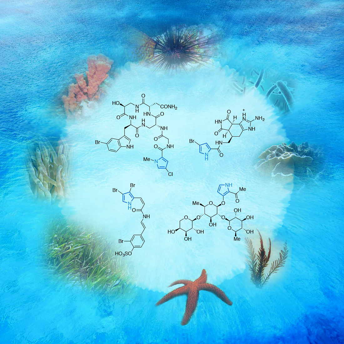
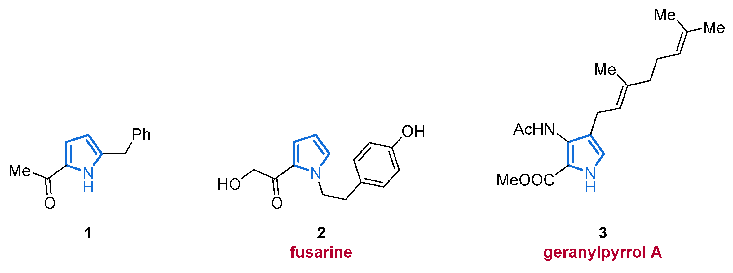
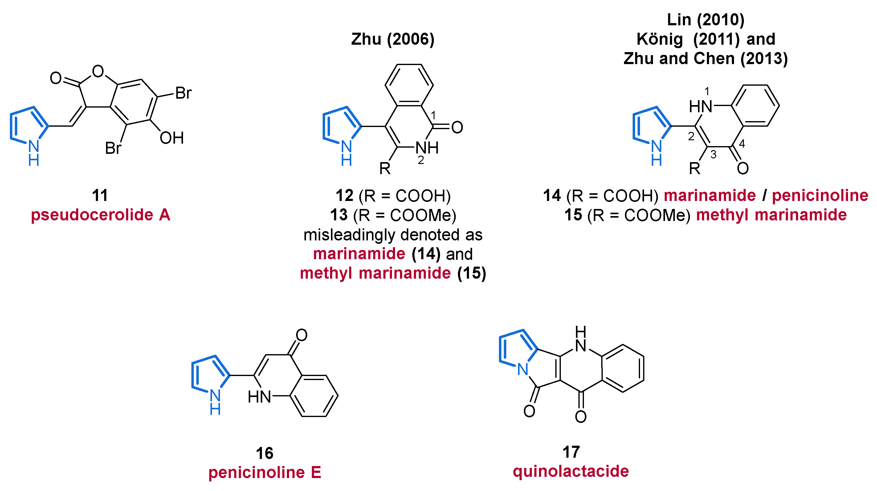
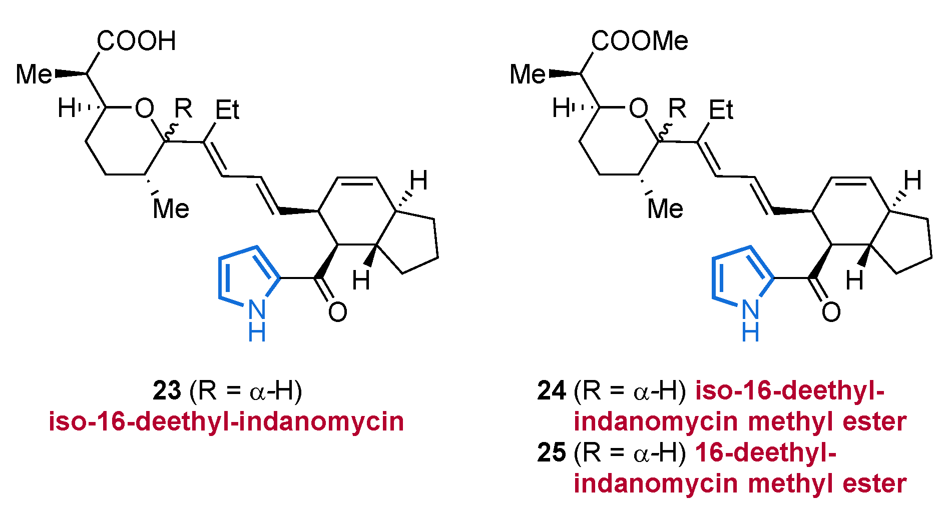
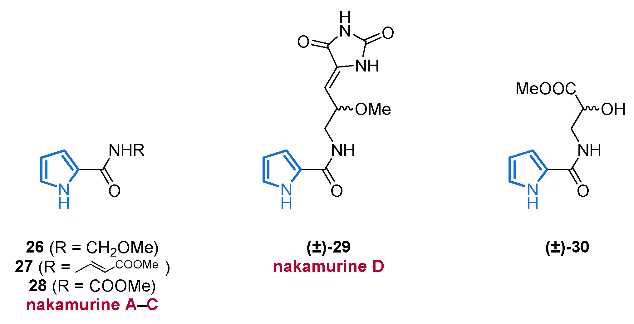
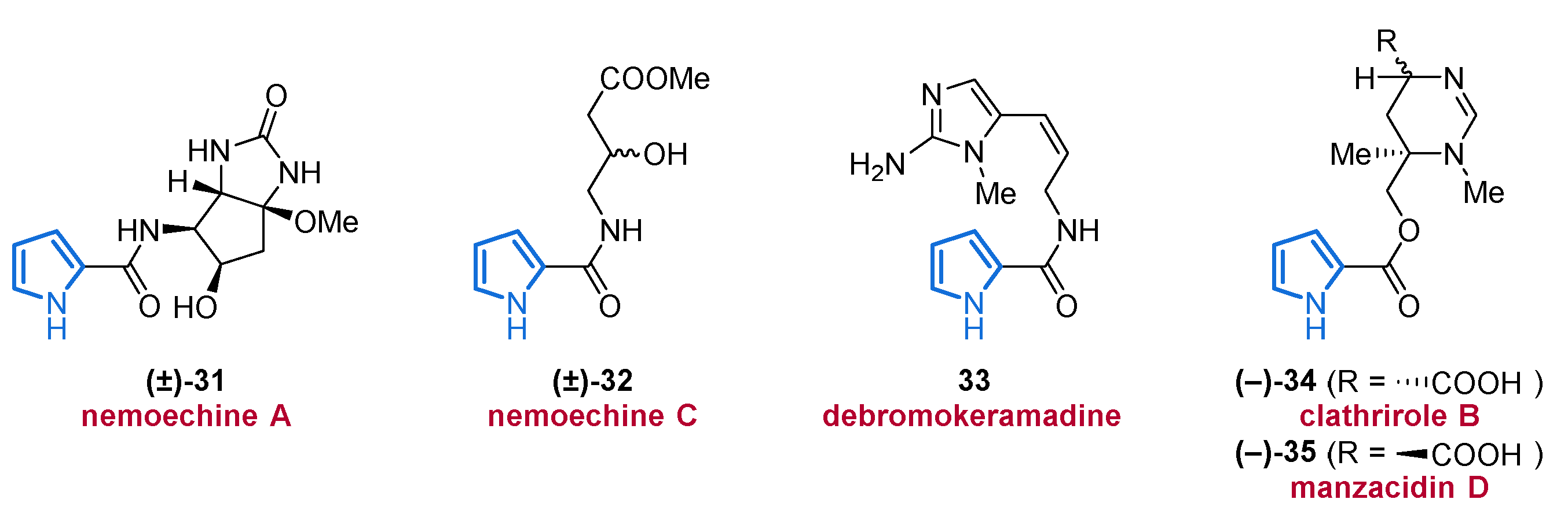
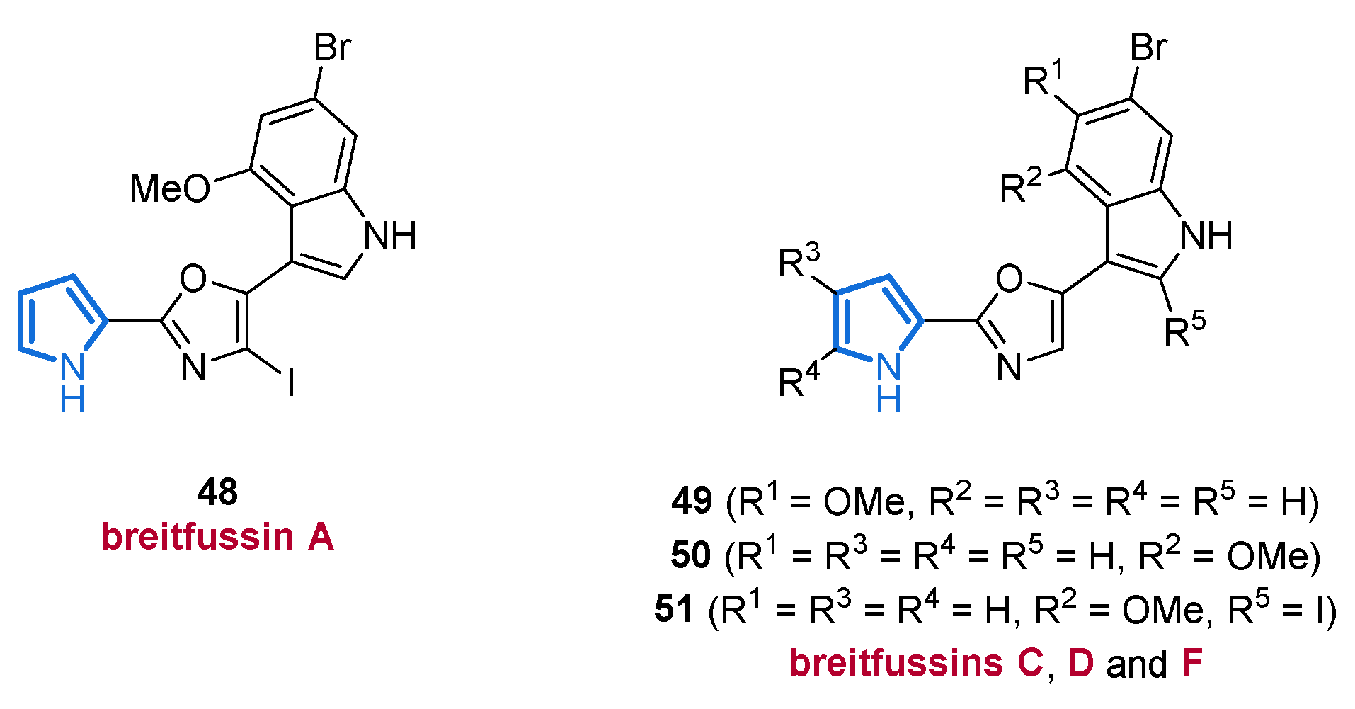
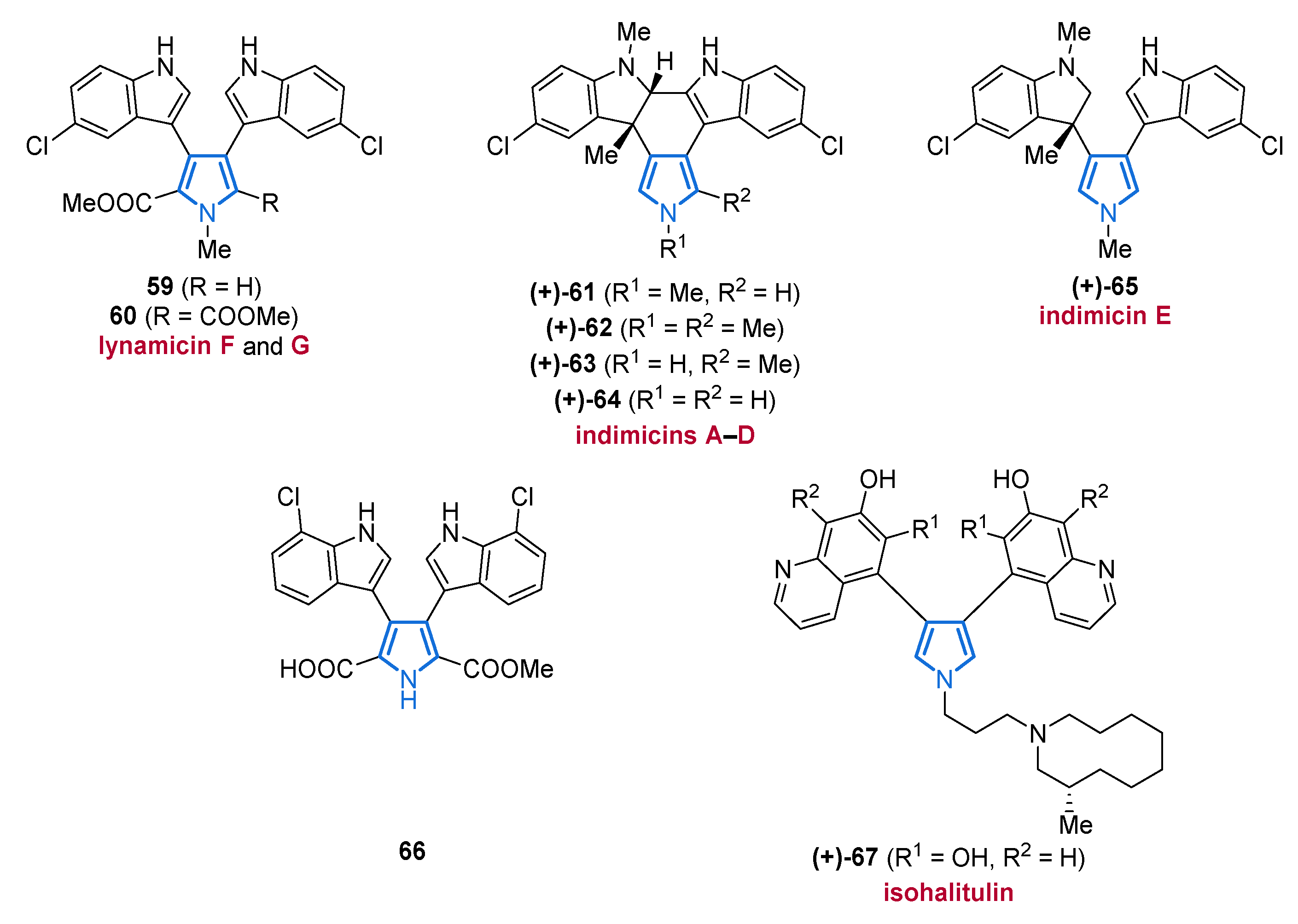
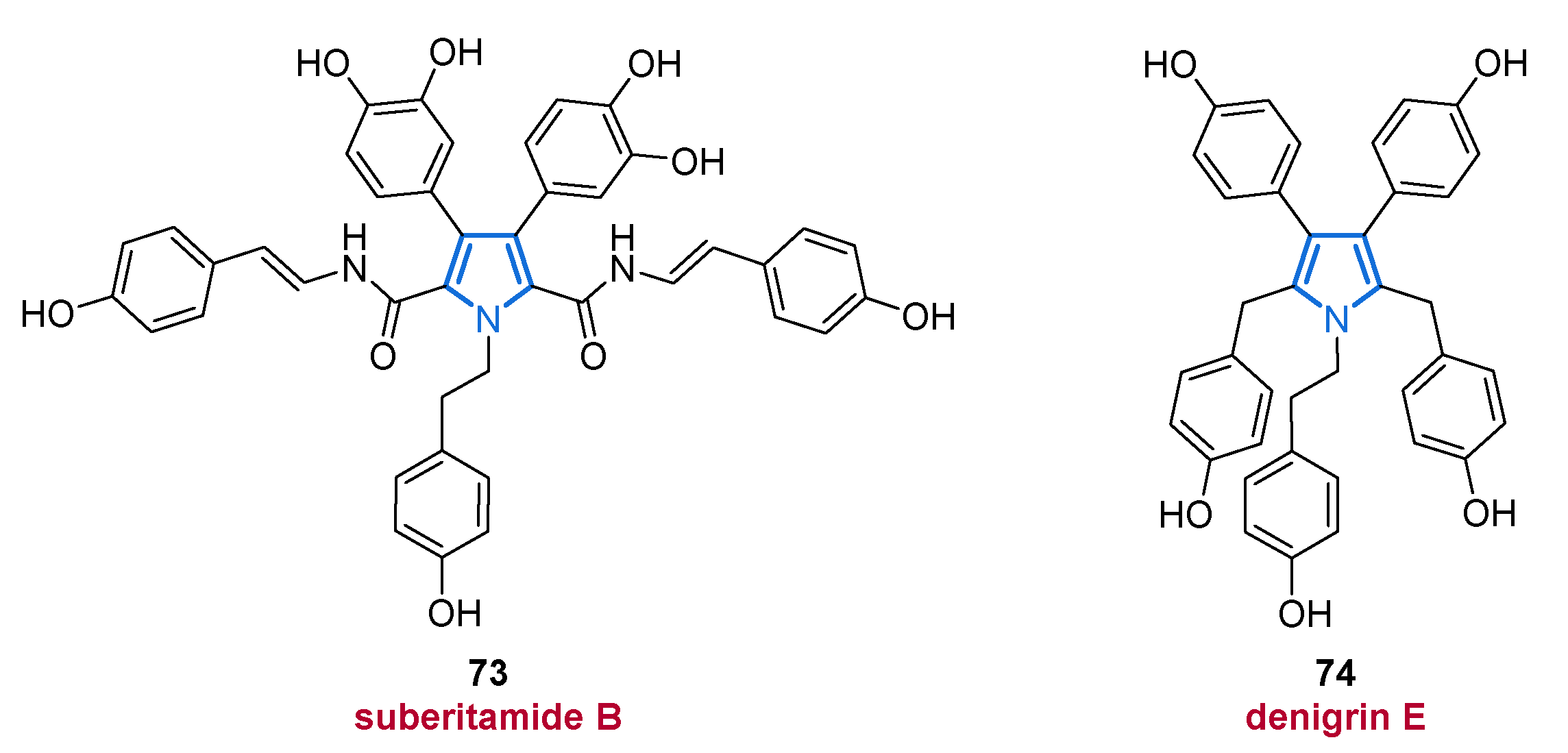
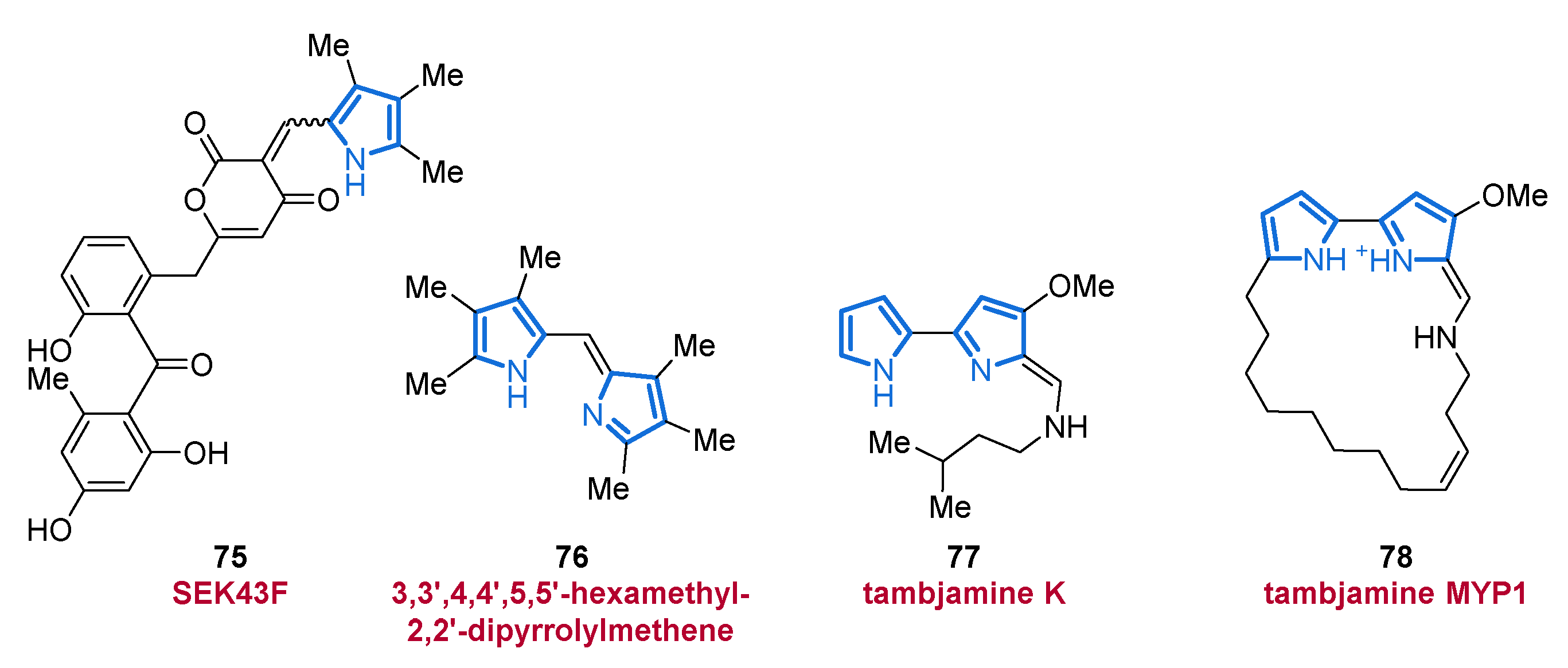
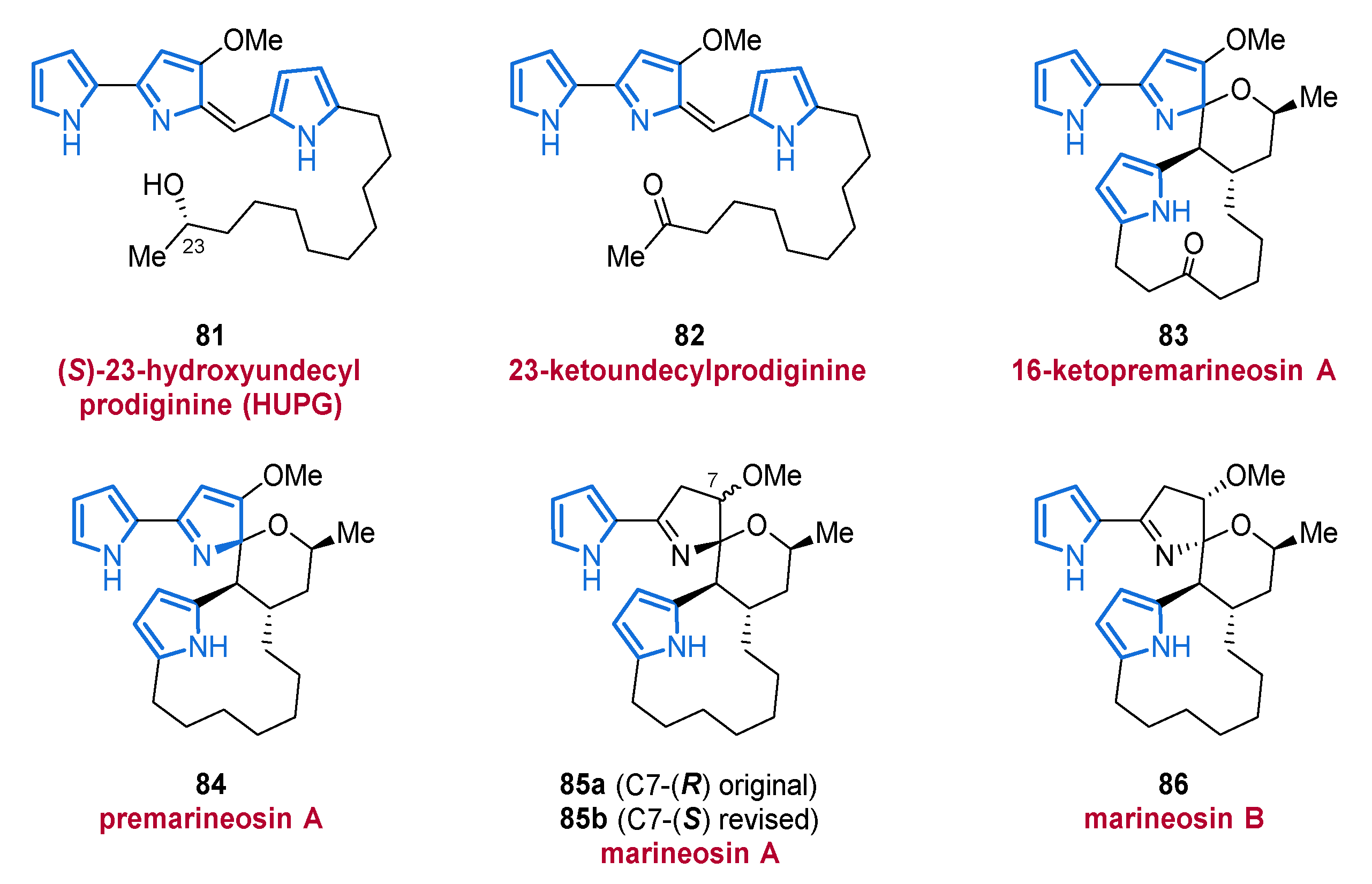
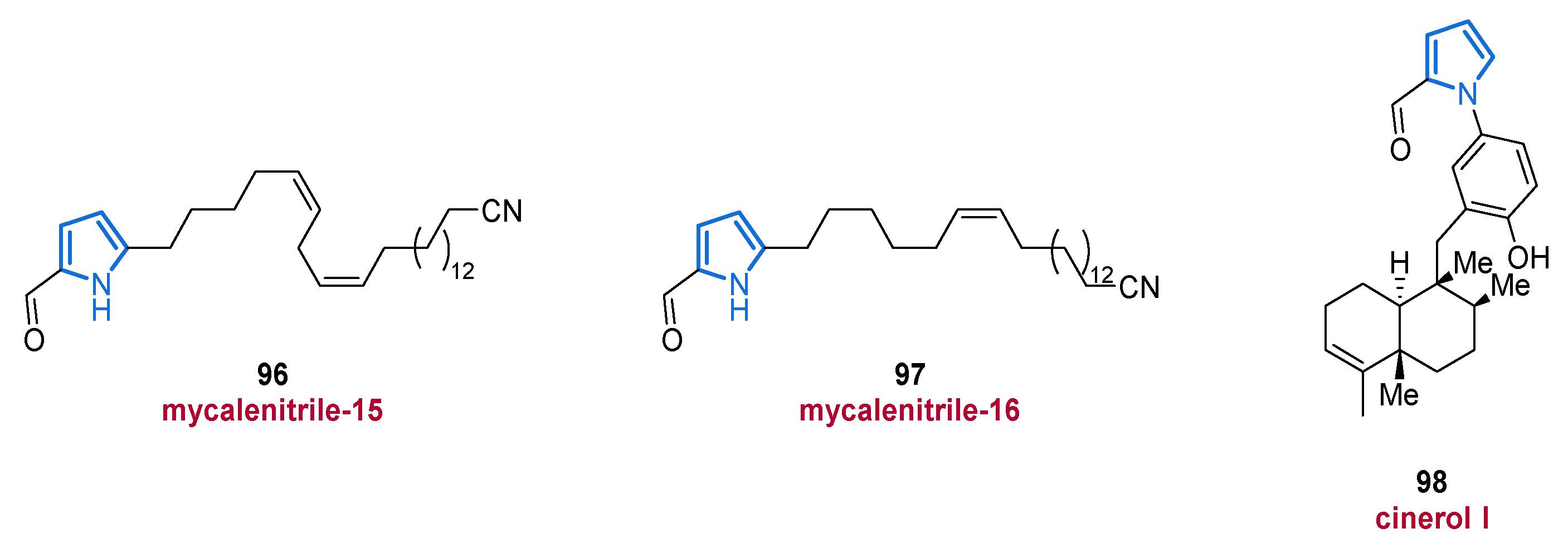
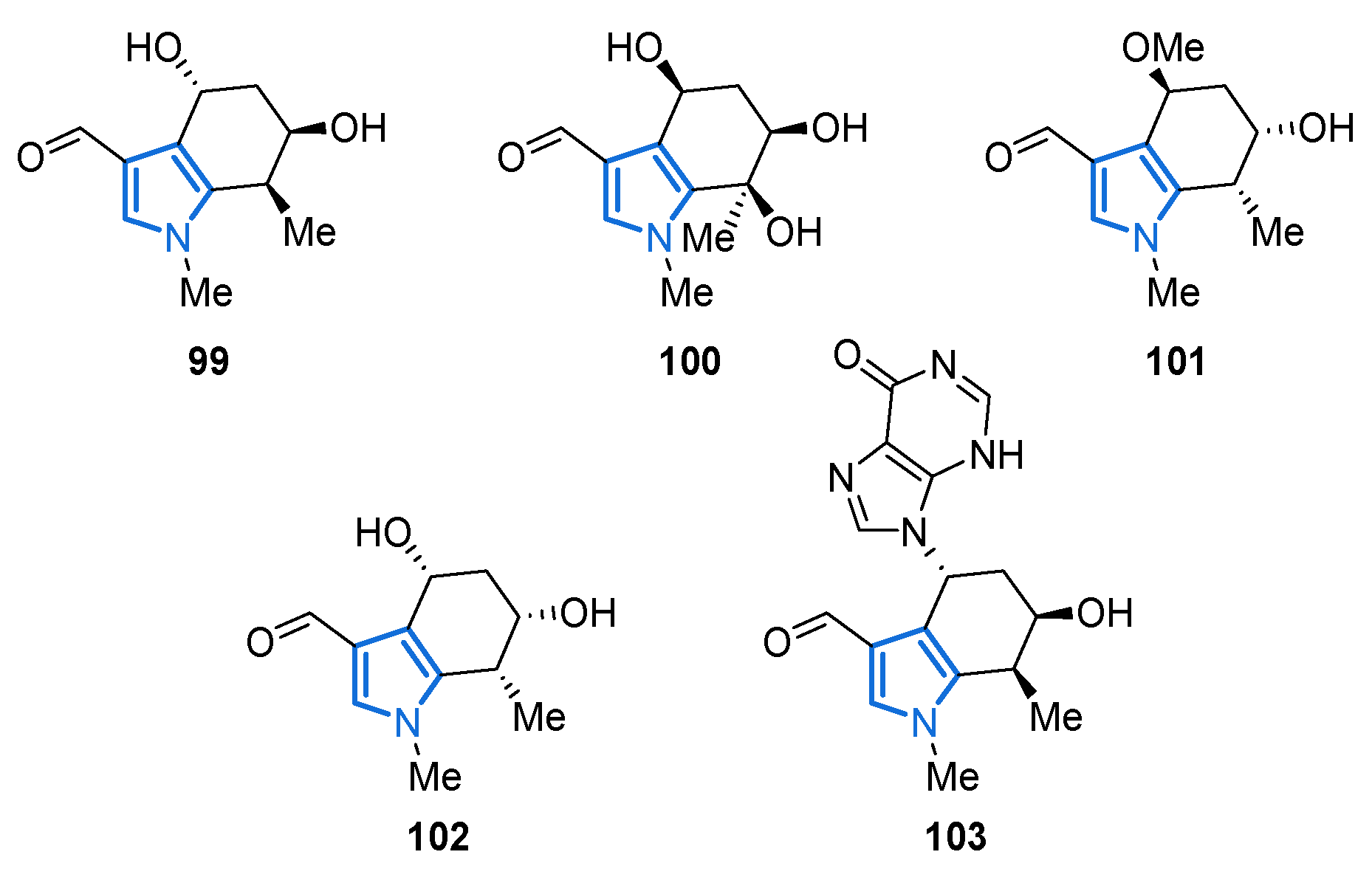
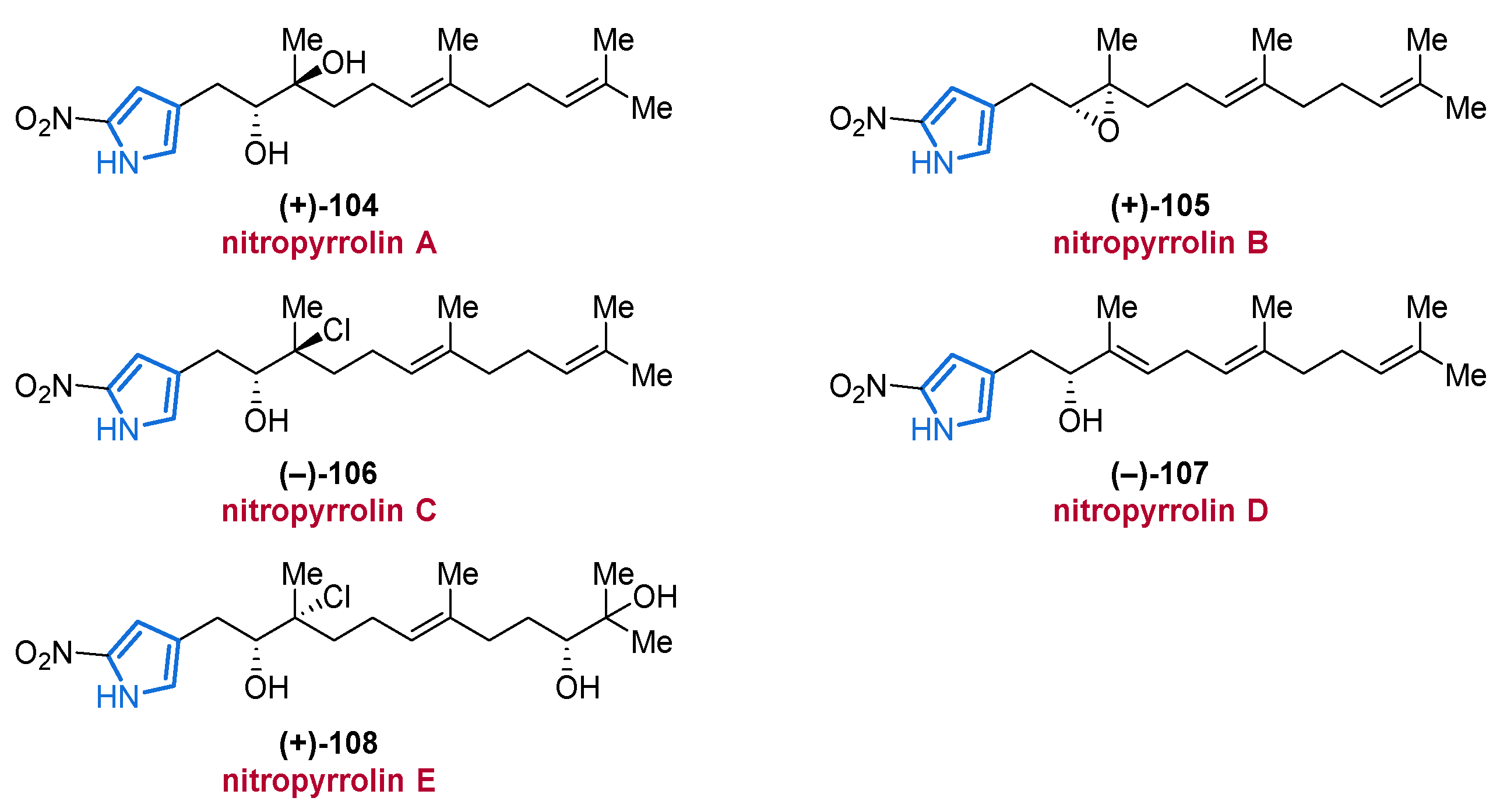
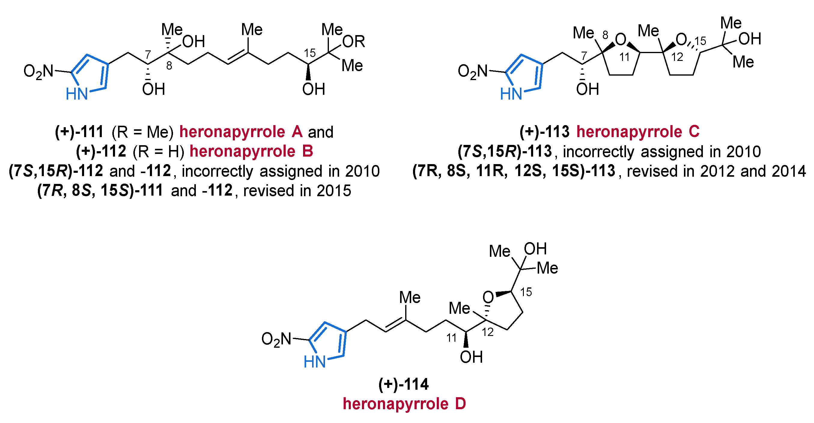
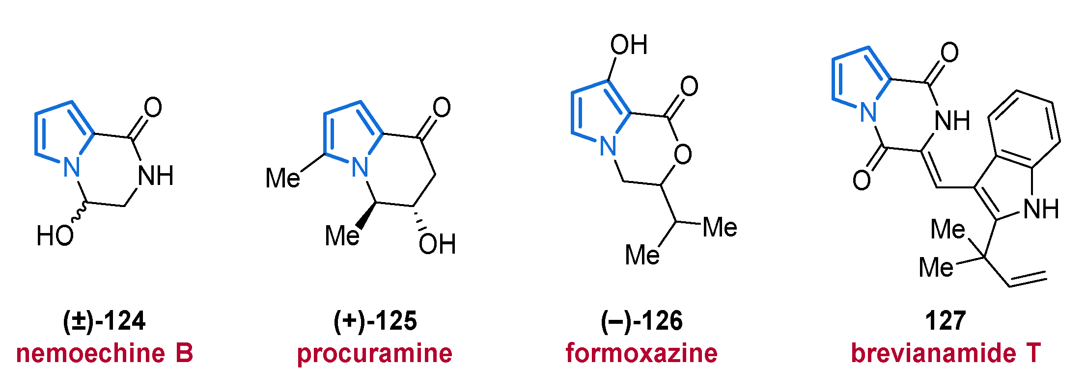
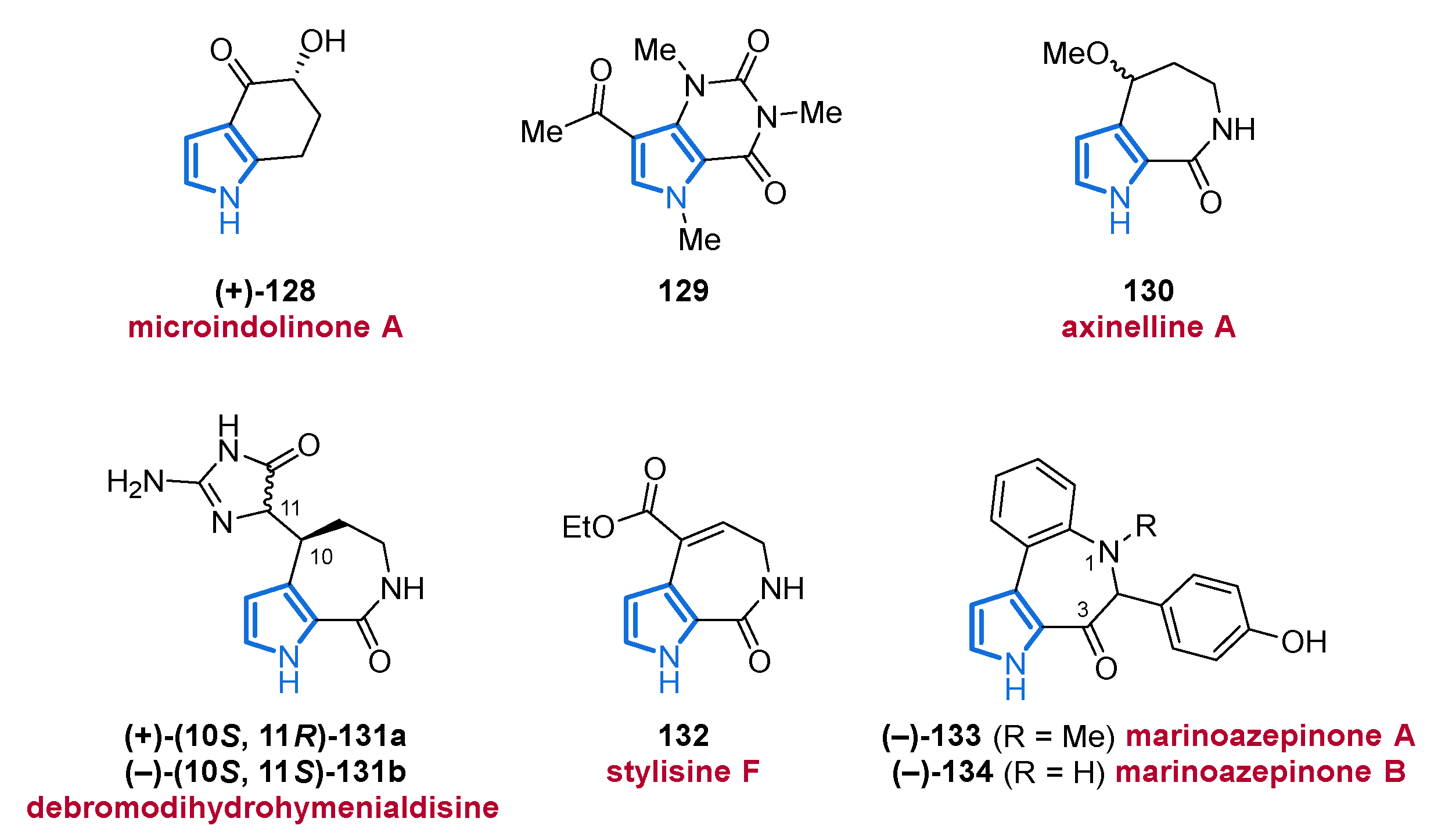
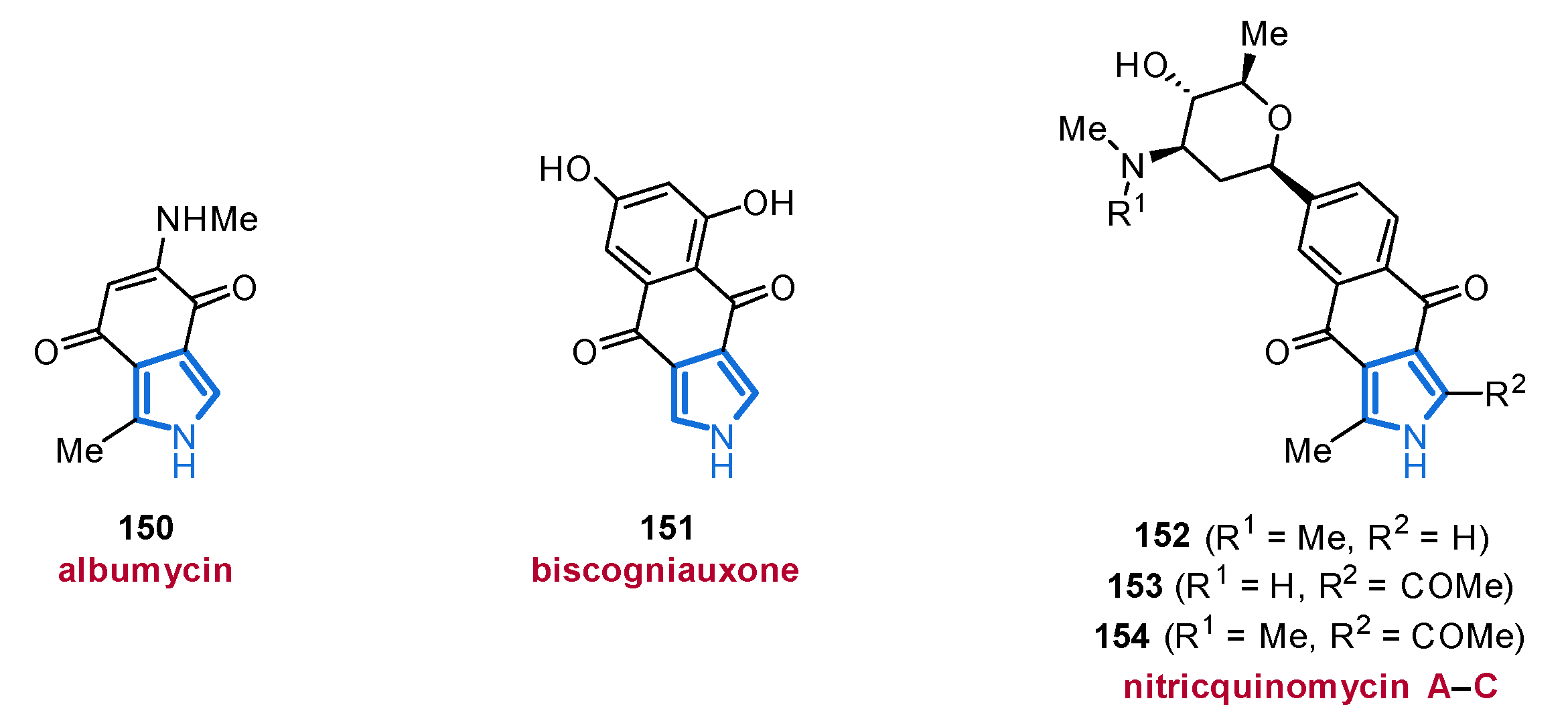
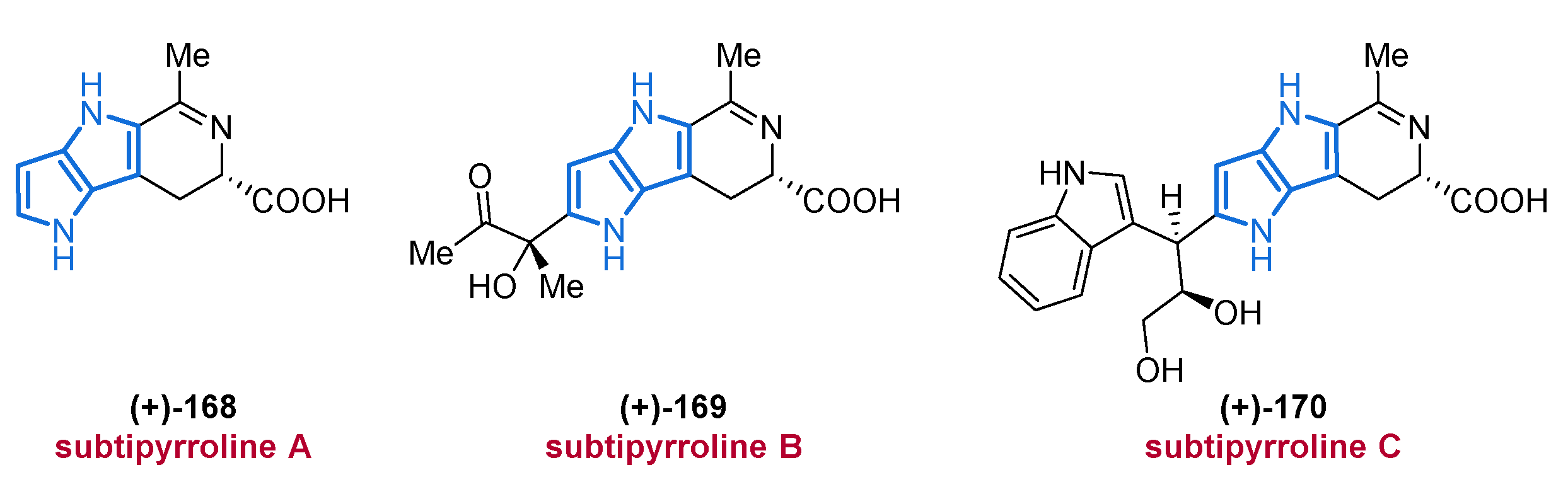
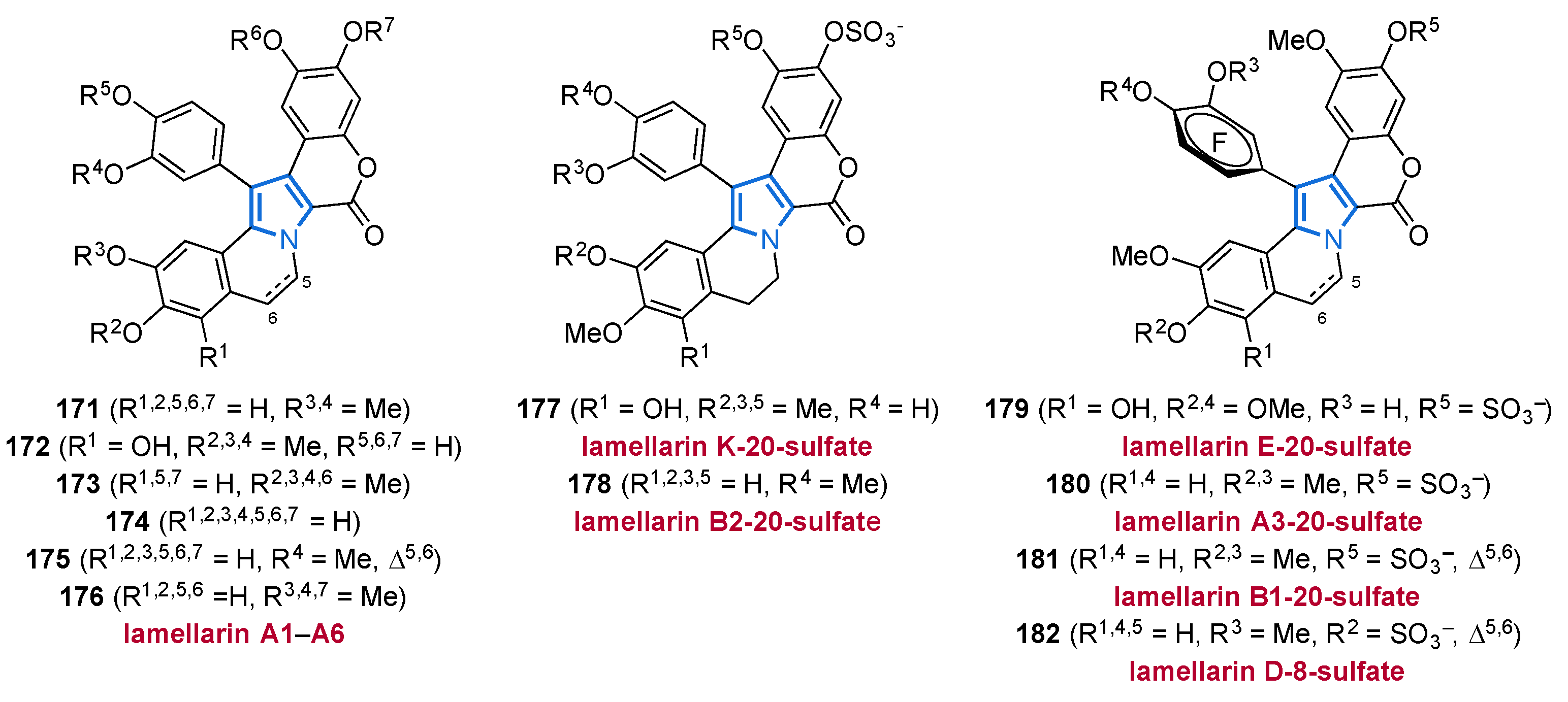
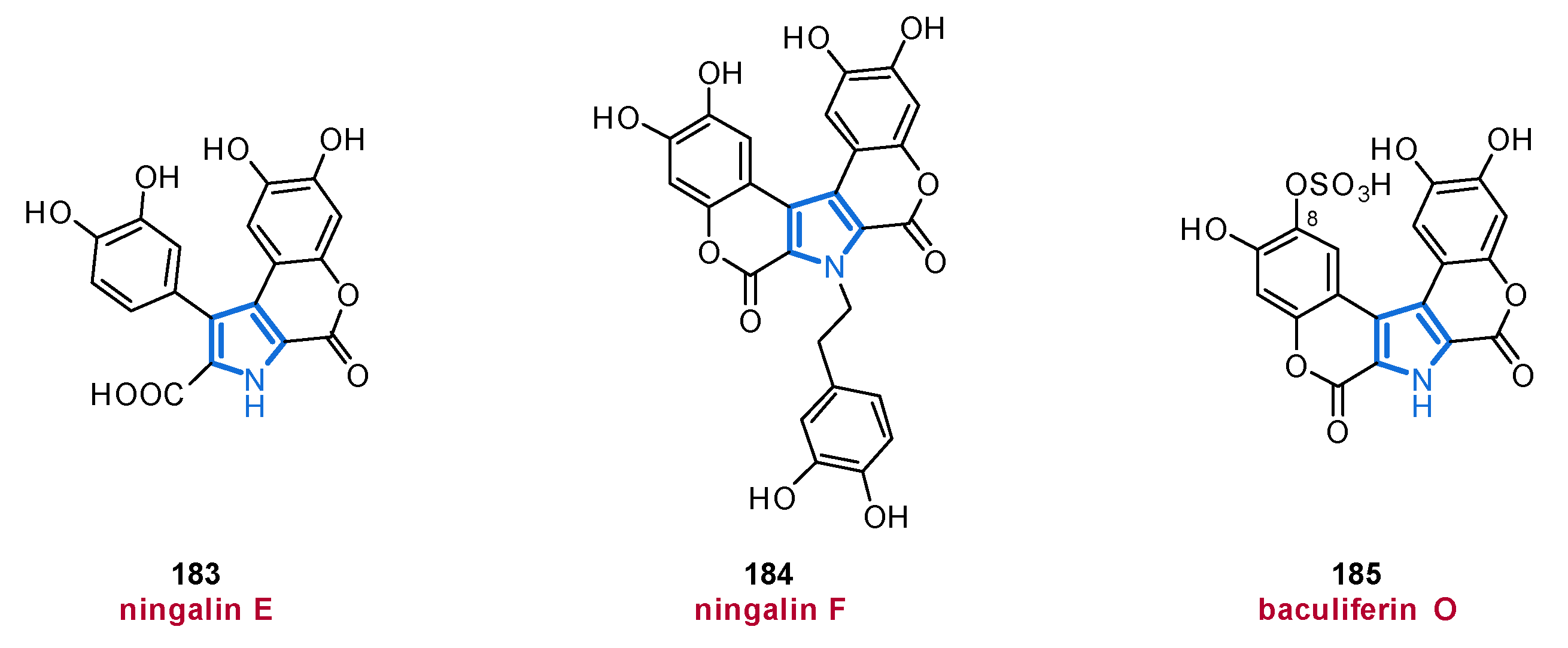
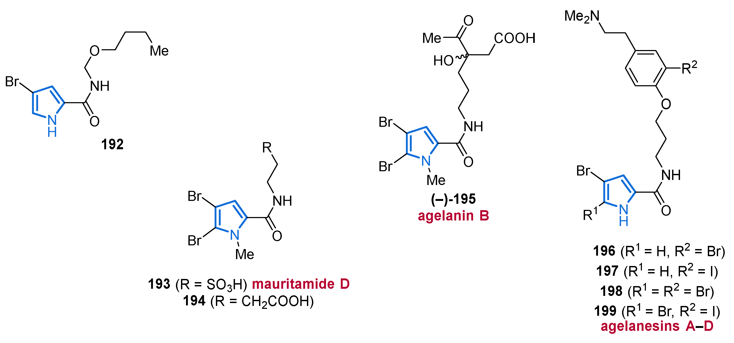
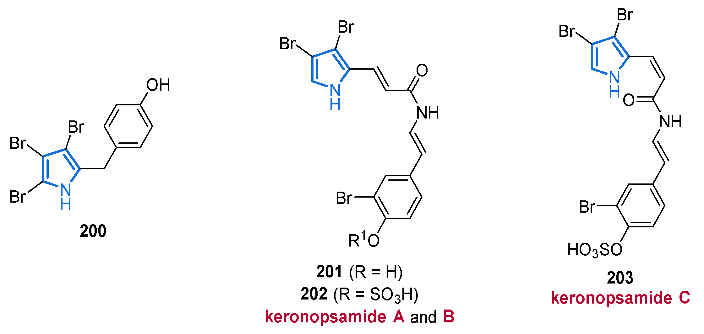
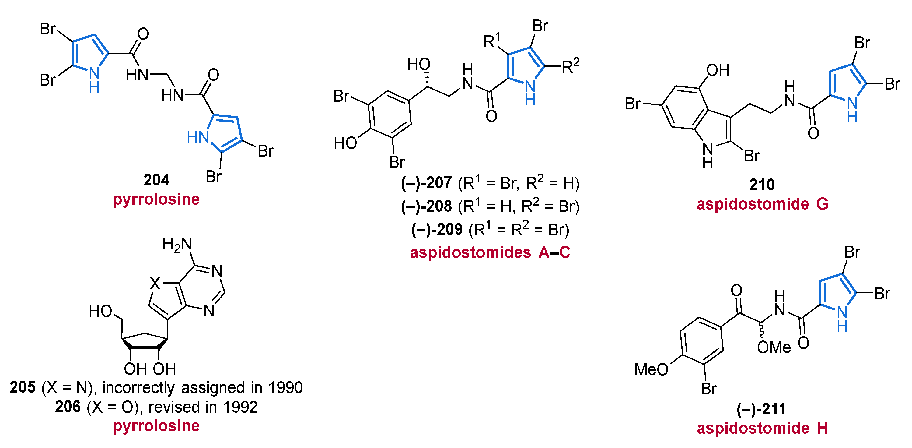
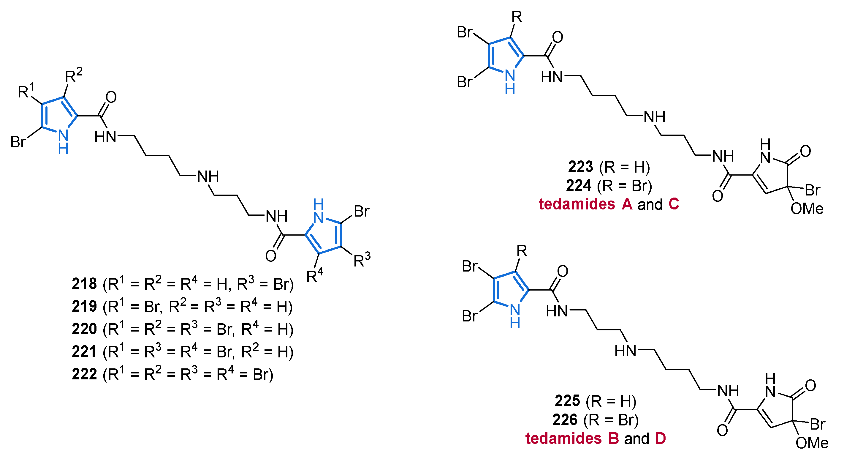
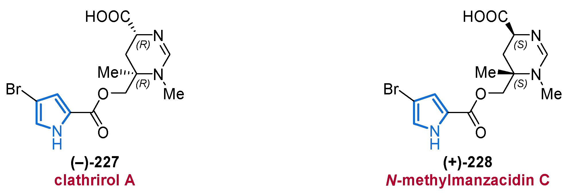
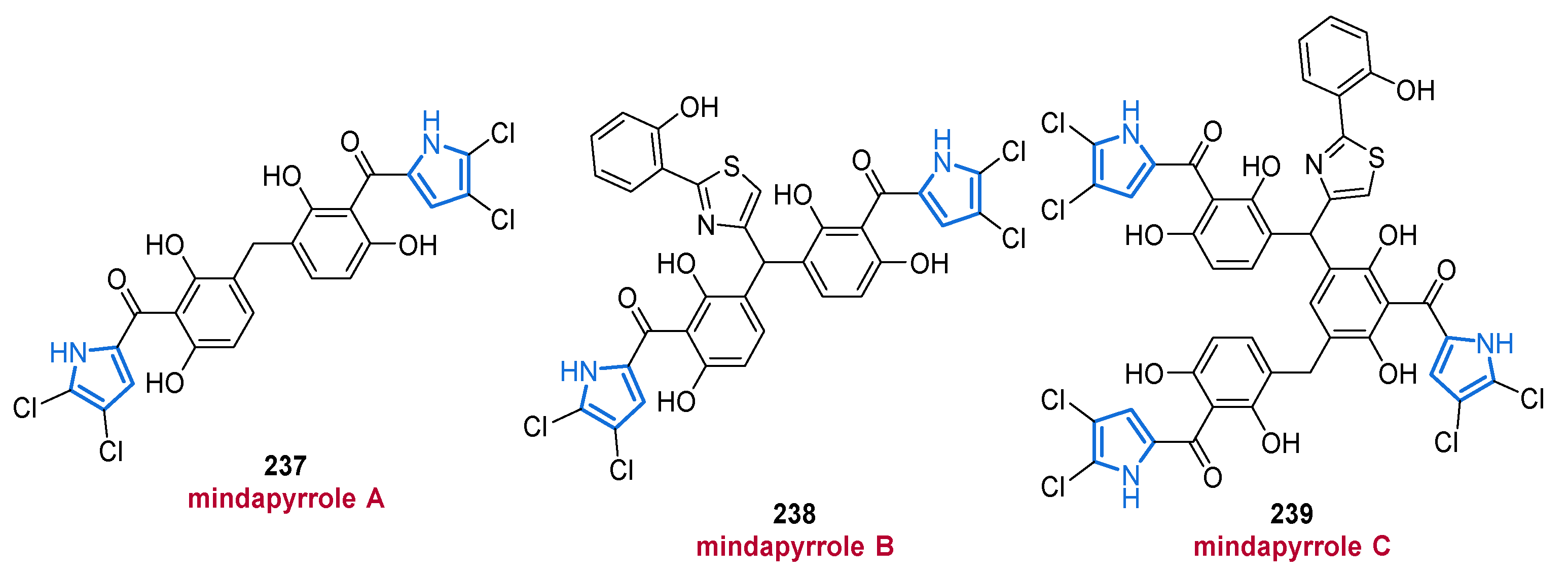
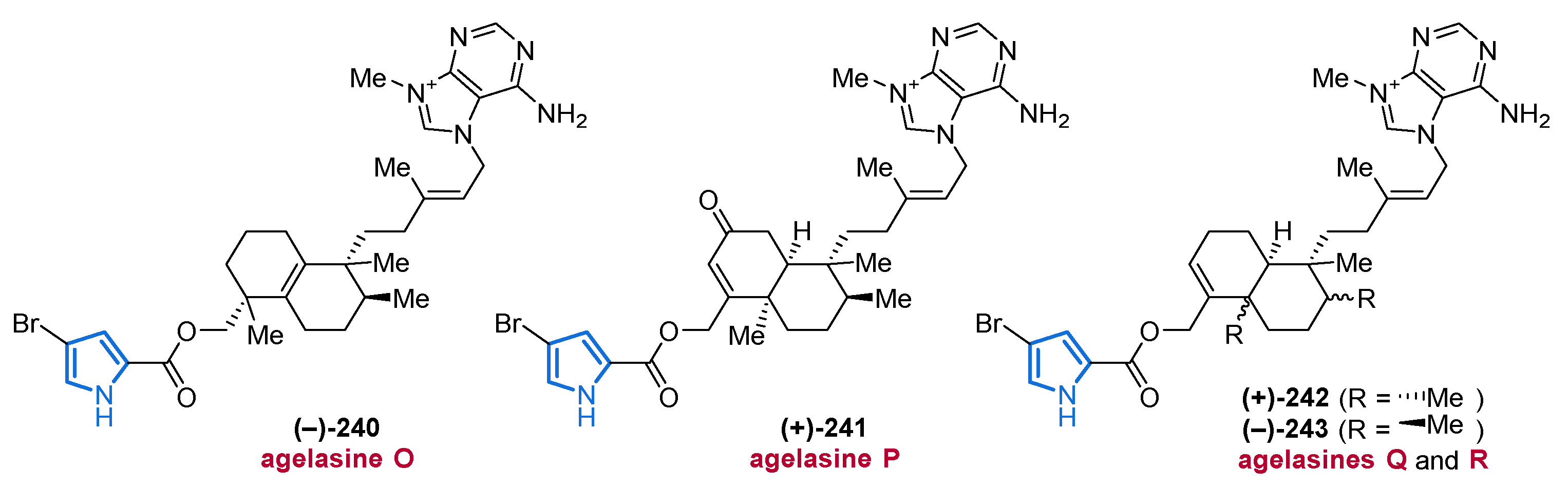
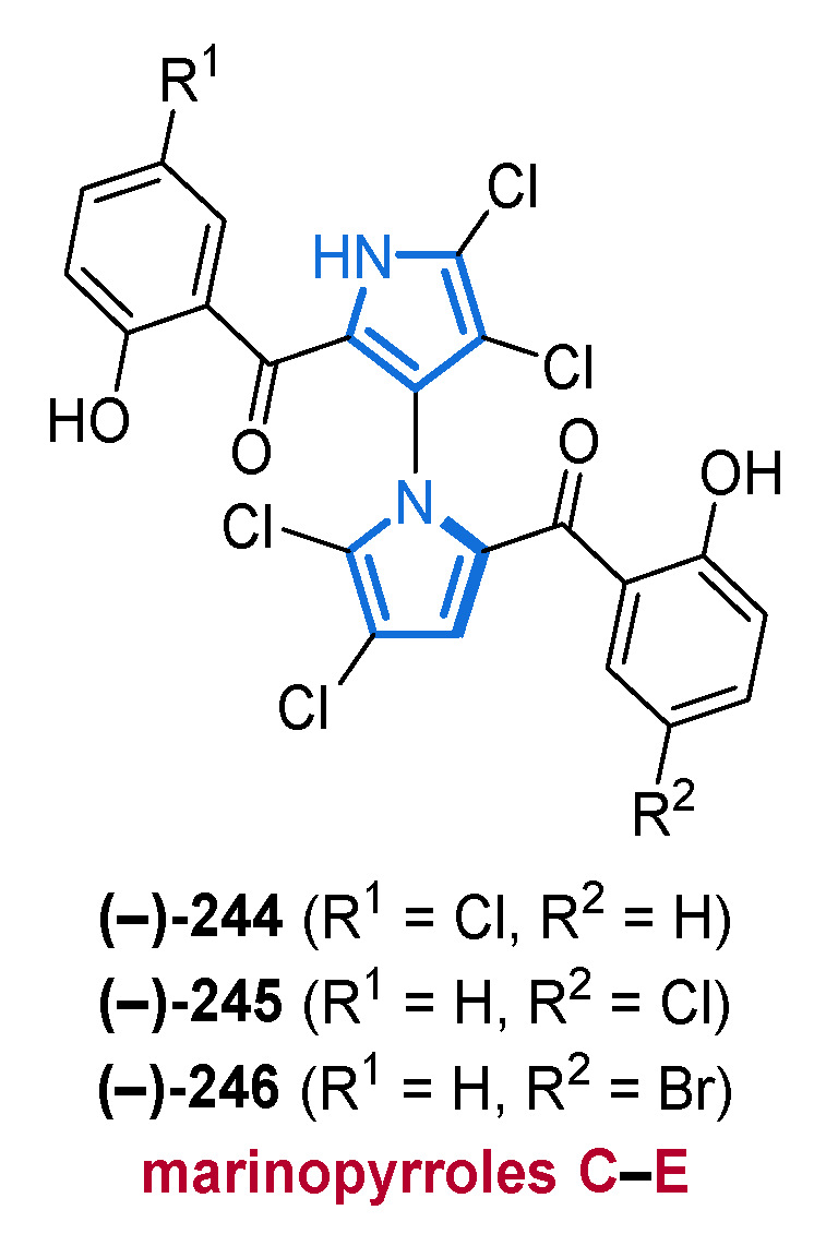
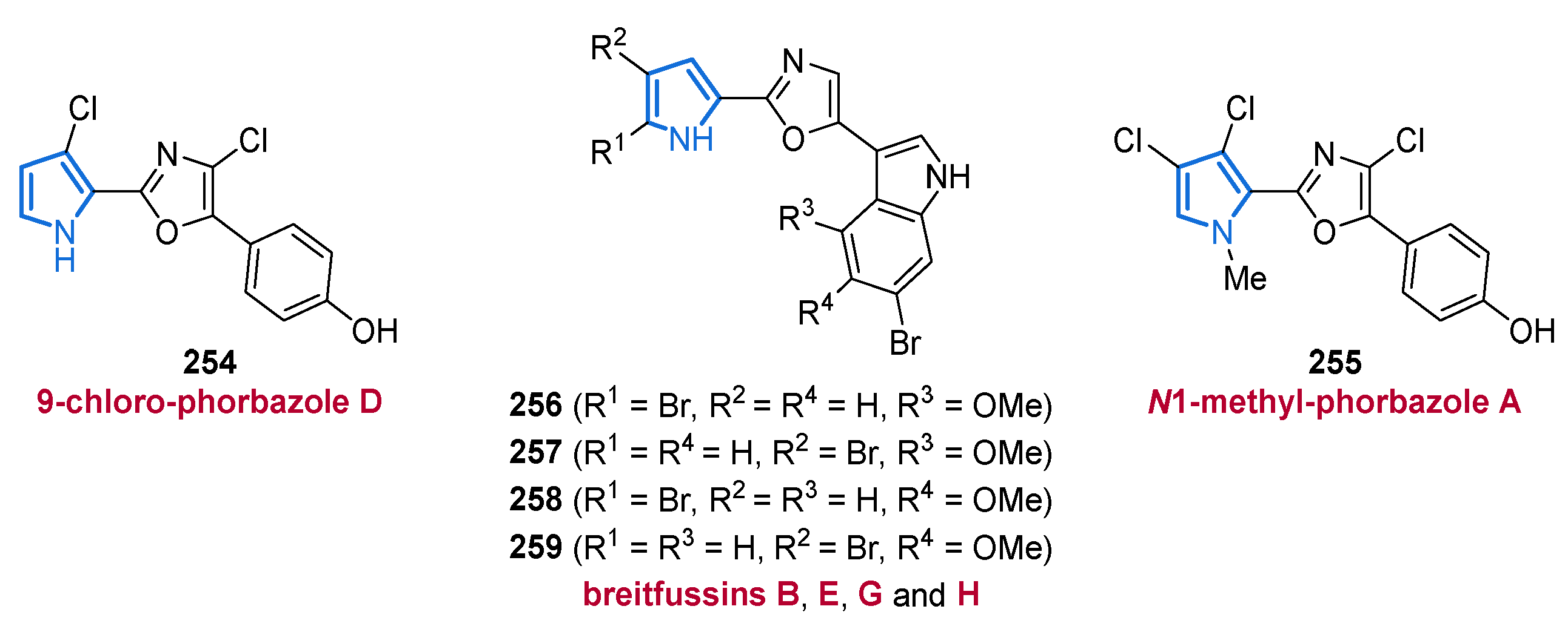
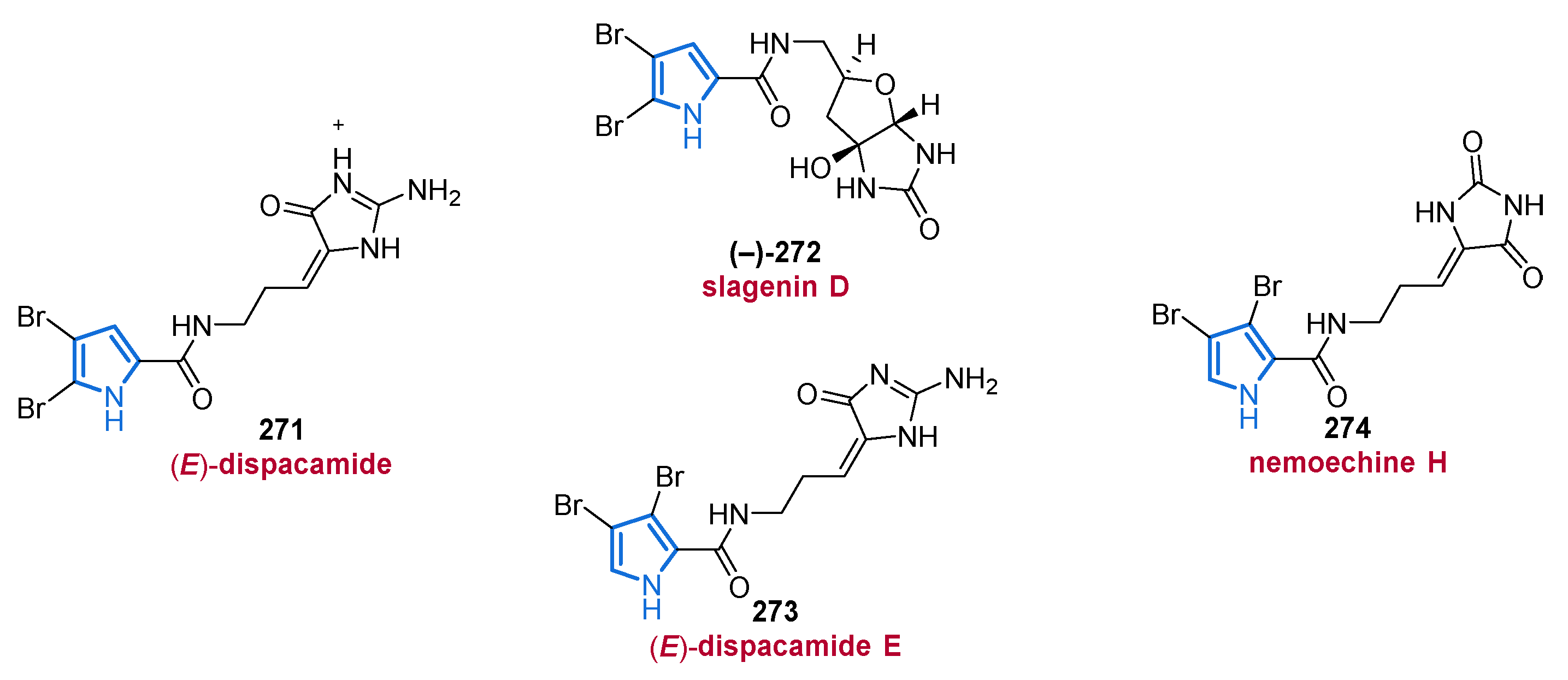
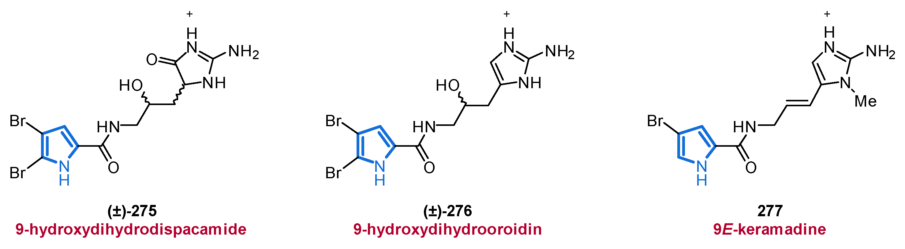
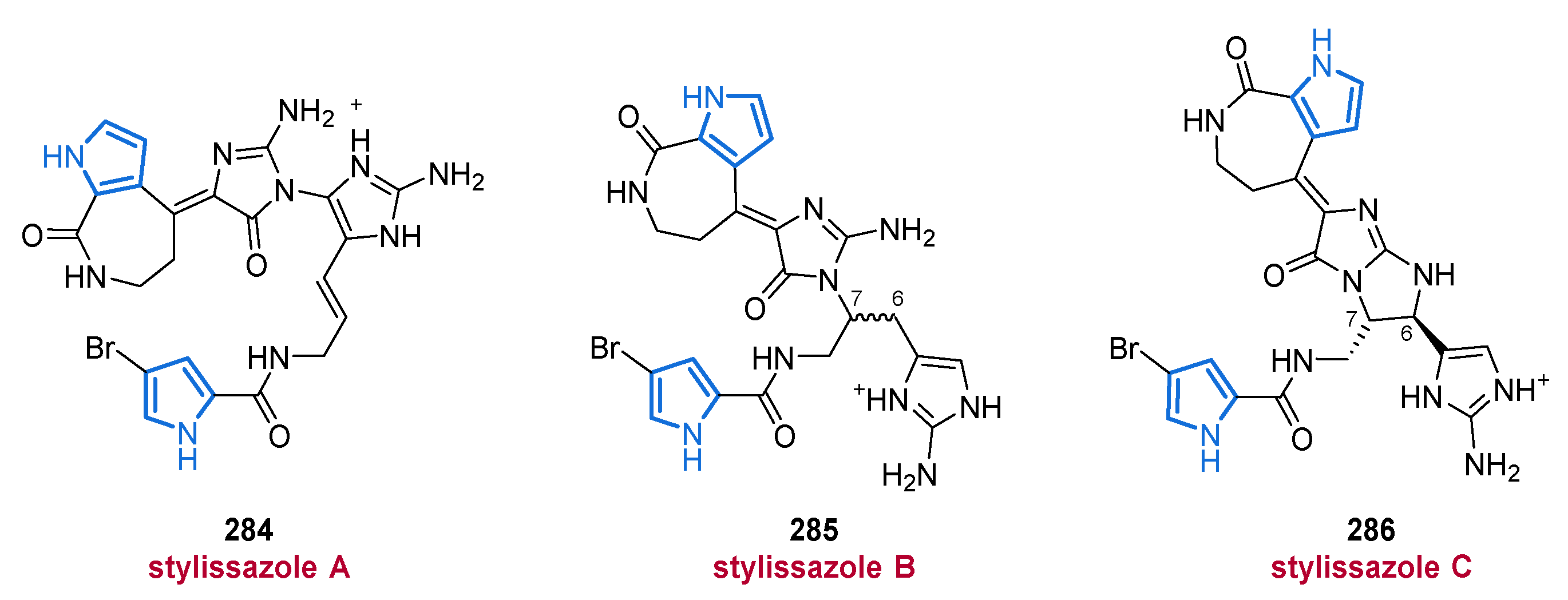
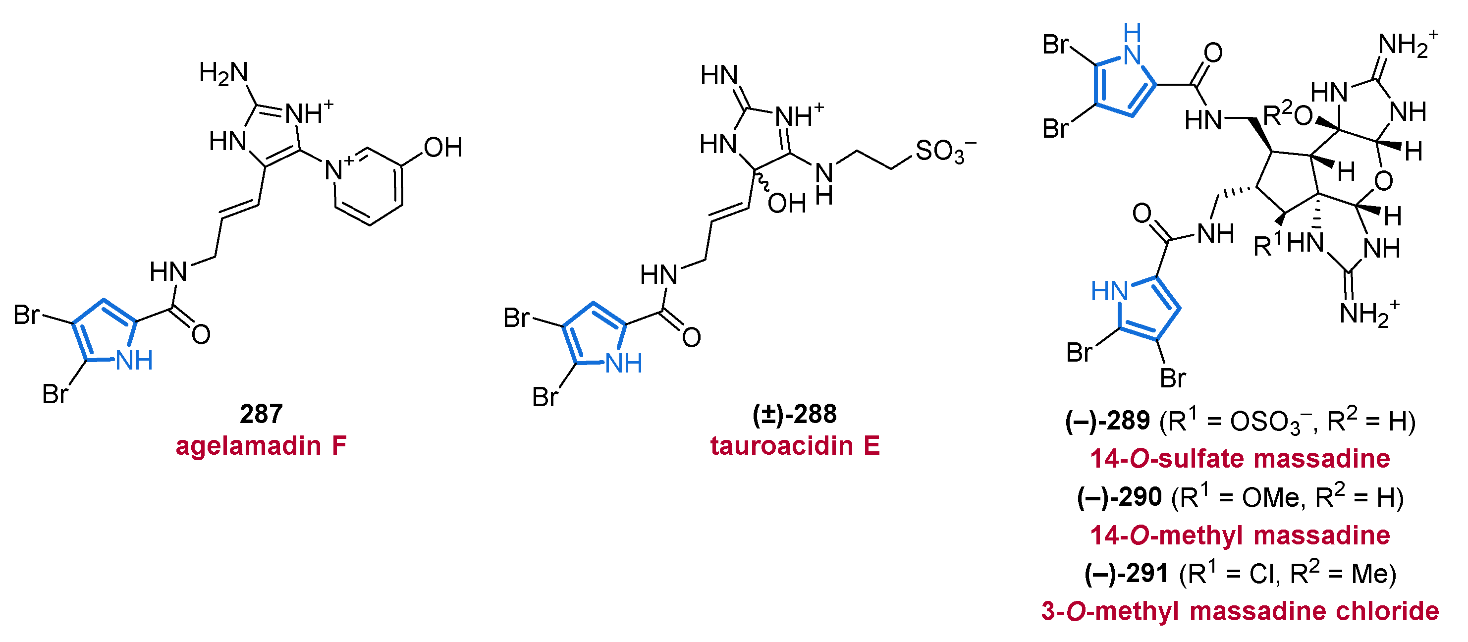
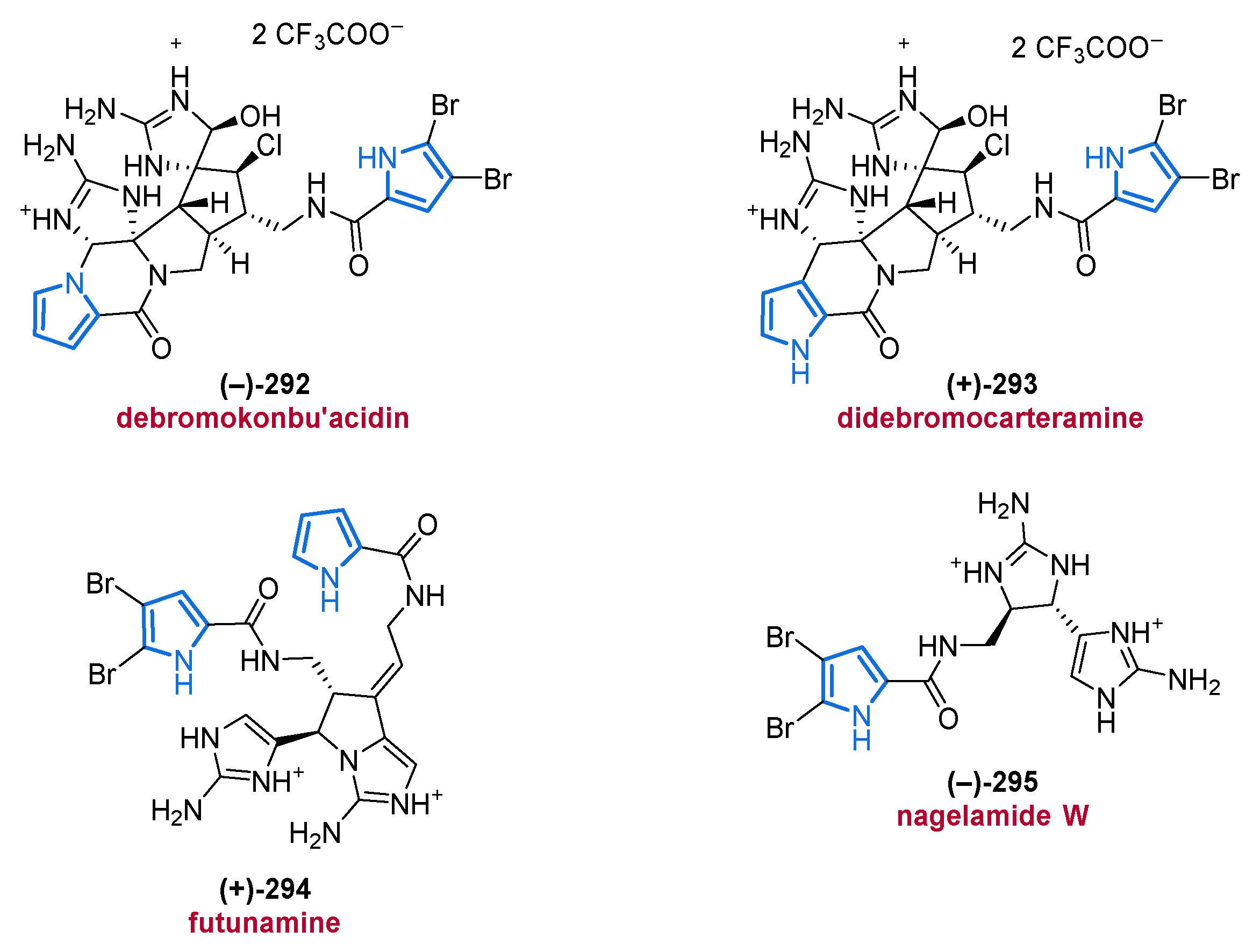
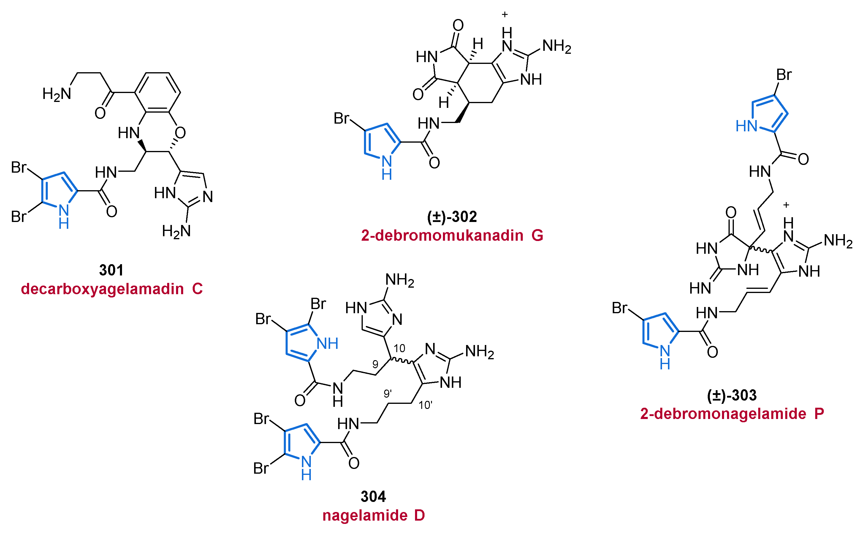
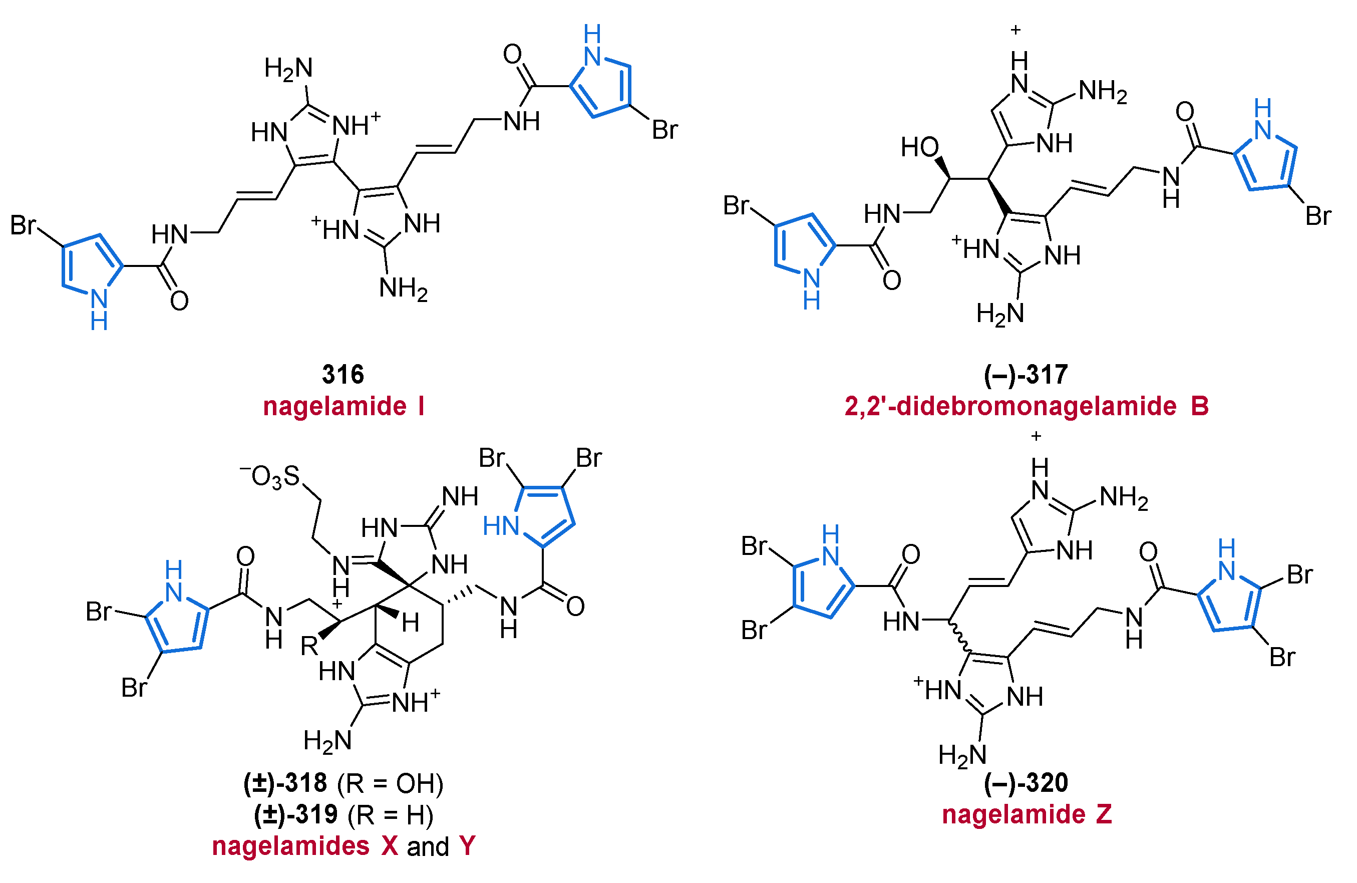
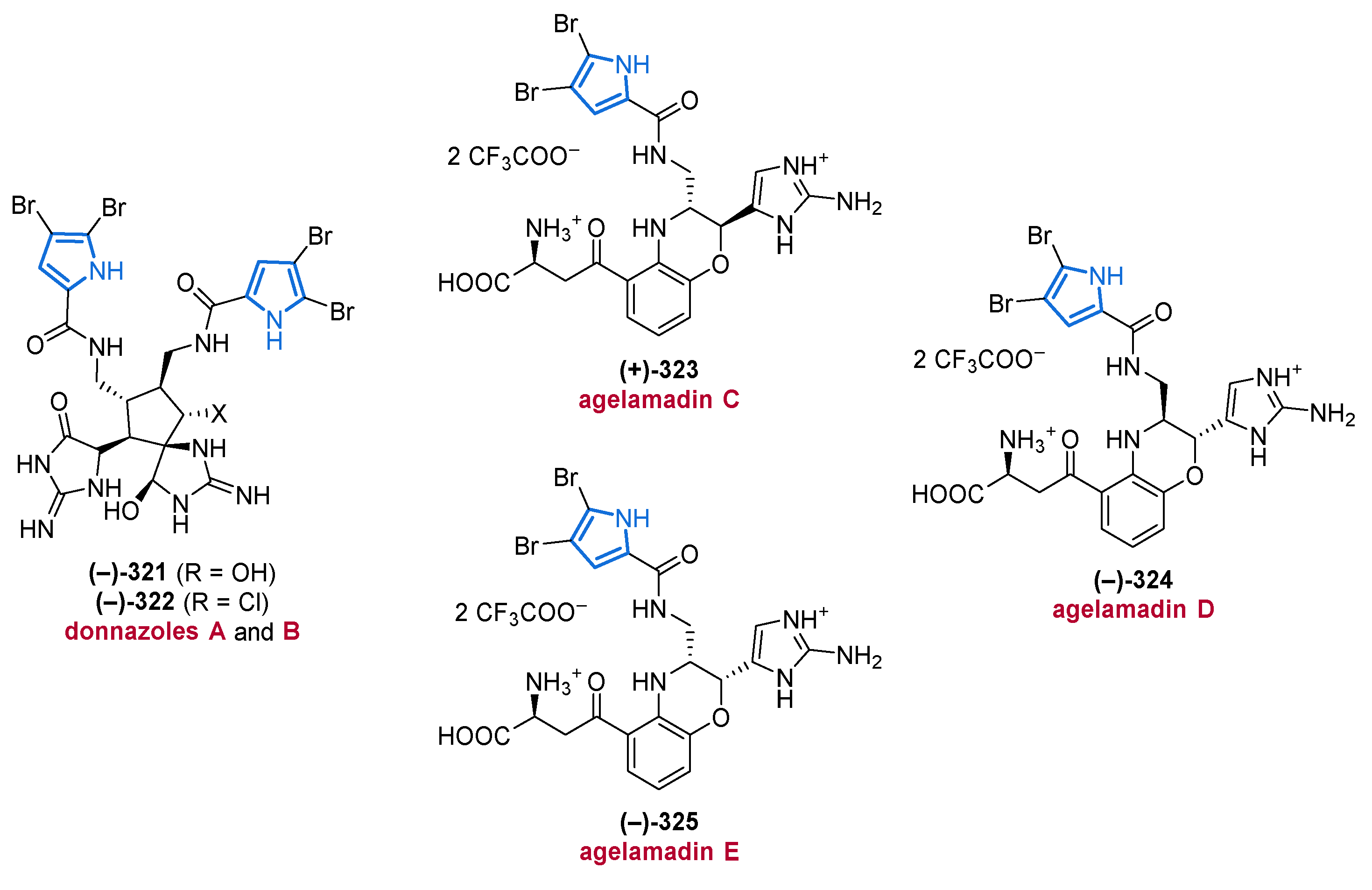
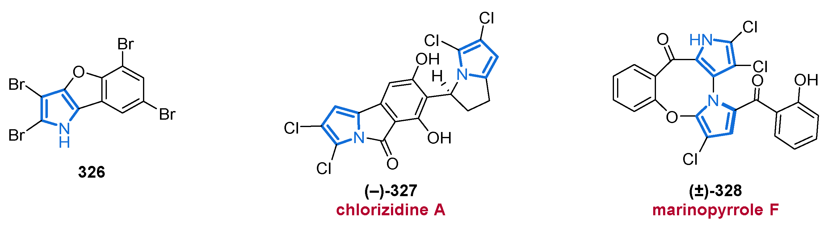
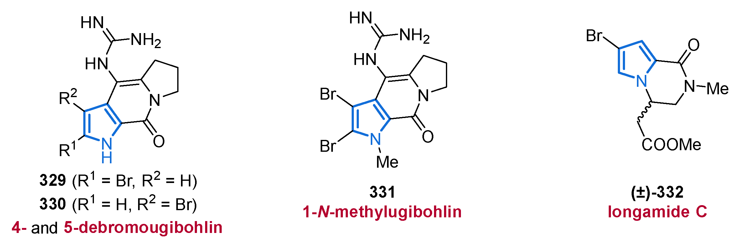
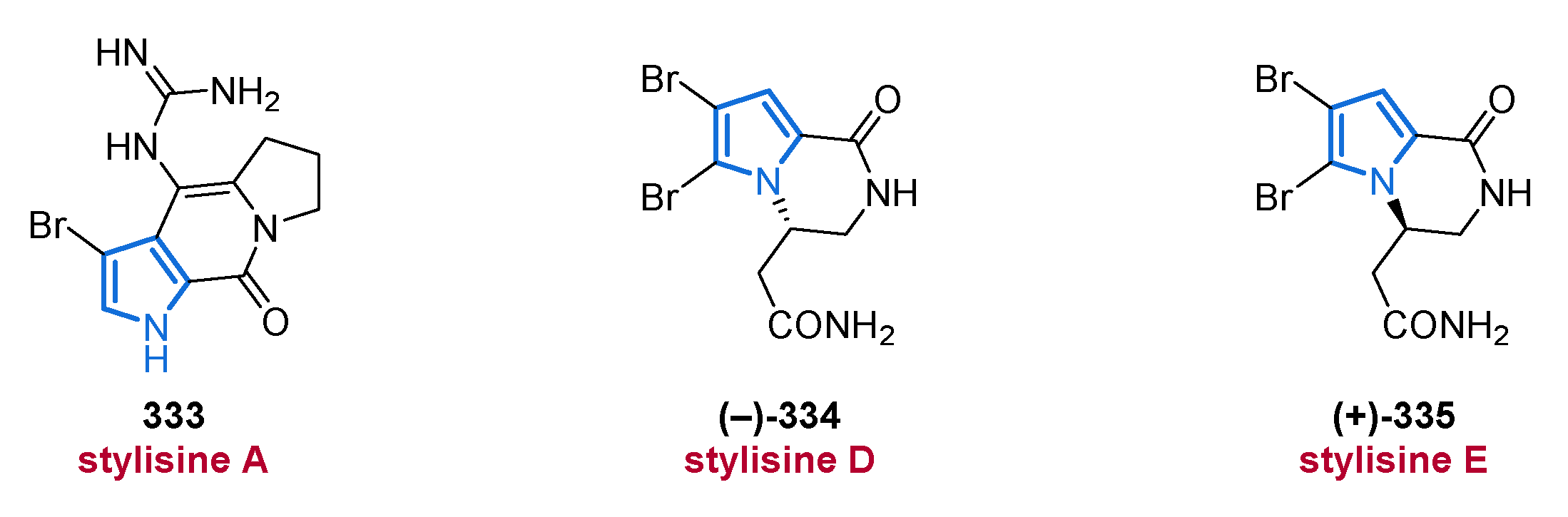
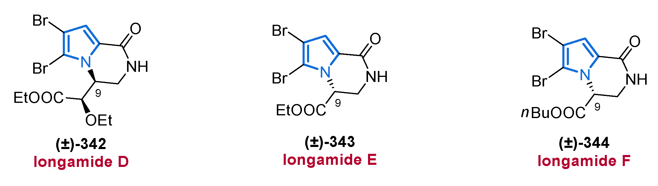
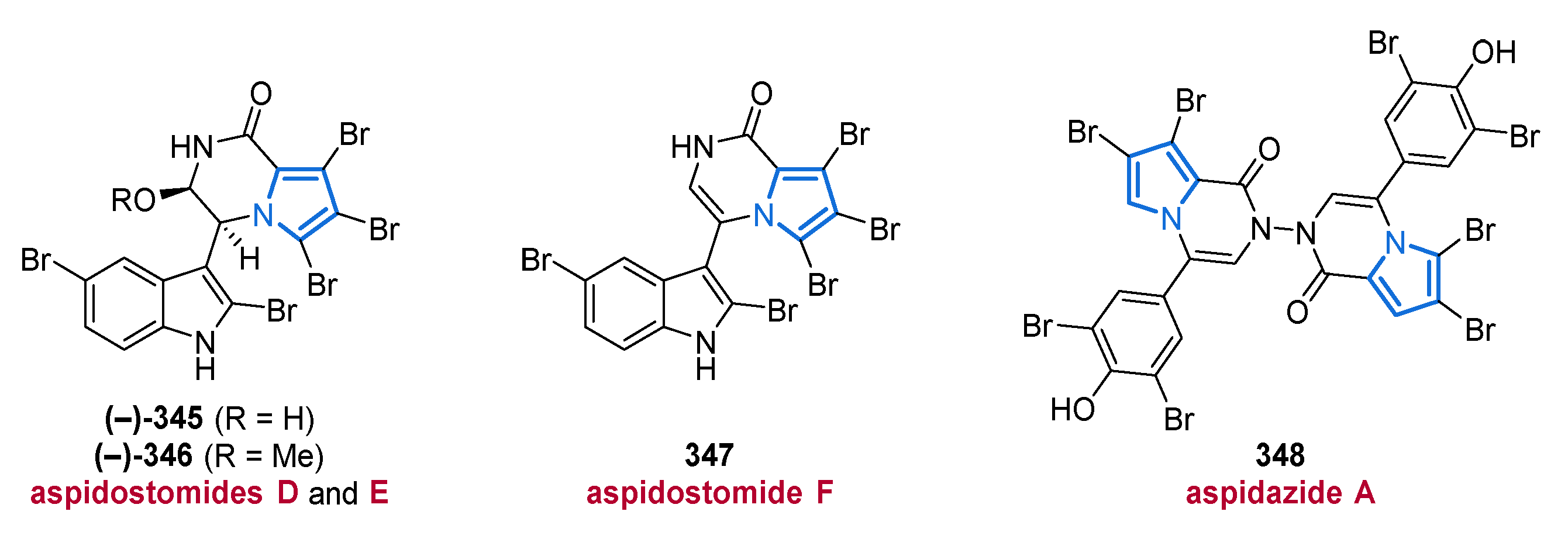
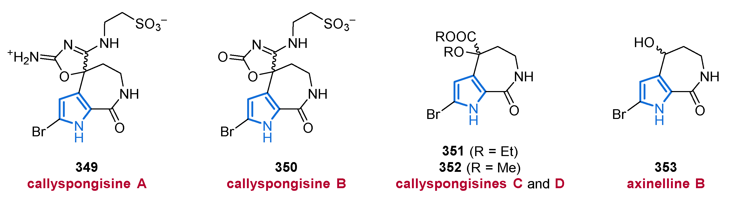
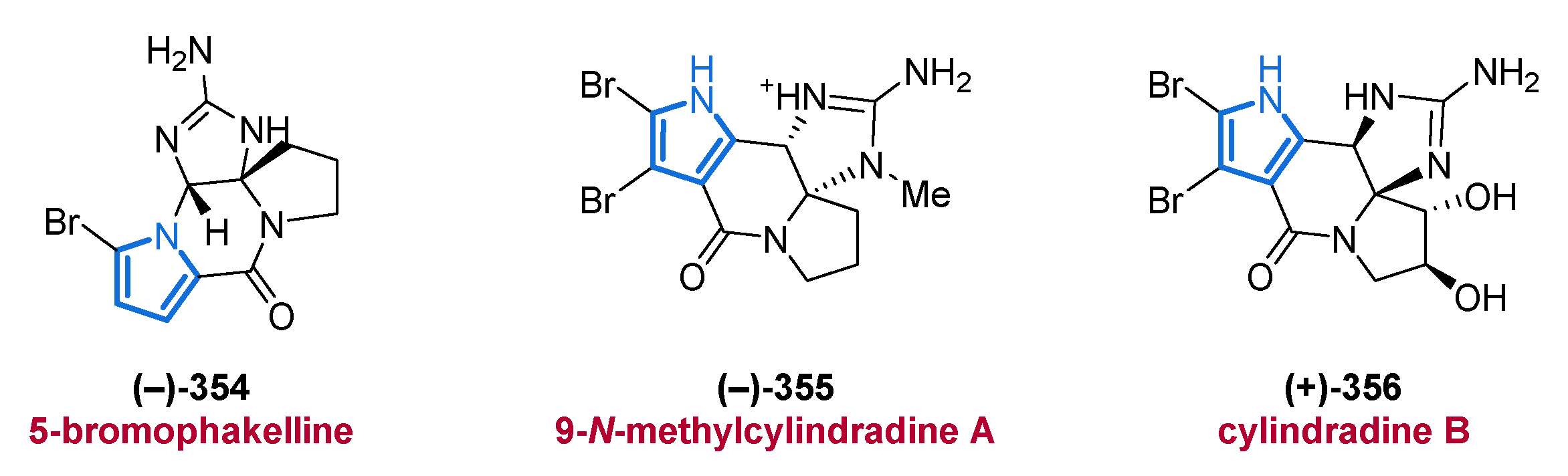
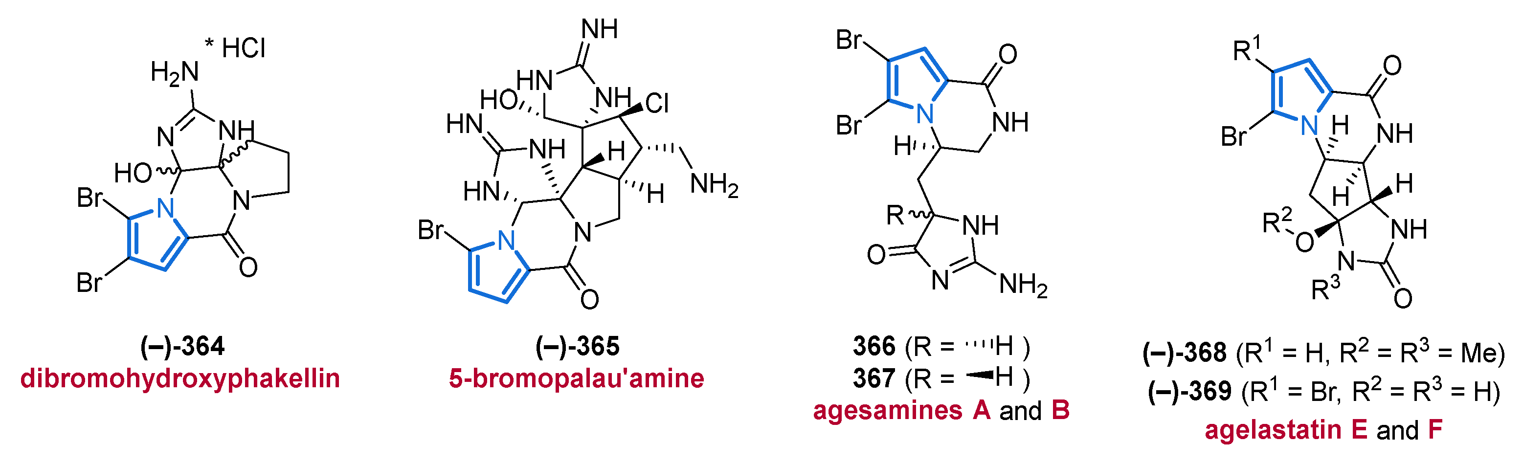
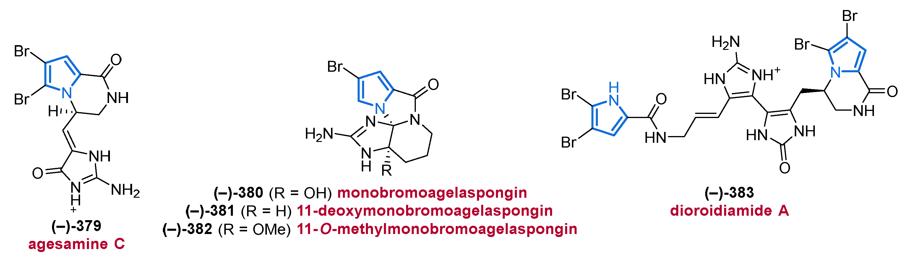
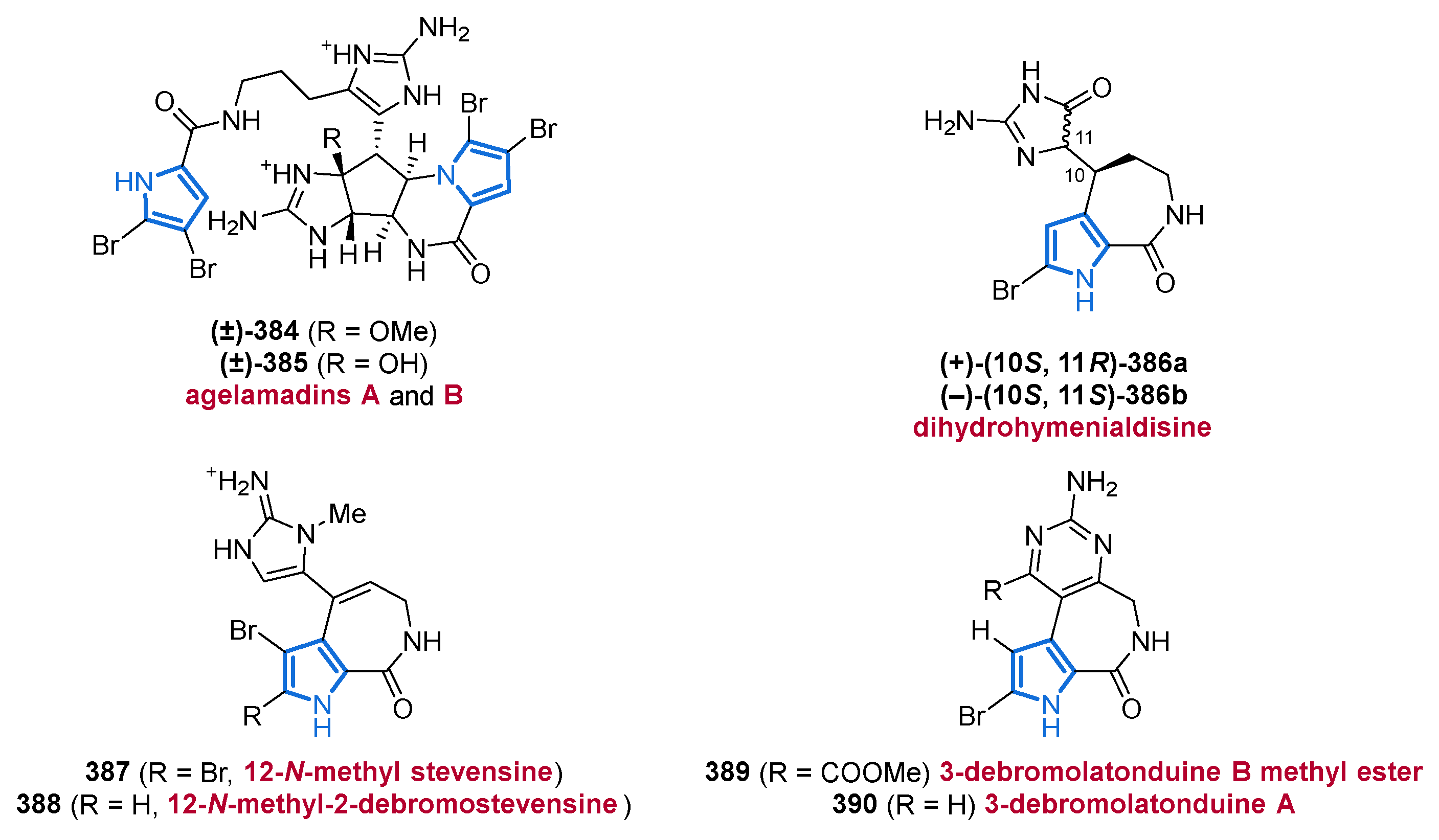
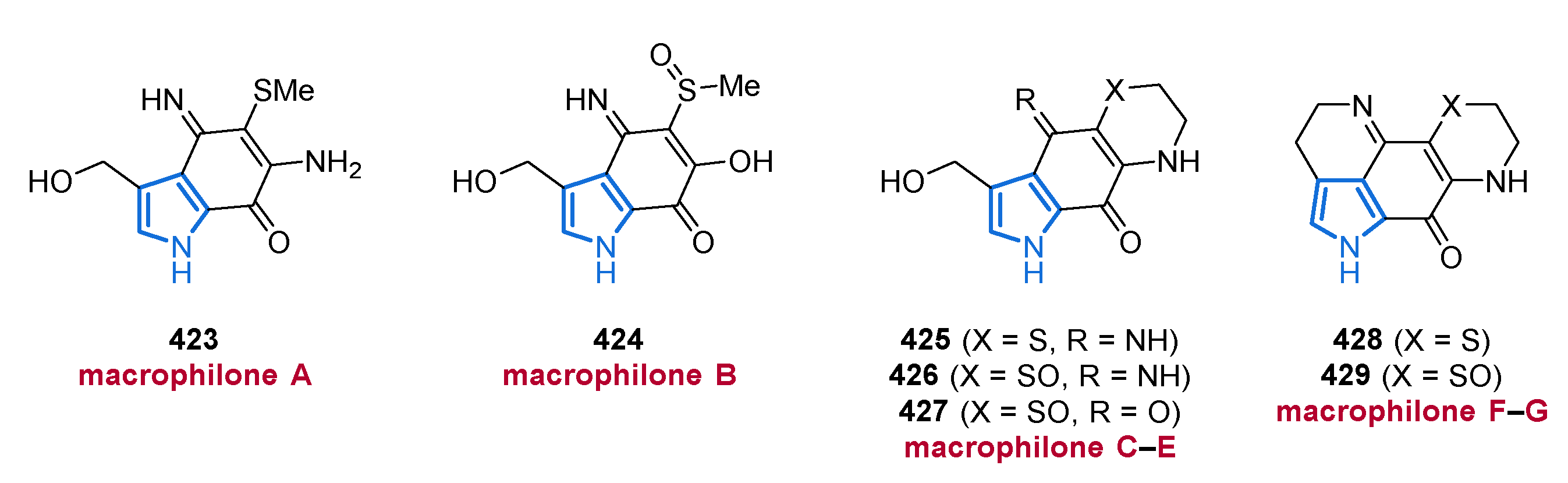
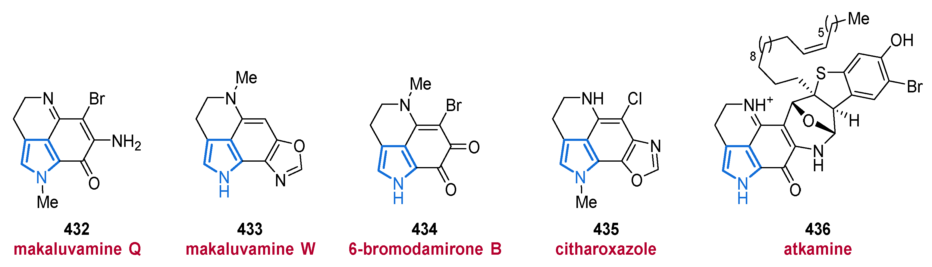
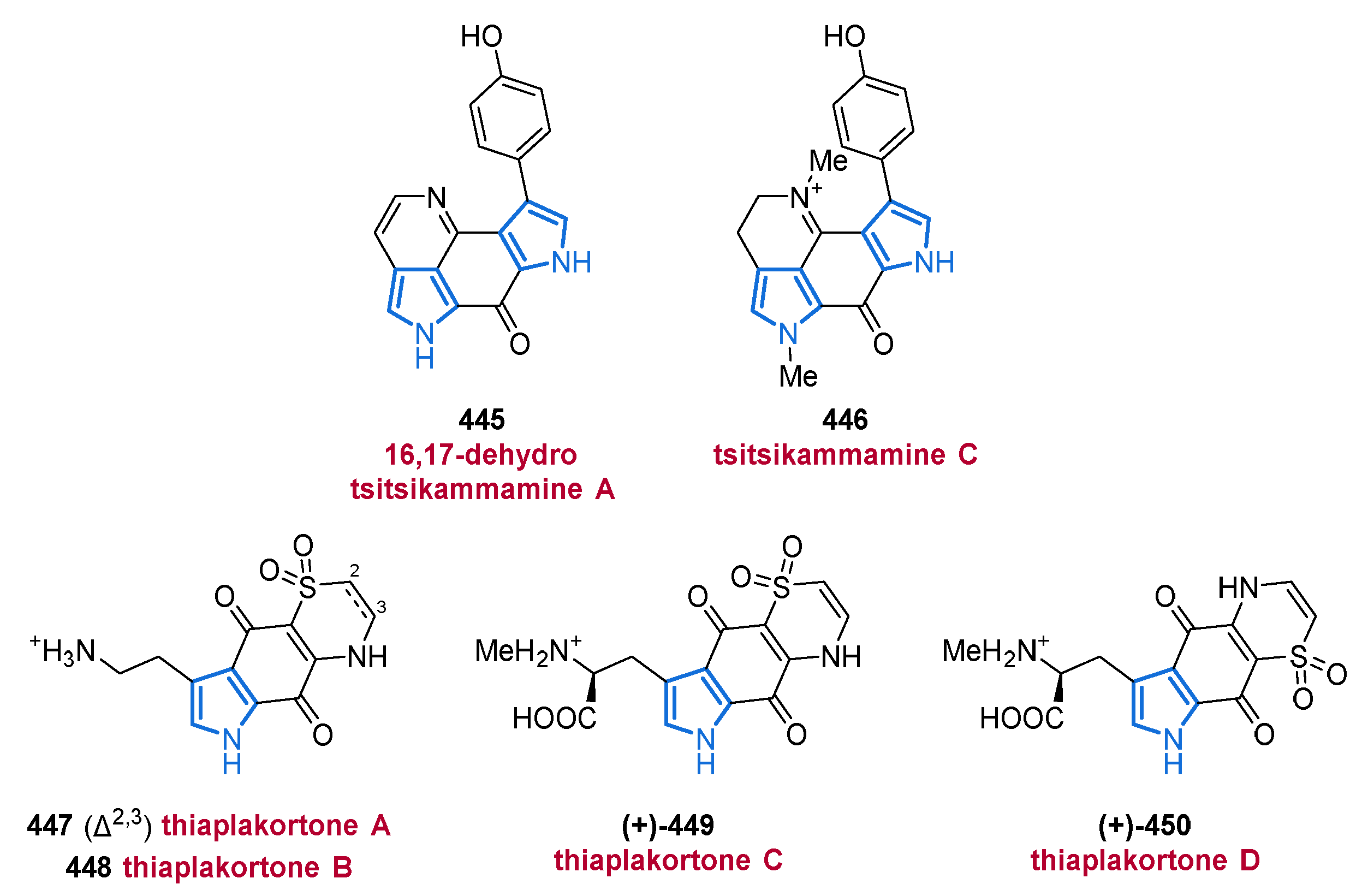
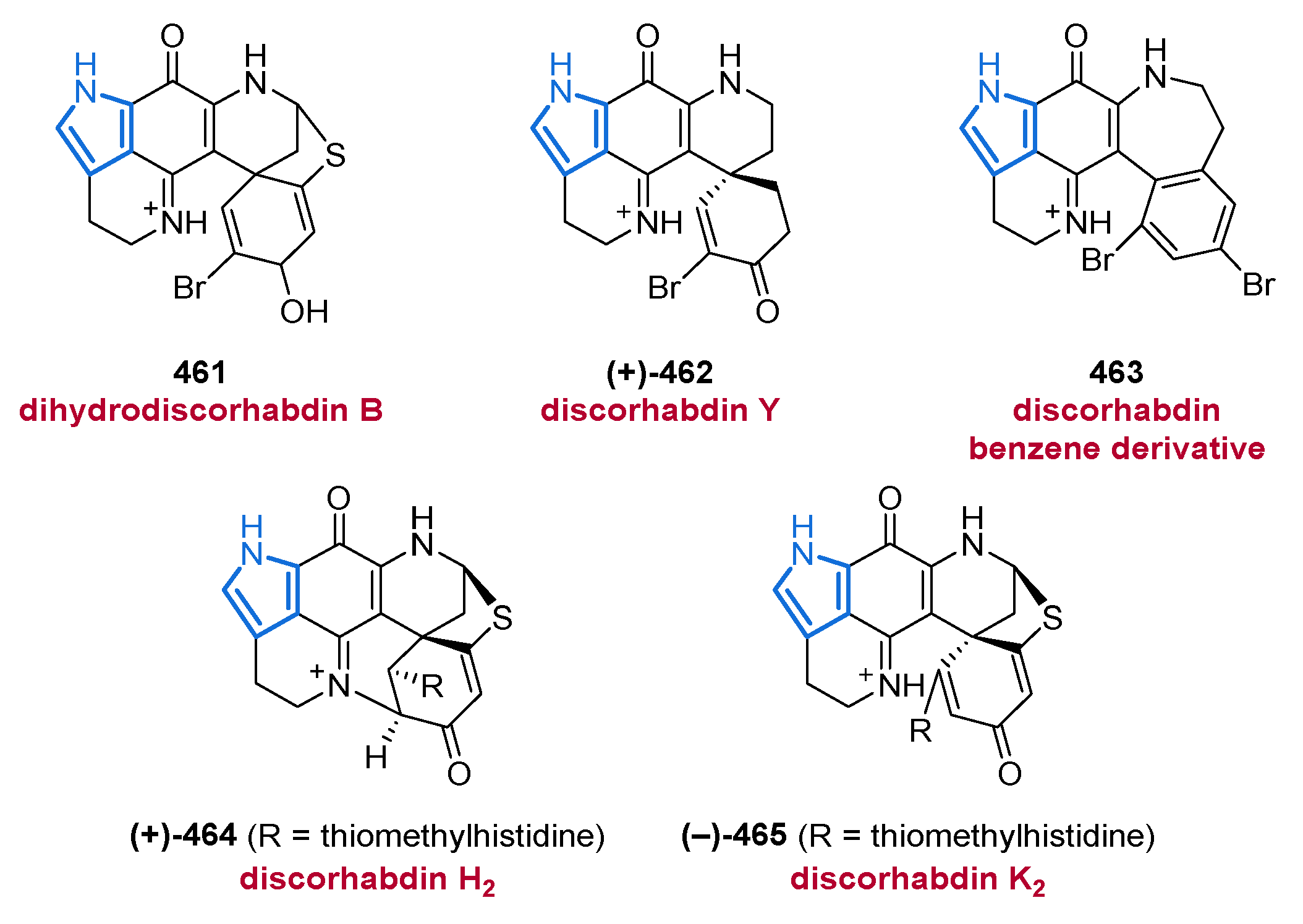
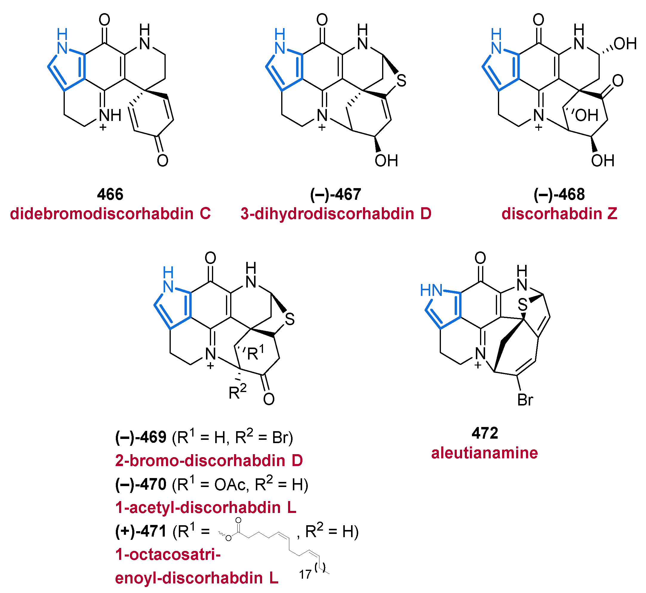
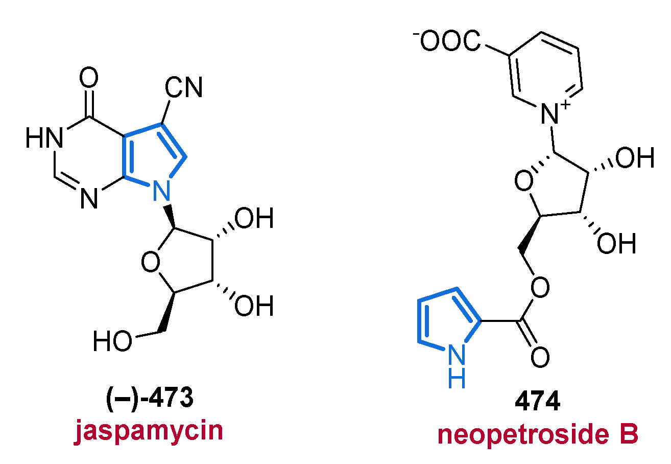
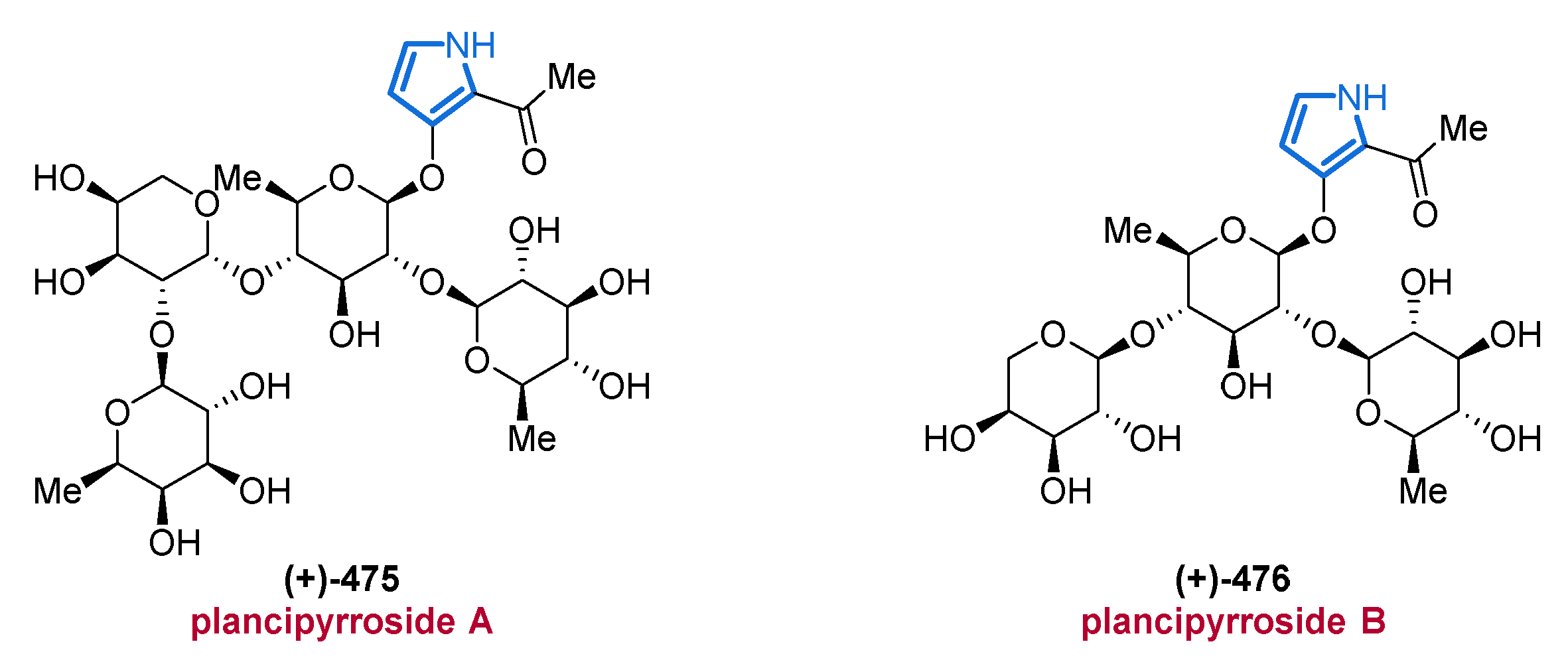
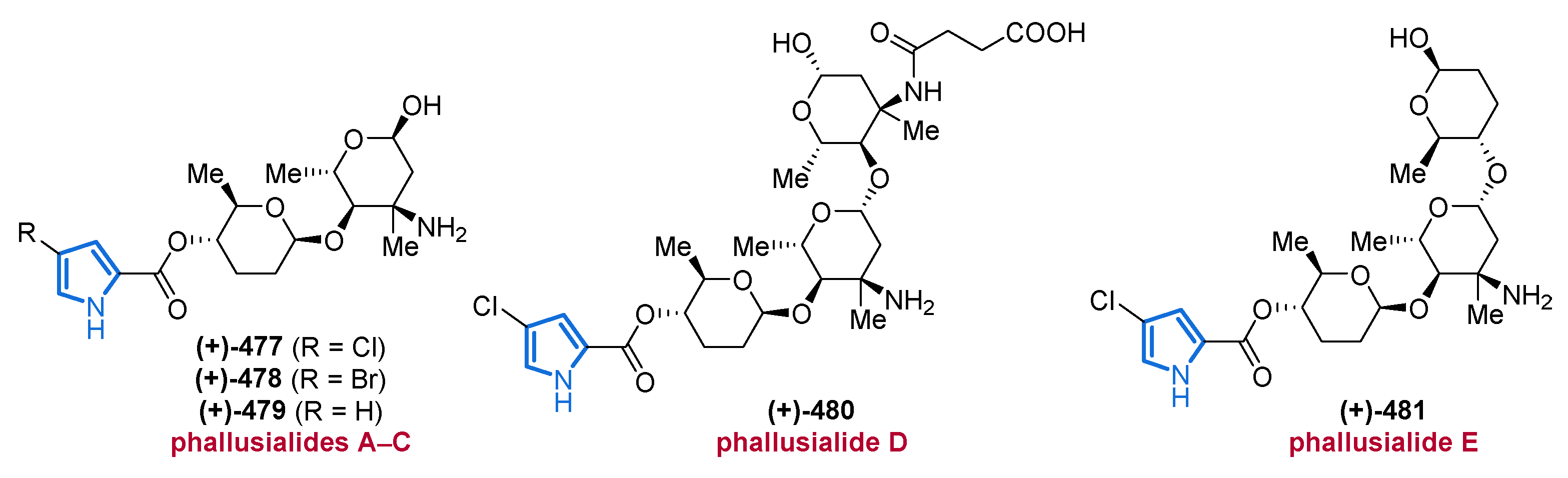
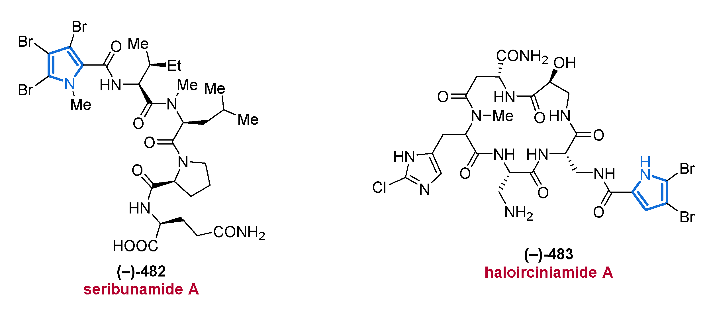
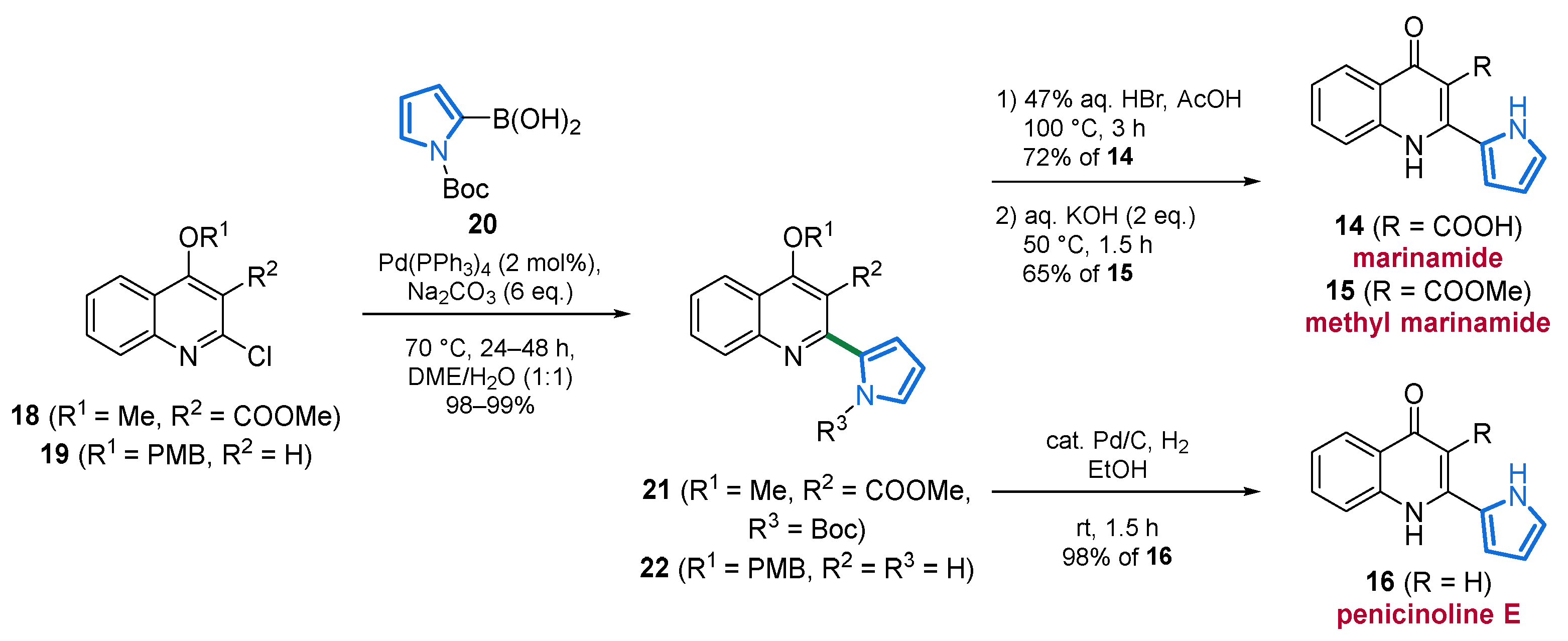
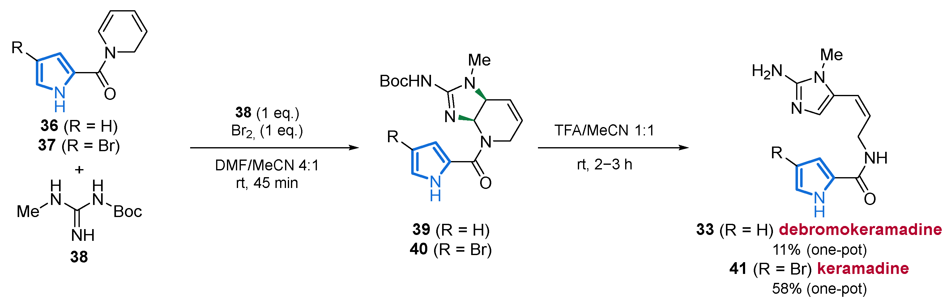
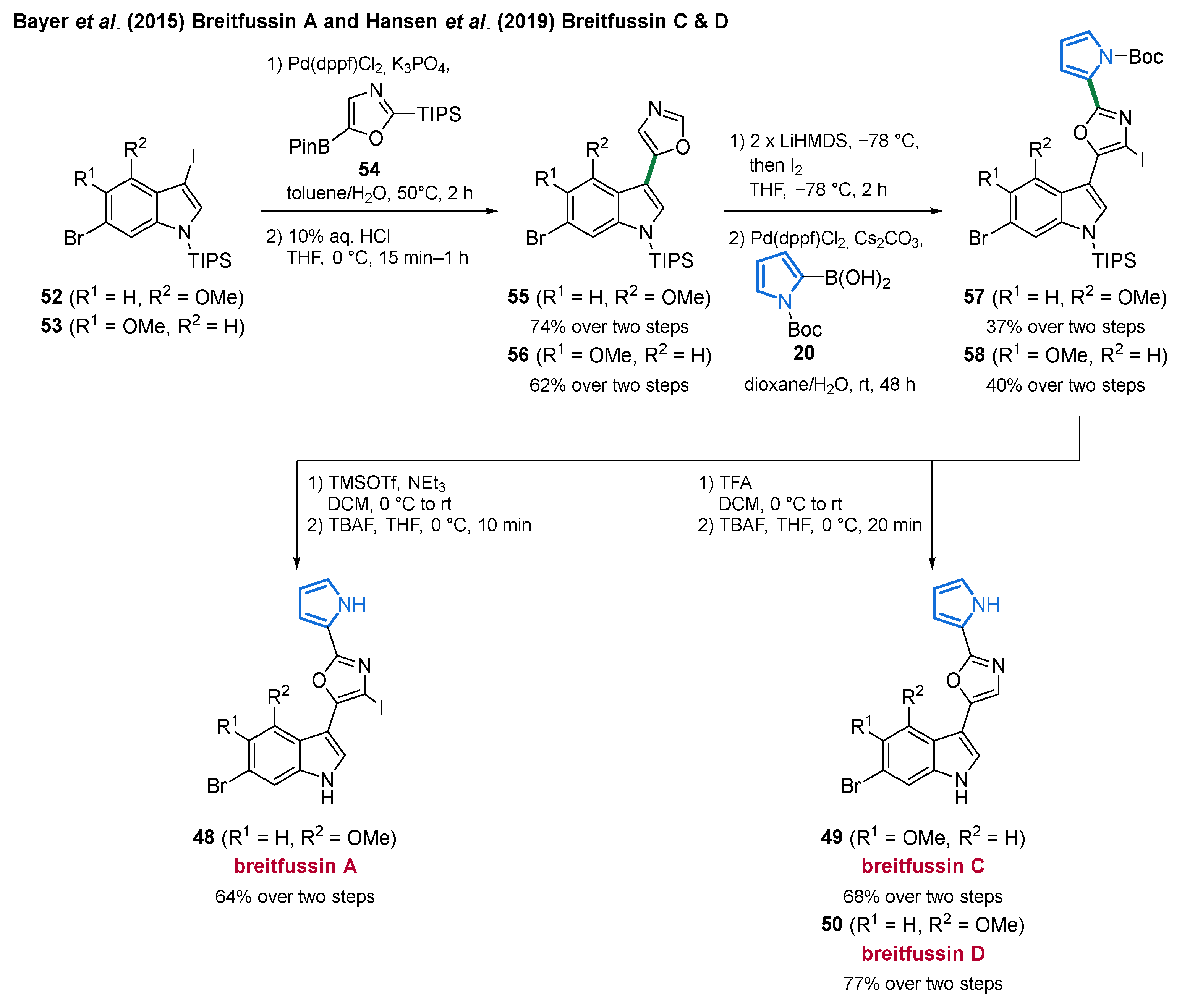
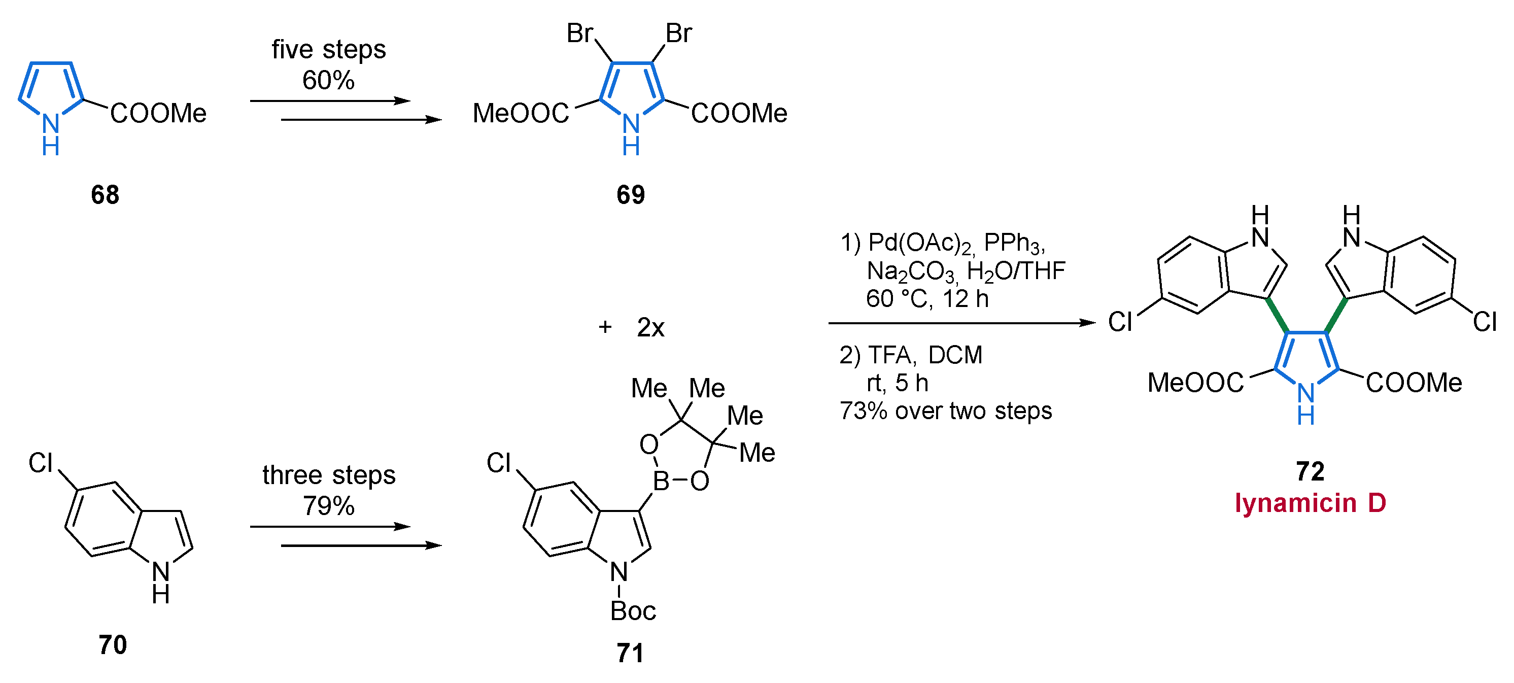
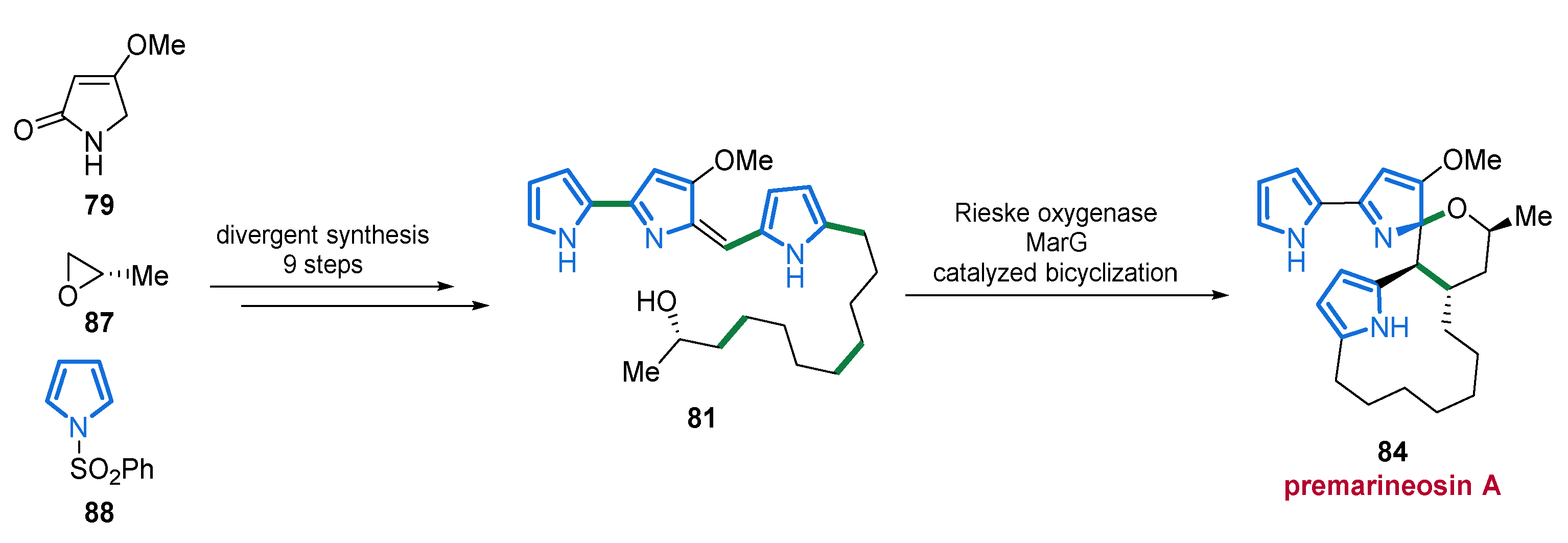
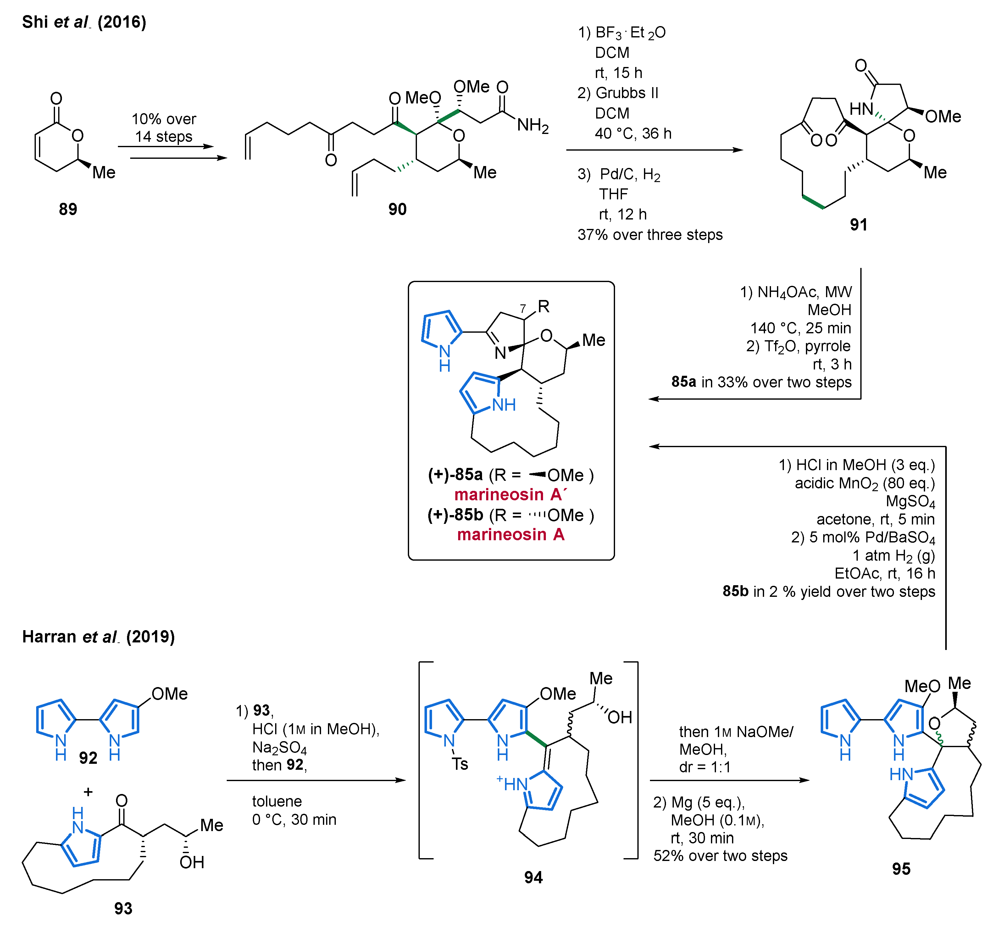
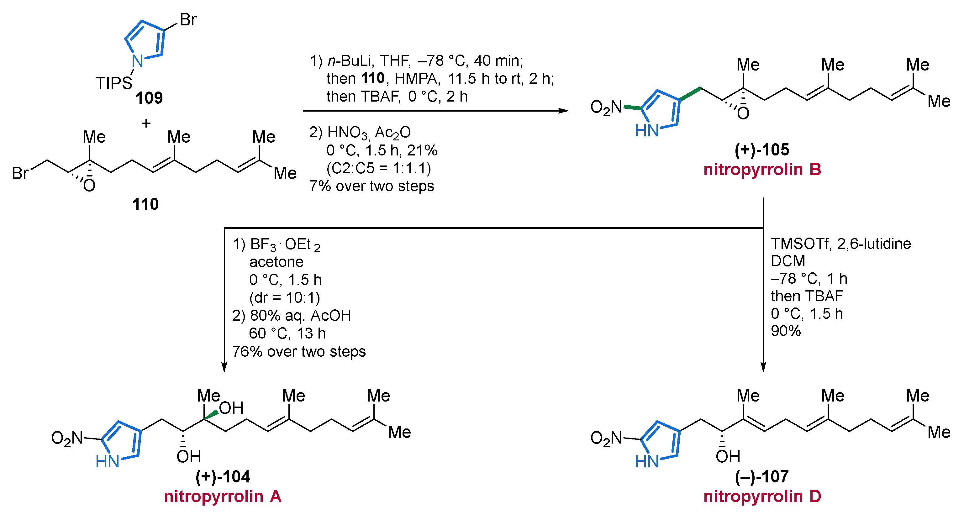
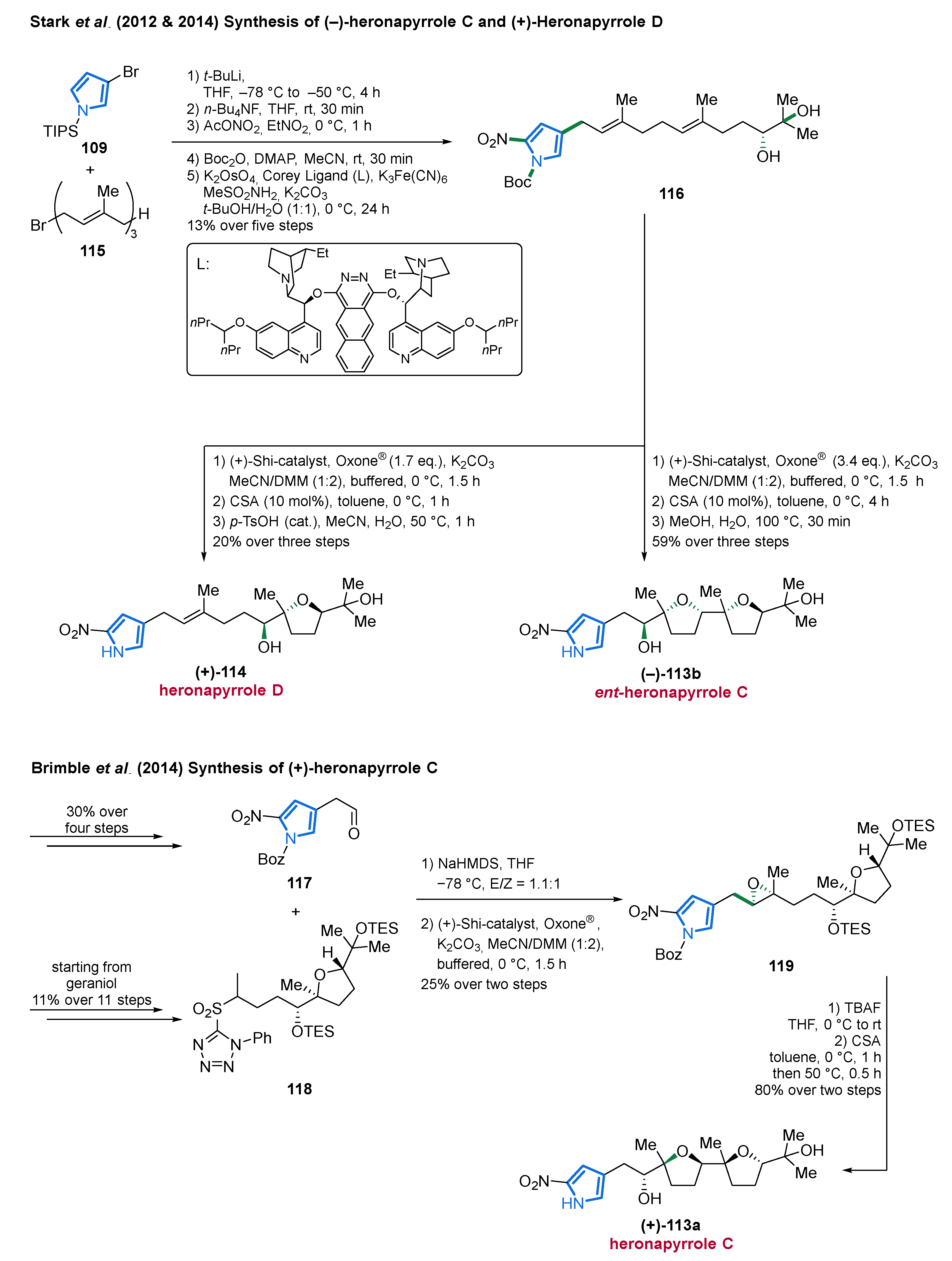
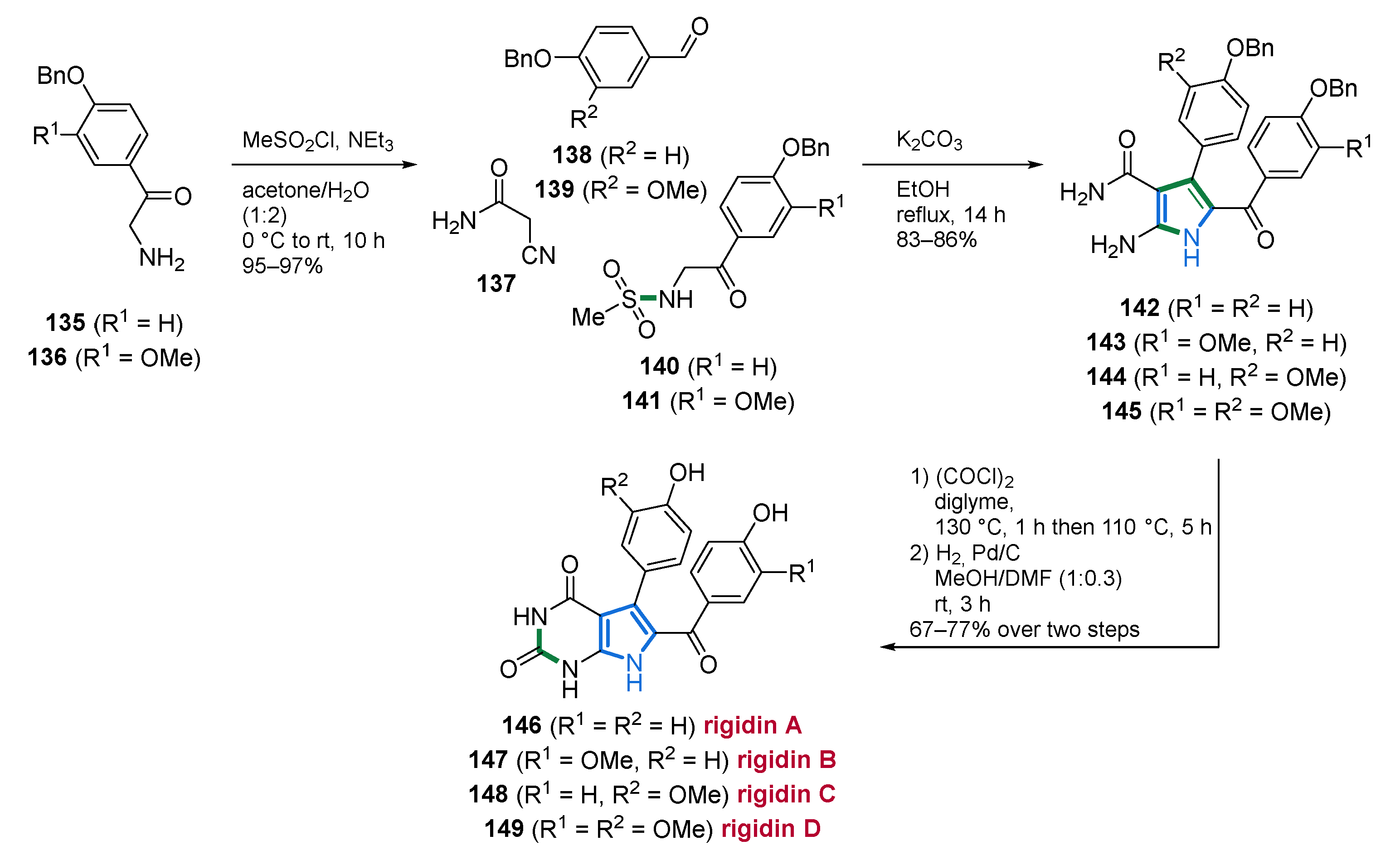
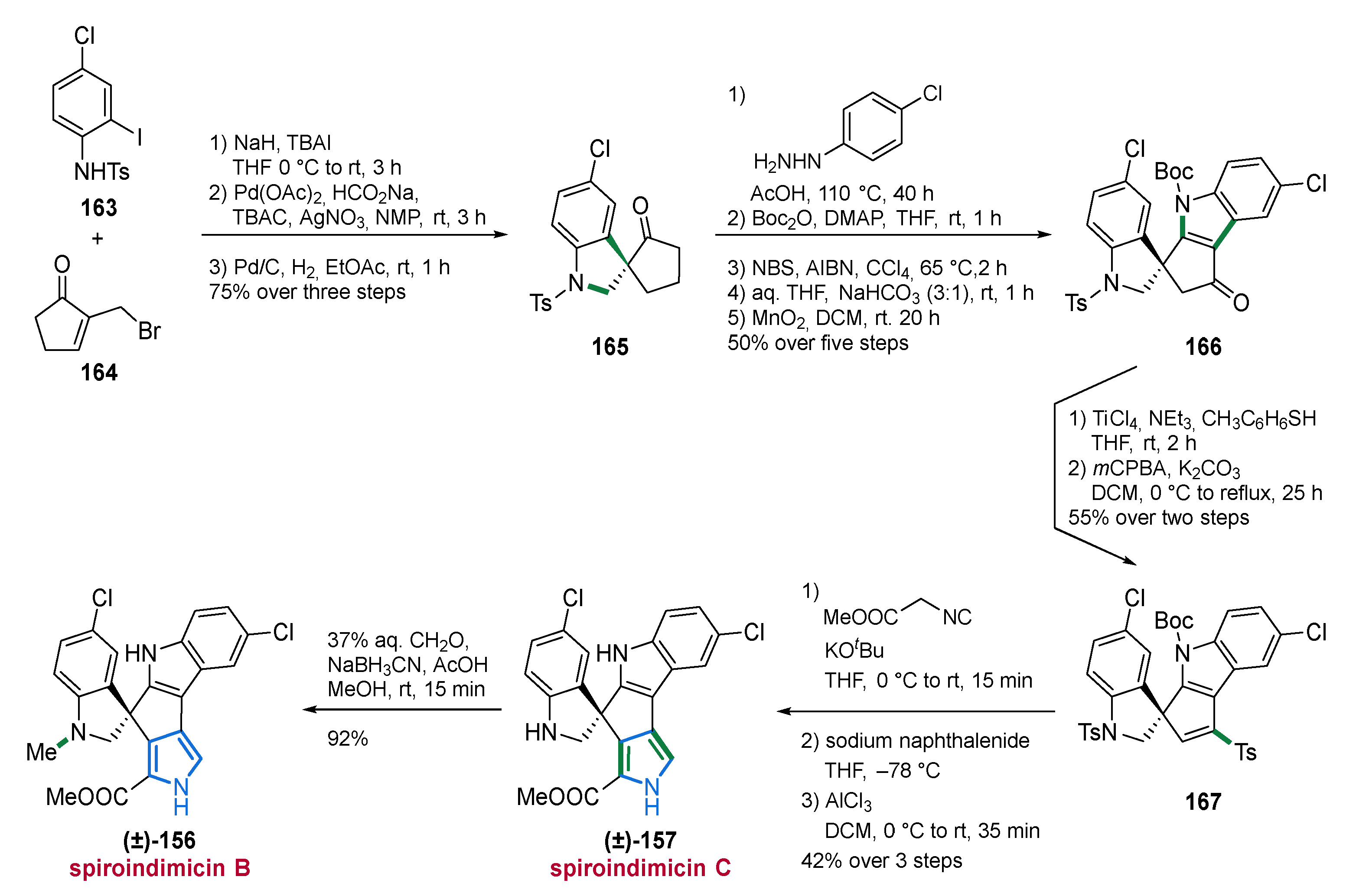
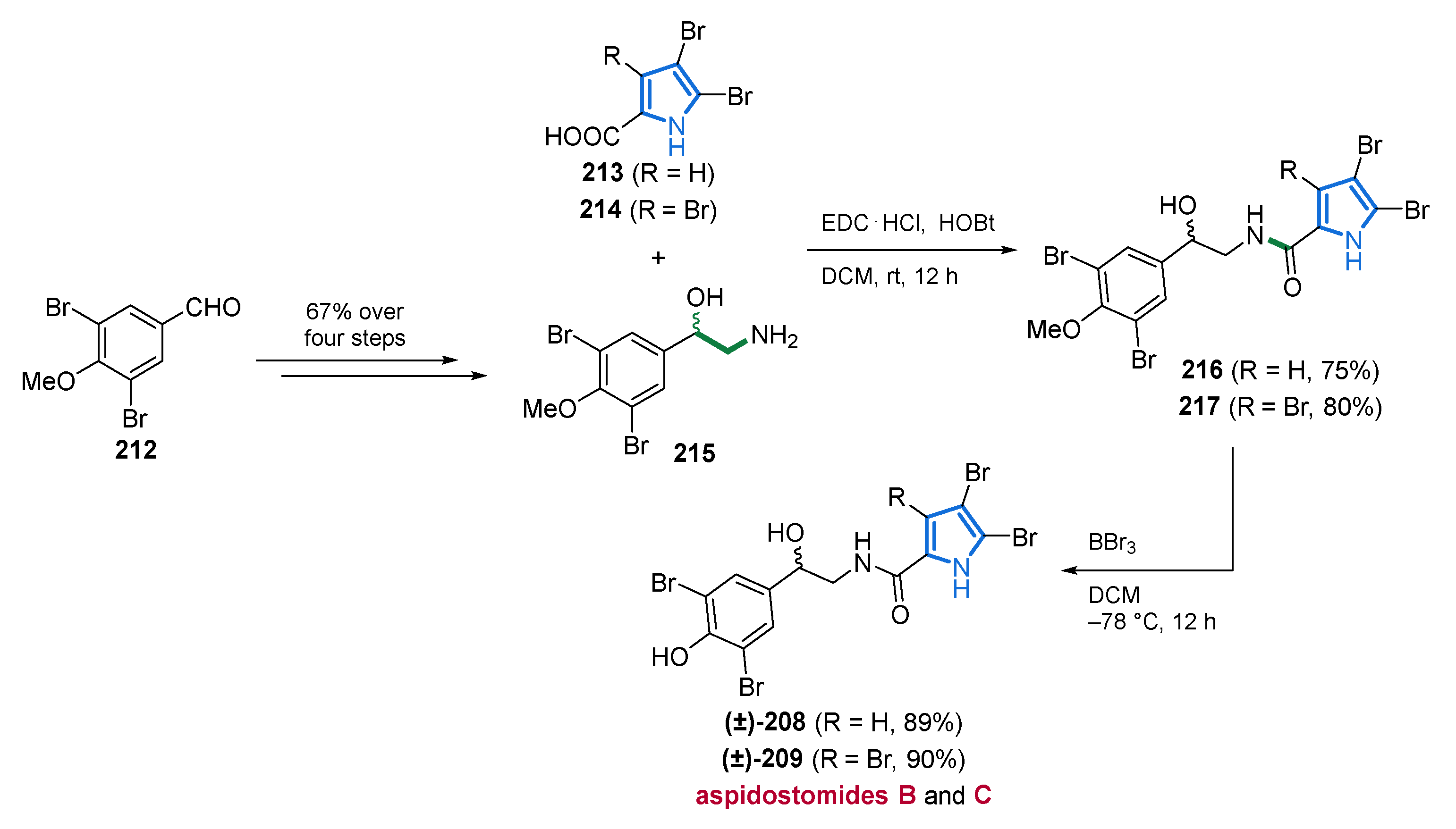
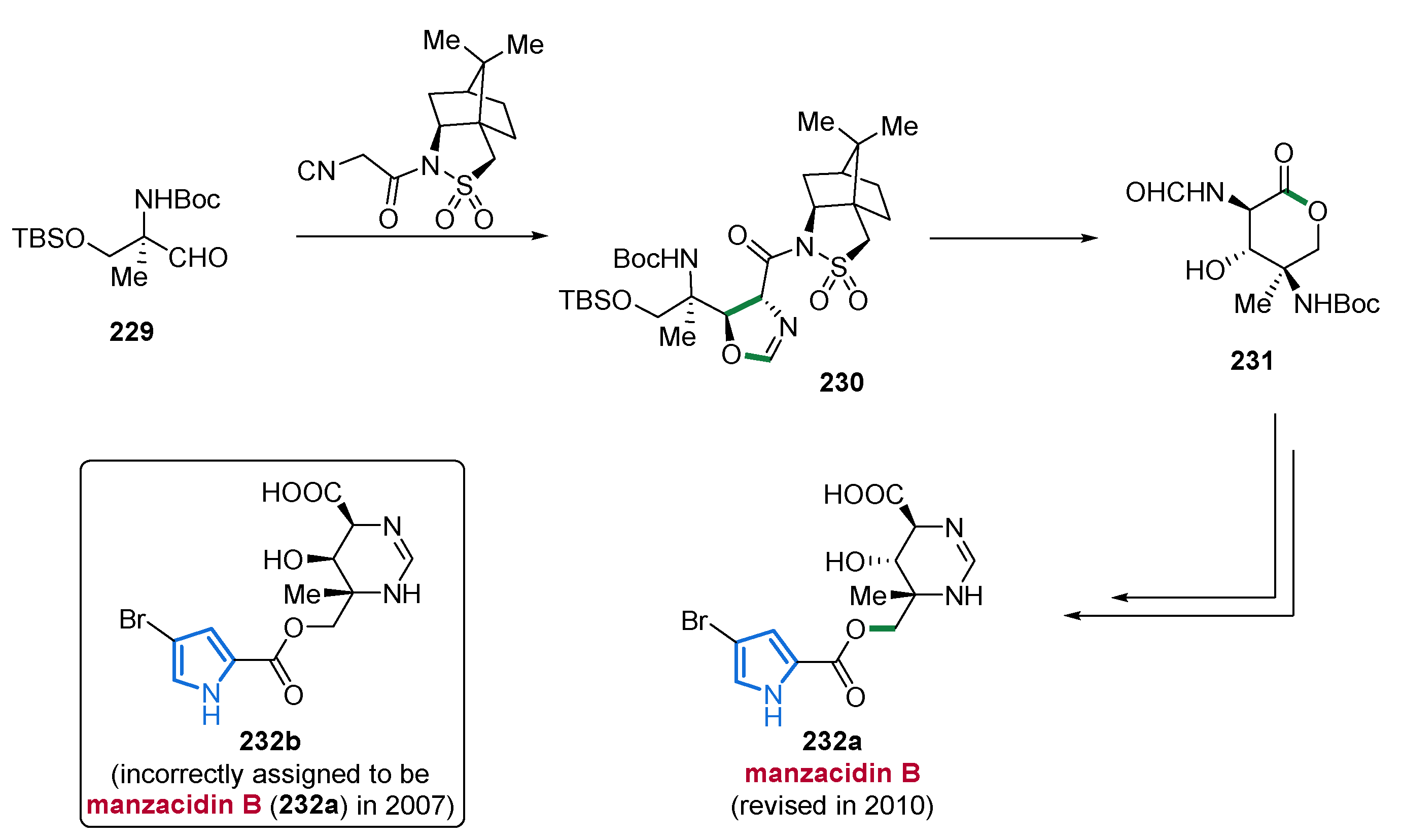
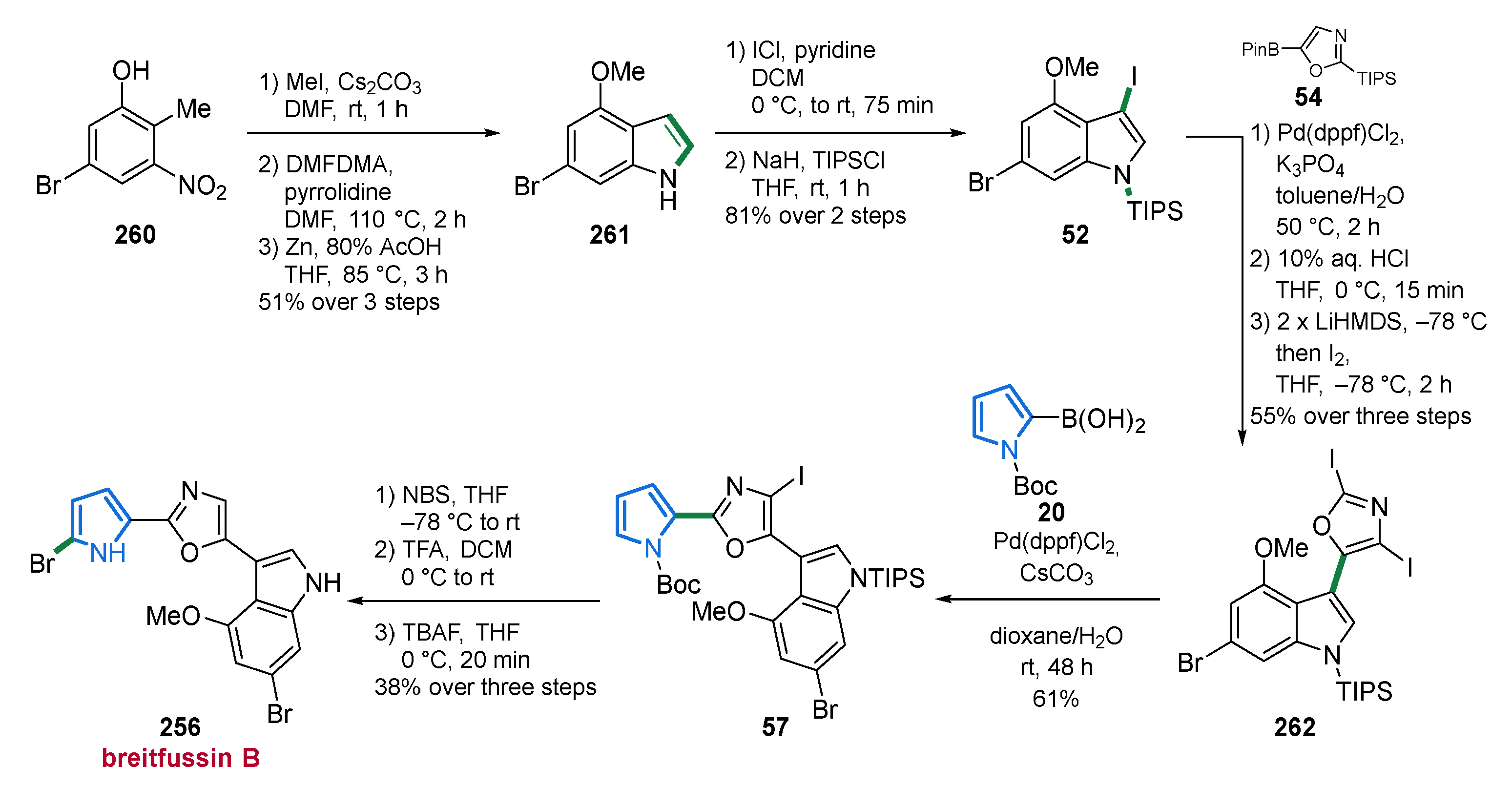
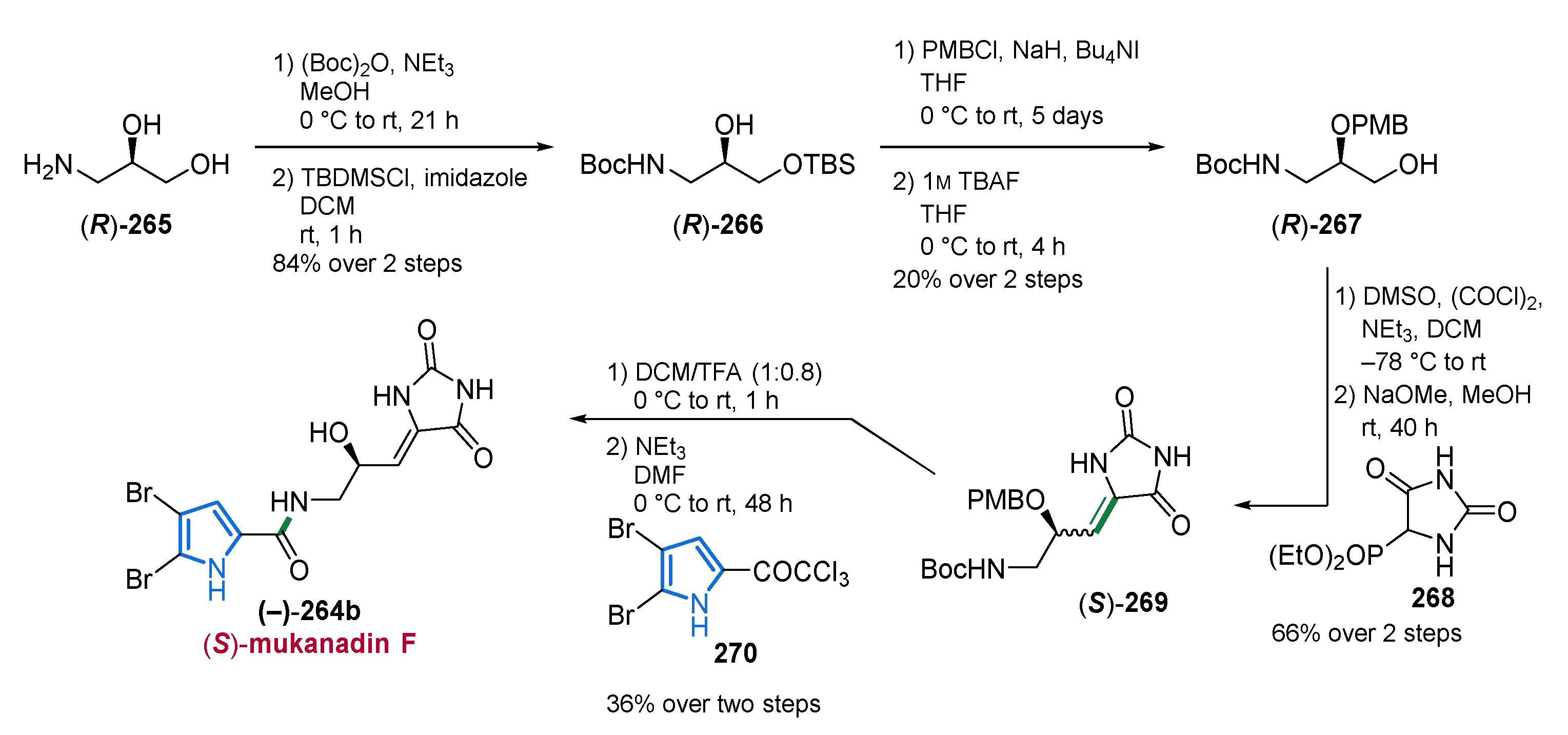
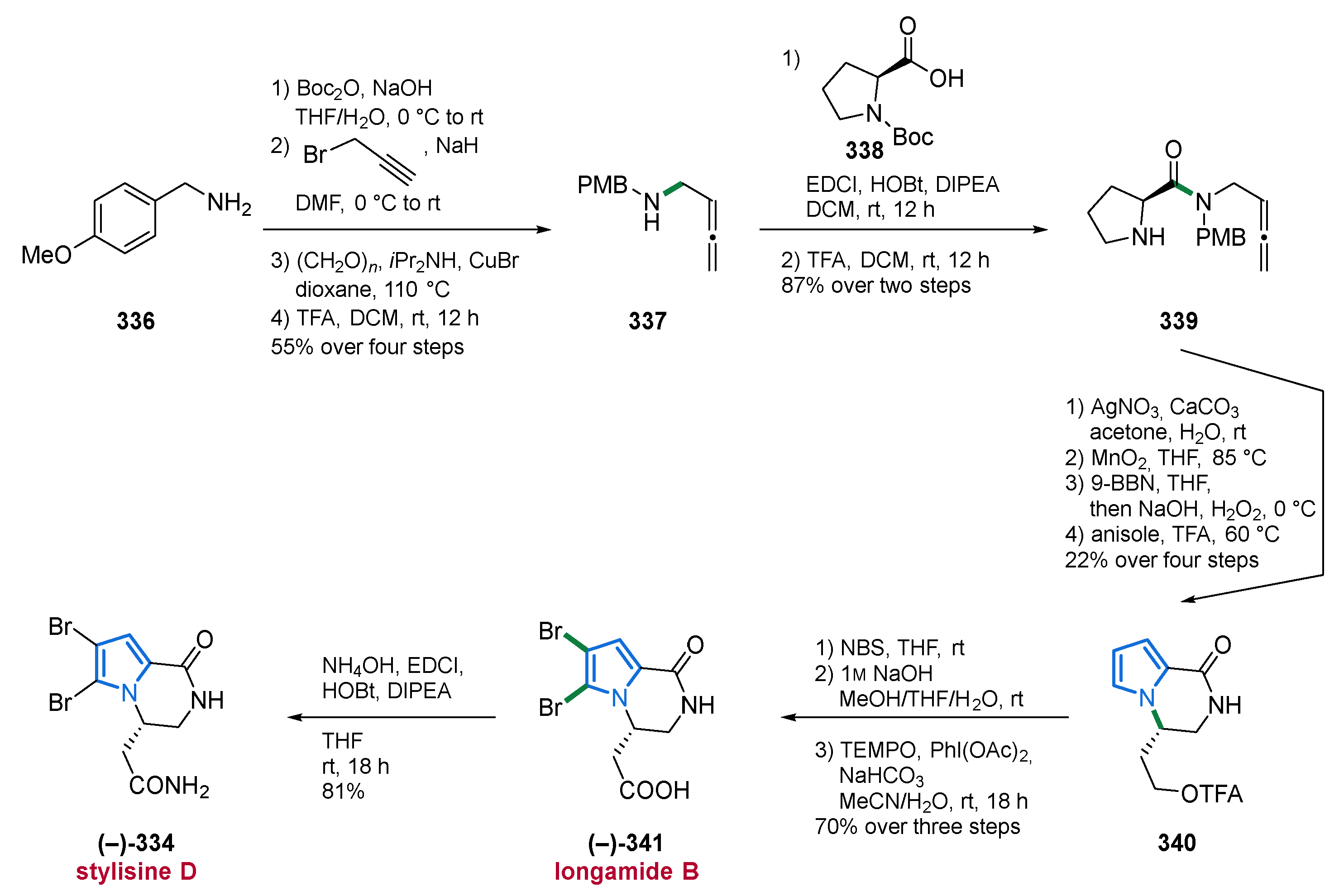
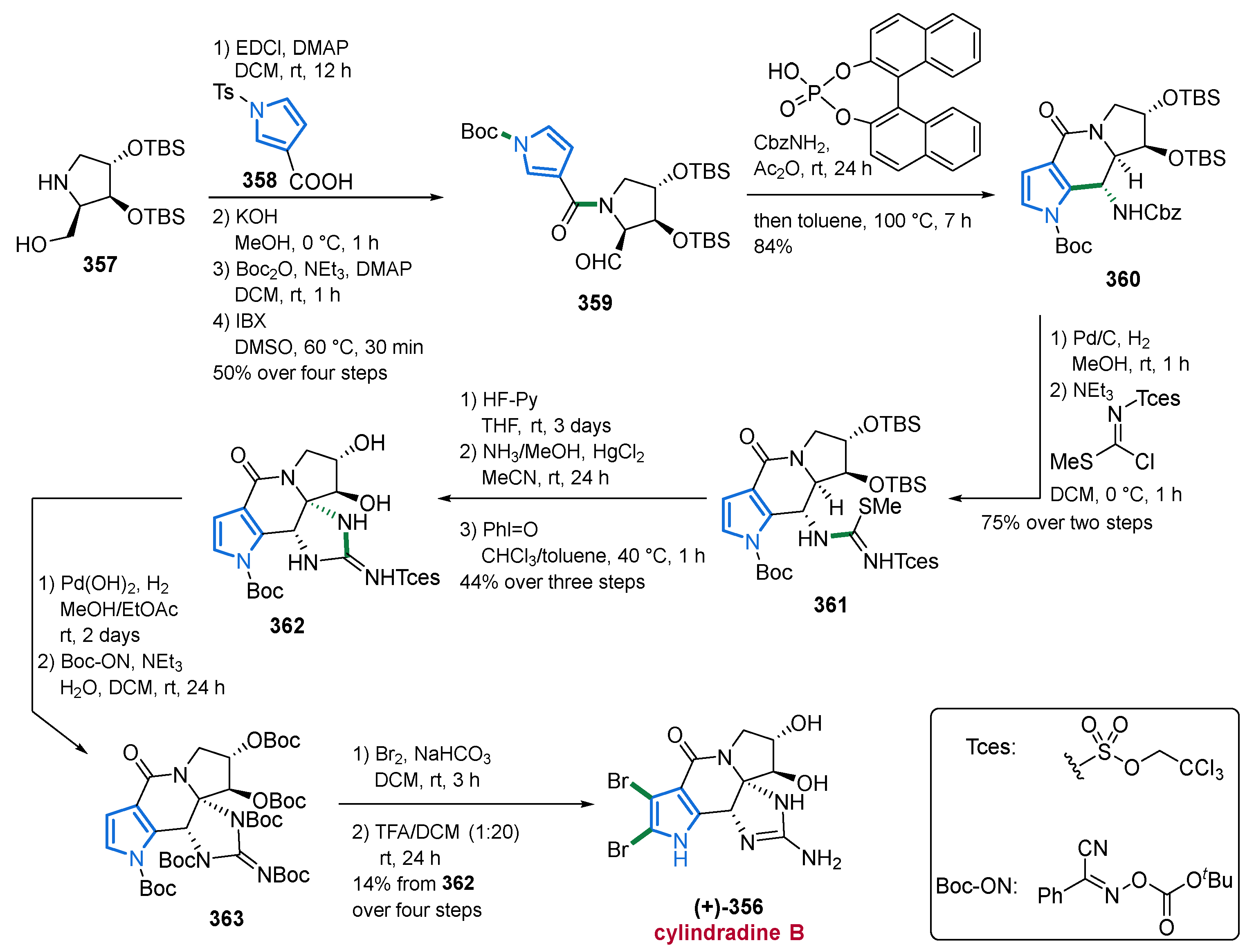
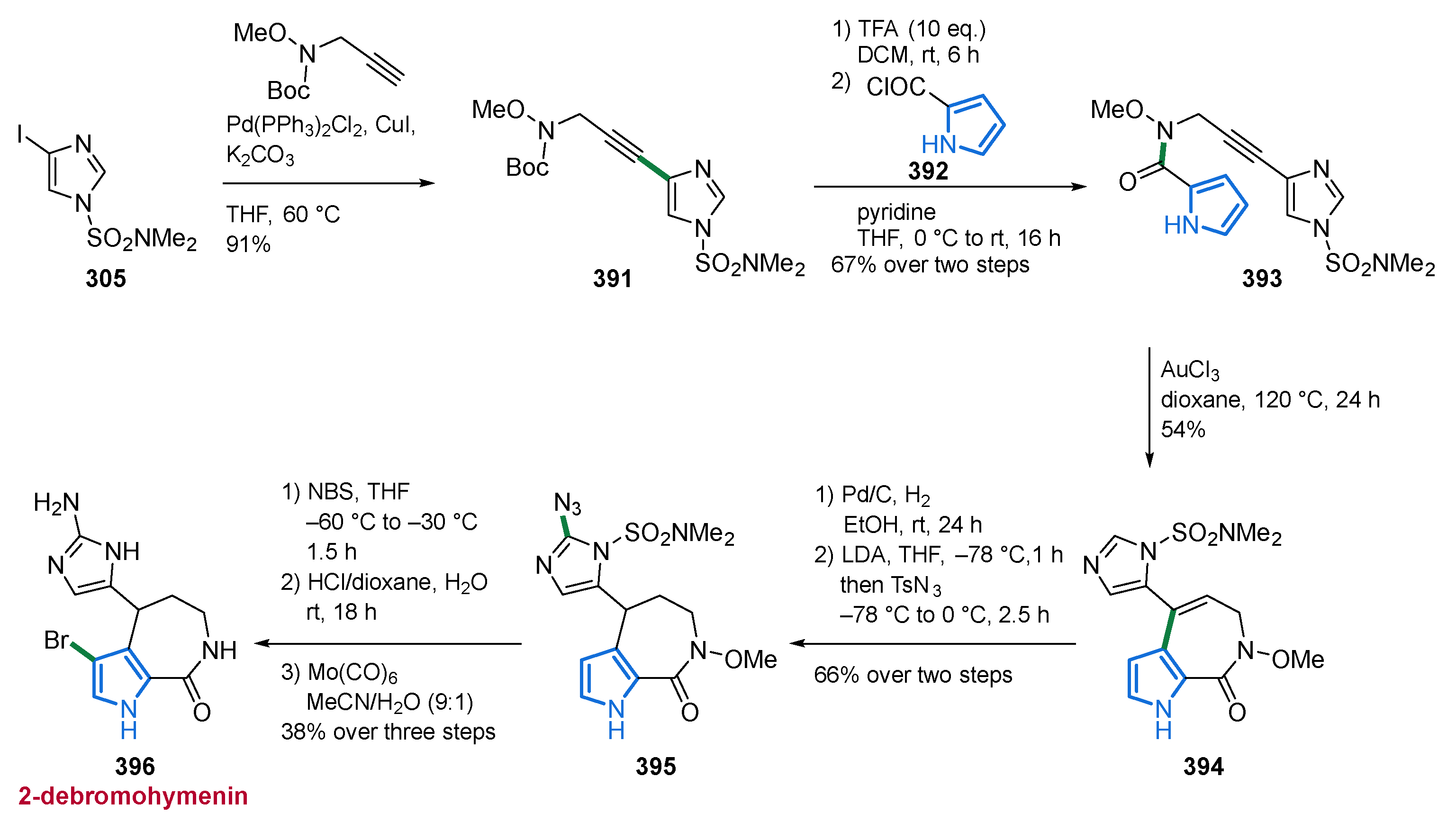
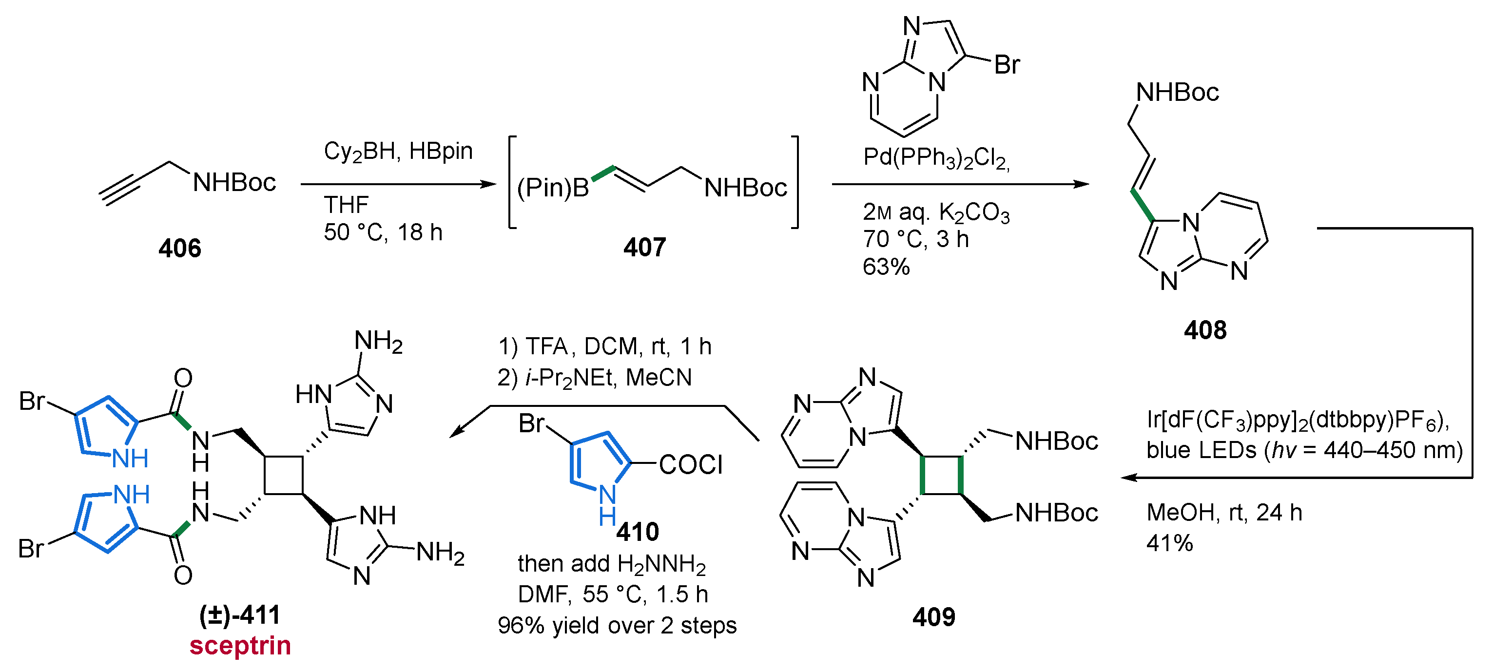
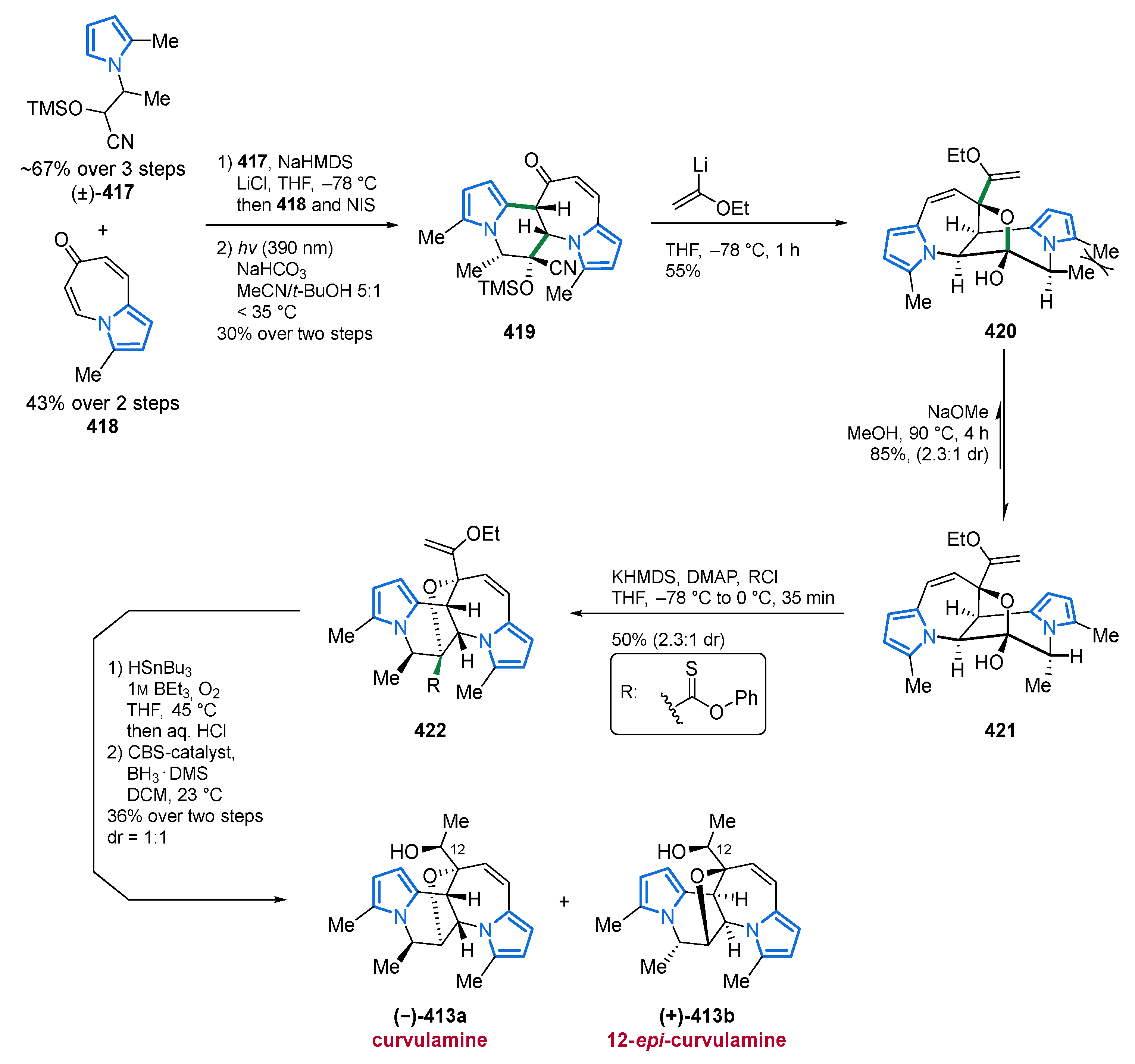
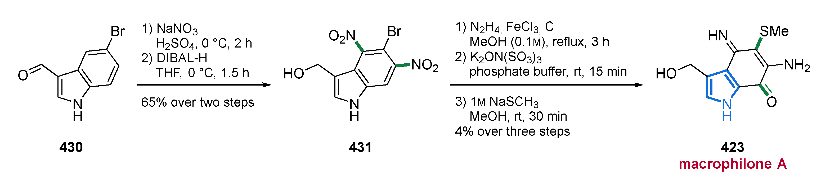
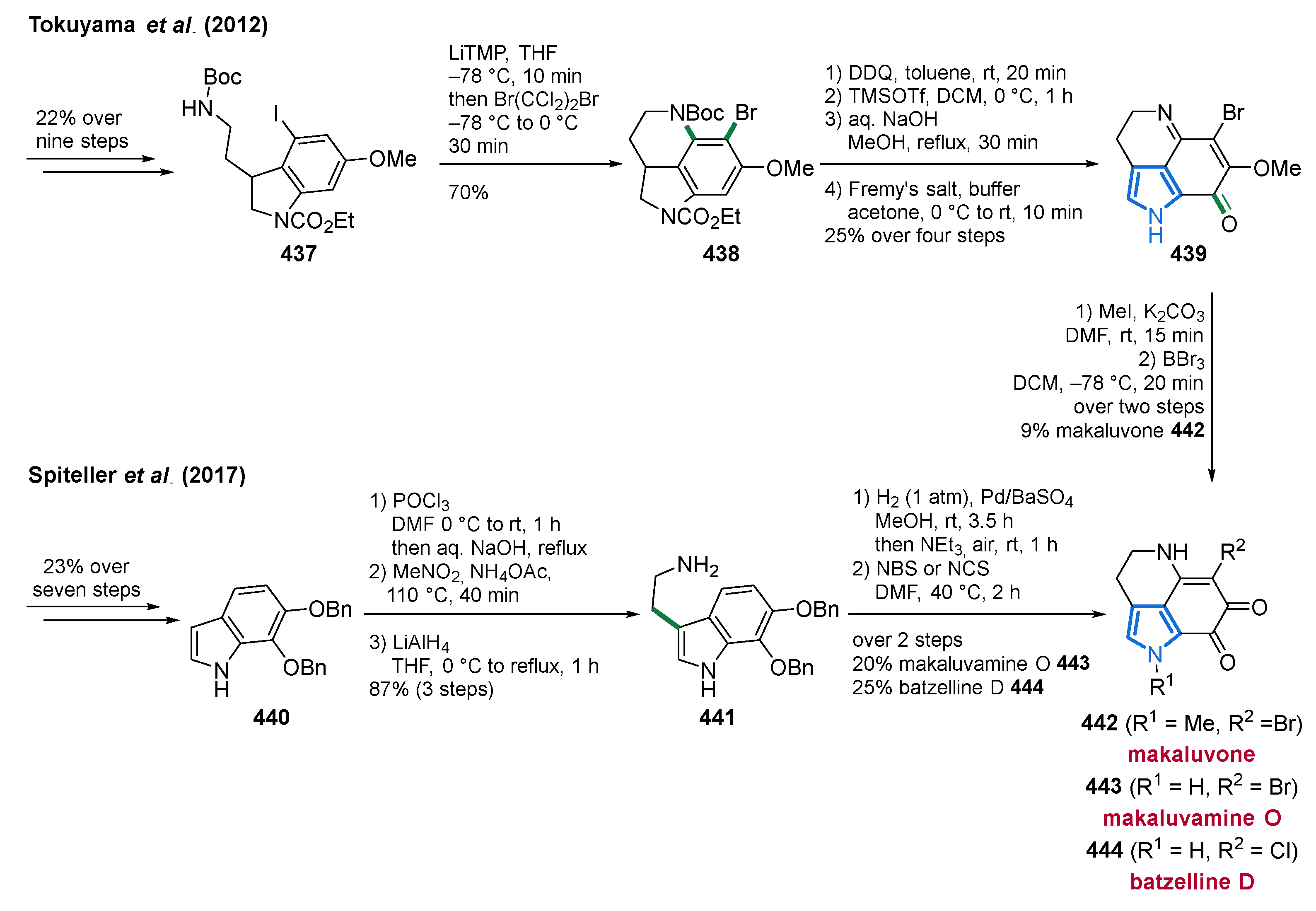
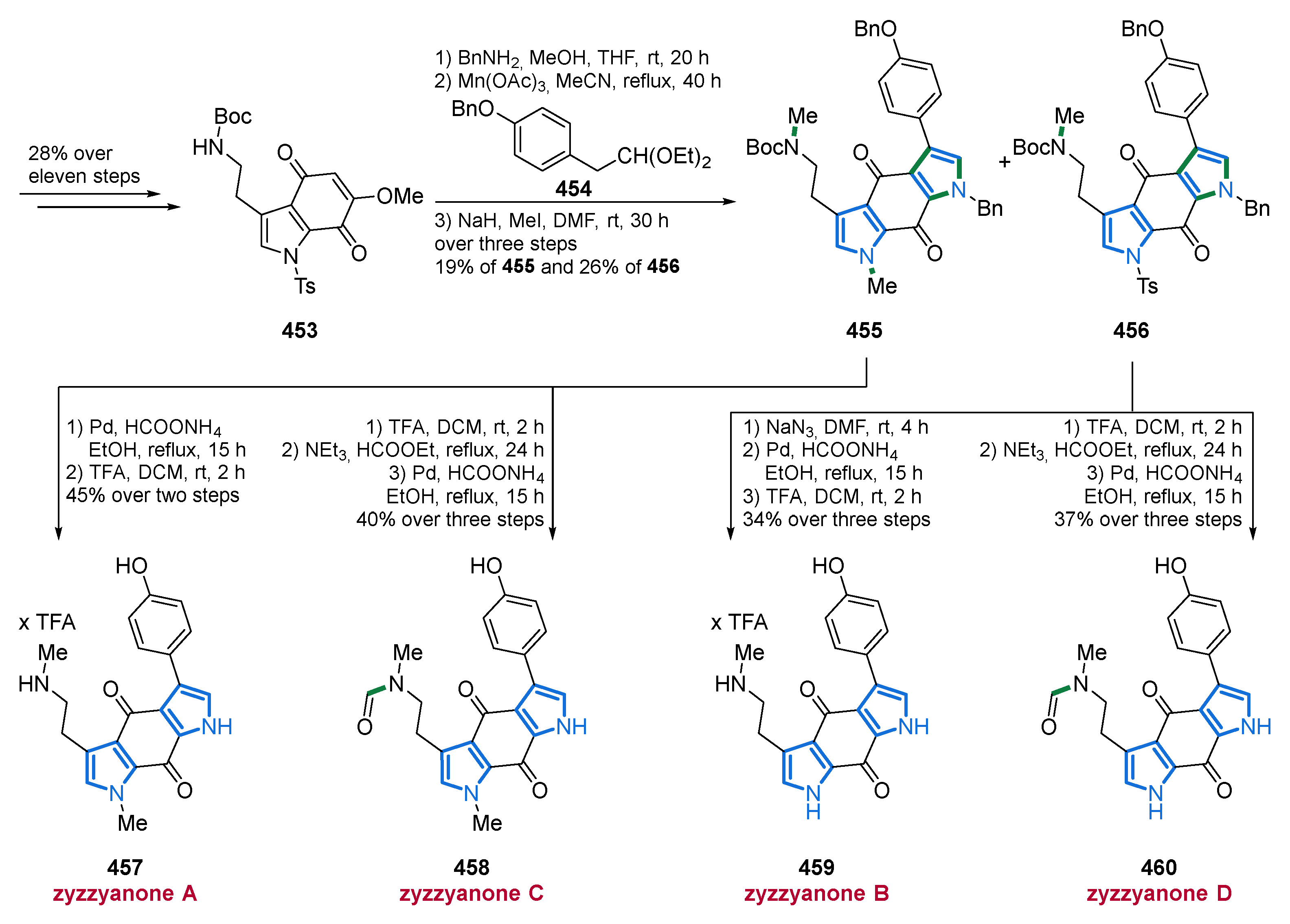
Table 1.
Summary of published synthesis of lamellarins and related analogs in the decade of 2010–2020.
Table 1.
Summary of published synthesis of lamellarins and related analogs in the decade of 2010–2020.
| Year | Author | Lamellarin and Related Congeners | Linear Steps i | Overall Yield |
|---|---|---|---|---|
| 2010 | Iwao [176] | Lamellarin α 20-sulfate | 15 | 6% |
| Lamellarin α 13-sulfate | 15 | 4% | ||
| Lamellarin α 13,20-disulfate | 14 | 9% | ||
| 2011 | Banwell [177] | G trimethyl ether | 10 | 3% |
| Lamellarin S | 11 | 6% | ||
| Jia [178] | Lamellarin D | 10 | 13% | |
| Lamellarin H | 10 | 13% | ||
| Lamellarin R | 5 | 53% | ||
| Ningalin B | 8 | 14% | ||
| 2012 | Vazquez [179] | Lamellarin Q | 6 | 28% |
| Lamellarin O | 7 | 25% | ||
| Banwell [180] | Lamellarin K | 9 | 57% | |
| Lamellarin T | 9 | 43% | ||
| Lamellarin U | 8 | 44% | ||
| Lamellarin W | 9 | 45% | ||
| 2013 | Opatz [181] | (Dihydro-)/lamellarin η | 8/9 | 62%/57% |
| Lamellarin G trimethyl ether | 7 | 69% | ||
| Iwao [182] | Lukianol A/B | 6/11 | 36%/11% | |
| 2014 | Yamaguchi [183] | Lamellarin C | 9 | 3% |
| Lamellarin I | 9 | 3% | ||
| Iwao [184] | Lamellarin N | 11;13 | 42%;34% | |
| Lamellarin L | 13 | 29% | ||
| 2015 | Iwao [185] | Lamellarin L | 10 | 14% |
| Lamellarin N | 10 | 12% | ||
| Opatz [186] | Lamellarin D trimethyl ether | 9 | 43% | |
| Lamellarin H | 10 | 41% | ||
| Ruchirawat [187] | Aza/lamellarin D | 13/13 | 12%/9% | |
| Aza/lamellarin N | 13/13 | 28%/15% | ||
| Tan and Yoshikai [188] | Lamellarin G trimethyl ether | 5 | 20% | |
| 2016 | Iwao [189] | Lamellarin U | 12 | 5% |
| Yang [190] | Lamellarin D trimethyl ether | 3 | 8% | |
| Lamellarin H | 4 | 7% | ||
| 2017 | Iwao [191] | Lamellarin N analogues | – | – |
| Azalamellarin N analogues | – | – | ||
| Iwao [192] | Lamellarin α | 12 | 22% | |
| Lamellarin η | 10 | 19% | ||
| Chandrasekhar [193] | Lamellarin D trimethyl ether | 6 | 44% | |
| Lamellarin D | 7 | 29% | ||
| Lamellarin H | 7 | 37% | ||
| Wu [194] | Lamellarin G trimethyl ether | 3 | 51% | |
| Yang [195] | Lamellarin D trimethyl ether | 2 | 37% | |
| Lamellarin H | 3 | 31% | ||
| Lamellarin D | 6;8 | 12–14% | ||
| Lamellarin χ | 6;8 | 12–14% | ||
| Ackermann [196] | Lamellarin D | 10 | 30% | |
| Lamellarin H | 10 | 29% | ||
| 2018 | Opatz [197] | Lamellarin G trimethyl ether | 7;8 | 19–42% |
| Chiu and Tonks [198] | Lamellarin R | 5 | 18% | |
| 2019 | Donohoe [199] | Lamellarin D | 7 | 22% |
| Lamellarin Q | 7 | 20% | ||
| Opatz and Michael [200] | Lamellarin G trimethyl ether | 6;7 | 56–73% | |
| Khan [201] | Lamellarin G trimethyl ether | 5 | 18% | |
| Lamellarin D trimethyl ether | 6 | 16% | ||
| Lamellarins H, U | 7/6 | 11%/11% | ||
| Dihydro/lamellarin η | 7/6 | 9%/10% | ||
| 2020 | Saito [202] | Lamellarin G trimethyl ether | 6 | 26% |
| Lamellarin H | 8 | 17% | ||
| Tsay [203] | Lamellarin R | 3 | 50% | |
| Liou [204] | Lamellarin R | 5 | 26% | |
| Lamellarin O | 5 | 10% | ||
| Lukianol A | 6 | 38% | ||
| Khan [205] | Lamellarins | 6/6/6 | 21%/21%/21% | |
| S,Z,G,L,N,D | 6/7/7 | 21%/19%/16% |
i The longest linear sequence in the synthesis was counted.
Publisher’s Note: MDPI stays neutral with regard to jurisdictional claims in published maps and institutional affiliations. |
© 2021 by the authors. Licensee MDPI, Basel, Switzerland. This article is an open access article distributed under the terms and conditions of the Creative Commons Attribution (CC BY) license (https://creativecommons.org/licenses/by/4.0/).
Share and Cite
MDPI and ACS Style
Seipp, K.; Geske, L.; Opatz, T. Marine Pyrrole Alkaloids. Mar. Drugs 2021, 19, 514. https://0-doi-org.brum.beds.ac.uk/10.3390/md19090514
AMA Style
Seipp K, Geske L, Opatz T. Marine Pyrrole Alkaloids. Marine Drugs. 2021; 19(9):514. https://0-doi-org.brum.beds.ac.uk/10.3390/md19090514
Chicago/Turabian StyleSeipp, Kevin, Leander Geske, and Till Opatz. 2021. "Marine Pyrrole Alkaloids" Marine Drugs 19, no. 9: 514. https://0-doi-org.brum.beds.ac.uk/10.3390/md19090514
Note that from the first issue of 2016, this journal uses article numbers instead of page numbers. See further details here.