Investigation the EMG Activities of Lower Limb Muscles When Doing Squatting Exercise in Water and on Land


Abstract
:1. Introduction
2. Materials and Methods
2.1. Study Design
2.2. Sample Size Planning
2.3. Participants
2.4. Experimental Setup
2.5. Procedures
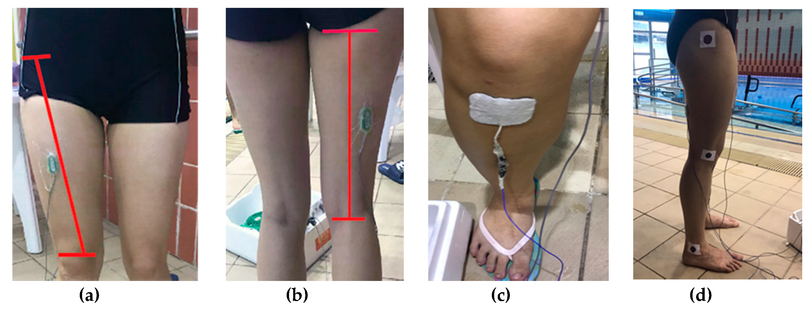
2.5.1. Electrodes and Markers
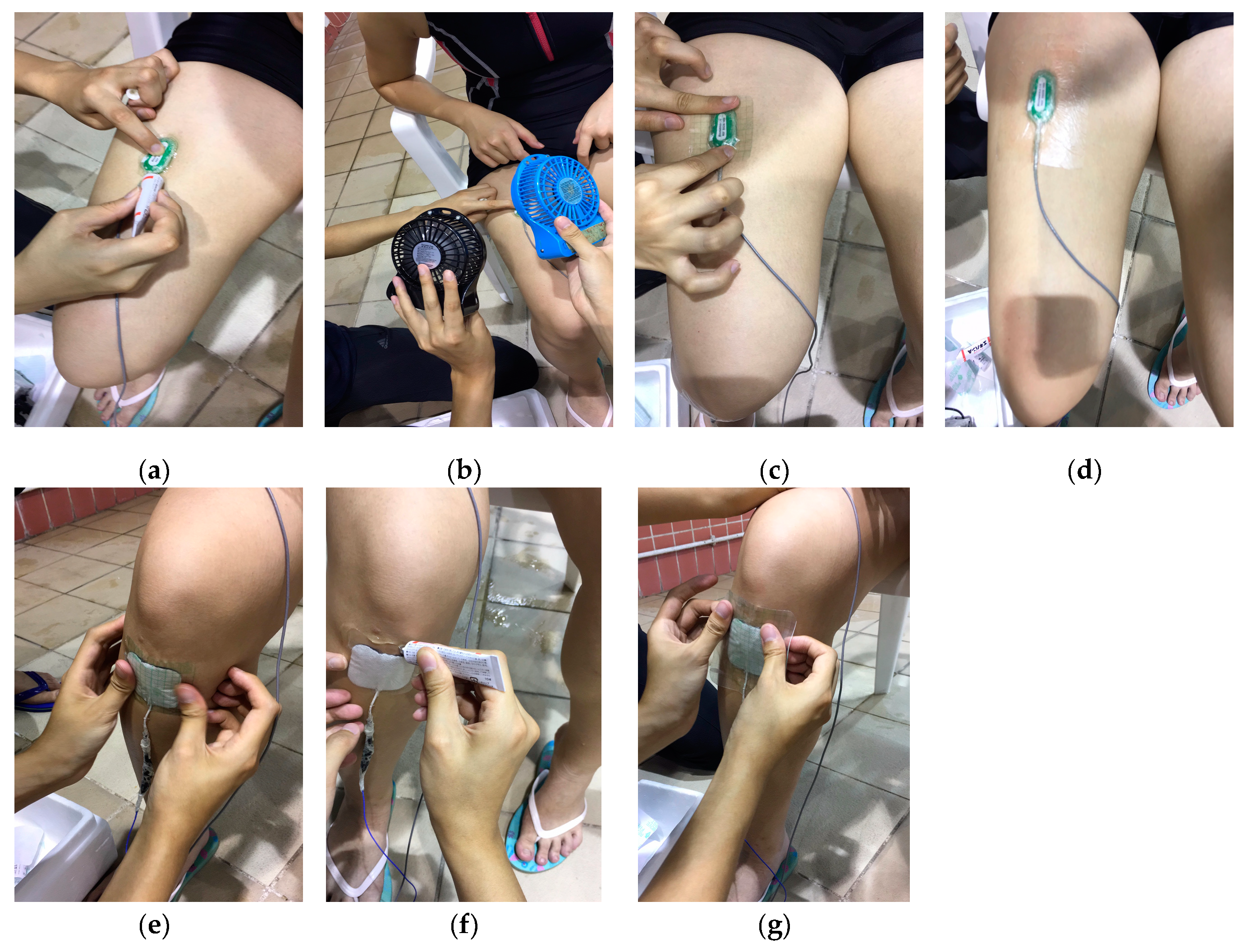
2.5.2. Waterproof Techniques
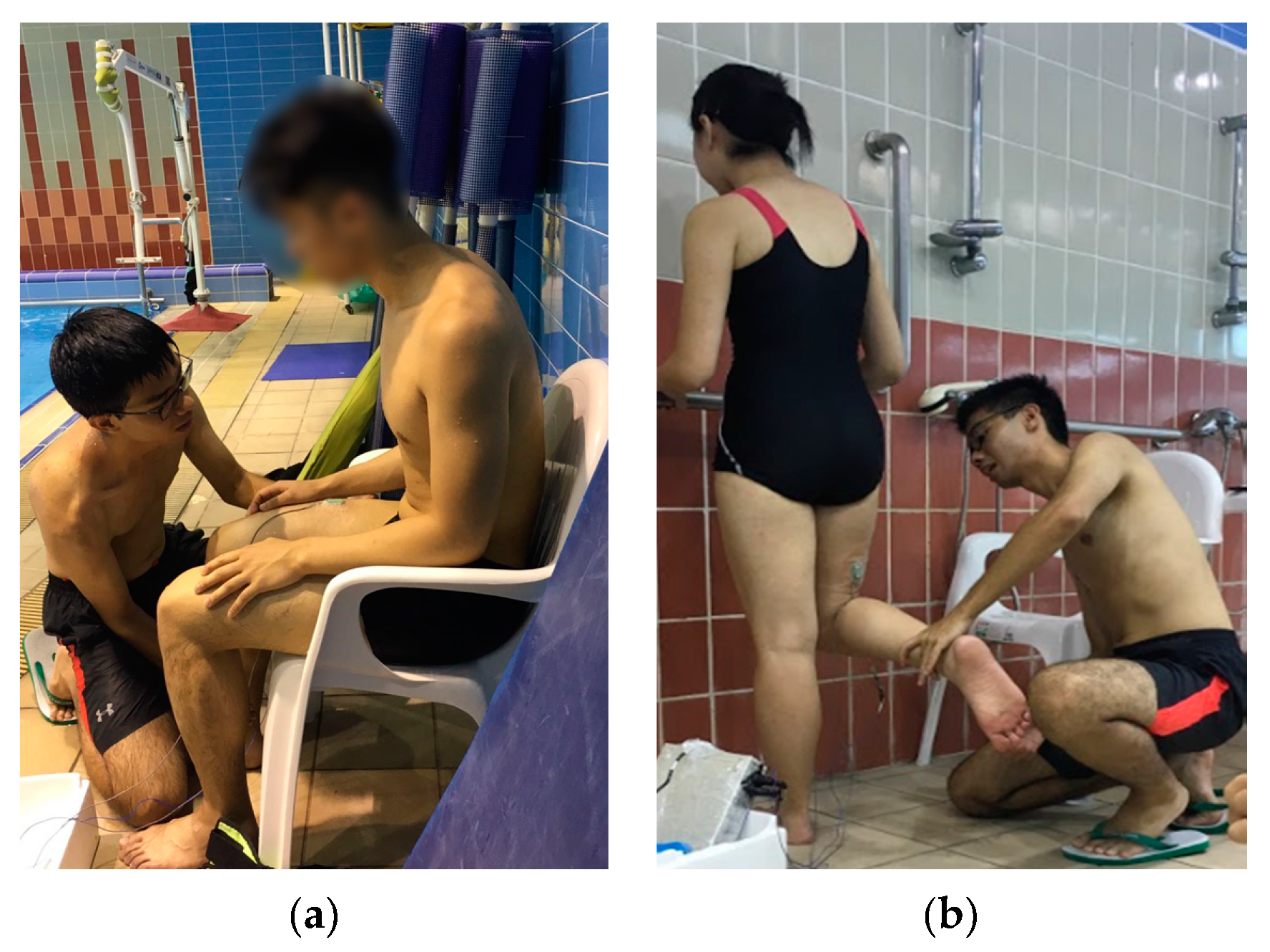
2.5.3. Squatting on Land
2.5.4. Squatting in Water
2.5.5. Maximal Voluntary Contractions (MVC)
RF MVC Testing
BF MVC Testing
2.6. Data Processing
2.7. Statistical Analysis
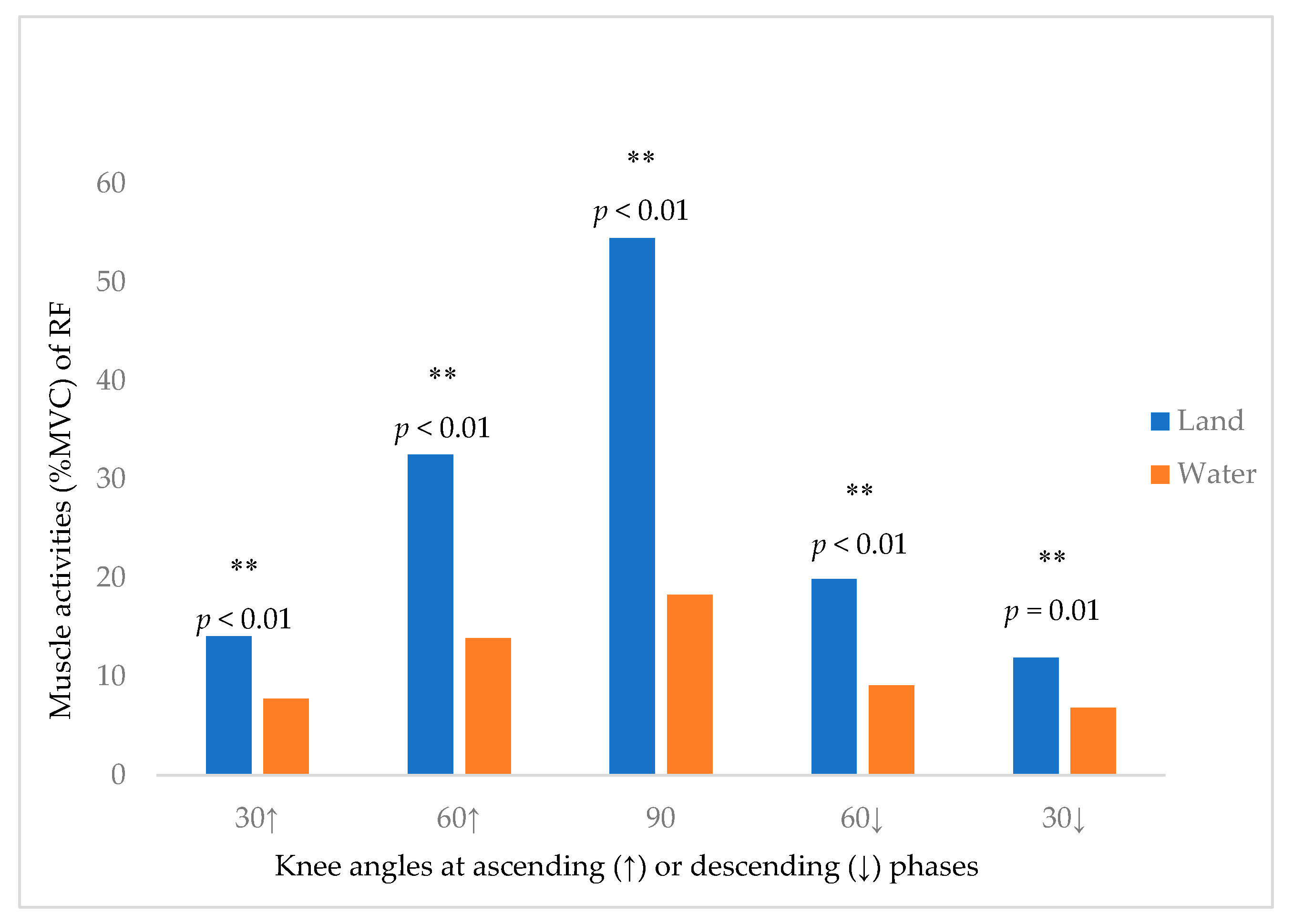
3. Results
4. Discussion
4.1. Difference of RF and BF Activity in Water and on Land
4.2. RF and BF Activity in Water and on Land at Different Squatting Phases and Knee Angles
4.3. The Effect of Different Water Properties on the RF and BF Muscle Activity
4.4. Limitation of Study
5. Conclusions
Author Contributions
Funding
Acknowledgments
Conflicts of Interest
References
- Heywood, S.; McClelland, J.; Geigle, P.; Rahmann, A.; Villalta, E.; Mentiplay, B.; Clark, R. Force during functional exercises on land and in water in older adults with and without knee osteoarthritis: Implications for rehabilitation. Knee 2019, 26, 61–72. [Google Scholar] [CrossRef]
- Heywood, S.; McClelland, J.; Mentiplay, B.; Geigle, P.; Rahmann, A.; Clark, R. Effectiveness of Aquatic Exercise in Improving Lower Limb Strength in Musculoskeletal Conditions: A Systematic Review and Meta-Analysis. Arch. Phys. Med. Rehabil. 2017, 98, 173–186. [Google Scholar] [CrossRef] [PubMed]
- Batterham, S.I.; Heywood, S.; Keating, J.L. Systematic review and meta-analysis comparing land and aquatic exercise for people with hip or knee arthritis on function, mobility and other health outcomes. BMC Musculoskelet. Disord. 2011, 12, 123. [Google Scholar] [CrossRef] [PubMed]
- Silva, L.E.; Valim, V.; Pessanha, A.P.C.; Oliveira, L.M.; Myamoto, S.; Jones, A.; Natour, J. Hydrotherapy versus conventional land-based exercise for the management of patients with osteoarthritis of the knee: A randomized clinical trial. Phys. Ther. 2008, 88, 12. [Google Scholar] [CrossRef] [PubMed]
- Momberg, B.L.; Louw, C.; Crous, L. Accelerated hydrotherapy and land-based rehabilitation in soccer players after anterior cruciate ligament reconstruction: A series of three single subject case studies. South Afr. J. Sports. Med. 2008, 20, 109. [Google Scholar] [CrossRef]
- Alberton, C.L.; Cadore, E.L.; Pinto, S.S.; Tartaruga, M.P.; Da Silva, E.M.; Kruel, L.F.M. Cardiorespiratory, neuromuscular and kinematic responses to stationary running performed in water and on dry land. Eur. J. Appl. Physiol. 2011, 111, 1157–1166. [Google Scholar] [CrossRef] [PubMed]
- Becker, B.E. Aquatic therapy: Scientific foundations and clinical rehabilitation applications. PM&R 2009, 1, 859–872. [Google Scholar]
- Harrison, R.; Bulstrode, S. Percentage Weight-Bearing during Partial Immersion in the Hydrotherapy Pool. Phys. Pract. 2009, 3, 60–63. [Google Scholar] [CrossRef]
- Heywood, S.; McClelland, J.; Geigle, P.; Rahmann, A.; Clark, R. Spatiotemporal, kinematic, force and muscle activation outcomes during gait and functional exercise in water compared to on land: A systematic review. Gait Posture 2016, 48, 120–130. [Google Scholar] [CrossRef]
- So, B.C.L.; Yuen, C.H.N.; Tung, K.L.H.; Lam, S.; Cheng, S.L.; Hung, Z.W.L.; Leung, R.W.K.; Szeto, G.P.Y. A Study on Trunk Muscle Activation of 2 Deep Water Running Styles (High-Knee and Cross-Country Style) and Land Walking. J. Sport. Rehabil. 2019, 1–6. [Google Scholar] [CrossRef]
- Cuesta-Vargas, A.I.; Cano-Herrera, C.L. Surface electromyography during physical exercise in water: A systematic review. BMC Sports Sci. Med. Rehabil. 2014, 6, 15. [Google Scholar] [CrossRef] [PubMed]
- Hoskin, K.; Dodd, K.; Chan, S.P.; Rosengarten, S.; Heywood, S. Aquatic Exercise Compared to Contrast Therapy With Shallow Water Treadmill Running to Assist. Recovery in Elite Australian Rules Footballers. Int. J. Aquat. Res. Educ. 2013, 7, 314–331. [Google Scholar] [CrossRef]
- Cuesta-Vargas, A.I.; Cano-Herrera, C.L.; Heywood, S. Analysis of the neuromuscular activity during rising from a chair in water and on dry land. J. Electromyogr. Kinesiol. 2013, 23, 1446–1450. [Google Scholar] [CrossRef] [PubMed]
- Lee, N.K.; Kwon, J.W.; Son, S.M.; Kang, K.W.; Kim, K.; Hyun-Nam, S. The effects of closed and open kinetic chain exercises on lower limb muscle activity and balance in stroke survivors. NeuroRehabilitation 2013, 33, 177–183. [Google Scholar] [PubMed]
- Cuesta-Vargas, A.I.; Cano-Herrera, C.L. EMG Analysis of the Neuromuscular Activity during Sit-to-Stand from Different Height Chairs in Water. Int. J. Aquat. Res. Educ. 2019, 12, 6. [Google Scholar]
- Pöyhönen, T.; Kyröläinen, H.; Keskinen, K.L.; Hautala, A.; Savolainen, J.; Mälkiä, E. Electromyographic and kinematic analysis of therapeutic knee exercises under water. Clin. Biomech. 2001, 16, 496–504. [Google Scholar] [CrossRef]
- Pöyhönen, T.; Avela, J. Effect of head-out water immersion on neuromuscular function of the plantarflexor muscles. Aviat. Space Environ. Med. 2002, 73, 1215–1218. [Google Scholar]
- Shields, R.K.; Madhavan, S.; Gregg, E.; Leitch, J.; Petersen, B.; Salata, S.; Wallerich, S. Neuromuscular control of the knee during a resisted single-limb squat exercise. Am. J. Sports Med. 2005, 33, 1520–1526. [Google Scholar] [CrossRef] [PubMed]
- Escamilla, R.F.; Fleisig, G.S.; Zheng, N.; Barrentine, S.W.; Wilk, K.E.; Andrews, J.R. Biomechanics of the knee during closed kinetic chain and open kinetic chain exercises. Med. Sci. Sports Exerc. 1998, 30, 556–569. [Google Scholar] [CrossRef] [PubMed]
- Gullett, J.C.; Tillman, M.D.; Gutierrez, G.M.; Chow, J.W. A biomechanical comparison of back and front squats in healthy trained individuals. J. Strength Cond. Res. 2009, 23, 284–292. [Google Scholar] [CrossRef] [PubMed]
- Hinman, R.S.; Heywood, S.E.; Day, A.R. Aquatic physical therapy for hip and knee osteoarthritis: Results of a single-blind randomized controlled trial. Phys. Ther. 2007, 87, 32–43. [Google Scholar] [CrossRef] [PubMed]
- Von Elm, E.; Altman, D.G.; Egger, M.; Pocock, S.; Gøtzsche, P.C.; Vandenbroucke, J.P. The Strengthening the Reporting of Observational Studies in Epidemiology (STROBE) Statement: Guidelines for reporting observational studies. Int. J. Surg. 2014, 12, 1495–1499. [Google Scholar] [CrossRef] [PubMed]
- Silvers, W.M.; Dolny, D.G. Comparison and reproducibility of sEMG during manual muscle testing on land and in water. J. Electromyogr. Kinesiol. 2011, 21, 95–101. [Google Scholar] [CrossRef] [PubMed]
- Nishiwaki, G.A.; Urabe, Y.; Tanaka, K. EMG Analysis of Lower Extremity Muscles in Three Different Squat Exercises. J. Jpn. Phys. Ther. Assoc. 2006, 9, 21–26. [Google Scholar] [CrossRef] [PubMed]
- Torres-Ronda, L.; Del Alcazar, X.S. The Properties of Water and their Applications for Training. J. Hum. Kinet. 2014, 44, 237–248. [Google Scholar] [CrossRef] [PubMed]
- Masumoto, K.; Takasugi, S.I.; Hotta, N.; Fujishima, K.; Iwamoto, Y. Muscle activity and heart rate response during backward walking in water and on dry land. Eur. J. Appl. Physiol. 2005, 94, 54–61. [Google Scholar] [CrossRef] [PubMed]
- Roca-Dols, A.; Losa-Iglesias, M.E.; Sánchez-Gómez, R.; Becerro-de-Bengoa-Vallejo, R.; López-López, D.; Palomo-López, P.; Rodríguez-Sanz, D.; Calvo-Lobo, C. Electromyography activity of triceps surae and tibialis anterior muscles related to various sports shoes. J. Mech. Behav. Biomed. Mater. 2018, 86, 158–171. [Google Scholar] [CrossRef]
- Roca-Dols, A.; Losa-Iglesias, M.E.; Sánchez-Gómez, R.; López-López, D.; Becerro-de-Bengoa-Vallejo, R.; Calvo-Lobo, C. Electromyography comparison of the effects of various footwear in the activity patterns of the peroneus longus and brevis muscles. J. Mech. Behav. Biomed. Mater. 2018, 82, 126–132. [Google Scholar] [CrossRef]
- Criswell, E.; Cram, J.R. Cram’s Introduction to Surface Electromyography, 2nd ed.; Jones and Bartlett Press: Sudbury, MA, USA, 2011. [Google Scholar]
- Chang, K.V.; Yang, K.C.; Wu, W.T.; Huang, K.C.; Han, D.S. Association between metabolic syndrome and limb muscle quantity and quality in older adults: A pilot ultrasound study. Diabetes Metab. Syndr. Obes. 2019, 12, 1821–1830. [Google Scholar] [CrossRef] [Green Version]


| Demographic Factors | Minimum | Maximum | Mean | Std. Deviation |
|---|---|---|---|---|
| Age (year) | 20 | 24 | 21.25 | 1.0 |
| Height (cm) | 150 | 178 | 168.1 | 6.9 |
| Weight (kg) | 46 | 70 | 58.7 | 7.9 |
| Body mass index (kg/m2) | 17.5 | 24.6 | 20.7 | 2.0 |
| Phase/Muscles | Land (%MVC, Mean ± SD) | Water (%MVC, Mean ± SD) | % MVC Differences in Water Compared to Land (Mean ± SD) | |||
|---|---|---|---|---|---|---|
| RF | BF | RF | BF | RF | BF | |
| Total phase | 26.45 ± 13.51 | 20.62 ± 13.79 | 11.44 ± 6.12 | 9.97 ± 13.93 | 15.01 ± 10.47 | 10.68 ± 16.64 |
| ↑phase | 21.45 ± 11.87 | 19.93 ± 11.87 | 10.20 ± 6.08 | 10.60 ± 14.12 | 11.25 ± 5.79 | 9.33 ± 15.46 |
| ↓phase | 28.88 ± 18.29 | 19.38 ± 12.69 | 12.87 ± 8.28 | 9.67 ± 14.07 | 16.01 ± 10.01 | 9.71 ± 15.64 |
| 30° ↓ | 14.07 ± 6.76 | 19.02 ± 15.83 | 7.76 ± 4.17 | 10.20 ± 13.20 | 6.31 ± 6.53 | 8.82 ± 17.09 |
| 60° ↓ | 32.54 ± 19.83 | 21.60 ± 16.11 | 13.86 ± 10.54 | 9.55 ± 13.76 | 18.68 ± 20.79 | 12.06 ± 18.28 |
| 90° | 54.5 ± 31.09 | 25.70 ± 18.01 | 18.29 ± 11.08 | 8.77 ± 14.28 | 36.20 ± 26.12 | 16.93 ± 20.56 |
| 60° ↑ | 19.90 ± 12.61 | 21.46 ± 14.30 | 9.12 ± 8.06 | 10.78 ± 14.65 | 10.78 ± 12.69 | 10.68 ± 17.33 |
| 30° ↑ | 11.88 ± 7.46 | 20.04 ± 12.87 | 6.81 ± 4.68 | 11.50 ± 13.87 | 5.07 ± 6.38 | 8.54 ± 15.67 |
| Phase/Muscles | RF (%MVC, Mean ± SD) | Difference in % MVC of RF at Two Media (Mean ± SD, p-Value) | ||
|---|---|---|---|---|
| Land | Water | |||
| Total phase | 26.45 ± 13.51 | 11.44 ± 6.12 | 15.01 ± 10.47 | 0.01 ** |
| ↑phase | 21.45 ± 11.87 | 10.20 ± 6.08 | 11.25 ± 5.79 | <0.01 ** |
| ↓phase | 28.88 ± 18.29 | 12.87 ± 8.28 | 16.01 ± 10.01 | <0.01 ** |
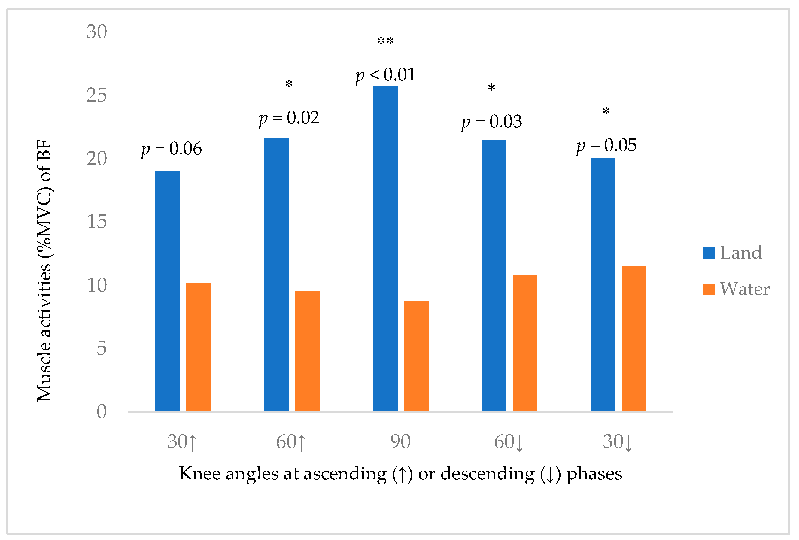
| Phase/Muscles | BF (%MVC, Mean ± SD) | Difference in % MVC of BF at Two Media (Mean ± SD, p-Value) | ||
|---|---|---|---|---|
| Land | Water | |||
| Total phase | 20.62 ± 13.79 | 9.97 ± 13.93 | 10.68 ± 16.64 | 0.01 ** |
| ↑phase | 19.93 ± 11.87 | 10.60 ± 14.12 | 9.33 ± 15.46 | <0.01 ** |
| ↓phase | 19.38 ± 12.69 | 9.67 ± 14.07 | 9.71 ± 15.64 | <0.01 ** |
© 2019 by the authors. Licensee MDPI, Basel, Switzerland. This article is an open access article distributed under the terms and conditions of the Creative Commons Attribution (CC BY) license (http://creativecommons.org/licenses/by/4.0/).
Share and Cite
Yuen, C.H.N.; Lam, C.P.Y.; Tong, K.C.T.; Yeung, J.C.Y.; Yip, C.H.Y.; So, B.C.L. Investigation the EMG Activities of Lower Limb Muscles When Doing Squatting Exercise in Water and on Land. Int. J. Environ. Res. Public Health 2019, 16, 4562. https://0-doi-org.brum.beds.ac.uk/10.3390/ijerph16224562
Yuen CHN, Lam CPY, Tong KCT, Yeung JCY, Yip CHY, So BCL. Investigation the EMG Activities of Lower Limb Muscles When Doing Squatting Exercise in Water and on Land. International Journal of Environmental Research and Public Health. 2019; 16(22):4562. https://0-doi-org.brum.beds.ac.uk/10.3390/ijerph16224562
Chicago/Turabian StyleYuen, Calvin H.N., Christine P.Y. Lam, Kate C.T. Tong, Jessica C.Y. Yeung, Chloe H.Y. Yip, and Billy C.L. So. 2019. "Investigation the EMG Activities of Lower Limb Muscles When Doing Squatting Exercise in Water and on Land" International Journal of Environmental Research and Public Health 16, no. 22: 4562. https://0-doi-org.brum.beds.ac.uk/10.3390/ijerph16224562