Article

The Development of Keratopathy after Surgery-Indicated Chronic Rhinosinusitis: A Population-Based Cohort Study

1
Institute of Medicine, Chung Shan Medical University, Taichung 402, Taiwan
2
Department of Ophthalmology, Show Chwan Memorial Hospital, Changhua 500, Taiwan
3
Department of Optometry, College of Medicine and Life Science, Chung Hwa University of Medical Technology, Tainan 717, Taiwan
4
Department of Otolaryngology–Head and Neck Surgery, Kaohsiung Chang Gung Memorial Hospital, Kaohsiung 833, Taiwan
5
Department of Ophthalmology, Chang Gung Memorial Hospital, Linkou 333, Taiwan
6
Department of Medicine, Chang Gung University College of Medicine, Taoyuan 333, Taiwan
7
Center for Tissue Engineering, Chang Gung Memorial Hospital, Linkou 333, Taiwan
8
Department of Ophthalmology, Chang Gung Memorial Hospital, Keelung 204, Taiwan
9
Department of Chinese Medicine, Chang Gung University, Taoyuan 333, Taiwan
10
Department of Medical Research, Chung Shan Medical University Hospital, Taichung 402, Taiwan
11
Department of Optometry, Chung Shan Medical University, Taichung 402, Taiwan
12
Department of Exercise and Health Promotion, Chung Chou University of Science and Technology, Changhua 500, Taiwan
*
Author to whom correspondence should be addressed.
These authors contributed equally to the work.
Int. J. Environ. Res. Public Health 2019, 16(7), 1218; https://0-doi-org.brum.beds.ac.uk/10.3390/ijerph16071218
Submission received: 25 March 2019 / Revised: 3 April 2019 / Accepted: 3 April 2019 / Published: 5 April 2019

Abstract

:
The aim of the present study was to evaluate the risk of developing keratopathy in patients with surgery-indicated chronic rhinosinusitis (CRS) via the National Health Insurance Research Database in Taiwan. Patients with a diagnostic code of CRS and who received functional endoscopic sinus surgery (FESS) were considered to have surgery-indicated CRS. The exclusion criteria were legal blindness, an ocular tumor, eyeball removal or previous keratopathy, and each individual in the study group was matched to four non-CRS patients by age and sex. The outcome was set as the occurrence of keratopathy according to the diagnostic codes after the index date. Cox proportional hazard regression was used for statistical analysis. A total of 6053 patients with surgery-indicated CRS and another 24,212 non-CRS individuals were enrolled after exclusions. The age and sex distributions were identical between the two groups due to matching, while comorbidities, including hypertension, diabetes mellitus, and other cardiovascular disorders, were significantly higher in the study group. There were 231 episodes of keratopathy in the study group, and 695 episodes of keratopathy in the control group after the index date, for which study group showed a significantly higher rate of developing keratopathy with an adjusted hazard ratio of 1.208 and a higher cumulative probability. In subgroup analysis, female sex with surgery-indicated CRS showed a significantly greater risk of developing keratopathy. In conclusion, surgery-indicated CRS that needs FESS to relieve symptoms is a potential risk factor for keratopathy.

1. Introduction

Chronic rhinosinusitis (CRS) is an inflammatory disease in the paranasal sinuses that persists for at least 12 weeks [1] and is found in more than 10 percent of the population [2]. The clinical presentation of CRS includes nasal discharge, pain in the facial region, reduction in the smell, headache, and shortness of breath [1,3]. Cranial nerve disorders, such as the alteration of trigeminal function, may occur in patients with CRS [4,5]. In its severe form, the infection and inflammation of the paranasal sinus in CRS may lead to a dreaded intracranial infection [6].
Both medical and surgical management have been utilized to treat CRS [7]. Topical corticosteroid therapy, oral corticosteroid administration, and antibiotic treatment with macrolide have been applied to treat CRS with acceptable outcomes [3,7]. Functional endoscopic sinus surgery (FESS) is a well-established intervention for CRS that is severe and refractory to medical management [8,9,10]. Nevertheless, the recovery of nasal sinus mucosa in patients with CRS is incomplete one year after FESS management [3]. Moreover, patients with some risk factors, such as higher Lund-Mackay computed tompgraphy scores and fungal-induced CRS, may still experience poor quality of life or persistent nasal polyp formation even after successful FESS intervention [11,12]. The above evidence suggests that the effect of severe CRS could persist despite FESS.
For ocular complications in CRS, both preseptal and visual-threatening orbital cellulitis were found in previous research [13]. Besides, dacryocystitis has also been reported after the development of CRS [14]. For the external eye region, however, only one case with concurrent keratoconjunctivitis and CRS was found previously [15]. Because severe CRS can influence adjacent structures, it is possible that severe CRS may result in certain corneal and conjunctival lesions.
The aim of the current study was to evaluate the development of keratopathy after surgery-indicated CRS that received FESS via the National Health Insurance Research Database (NHIRD) in Taiwan. The effect of CRS disease interval and other potential risk factors for developing keratopathy were also investigated.

2. Materials and Methods

2.1. Data Source

This retrospective, population-based cohort study was approved by the National Health Insurance Administration and the Institutional Review Board of Chung Shan Medical University. Provided by the Taiwan National Health Research Institutes, the NHIRD contains data on insurance claims from more than 99% of Taiwan’s population. The claims data used in the current study were obtained from the 2016 version of the Longitudinal Health Insurance Database (LHID 2016). The LHID 2016 contains data on two million patients randomly sampled from the NHIRD registry since 2005. The LHID 2016 data were linked from 1 January 2000 to 31 December 2016, and both the International Classification of Diseases, Ninth Revision (ICD-9) and International Classification of Diseases, Tenth Revision (ICD-10) were used for disease diagnosis. Details on the medications prescribed to the patients and patient demographics, socioeconomic status, and residence are also available in the NHIRD.

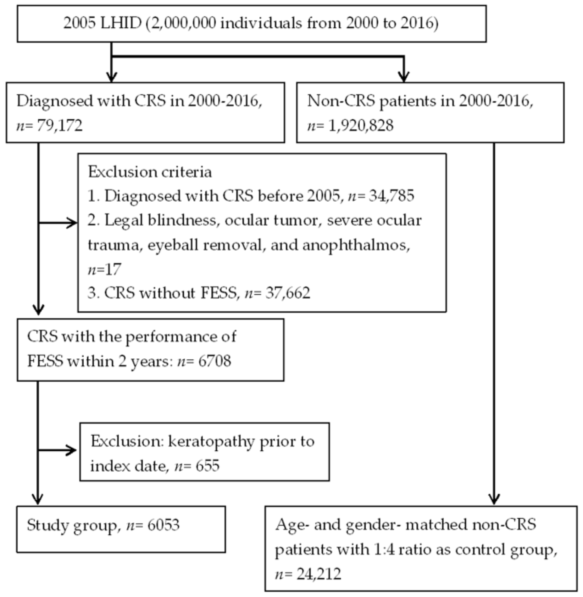
2.2. Patient Selection

Patients were defined as having surgery-indicated CRS if their medical records indicated (1) a history of CRS (ICD-9 codes: 473.x, ICD-10 codes: J32.x), (2) arrangement for FESS (procedure codes: 65013B, 65015B, 65063B, 65064B) within two years of diagnosis of CRS, (3) the usage of corticosteroids or antibiotics for at least two years from the diagnosis of CRS, and (4) receipt of the CRS diagnosis by an otorhinolaryngologist (department code: 09). The index date was set as the date two years after the diagnosis of CRS. To more accurately elucidate the association between surgery-indicated CRS and keratopathy, the following exclusion criteria were applied: (1) a diagnosis of legal blindness (ICD-9 codes: 369.4, ICD-10 codes: H54.0x, H54.1x, H54.4x, H54.8) at any time, (2) a diagnosis of ocular tumors (ICD-9 codes: 190.0–190.9, ICD-10 codes: C69.x) at any time, (3) a diagnosis of severe ocular trauma (ICD-9 codes: 871.0–871.2, 871.4–871.9, ICD-10 codes: S05.2x–S05.6x) at any time, (4) any type of eyeball removal surgery or diagnosis of anophthalmos (ICD-9 codes: 16.3x, 16.4x, 16.5x, 871.3, ICD-10 codes: Q11.1, S05.7x, Z90.01 plus procedure codes: 85001C, 85002C, 86808B) before the index date, or (5) a diagnosis of any type of keratopathy (ICD-9 codes: 370.x, 371.x, ICD-10: H16.x, H17.x, H18.x) before the index date. In addition, each individual in the study group was age- and sex-matched with four non-CRS individuals, as discussed in the following sections, which constituted the control group. Patients with surgery-indicated CRS who could not be matched with four non-CRS patients were excluded.

2.3. Main Outcome Measurement

The development of keratopathy is the main outcome in the current study and was based on the keratopathy-related diagnostic codes (ICD-9 codes: 370.0x, 370.2x, 370.3x, 370.4x, 370.5x, 370.6x, 370.8, 370.9, 371.0x, 371.21-371.23, ICD-10 codes: H16.0x H16.1x, H16.20x, H16.23x–H16.29x, H16.3x–H16.9, H17.1x, H17.8x, H17.9, H18.1x, H18.22x, H18.23x). In practice, ICD-9 codes for “unspecific corneal disorder (ICD-9 codes: 371.9, ICD-10 codes: H18.9)” may also be considered for some forms of keratopathy, but these codes were eliminated to prevent overestimation and confusion. Furthermore, only patients who received the abovementioned diagnostic codes by an ophthalmologist (department code: 10) were considered to have the outcome and included in the study.

2.4. Demographic Variables and Co-Morbidities

To standardize the health condition of participants, we also considered the effects of demographic conditions (i.e., age, sex, and income level) and the following systemic co-morbidities, according to our Modified Deyo–Charlson co-morbidity index in the multivariate analysis model: hypertension (ICD-9 codes: 401.x–405.x, ICD-10 codes: I10, I11.x, I13.x, I15.x, I16.x, I87.3x, I97.3x, O10.x, O11.x, O13.x, O16.x), diabetes mellitus (ICD-9 codes: 250.x, 277.7, ICD-10 codes: O24.4, E11.x, E13.x, E88.81), ischemic heart diseases (ICD-9 codes: 410.x, 412.x, 414.0, 414.0x, 414.2, 414.3, 414.4, 414.8, 414.9, ICD-10 codes: I20.x–I25.x), hyperlipidemia (ICD-9 codes: 272.0, 272.1, 272.2, 272.4, 272.9, ICD-10 codes: E78.0x, E78.1, E78.2, E78.3, E78.4x, E78.5, E78.70, E78.79, E78.89, E78.9), congestive heart failure (ICD-9 codes: 398.91, 402.01, 402.11, 402.91, 404.01, 404.03, 404.11, 404.13, 404.91, 404.93, 425.4–425.9, 428.x, ICD-10 codes: I50.2x, I50.3x, I50.4x, I50.84, I50.89, I50.9), peripheral vascular disease (ICD-9 codes: 093.0, 437.3, 440.x, 441.x, 443.1–443.9, 47.1, 557.1, 557.9, V43.4, ICD-10 codes: E75.21, I73.x, I79.8, T82.856, Z95.82x, Z98.62), cerebrovascular disease (ICD-9 codes: 362.34, 430.x–438.x, ICD-10 codes: G46.x, I60.x-I66.x, I67.0, I67.1, I67.2, I67.6, I67.81, I67.82, I67.84x, I67.89, I67.9), dementia (ICD-9 codes 290.x, 294.1, 331.2, ICD-10 codes: F01.x, F02.x, F03.x, G31.x), chronic pulmonary disease, including asthma (ICD-9 codes: 416.8, 416.9, 490.x–505.x, 506.4, 508.1, 508.8, ICD-10 codes: J41.x, J42, J43.x, J44.x, J47.x), rheumatic disease (ICD-9 codes: 446.5, 710.0, 710.1, 710.3, 710.4, 714.0–714.2, 714.8, 725.x, ICD-10 codes: M05.1x, M05.2x-M05.9, M31.6, M32.1x, M32.8, M32.9, M33.03, M33.13, M33.2x, M33.90, M33.93, M34.0x, M34.1x, M34.9, M35.3), and peptic ulcer disease (ICD-9 codes: 531.x–534.x, ICD-10 codes: K25.x-K28.x). To further standardize the ocular condition, dry eye disease (DED) (ICD-9 codes: 370.33, 370.34, 372.53, 375.15, 710.2, ICD-10 codes: H04.12x, H11.14x, H16.21x, H16.22x, M35.00, M35.01), uveitis (ICD-9 codes: 360.12, 363.0x, 363.1x, 363.20, 363.21, 363.22, 364.0x, 364.1x, 364.2x, 364.3, ICD-10 codes: H20.x, H30.0x, H30.1x, H30.2x, H30.8x, H30.9x, H44.11x), and glaucoma (ICD-9 codes: 365.1x, 365.2x, 365.7x, 365.9, ICD-10 codes: H40.1x, H40.2x, H40.89, H40.9) were also considered in the multivariate model. We longitudinally traced the data from the index date until the date of keratopathy diagnosis, withdrawal from the National Health Insurance program, or 31 December 2016.

2.5. Statistical Analysis
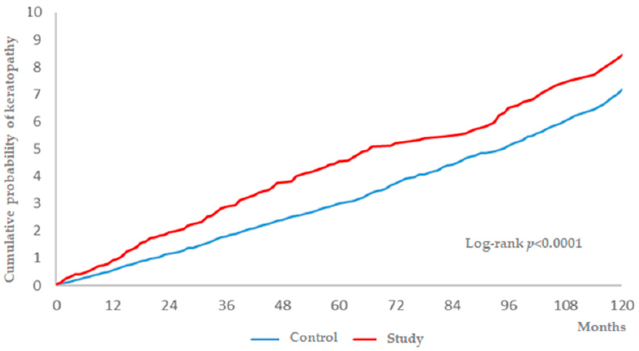
SAS version 9.4 (SAS Institute Inc, Cary, NC, USA) was employed for all analyses. After age and sex-matching at 1:4 ratio of the study and control groups, the incidence rate, crude relative risk, and corresponding 95% confidence intervals (CI) were calculated using Poisson regression. In the next step, Cox proportional hazard regression was adopted to compute adjusted hazard ratios (aHR) by incorporating the aforementioned demographic data, prominent ocular diseases, and systemic comorbidities in the multivariate model. Moreover, the sensitivity analysis with aHR which stratified by the sex and age subgroups was also conducted for the subgroup analysis. We plotted Kaplan–Meier curves to indicate the cumulative incidence proportion of keratopathy between the study and control groups, and used the log rank test to determine the significant difference between the survival curves. Because most patients in the NHIRD are Han Taiwanese, the race was not considered a covariate. Statistical significance was set at p < 0.05.

3. Results

A total of 6053 patients with surgery-indicated CRS were enrolled in the study group after exclusion, and other 24,212 individuals were enrolled in the control group. The flowchart of patient selection is shown in Figure 1. The age and sex ratios were identical due to the matching process, and the different comorbidities in the study and control group are shown in Table 1.
There were 231 and 695 events of keratopathy in the study group and the control group, while the study group had higher incidence rate (74.35) and crude relative risk (1.345) (Table 2). Moreover, the study group showed a significant aHR (1.208, 95% CI: 1.038–1.406) compared to the control group after adjusting for demographic data, disease interval, prominent ocular diseases, and systemic comorbidities (Table 3), and a significantly higher cumulative probability was also revealed in the study group (p < 0.0001) (Figure 2). In addition to surgery-indicated CRS, hypertension, chronic pulmonary diseases, peptic ulcer disease, and DED were also prominently related to the development of keratopathy (Table 3). In the subgroup analysis, females with surgery-indicated CRS had a significantly higher aHR (1.287, 95% CI: 1.038–1.595) for developing keratopathy, while only a marginally significant value was found for males with surgery-indicated CRS (Table 4).

4. Discussion

Briefly, CRS that requires surgical management to relieve symptoms significantly increased the risk of developing keratopathy in the current study after adjusting for multiple potential risk factors. Moreover, the chance of developing keratopathy was elevated in several diseases, including cardiovascular disorders, pulmonary diseases, and DED in the multivariate model.
Several mechanisms may lead to keratopathy. At first, inflammatory status is an important part of the pathophysiology of keratopathy, in which the elevated inflammatory mediators can be found in both the auto-immune and infectious keratopathies [16,17]. Similarly, the CRS has been regarded as an inflammatory disease recently, and both the inflammatory-related epithelial barrier dysfunction and immunoglobulin E production were proposed as the etiologic mechanisms for CRS [18]. Moreover, same inflammatory mediators, such as interleukin and tumor necrosis factor-alpha, are shown to be elevated in CRS and keratopathy in preceding researches [17,18,19]. Accordingly, the elevated inflammatory process in CRS might also raise the possibility of corneal damage. In addition, the infections accounting for certain types of keratopathies [20,21] and lesions like liver abscesses and orbital infections could lead to keratitis via hematogenous route [22,23]. Although accounting for much fewer cases compared to inflammatory disorders, infection is still one of the etiologies of CRS [24]. Thus, it is possible that the microorganisms that spread from infectious CRS to the cornea via either the anastomosis of an anterior ethmoidal artery-posterior ethmoidal artery-sphenopalatine artery or cavernous sinus-ophthalmic vein route then lead to keratopathy. On the other hand, some autoimmune diseases, such as Wegener granulomatosis, lead to both corneal lesions and CRS [25], and CRS might indicate an underlying autoimmune disease that may lead to keratopathy. As a consequence, the presence of CRS, especially severe cases that need surgery to manage, may lead to keratopathy, which was supported by the current study.
For the relationship between CRS and keratopathy, only concurrent keratoconjunctivitis and CRS had been found in previous research [15]. In the current study, surgery-indicated CRS that required FESS management showed an increased risk of developing keratopathy with a significantly higher aHR after controlling the effect of several potential risk factors in the multivariate analysis. To our knowledge, this study is a preliminary investigation that demonstrated a correlation between surgery-indicated CRS and keratopathy. In addition, only those episodes of keratopathy that occurred after arranging for FESS were included, and the increase in the risk of developing keratopathy with longer disease period of CRS was also evaluated which presented with the higher cumulative probability in the study group. Thus, a causal relationship is supported. For the difference in correlation between the two diseases in the different sexes, although the aHR in males was non-significant, the aHR was similar to that in the female population with a similar interaction p-value, and the lower limit also showed marginal significance. We speculated that surgery-indicated CRS elevated the risk of developing keratopathy in males with a longer follow-up interval.
Several disorders were also related to the development of keratopathy according to the multivariate analysis in the current study. DED revealed significantly higher aHR for the occurrence of keratopathy in the current study. Because DED will lead to inflammation and damage of the ocular surface [26], the relationship between DED and keratopathy in the current study is reasonable and has been illustrated in a previous study [27]. Concerning the influence of glaucoma on the cornea, an acute attack of glaucoma may lead to corneal decompensation in patients with an impaired corneal endothelium [28]. Certain anti-glaucomatous agents can also contribute to damage the ocular surface and keratoepitheliopathy, such as superficial punctate keratitis [29]. In the current study, the aHR of glaucoma was also elevated, although not significantly. On the other hand, uveitis also did not show a significant effect on the occurrence of keratopathy. This phenomenon might result from the exclusion of patients with pre-existing keratopathy, thus excluding numbers of patients with uveitis because coexisting uveitis and keratopathy in viral infections are not uncommon [30,31]. Among systemic conditions, cardiovascular as well as pulmonary disorders are the major disorders associated with a higher probability of keratopathy, but the exact mechanism needs further validation.
There are several limitations to the current study. First, the observational and retrospective nature of the study design may diminish the standardization of the patient population, even after multivariate analysis. In addition, we used claim data rather than real medical documents, and some important information, such as the laterality and severity of keratopathy and the postoperative condition of CRS after the FESS procedure, could not be accessed. Moreover, there are different types of keratopathy (e.g., infectious keratitis or corneal edema), and we failed to analyze the effect of surgery-indicated CRS on each type of keratopathy due to extremely small numbers for certain types of keratopathy.

5. Conclusions

In conclusion, surgery-indicated CRS that requires FESS to relieve symptoms is a potential risk factor for keratopathy after adjusting for multiple potential risk factors. Furthermore, the risk of developing keratopathy in patients with surgery-indicated CRS is correlated to a longer disease period of CRS. Further study to investigate whether surgery-indicated CRS leads to a worse prognosis for keratopathy is advocated.

Author Contributions

Conceptualization, H.-J.H., C.-Y.L., and S.-F.Y.; methodology, C.-Y.L. and K.-L.Y.; formal analysis, H.-C.C., C.-C.S., and J.-Y.H.; resources, H.-Y.L., and S.-F.Y.; writing—original draft preparation, H.-J.H., C.-Y.L., and H.-C.C.; writing—review and editing, H.-J.H., C.-Y.L., H.-C.C., and S.-F.Y.

Funding

This research received no external funding.

Conflicts of Interest

The authors declare no conflict of interest.

References

  1. Siedek, V.; Stelter, K.; Betz, C.S.; Berghaus, A.; Leunig, A. Functional endoscopic sinus surgery—A retrospective analysis of 115 children and adolescents with chronic rhinosinusitis. Int. J. Pediatr. Otorhinolaryngol. 2009, 73, 741–745. [Google Scholar] [CrossRef]
  2. Gulati, S.P.; Chaudhry, D.; Kalra, V.; Wadhera, R.; Garg, A. The role of functional endoscopic sinus surgery (fess) in patients with asthma with chronic sinusitis. Indian J. Otolaryngol. Head Neck Surg. 2008, 60, 152–155. [Google Scholar] [CrossRef] [PubMed]
  3. Anselmo-Lima, W.T.; Ferreira, M.D.; Valera, F.C.; Rossato, M.; de Mello, V.R.; Demarco, R.C. Histological evaluation of maxillary sinus mucosa after functional endoscopic sinus surgery. Am. J. Rhinol. 2007, 21, 719–724. [Google Scholar] [CrossRef] [PubMed]
  4. Poletti, S.C.; Cuevas, M.; Weile, S.; Hummel, T. Trigeminal sensitivity in chronic rhinosinusitis: Topographical differences and the effect of surgery. Rhinology 2017, 55, 70–74. [Google Scholar] [CrossRef] [PubMed]
  5. Saliba, J.; Fnais, N.; Tomaszewski, M.; Carriere, J.S.; Frenkiel, S.; Frasnelli, J.; Tewfik, M.A. The role of trigeminal function in the sensation of nasal obstruction in chronic rhinosinusitis. Laryngoscope 2016, 126, E174–E178. [Google Scholar] [CrossRef] [PubMed]
  6. Constantin, F.; Niculescu, P.A.; Petre, O.; Balasa, D.; Tunas, A.; Rusu, I.; Lupascu, M.; Orodel, C. Orbital cellulitis and brain abscess—Rare complications of maxillo-spheno-ethmoidal rhinosinusitis. Rom. J. Ophthalmol. 2017, 61, 133–136. [Google Scholar] [CrossRef] [PubMed]
  7. Fokkens, W.J.; Lund, V.J.; Mullol, J.; Bachert, C.; Alobid, I.; Baroody, F.; Cohen, N.; Cervin, A.; Douglas, R.; Gevaert, P.; et al. EPOS 2012: European position paper on rhinosinusitis and nasal polyps 2012. A summary for otorhinolaryngologists. Rhinology 2012, 50, 1–12. [Google Scholar] [CrossRef] [PubMed]
  8. Tan, B.K.; Lane, A.P. Endoscopic sinus surgery in the management of nasal obstruction. Otolaryngol. Clin. North Am. 2009, 42, 227–240. [Google Scholar] [CrossRef] [PubMed]
  9. Ramakrishnan, V.R.; Kennedy, D.W. Advances in the surgical management of chronic sinusitis and nasal polyps. Curr. Allergy Asthma Rep. 2011, 11, 220–229. [Google Scholar] [CrossRef] [PubMed]
  10. Welch, K.C.; Stankiewicz, J.A. A contemporary review of endoscopic sinus surgery: Techniques, tools, and outcomes. Laryngoscope 2009, 119, 2258–2268. [Google Scholar] [CrossRef]
  11. Barac, A.; Pekmezovic, M.; Spiric, V.T.; Trivic, A.; Marinkovic, J.; Pekic, S.; Arsenijevic, V.A. Chronic rhinosinusitis: Association of recalcitrant nasal polyposis and fungal finding in polyp’s single-cell suspension. Euro. Arch. Otorhinolaryngol. 2015, 272, 3727–3734. [Google Scholar] [CrossRef] [PubMed]
  12. Brooks, S.G.; Trope, M.; Blasetti, M.; Doghramji, L.; Parasher, A.; Glicksman, J.T.; Kennedy, D.W.; Thaler, E.R.; Cohen, N.A.; Palmer, J.N.; et al. Preoperative lund-mackay computed tomography score is associated with preoperative symptom severity and predicts quality-of-life outcome trajectories after sinus surgery. Int. Forum Allergy Rhinol. 2018, 8, 668–675. [Google Scholar] [CrossRef] [PubMed]
  13. Chang, Y.S.; Chen, P.L.; Hung, J.H.; Chen, H.Y.; Lai, C.C.; Ou, C.Y.; Chang, C.M.; Wang, C.K.; Cheng, H.C.; Tseng, S.H. Orbital complications of paranasal sinusitis in Taiwan, 1988 through 2015: Acute ophthalmological manifestations, diagnosis, and management. PLoS ONE 2017, 12, e0184477. [Google Scholar] [CrossRef] [PubMed]
  14. Cazzavillan, A.; Gaini, R.M.; Pignataro, L.; Piacentini, E.; Leo, G. Treatment of rhinosinusitis: The role of surgery. Int. J. Immunopathol. Pharmacol. 2010, 23, 74–77. [Google Scholar] [PubMed]
  15. Rossi, P.; Urbani, C.; Donelli, G.; Pozio, E. Resolution of microsporidial sinusitis and keratoconjunctivitis by itraconazole treatment. Am. J. ophthalmol. 1999, 127, 210–212. [Google Scholar] [CrossRef]
  16. Cao, Y.; Zhang, W.; Wu, J.; Zhang, H.; Zhou, H. Peripheral ulcerative keratitis associated with autoimmune disease: Pathogenesis and treatment. J. Ophthalmol. 2017, 2017, 7298026. [Google Scholar] [CrossRef] [PubMed]
  17. Maertzdorf, J.; Osterhaus, A.D.; Verjans, G.M. Il-17 expression in human herpetic stromal keratitis: Modulatory effects on chemokine production by corneal fibroblasts. J. Immunol. 2002, 169, 5897–5903. [Google Scholar] [CrossRef] [PubMed]
  18. Ramakrishnan, V.R.; Gonzalez, J.R.; Cooper, S.E.; Barham, H.P.; Anderson, C.B.; Larson, E.D.; Cool, C.D.; Diller, J.D.; Jones, K.; Kinnamon, S.C. Rna sequencing and pathway analysis identify tumor necrosis factor alpha driven small proline-rich protein dysregulation in chronic rhinosinusitis. Am. J. Rhinol. Allergy 2017, 31, 283–288. [Google Scholar] [CrossRef] [PubMed]
  19. Olcott, C.M.; Han, J.K.; Cunningham, T.D.; Franzese, C.B. Interleukin-9 and interleukin-17c in chronic rhinosinusitis. Int. Forum Allergy Rhinol. 2016, 6, 841–847. [Google Scholar] [CrossRef] [PubMed]
  20. Lee, B.; Cai, C.X.; Srikumaran, D.; Woreta, F.A. Severe achromobacter xylosoxidans keratitis with deep corneal involvement. Am. J. Ophthalmol. Case Rep. 2018, 11, 128–130. [Google Scholar] [CrossRef] [PubMed]
  21. Wang, J.Y.; Wang, D.Q.; Qi, X.L.; Cheng, J.; Xie, L.X. Modified ulcer debridement in the treatment of the superficial fungal infection of the cornea. Int. J. Ophthalmol. 2018, 11, 223–229. [Google Scholar] [PubMed]
  22. Djalilian, A.R.; Smith, J.A.; Walsh, T.J.; Malech, H.L.; Robinson, M.R. Keratitis caused by candida glabrata in a patient with chronic granulomatous disease. Am. J. Ophthalmol. 2001, 132, 782–783. [Google Scholar] [CrossRef]
  23. Clark, J.D.; Fernandez de Castro, J.P.; Compton, C.; Lee, H.; Nunery, W. Orbital cellulitis and corneal ulcer due to cedecea: First reported case and review of the literature. Orbit 2016, 35, 140–143. [Google Scholar] [CrossRef] [PubMed]
  24. Drago, L.; Pignataro, L.; Torretta, S. Microbiological aspects of acute and chronic pediatric rhinosinusitis. J. Clin. Med. 2019, 8, 149. [Google Scholar] [CrossRef] [PubMed]
  25. Biswas, J.; Babu, K.; Gopal, L.; Krishnakumar, S.; Suresh, S.; Ramakrishnan, S. Ocular manifestations of wegener’s granulomatosis. Analysis of nine cases. Indian J. Ophthalmol. 2003, 51, 217–223. [Google Scholar] [PubMed]
  26. Tsubota, K.; Yokoi, N.; Shimazaki, J.; Watanabe, H.; Dogru, M.; Yamada, M.; Kinoshita, S.; Kim, H.M.; Tchah, H.W.; Hyon, J.Y.; et al. New perspectives on dry eye definition and diagnosis: A consensus report by the asia dry eye society. Ocul. Surf. 2017, 15, 65–76. [Google Scholar] [CrossRef] [PubMed]
  27. Milner, M.S.; Beckman, K.A.; Luchs, J.I.; Allen, Q.B.; Awdeh, R.M.; Berdahl, J.; Boland, T.S.; Buznego, C.; Gira, J.P.; Goldberg, D.F.; et al. Dysfunctional tear syndrome: Dry eye disease and associated tear film disorders—New strategies for diagnosis and treatment. Curr. Opin. Ophthalmol. 2017, 27 (Suppl. 1), 3–47. [Google Scholar] [CrossRef]
  28. Krontz, D.P.; Wood, T.O. Corneal decompensation following acute angle-closure glaucoma. Ophthalmic Surg. 1988, 19, 334–338. [Google Scholar] [PubMed]
  29. Inoue, K.; Okugawa, K.; Kato, S.; Inoue, Y.; Tomita, G.; Oshika, T.; Amano, S. Ocular factors relevant to anti-glaucomatous eyedrop-related keratoepitheliopathy. J. Glaucoma 2003, 12, 480–485. [Google Scholar] [CrossRef] [PubMed]
  30. Wensing, B.; Mochizuki, M.; De Boer, J.H. Clinical characteristics of herpes simplex virus associated anterior uveitis. Ocular Immunol. Inflamm. 2018, 26, 333–337. [Google Scholar] [CrossRef] [PubMed]
  31. Chan, N.S.; Chee, S.P.; Caspers, L.; Bodaghi, B. Clinical features of cmv-associated anterior uveitis. Ocular Immunol. Inflamm. 2018, 26, 107–115. [Google Scholar] [CrossRef] [PubMed]
Figure 1. The flowchart of patient selection. LHID: Longitudinal Health Insurance Database; CRS: chronic rhinosinusitis; FESS: functional endoscopic sinus surgery.
Ijerph 16 01218 g001
Figure 2. The cumulative probability of keratopathy between the study and control groups.
Ijerph 16 01218 g002
Table 1. Baseline characteristics between the study and control groups.
Baseline CharacteristicsStudy, n = 6053Control, n = 24,212p Value
Age 1.0000
  <402177 (35.97%)8708 (35.97%)
  40–592643 (43.66%)10,572 (43.66%)
  60–791168 (19.3%)4672 (19.3%)
  ≥8065 (1.07%)260 (1.07%)
Sex 1.0000
  Male3812 (62.98%)15,248 (62.98%)
  Female2241 (37.02%)8964 (37.02%)
Co-morbidities
  Hypertension1662 (27.46%)5654 (23.35%)<0.0001
  Diabetes mellitus823 (13.6%)2838 (11.72%)0.0047
  Ischemic heart diseases634 (10.47%)1901 (7.85%)<0.0001
  Hyperlipidemia1466 (24.22%)4736 (19.56%)<0.0001
  Heart failure223 (3.68%)675 (2.79%)0.0002
  Peripheral vascular disease165 (2.73%)554 (2.29%)0.0454
  Cerebrovascular disease432 (7.14%)1408 (5.82%)0.0001
  Dementia37 (0.61%)167 (0.69%)0.5045
  Chronic pulmonary diseases1701 (28.1%)3920 (16.19%)<0.0001
  Rheumatic disease154 (2.54%)414 (1.71%)<0.0001
  Peptic ulcer disease1839 (30.38%)5352 (22.1%)<0.0001
  DED401 (6.62%)1024 (4.23%)<0.0001
  Uveitis54 (0.89%)179 (0.74%)0.2237
  Glaucoma161 (2.66%)386 (1.59%)<0.0001
DED = dry eye disease.
Table 2. The incidence of keratopathy in the study and control groups.
IncidenceStudy, n = 6053Control, n = 24,212
Follow-up person months310,7091,258,744
New keratopathy events231695
Incidence rate * (95% CI)74.35 (65.35–84.58)55.21 (51.26–59.48)
Crude Relative risk (95% CI)1.345 (1.159–1.561)Reference
CI = confidence interval; * Incidence rate, per 100,000 person months.
Table 3. Multiple Cox proportional hazard regression for the estimation of adjusted hazard ratios on keratopathy.
VariableaHR (95% CI)
Surgery-indicated CRS1.208 (1.038–1.406)
Age (Reference: 40–59)
  <401.044 (0.885–1.231)
  60–791.487 (1.249–1.77)
  ≥801.06 (0.535–2.099)
Sex (Reference: Female)
  Male0.648 (0.568–0.739)
Co-morbidities
  Hypertension1.235 (1.034–1.475)
  Diabetes mellitus0.946 (0.771–1.161)
  Ischemic heart diseases0.983 (0.783–1.235)
  Hyperlipidemia1.085 (0.906–1.300)
  Heart failure0.852 (0.595–1.219)
  Peripheral vascular disease1.297 (0.921–1.827)
  Cerebrovascular disease0.953 (0.732–1.241)
  Dementia0.874 (0.427–1.792)
  Chronic pulmonary diseases1.335 (1.142–1.561)
  Rheumatic disease1.261 (0.874–1.821)
  Peptic ulcer disease1.105 (0.946–1.292)
  DED2.085 (1.668–2.607)
  Uveitis1.414 (0.792–2.524)
  Glaucoma1.334 (0.93–1.914)
aHR = adjusted hazard ratio; CI = confidence interval; DED = dry eye disease.
Table 4. The sensitivity analysis for the adjusted hazard ratio stratified by sex and age groups.
SubgroupsIncidence Rate (95% CI) of KeratopathyaHR (95% CI)
StudyControl
Sex subgroups
  Male55.88 (46.39–67.3)44.87 (40.47–49.76)1.14 (0.919–1.415)
  Female107.09 (89.54–128.07)73.39 (65.94–81.69)1.287 (1.038–1.595)
  p for interaction 0.5209
Age subgroups (at index date)
  <4052.03 (40.56–66.74)44.56 (38.97–50.94)1.117 (0.838–1.488)
  40–5972.26 (59.34–87.99)48.89 (43.42–55.04)1.295 (1.024–1.637)
  60–79125.9 (98.91–160.26)96.41 (84.02–110.63)1.148 (0.862–1.527)
  ≥80189.49 (71.12–504.88)53.07 (22.09–127.49)12.006 (1.004–143.496)
  p for interaction 0.4097
aHR = adjusted hazard ratio; CI = confidence interval.

