Article

Analysis of Genetic Characterization and Clonality of Legionella pneumophila Isolated from Cooling Towers in Japan

Department of Infectious Diseases, Kobe Institute of Health, 4-6-5 Minatojima-nakamichi, Chuo-ku, Kobe 650-0046, Japan
*
Authors to whom correspondence should be addressed.
Int. J. Environ. Res. Public Health 2019, 16(9), 1664; https://0-doi-org.brum.beds.ac.uk/10.3390/ijerph16091664
Submission received: 25 April 2019 / Revised: 9 May 2019 / Accepted: 10 May 2019 / Published: 13 May 2019
(This article belongs to the Special Issue Environmental Microbiology and Urban Health)

Abstract

:
We investigated the genetic characteristics of 161 Legionella pneumophila strains isolated over a period of 10 years from cooling towers in Japan. Minimum spanning tree analysis based on the sequence-based typing (SBT) of them identified three clonal complexes (CCs); CC1 (105/161, 65.2%), CC2 (22 /161, 13.7%), and CC3 (20/161, 12.4%). CC1 was formed by serogroup (SG) 1 and SG7, whereas CC2 was mainly formed by SG1. All of the CC3 isolates except two strains were SG13. The major sequence types (STs) in CC1 and CC2 were ST1 (88/105, 83.8%) and ST154 (15/22, 68.2%), respectively. These STs are known as typical types of L. pneumophila SG1 in Japanese cooling tower. Additionally, we identified 15 strains of ST2603 as the major type in CC3. This ST has not been reported in Japanese cooling tower. Whole genome sequencing (WGS) analysis of the representative strains in the three CCs, which were isolated from various cooling towers over the 10 years, elucidated high clonal population of L. pneumophila in Japanese cooling tower. Moreover, it revealed that the strains of CC2 are phylogenetically distant compared to those of CC1 and CC3, and belonged to L. pneumophila subsp. fraseri.

1. Introduction

Legionella pneumophila is a major pathogen causing Legionnaires’ disease, whose severity varies from a mild febrile illness to a serious and potentially fatal form of pneumonia [1,2]. L. pneumophila is a ubiquitous bacterium generally found in natural freshwater environments, but can also contaminate many artificially made water systems, such as cooling towers, baths, showers, spa pools, decorative fountains, and hot- and cold-potable water systems of large buildings [3]. Legionnaires’ disease is caused by the inhalation of aerosols containing Legionella. It is important to monitor the contamination of water systems with these bacteria to prevent legionellosis.
In Japan, several outbreaks of Legionnaires ‘disease associated with public bath facilities have been reported. Public baths are known to be major sources of infection in Japan [4]. However, it is likely that there are many unidentified sources of Legionella infections. In addition, cooling towers contaminated with Legionella have caused large community outbreaks in several countries [5,6,7,8,9]. Lijie et al. have reported high prevalence and genetic polymorphism in industrial cooling towers in China [10]. Amemura-Maekawa et al. elucidated the differences in the distribution of sequence types (STs) of L. pneumophila SG1 isolated from bathwater, cooling tower water, and soil in Japan [11]. However, there exists no surveillance system in cooling towers in Japan focusing on the population structure of L. pneumophila.
We have surveyed the presence of Legionella in large buildings and hospital cooling towers in Kobe city, Japan, with yearly regular monitoring. We have collected Legionella strains from cooling towers through our routine environmental monitoring from 2003–2012. In this study, we investigated the population structure and genetic characteristics of L. pneumophila in cooling towers water system over a period of 10 years, using sequence-based typing (SBT) methods. The detailed characteristics of the representative isolates from the major clonal groups were further identified by whole genome sequencing (WGS).

2. Materials and Methods

2.1. Bacterial Strains

A total of 161 L. pneumophila strains isolated from cooling towers in 62 facilities, including large buildings and hospitals in Kobe City region, Japan from 2003 to 2012, were analyzed in this study (Table S1). Locations and numbers of cooling towers are listed as follows: hospitals (n = 36); office buildings (n = 27); accommodations (n = 21); multipurpose buildings (n = 12); apartment houses (n = 10); shopping centers (n = 8); factories (n = 5); schools (n = 4); wedding halls (n = 2); golf clubs (n = 2); public facility (n = 1); and unknown (n = 33), in which the location of cooling towers could not be identified. We collected L. pneumophila strains by using one of two methods. One method was that water samples (500 mL) were filtrated with 0.2 μm pore-size polycarbonate membrane (catalog no.GTTP04700; Millipore, Billerica, MA, USA) and the membrane was resuspended in 5 mL of distilled water. The other was that water samples (200 ml) were centrifuged at 6000 rpm for 30 min at 15 °C and precipitation was resuspended in 2 mL of distilled water. After the concentrated samples were mixed with equal volumes of 0.2 mol/liter KCl-HCl buffer (pH 2.2) for 5 min at room temperature, they were spread onto glycine-vancomycin-polymyxin B cycloheximide agar plates (Oxoid, Basingstoke, Hampshire, United Kingdom) and modified Wadowsky Yee agar plates (Oxoid, Basingstoke, Hampshire, United Kingdom). The agar plates were incubated at 36 °C for 7 days in a moist chamber. Colonies were subcultured onto buffered charcoal yeast extract agar plates (Oxoid, Basingstoke, Hampshire, United Kingdom) and identified as Legionella using L-cysteine requirement test and an antiserum kit (Denka Seiken, Japan). The Bacterial isolates from buffered charcoal yeast extract agar plates were suspended in 100 µL TE buffer and incubated for 10 min at 95 °C. After centrifugation to remove cell debris, the supernatant was promptly used as DNA template for the PCRs.

2.2. SBT

Genotyping was conducted by the SBT method of the European working group for Legionella infections (EWGLI) using 7 genes (flaA, pliE, asd, mip, mompS, proA, and neuA), as described previously [12,13,14]. The SBT database that was available on the EWGLI website was used for nucleotide analysis, and the sequences were compared with those in the SBT database, which were also available on the website (http://www.hpa-bioinformatics.org.uk/legionella/legionella_sbt/php/sbt_homepage.php).

2.3. Data Analysis

A minimum spanning tree (MST) based on SBT types was constructed using Bionumerics software (Bionumerics ver.7.5; Applied Math., Sint-Martens-Latem, Beigium). An MST had categorical coefficients of similarity and a priority rule for the highest number of single-locus variants as parameters, which were used to indicate differences in the number of loci among operational taxonomic units.

2.4. Genome Sequencing of the 25 L. pneumophila Strains

Twenty-five representative strains with different separation locations, years, and STs were selected and subjected to genomic analysis (Table 2). WGS DNA libraries were constructed using a QIAseq FX DNA Library kit (QIAGEN, Hilden, Germany), and paired-end sequences (2 × 300 bp) were generated using the MiSeq system (Illumina, San Diego, CA, USA) with a MiSeq reagent kit v.3 (Illumina). All of the generated reads were assembled into contigs using A5-miseq [15]. For comparative analysis, a core SNP matrix and maximum-likelihood phylogenetic tree were generated using kSNP (v. 3.0) with a k-mer size of 19 [16]. Moreover, the degree of pairwise genome-based relatedness was calculated as an average nucleotide identity (ANI) value following the BLAST-based ANI calculation method using JSpecies software [17]. The genomic sequences of L. pneumophila Philadelphia and Paris strains (accession no. AE017354 and CR628336) were obtained from the NCBI database and were used as references. Additionally, two genomic sequences of L. pneumophila subsp. fraseri strains (accession no. CP017457 and CP017458) were obtained from NCBI database and used as an outgroup.

2.5. Nucleotide Sequence Accession Number

The WGS reads are available from the DDBJ/EMBL/GenBank Sequence Read Archive under accession number DRA007772 and DRA008124.

3. Results

3.1. Distribution of the SGs of L. pneumophila

Among the 161 L. pneumophila strains, SG1 was the most frequently isolated SG, accounting for 70.8% (114/161) of the strains, followed by SG13 (18/161, 11.2%), SG7 (13/161, 8.1%), and SG UT (untypable) (7/161, 4.3%) (Table 1).

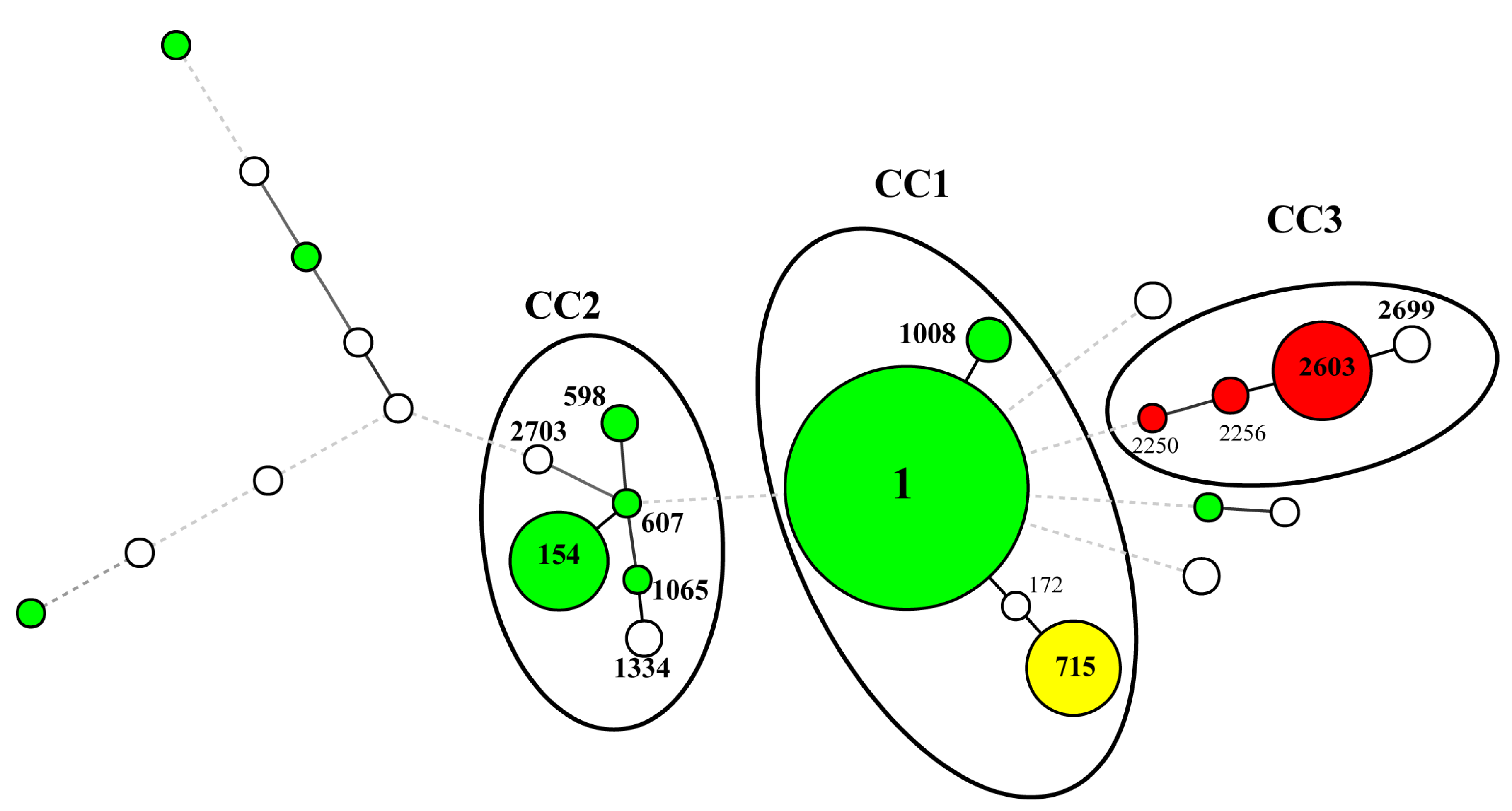
3.2. SBT Analysis of L. pneumophila

The 161 isolates were differentiated by SBT into 26 different STs (Table 1). The profiles of 20 of the 26 STs could be found in the EWGLI SBT database. Eighty-eight isolates belonged to ST1 (54.7%), which was the most frequently occurring ST, followed by ST2603 (15/161, 9.3%), ST154 (15/161, 9.3%), and ST715 (13/161, 8.1%) (Table 1). Table 1 shows the relationship between SGs and STs. MST-based SBT analysis identified three CCs (Figure 1). Of the 3 CCs, CC1, which was the most prevalent clonal group, consisted of 105 isolates assigned to ST1, ST715, ST1008, and ST172. CC2 was the second-most prevalent clonal group, consisting of 22 isolates that belonged to ST154, ST598, ST607, ST1065, ST1334, and ST2703.
The other CC3 (ST2603, ST2256, ST2250, and ST2699) consisted of 20 isolates. CC1 was formed by SG1 and SG7, while CC2 was mainly formed by SG1. SG13 isolates were contained in CC3. These results suggested that the specific genotypes in CC1, CC2, and CC3 are found in the cooling towers of Japan.

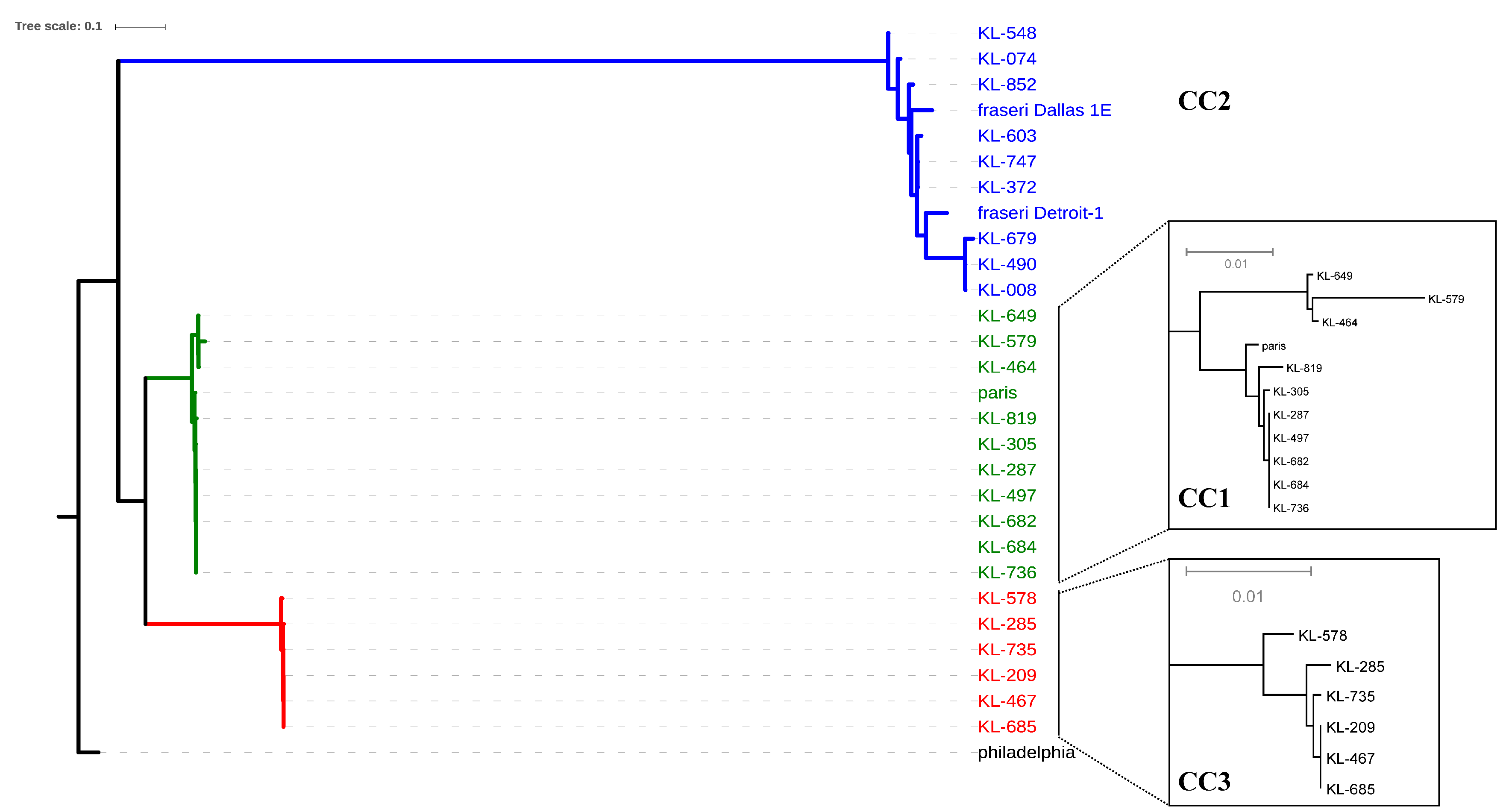
3.3. Whole Genome Analysis

To clarify the genetic relatedness among the strains belonging to the three CCs isolated from the different cooling towers in Japan during separate years, the whole-genome sequences were compared. In the phylogenetic tree based on the core genome SNPs, the three CCs were divided into three clades (Figure 2). Despite of KL852 assigned to ST2703, which differed from ST607 in triple-locus, the core genome SNPs phylogenetic tree indicated that KL852 was included in CC2 cluster (Table 2 and Figure 2).
For each clades containing each CCs (CC1, CC2, and CC3), 1093, 4558, and 245 of core SNPs were detected, respectively. CC1 contained the L. pneumophila strain Paris assigned to ST1. The CC1 cluster strains were classified into two sub-clades. In the sub-clades, closely related strain groups (e.g., KL464 and KL649; KL287, KL305, KL497, KL682, KL684, and KL736), with the number of SNPs being 0 or 1, were detected despite the separation years and/or sites. Likewise, the four strains of CC3 (KL209, KL467, KL685, and KL735) isolated from spatiotemporally separated cooling towers were also closely related to each other with 0 or 1 core SNPs. Surprisingly, even strains isolated from the same cooling towers were classified as CC1 or CC3, suggesting that several specific genetic strains of L. pneumophila could coexist in the same environment.
The phylogenetic tree based on the core genome SNPs demonstrated that CC2 is phylogenetically distant compared to CC1 and CC3 (Figure 2). Therefore, we calculated the ANI values among the strains. The ANI values among reference strains Paris and Philadelphia and our strains belonging to CC1 and CC3 ranged between 96.0 and 99.9%. These ANI values were higher than the 95% cut-off ANI value for bacterial species proposed by Goris et al. [18]. On the other hand, the ANI values in CC1 versus CC2 and CC3 versus CC2 were well below (91.8–92.8%) the proposed cut-off ANI value (95%) for bacterial species. Furthermore, the ANI value between CC2 and L. pneumophila subsp. fraseri was in the range of 98.4–99.8%. Therefore, when combined with the same cluster in the phylogenetic tree, the isolates of CC2 are concluded to belong to L. pneumophila subsp. fraseri.

4. Discussion

In this study, we analyzed L. pneumophila isolates obtained from cooling towers in Kobe city, Japan, during the past 10 years, by SBT and WGS. We revealed that the three high clonal groups, CC1, CC2, and CC3, have colonized the cooling towers in Japan for many years. Furthermore, we found that CC1 and CC3 strains could coexist in the same environment: e.g., KL287 (CC1) and KL285 (CC3); KL464 (CC1) and KL467 (CC3); KL579 (CC1) and KL578 (CC3); KL684 (CC1) and KL685 (CC3); KL736 (CC1) and KL735 (CC3) (Table 2). Our finding was suggested that these limited genetic groups of L. pneumophila might have been continuously inhabitant in Japanese cooling towers. Lijie et al., have reported high genetic polymorphism in industrial cooling towers in China [18]. However, we found lower genetic diversity in cooling towers in Japan through our monitoring over many years.
The major SGs constituting CC1, CC2, and CC3 were SG1, SG13, and SG7. In Japan, the main infection sources of legionellosis are hot springs and public baths [4]. SG1 has been found to be the major SG involved in bathwater-associated cases of legionellosis in Japan [19]. SG1 is also the predominant SG causing clinical Legionnaires’ disease cases, while legionellosis caused by SG13 and SG7 is very rare in Japan [20]. However, an outbreak of Legionnaires’ disease caused by SG13 and SG1 L. pneumophila strains in a spa house was recently reported in Japan [21]. Previous studies have revealed the variation of SGs and STs in bath water and shower water in public bath facilities and have reported them as infection sources in legionellosis cases [22]. On the other hand, as per a previous study and our findings, some definite SGs, SG1, SG13, and SG7, occupy niches in the cooling tower [23]. Therefore, it could be suggested that the distribution of SGs differed between public baths and cooling towers in Japan. In addition, a previous study has reported that the flaA genotypes in cooling towers were lower diversity than in bath water, and that flaA1 or flaA11 were the predominant flaA genotypes in cooling towers in Japan [23]. Similarly, in our study, we found that the dominant CC1 and CC2 strains harbored flaA1 and flaA11, respectively (Table S1). Conclusively, it can be suggested that limited genotypes colonize the cooling towers in Japan. This limited diversity might be due to the sources and properties of cooling towers water, which is chemically more homogeneous than public baths water from hot springs with different composition [23].
In a previous study, the distribution of STs in L. pneumophila SG1 differed between the water sources: hot springs, cooling towers, potable water systems, and soils in Japan, China, and South Korea [10,24,25]. Among the 26 STs detected in the 161 isolates from the cooling tower, ST1 in CC1 and ST154 in CC2 have been previously identified as STs in L. pneumophila SG1 isolated from a cooling tower in Japan [10]. ST1 (88/161, 54.7%) was the most dominant ST in this study. ST1 has been known to be the most prevalent worldwide [26,27,28]. Recent research has shown that ST1 is the major ST in clinical L. pneumophila in Japan [20]. The SG1-specific gene lag−1 is known to be a pathogenic marker [29]. Only two strains, ST2 and ST40, among the 114 SG1 strains were lag−1 positive, whereas the most prevalent ST1 isolated in this study were lag−1 negative (Table S1). Since the ST1 strains lacking lag−1 were reported as major clinical ST previously, it is considered necessary to focus on ST1 as sequence type which is at risk for clinical infection [20].
Phylogenetic trees based on the core genome SNPs showed that CC2 separated from CC1 and CC3. All 22 isolates belonging to the CC2 group possessed pilE14, consistent with a previous report [20]. Based on a genome phylogenetic tree, we showed that the CC2 strains are present in the same cluster as L. pneumophila subsp. fraseri strains. According to a previous study, the SBT pattern in L. pneumophila subsp. fraseri strains has been identified as the consensus pattern 11-x-16-x-x-13-x (flaA-pliE-asd-mip-mompS-proA-neuA) [30]. ST154, ST598, ST607, ST1065, ST1334, and ST2703 included in CC2 also exhibited the 11-x-16-x-x-13-x pattern. Using a combination of phylogeny and genome sequence comparison, CC2 has been identified to belong to L. pneumophila subsp. fraseri.
In this study, core SNPs were identified using kSNP software to further resolve the genome sequences. WGS based genotypes correlated with the ST classification by SBT. Hundreds of core SNPs are detected between strains of different genetic lineage. Furthermore, core SNPs genotypes were elucidated the genomic proximity and stability between strains within each CC, despite the difference in separation years and separation sites. Brian et al. have reported that isolates associated with specific Legionella outbreaks differed by <5 core SNPs and formed outbreak-specific clades [31]. In our research, despite the differences in separation years and separation sites, there are strains in which only 0 or 1 core SNP are detected among the strains within each CC (such as, KL287, KL305, KL497, KL682, KL684, and KL736). Our results are suggested that the identical genotype in three genetic lineages are adapted to cooling towers in Japan.

5. Conclusions

In conclusion, we found three high clonal groups, CC1, CC2, and CC3, which had colonized cooling towers in Japan over our ten year monitoring period. We elucidated that CC2 is phylogenetically distant compared to CC1 and CC3, and belonged to L. pneumophila subsp. fraseri. Our findings could be helpful for estimating the sources of infection in Japan and developing prevention strategies.

Supplementary Materials

The following are available online at https://0-www-mdpi-com.brum.beds.ac.uk/1660-4601/16/9/1664/s1, Table S1: Information of the 161 strains in this study.

Author Contributions

N.N. and R.N. designed the study methods and wrote the first draft of the manuscript; N.N., S.T., and K.A. did bacterial isolation and identification; S.T., N.N. and R.N. did the typing and PCR work; N.N. and R.N. analyzed the data; T.I. contributed to the writing of the manuscript. All authors have read and approved the final manuscript.

Funding

This work was supported by JSPS KAKENHI grant number 16K16219 and 19K12376.

Acknowledgments

We thank Norman K. Fry (Respiratory and Vaccine Preventable Bacteria Reference Unit, Public Health England) for assigning the newly identified alleles and STs.

Conflicts of Interest

The authors declare no conflict of interest.

Abbreviations

CCClonal complex
SBTSequence-based typing
WGSWhole genome sequencing
UTUntypable
STSequence type
SGSerogroup
ANIAverage nucleotide identity
SNPSingle nucleotide polymorphism

References

  1. Fields, B.S.; Benson, R.F.; Besser, R.E. Legionella and Legionnaires’ disease: 25 years of investigation. Clin. Microbiol. Rev. 2002, 15, 506–526. [Google Scholar] [CrossRef] [PubMed]
  2. Diederen, B.M.W. Legionella spp. and Legionnaires’ disease. J. Infect. 2008, 56, 1–12. [Google Scholar] [CrossRef] [PubMed]
  3. Fliermans, C.B.; Cherry, W.B.; Orrison, L.H.; Smith, S.J.; Tison, D.L.; Pope, D.H. Ecological distribution of Legionella pneumophila. Appl. Environ. Microbiol. 1981, 41, 9–16. [Google Scholar]
  4. National Institute of Infectious Diseases and Tuberculosis and Infectious Diseases Control Division, Ministry of Health, Labour, and Welfare. Legionellosis, April 1999–July 2000; IASR (Infectious Agents Surveillance Report); IASR: Tokyo, Japan, 2000; pp. 186–187. [Google Scholar]
  5. Fitzhenry, R.; Weiss, D.; Cimini, D.; Balter, S.; Boyd, C.; Alleyne, L.; Stewart, R.; McIntosh, N.; Econome, A.; Lin, Y.; et al. Legionnaires’ Disease Outbreaks and Cooling Towers, New York City, NY, USA. Emerg. Infect. Dis. 2017, 23, 1769. [Google Scholar] [CrossRef] [PubMed]
  6. Bhopal, R.S.; Fallon, R.J. Variation in time and space of non-outbreak Legionnaires’ disease in Scotland. Epidemiol. Infect. 1991, 106, 45–61. [Google Scholar] [CrossRef] [PubMed]
  7. García-Fulgueiras, A.; Navarro, C.; Fenoll, D.; García, J.; González-Diego, P.; Jiménez-Buñuales, T.; Pelaz, C. Legionnaires’ disease outbreak in Murcia, Spain. Emerg. Infect. Dis. 2003, 9, 915–921. [Google Scholar] [CrossRef]
  8. Shivaji, T.; Sousa Pinto, C.; San-Bento, A.; Oliveira Serra, L.A.; Valente, J.; Machado, J.; Vasconcelos, P. A large community outbreak of Legionnaires disease in Vila Franca de Xira, Portugal, October to November 2014. Eurosurveillance 2014, 19, 20991. [Google Scholar] [CrossRef]
  9. Maisa, A.; Brockmann, A.; Renken, F.; Lück, C.; Pleischl, S.; Exner, M.; Daniels-Haardt, I.; Jurke, A. Epidemiological investigation and case-control study: A Legionnaires’ disease outbreak associated with cooling towers in Warstein, Germany, August–September 2013. Eurosurveillance 2015, 20, 30064. [Google Scholar] [CrossRef]
  10. Li, L.; Qin, T.; Li, Y.; Zhou, H.; Song, H.; Ren, H.; Li, L.; Li, Y.; Zhao, D. Prevalence and Molecular Characteristics of Waterborne Pathogen Legionella in Industrial Cooling Tower Environments. Int. J. Environ. Res. Public Health 2015, 12, 12605–12617. [Google Scholar] [CrossRef] [PubMed]
  11. Amemura-Maekawa, J.; Kikukawa, K.; Helbig, J.H.; Kaneko, S.; Suzuki-Hashimoto, A.; Furuhata, K.; Chang, B.; Murai, M.; Ichinose, M.; Ohnishi, M.; et al. Distribution of monoclonal antibody subgroups and sequence-based types among Legionella pneumophila serogroup 1 isolates derived from cooling tower water, bathwater, and soil in Japan. Appl. Environ. Microbiol. 2012, 78, 4263–4270. [Google Scholar] [CrossRef]
  12. Farhat, C.; Mentasti, M.; Jacobs, E.; Fry, N.K.; Lück, C. The N-acylneuraminate cytidyltransferase gene, neuA, is heterogenous in Legionella pneumophila strains but can be used as a marker for epidemiological typing in the consensus sequence-based typing scheme. J. Clin. Microbiol. 2011, 49, 4052–4058. [Google Scholar] [CrossRef] [PubMed]
  13. Gaia, V.; Fry, N.K.; Afshar, B.; Lück, P.C.; Meugnier, H.; Etienne, J.; Peduzzi, R.; Harrison, T.G. Consensus sequence-based scheme for epidemiological typing of clinical and environmental isolates of Legionella pneumophila. J. Clin. Microbiol. 2005, 43, 2047–2052. [Google Scholar] [CrossRef] [PubMed]
  14. Ratzow, S.; Gaia, V.; Helbig, J.H.; Fry, N.K.; Lück, P.C. Addition of neuA, the gene encoding N-acylneuraminate cytidylyl transferase, increases the discriminatory ability of the consensus sequence-based scheme for typing Legionella pneumophila serogroup 1 strains. J. Clin. Microbiol. 2007, 45, 1965–1968. [Google Scholar] [CrossRef]
  15. Coil, D.; Jospin, G.; Darling, A.E. A5-miseq: An updated pipeline to assemble microbial genomes from Illumina MiSeq data. Bioinformatics 2015, 31, 587–589. [Google Scholar] [CrossRef]
  16. Gardner, S.N.; Slezak, T.; Hall, B.G. kSNP3.0: SNP detection and phylogenetic analysis of genomes without genome alignment or reference genome. Bioinformatics 2015, 31, 2877–2878. [Google Scholar] [CrossRef]
  17. Richter, M.; Rosselló-Móra, R. Shifting the genomic gold standard for the prokaryotic species definition. Proc. Natl. Acad. Sci. USA 2009, 106, 19126–19131. [Google Scholar] [CrossRef] [Green Version]
  18. Goris, J.; Konstantinidis, K.T.; Klappenbach, J.A.; Coenye, T.; Vandamme, P.; Tiedje, J.M. DNA–DNA hybridization values and their relationship to whole-genome sequence similarities. Int. J. Syst. Evol. Microbiol. 2007, 57, 81–91. [Google Scholar] [CrossRef] [PubMed] [Green Version]
  19. Kuroki, T.; Ishihara, T.; Ito, K.; Kura, F. Bathwater-associated cases of legionellosis in Japan, with a special focus on Legionella concentrations in water. Jpn. J. Infect. Dis. 2009, 62, 201–205. [Google Scholar]
  20. Amemura-Maekawa, J.; Kura, F.; Chida, K.; Ohya, H.; Kanatani, J.I.; Isobe, J.; Tanaka, S.; Nakajima, H.; Hiratsuka, T.; Yoshino, S.; et al. Legionella pneumophila and Other Legionella Species Isolated from Legionellosis Patients in Japan between 2008 and 2016. Appl. Environ. Microbiol. 2018, 84, e00721-18. [Google Scholar] [CrossRef] [PubMed]
  21. Kuroki, T.; Amemura-Maekawa, J.; Ohya, H.; Furukawa, I.; Suzuki, M.; Masaoka, T.; Aikawa, K.; Hibi, K.; Morita, M.; Lee, K.I.; et al. Outbreak of Legionnaire’s Disease Caused by Legionella pneumophila Serogroups 1 and 13. Emerg. Infect. Dis. 2017, 23, 349–351. [Google Scholar] [CrossRef] [PubMed]
  22. Kanatani, J.I.; Isobe, J.; Norimoto, S.; Kimata, K.; Mitsui, C.; Amemura-Maekawa, J.; Kura, F.; Sata, T.; Watahiki, M. Prevalence of Legionella species isolated from shower water in public bath facilities in Toyama Prefecture, Japan. J. Infect. Chemother. 2017, 23, 265–270. [Google Scholar] [CrossRef]
  23. Amemura-Maekawa, J.; Kura, F.; Chang, B.; Suzuki-Hashimoto, A.; Ichinose, M.; Endo, T.; Watanabe, H. Distinct difference of flaA genotypes of Legionella pneumophila between isolates from bath water and cooling tower water. Microbiol. Immunol. 2008, 52, 460–464. [Google Scholar] [CrossRef]
  24. Qin, T.; Zhou, H.; Ren, H.; Guan, H.; Li, M.; Zhu, B.; Shao, Z. Distribution of sequence-based types of legionella pneumophila serogroup 1 strains isolated from cooling towers, hot springs, and potable water systems in China. Appl. Environ. Microbiol. 2014, 80, 2150–2157. [Google Scholar] [CrossRef]
  25. Lee, H.K.; Shim, J.I.; Kim, H.E.; Yu, J.Y.; Kang, Y.H. Distribution of Legionella species from environmental water sources of public facilities and genetic diversity of L. pneumophila serogroup 1 in South Korea. Appl. Environ. Microbiol. 2010, 76, 6547–6554. [Google Scholar] [CrossRef]
  26. Kozak-Muiznieks, N.A.; Lucas, C.E.; Brown, E.; Pondo, T.; Taylor, T.H., Jr.; Frace, M.; Miskowski, D.; Winchell, J.M. Prevalence of sequence types among clinical and environmental isolates of Legionella pneumophila serogroup 1 in the United States from 1982 to 2012. J. Clin. Microbiol. 2014, 52, 201–211. [Google Scholar] [CrossRef]
  27. Harrison, T.G.; Afshar, B.; Doshi, N.; Fry, N.K.; Lee, J.V. Distribution of Legionella pneumophila serogroups, monoclonal antibody subgroups and DNA sequence types in recent clinical and environmental isolates from England and Wales (2000–2008). Eur. J. Clin. Microbiol. Infect. Dis. 2009, 28, 781–791. [Google Scholar] [CrossRef] [PubMed]
  28. David, S.; Sánchez-Busó, L.; Harris, S.R.; Marttinen, P.; Rusniok, C.; Buchrieser, C.; Harrison, T.G.; Parkhill, J. Dynamics and impact of homologous recombination on the evolution of Legionella pneumophila. PLoS Genet. 2017, 13, e1006855. [Google Scholar] [CrossRef] [PubMed]
  29. Kozak, N.A.; Benson, R.F.; Brown, E.; Alexander, N.T.; Taylor, T.H., Jr.; Shelton, B.G.; Fields, B.S. Distribution of lag-1 alleles and sequence-based types among Legionella pneumophila serogroup 1 clinical and environmental isolates in the United States. J. Clin. Microbiol. 2009, 47, 2525–2535. [Google Scholar] [CrossRef]
  30. Kozak-Muiznieks, N.A.; Morrison, S.S.; Mercante, J.W.; Ishaq, M.K.; Johnson, T.; Caravas, J.; Lucas, C.E.; Brown, E.; Raphael, B.H.; Winchell, J.M. Comparative genome analysis reveals a complex population structure of Legionella pneumophila subspecies. Infect. Genet. Evol. 2018, 59, 172–185. [Google Scholar] [CrossRef] [PubMed]
  31. Raphael, B.H.; Baker, D.J.; Nazarian, E.; Lapierre, P.; Bopp, D.; Kozak-Muiznieks, N.A.; Morrison, S.S.; Lucas, C.E.; Mercante, J.W.; Musser, K.A.; et al. Genomic resolution of outbreak-associated Legionella pneumophila serogroup 1 isolates from New York State. Appl. Environ. Microbiol. 2016, 82, 3582–3590. [Google Scholar] [CrossRef]
Figure 1. A minimum spanning tree of 161 L. pnuemohila strains from cooling towers in Kobe, Japan, based on SBT. The strains belonged to serogroup (SG)1 (n = 114), SG13 (n = 18), SG7 (n = 13) and the other SGs (n = 16) were shown as the circles with green, red, yellow, and white respectively. The circle sizes are proportional to the numbers of isolates sharing an identical pattern. The length of the thin line is proportional with single-, double- or triple-locus variants. The dotted lines indicate four loci variants or higher. The clonal complexes (CCs) (CC1, CC2, and CC3) generated with single- and double variants are indicated by the circle surrounding the ST. Whereas KL852 was assigned to ST2703, which differed from ST607 in triple-locus, the strain was included in CC2.
Ijerph 16 01664 g001
Figure 2. Phylogenetic tree based on a 32586-core-SNP matrixgenome-wide SNPs among the main strains assigned to the three CCs listed in Table 2. A maximum-likelihood tree was constructed using kSNP 3.0 [16]. The lineages including CCs are highlighted by green (CC1), blue (CC2), and red (CC3). Insets show subtrees of CC1 and CC3 isolates.
Ijerph 16 01664 g002
Table 1. Distribution of SGs and STs of 161 L. pneumophila isolated in cooling tower, Kobe, Japan form 2003–2012.
SGNo. of Isolates (%)STs (No. of Isolates)
SG1114(70.8)ST1 (88), ST154 (15), ST1008 (3), ST598 (2), ST2 (1), ST40 (1), ST45 (1), ST59 (1), ST607 (1), ST1065 (1)
SG1318(11.2)ST2603 (15), ST2256 (2), ST2250 (1)
SG713(8.1)ST715 (13)
SG UT7(4.3)ST1334 (2), ST2699 * (2), ST172 (1), ST1916 (1), ST2701 * (1)
SG54(2.5)ST2700 * (2), ST2704 * (2)
SG62(1.2)ST68 (1), ST242 (1)
SG21(0.6)ST2702 * (1)
SG81(0.6)ST1324 (1)
SG91(0.6)ST2703 * (1)
* We identified as new sequence types (STs) in sequence-based typing (SBT) database.
Table 2. Information of the 25 strains assigned to three complexes on genome analysis.
ComplexesNo. of StrainYearsSite in CT aSGSTDDBJ Accession No. of Read Data
CC1KL2872006F11DRR163670
KL3052006A11DRR163671
KL4642008A11DRR163673
KL4972008E11DRR163675
KL5792009H11DRR163677
KL6492010A11DRR163679
KL6822010M11008DRR170776
KL6842010D11DRR163680
KL7362011J11DRR163683
KL8192012Q7715DRR170777
CC2KL0082003KUT1334DRR170771
KL0742004L1607DRR170772
KL3722007C1154DRR163672
KL4902008NUT1334DRR170773
KL5482009O1598DRR170774
KL6032009I1154DRR163678
KL6792010P11065DRR170775
KL7472011G1154DRR163684
KL852 b2012L92703DRR170778
CC3KL2092005B132603DRR163668
KL2852006FUT2699DRR163669
KL4672008A132603DRR163674
KL5782009H132603DRR163676
KL6852010D132603DRR163681
KL7352011J132603DRR163682
a Locations of cooling towers are as follows: hospital (A); unknown (B, K); office buildings (C, F, L, Q); apartment house (D); accommodations (E, H, J, M, N, O); shopping center (G); wedding hall (I). b Whereas KL852 was assigned to ST2703, which differed from ST607 in triple-locus, the strain was included in CC2.

