Article

Algicidal Efficiency and Genotoxic Effects of Phanerochaete chrysosporium against Microcystis aeruginosa

1
Chongqing Engineering Laboratory of Nano/Micro Biological Medicine Detection Technology, School of Architecture and Engineering, Chongqing University of Science and Technology, Chongqing 401331, China
2
Institute of Life Sciences & Biomedicine Collaborative Innovation Center, Wenzhou University, Wenzhou 325035, China
*
Author to whom correspondence should be addressed.
These two authors contributed equally to this work.
Int. J. Environ. Res. Public Health 2020, 17(11), 4029; https://0-doi-org.brum.beds.ac.uk/10.3390/ijerph17114029
Submission received: 5 May 2020 / Revised: 29 May 2020 / Accepted: 30 May 2020 / Published: 5 June 2020
(This article belongs to the Section Environmental Microbiology)

Abstract

:
Eutrophication has become a severe environmental problem. This study evaluated the algicidal efficiency and genotoxic effects of Microcystis aeruginosa co-cultured with Phanerochaete chrysosporium for 48 h under the optimum conditions of 250 mg/L of P. chrysosporium at 25 °C with dissolved oxygen content of 7.0 mg/L. The results showed that the activity of algal dehydrogenase, superoxide dismutase, and peroxidase were all decreased and the malondialdehyde content increased after co-culturing. Fourier transform infrared spectroscopy and scanning electron microscopy observations showed that the functional group and structure of algal cells were significantly changed. Compared with those of control tadpoles, blood cells of Fejervarya multistriata tadpoles had increased micronucleus frequency (from 1.05 ± 0.09 to 1.99 ± 0.05) and abnormal nuclei (from 2.45 ± 0.06 to 5.83 ± 0.07). The tail length of M. aeruginosa co-cultured with P. chrysosporium increased from 1.12 ± 0.21 to 21.68 ± 0.34, and the comet length increased from 6.45 ± 0.09 to 36.45 ± 0.67 within 48 h. Micronucleus assay and Comet assay results demonstrated that P. chrysosporium might effectively remove algae and reduce genotoxic effects and may be safe for aquatic ecosystems.

1. Introduction

Eutrophication results from the enrichment with nitrogen, phosphorus, and other nutrients, and may cause abnormal reproduction in algae and other aquatic plants, decreased water transparency and dissolved oxygen content, poor water quality, and death of aquatic organisms [1]. It has become a global environmental problem and threatens to destroy the balance of aquatic ecosystems such as lakes, reservoirs, and closed and semi-enclosed rivers [2,3].
Microcystis aeruginosa is a common algae involved in water eutrophication. Its growth, metabolism and death will not only pose a serious threat to the aquatic environment and ecological security, and have an inestimable impact on human survival and health [4]. Therefore, treating M. aeruginosa has important social and practical significance. Three methodologies are commonly used in the removal of algae and algal toxins: Physical, chemical, and biological methods. The physical method is not comprehensive or cost effective, and the chemical methods are prone to secondary pollution and poor security. However, biological remediation methods are relatively cost effective and have low risk when compared with the physical and chemical methods; making them promising candidates for future research [5]. White-rot fungi can degrade or decrease various environmental pollutants, including dyes, lignin, and toxic wastes [6]. Previous studies indicated that their suppressive effect on algal species could be a result of enzymes secreted during incubation, such as laccase [7,8,9,10,11]. However, the mechanisms underlying the ecotoxicological relationships between white-rot fungi and algae still need to be studied.
Modern molecular biology methods provide a new avenue for investigating ecological toxicology mechanisms at a cellular and molecular level [12,13]. A variety and combination of physical and chemical factors detrimentally affect the structure and function of DNA, resulting in genetic diseases, cell mutation, and ultimately death [14]. Comet assays can quantitatively determine DNA integrity at the single-cell level are suitable proxies for monitoring genetic damage [15,16,17]. The micronucleus is an abnormal structure and chromosome aberration in eukaryotes [18,19,20]. Pollution can be monitored by observing the cell micronuclei of plant and animal tissue. In recent years, it has been widely used in radiation injury, chemical mutagenesis, and environmental monitoring [21,22,23].
Some algae-lysing microorganisms of mature or artificial domestication may produce endotoxins or exotoxins during the process of algal cell metabolism or death, which poses a severe threat to a body of water. The algicidal efficiency and toxicity of the algae-control system of white- rot fungi need to be determined for it to be safely applied to the treatment of eutrophic water. In this study, we evaluated the genotoxic effects of Phanerochaete chrysosporium, a typical white-rot fungi, on M. aeruginosa in Fejervarya multistriata tadpoles by a comet and micronucleus test. Structural characteristics, including the algal cell and surface morphology, were also assessed and compared using Fourier transform infrared spectroscopy (FTIR) and scanning electron microscopy (SEM) to investigate the algicidal efficiency of P. chrysosporium.

2. Materials and Methods

2.1. Algal Strains and Cultivation

M. aeruginosa, which as research target, was provided by the Freshwater Algae Culture Collection of the Chinese Academy of Sciences. All stock and experimental cultures were maintained at 25 °C under a 12:12 h (light:dark) cycle at approximately 90 µmol photons m−2·s−1 provided by cool-white fluorescent lamps to achieve exponential growth. The growth medium for M. aeruginosa was BG-11 and 1000 mL distilled water.

2.2. Fungal Strains

P. chrysosporium was provided by the Institute of Microbiology (China). The strain was maintained on potato dextrose agar (PDA) plates and cultured at 35 ℃ for 5 days. It was then used in further experiments.

2.3. Animal and Experimental Design

One hundred of F. multistriata tadpoles, which were used for toxicology testing, were obtained from a Chongqing farmland suburb and then placed in a non-polluted eco-pond (27–28 °C). The healthy tadpoles were of average uniform size at 32–36 mm, with an average body length and weight of 26.28 ± 0.34 mm and 0.19 ± 0.019 g, respectively.
F. multistriata tadpoles were fed to M. Aeruginosa. The M. aeruginosa was treated with P. chrysosporium under the optimum conditions of 250 mg/L P. chrysosporium at dissolved oxygen content of 7.0 mg/L at 25 °C and 12:12 h (light:dark) cycle for 24 h and 48 h. Control cultures were maintained concurrently throughout the experiment at identical conditions in the absence of P. chrysosporium and no feeding during the experiment. All experiments were conducted in triplicate for the micronucleus assay and the comet assay.

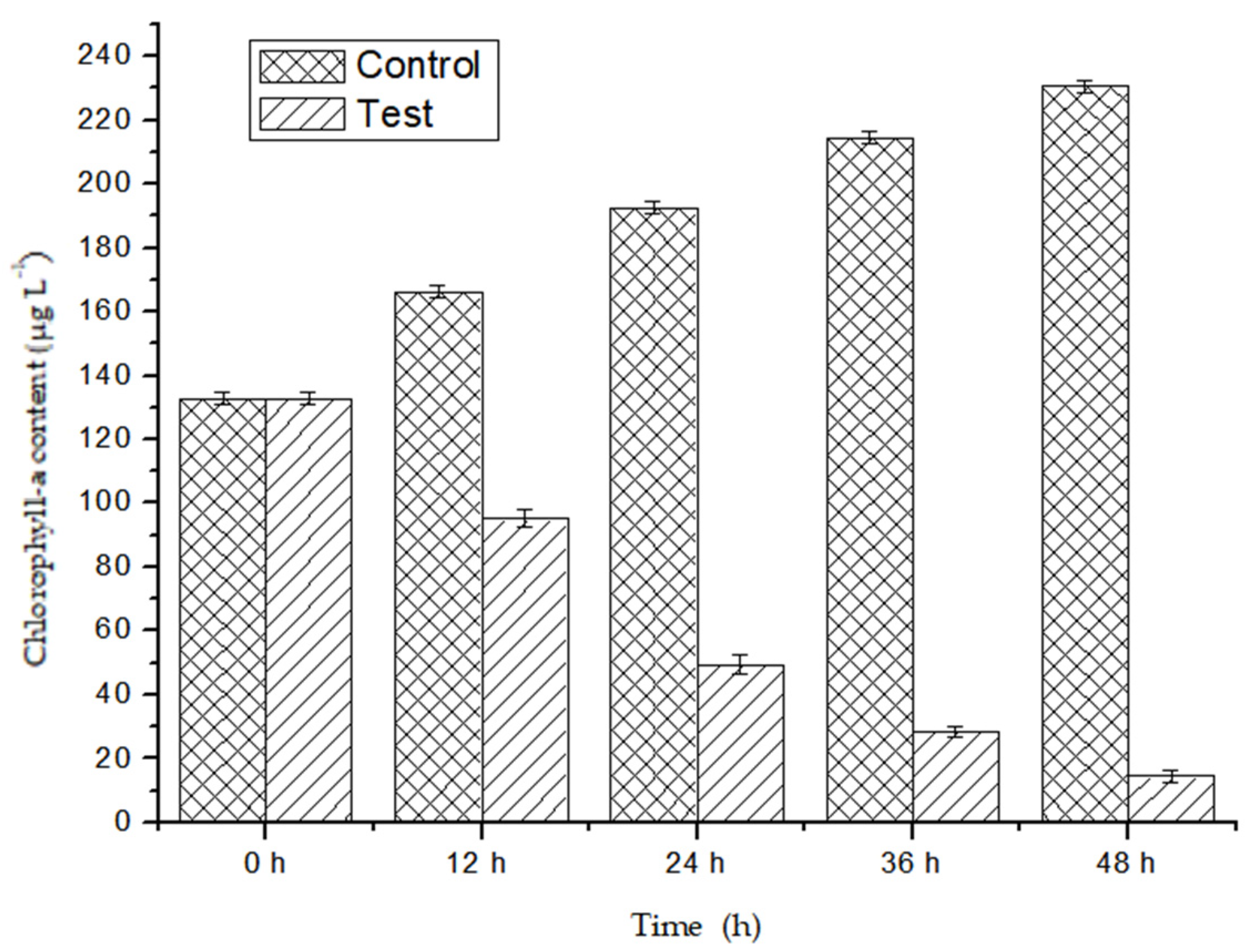
2.4. Chlorophyll—A Content Test

The algicidal tests were conducted under the conditions of 25 °C, pH 7.0, 7.0·mg·L−1 DO, and 12:12 h (light:dark) cycle. All experiments were conducted in triplicate. The total chlorophyll-a was extracted with 90% acetone and quantified using the repeated freezing–thawing extraction method [24].

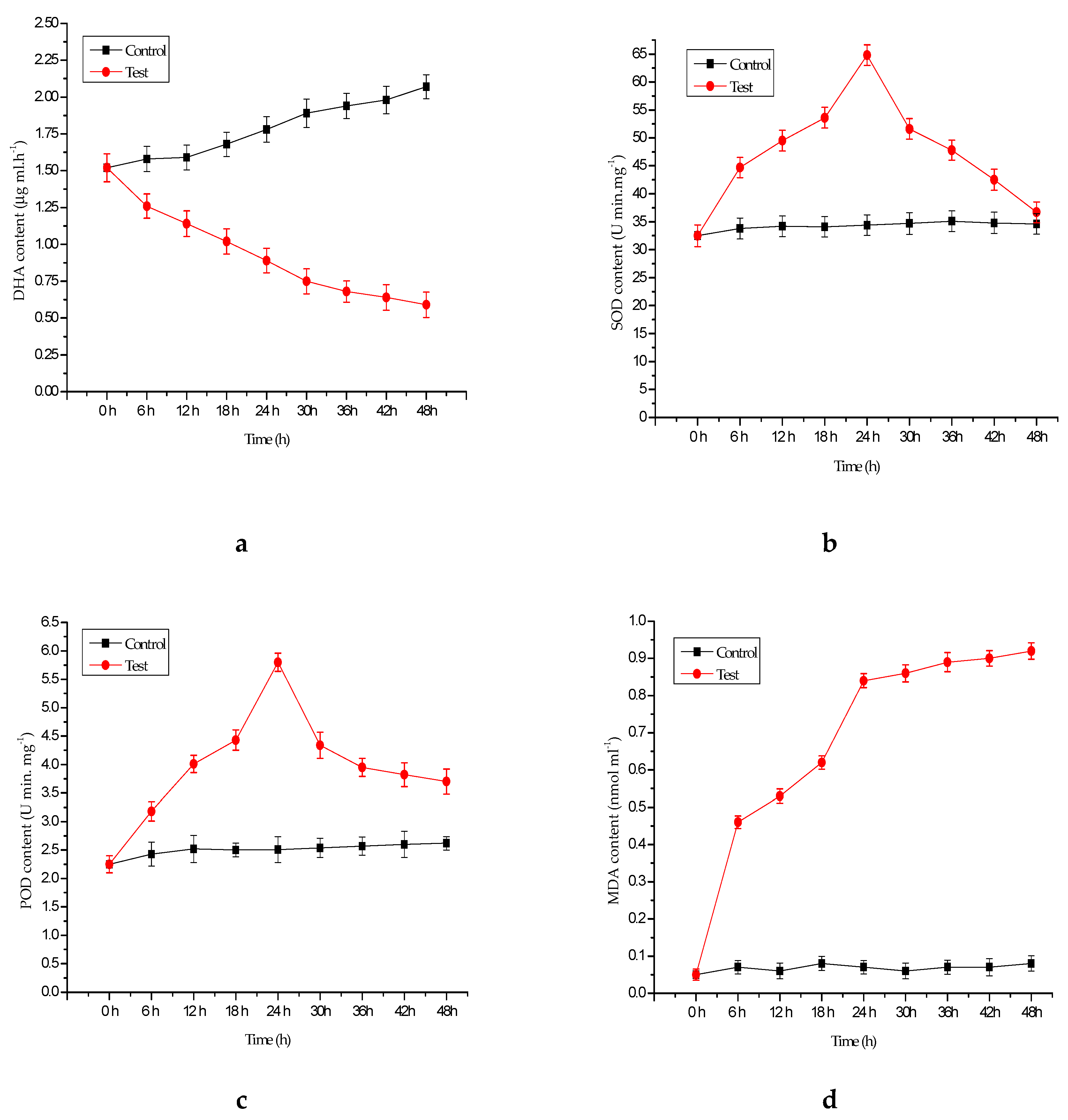
2.5. Antioxidant Enzyme Assays

Aliquots of 25 µL of the samples were collected and centrifuged at 8000 rpm for 20 min for enzyme activity tests. The activities of dehydrogenase (DHA), superoxide dismutase (SOD), and peroxidase (POD) in the supernatant were determined based on the triphenyl tetrazolium chloride- dehydrogenase reduction [25], nitrogen blue tetrazole [26], and the guaiacol method [27], respectively. The content of malondialdehyde (MDA) was measured using the thiobarbituric acid method [28].
Fourier transform infrared spectroscopy disks were prepared by mixing 2 mg of lyophilized sample using a freeze-dry system (FREEZONE-6, Hitachi, Japan) with 150 mg of KBr. The samples were then pressed in a standard device using a pressure of 16 MPa to produce 13-mm-diameter pellets. Fourier transform infrared (FTIR) spectra were recorded between 4000 and 400 cm−1 using a Spectrum GX (Perkin Elmer, Waters, US).

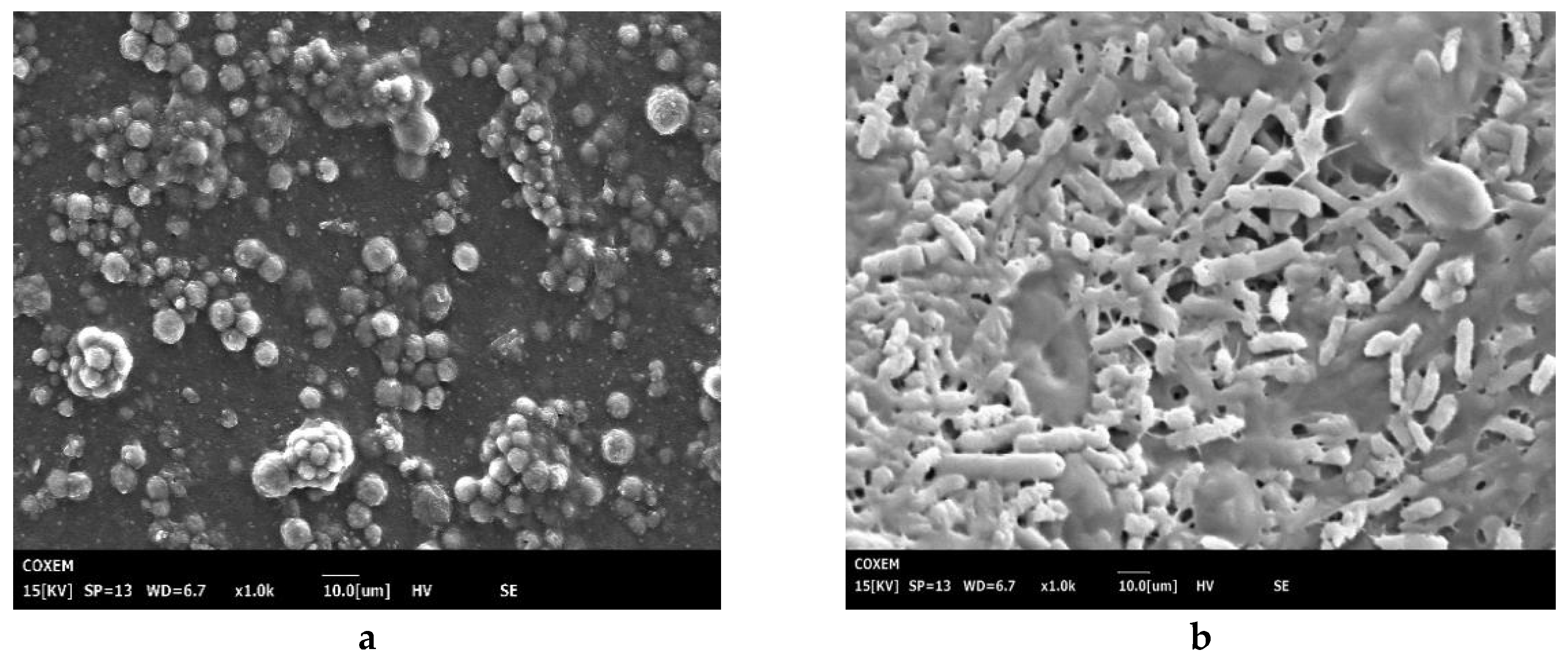
2.6. Scanning Electron Microscopy

The microstructural changes and surface characteristics of the M. aeruginosa and M. aeruginosa treated with P. chrysosporium were tested through a SEM TESCAN VEGA II LMU (COXEM, Hitachi, Japan) at 20 kV accelerating voltage.

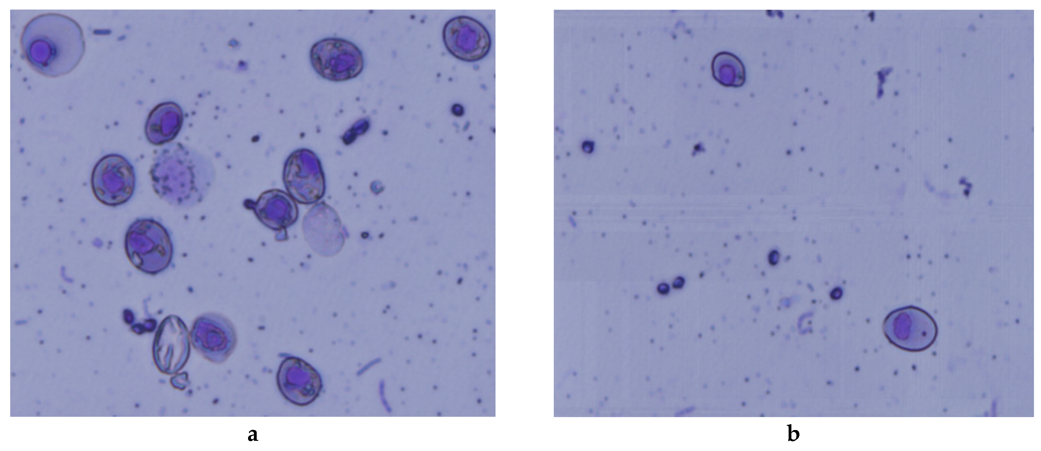
2.7. Micronucleus Assay
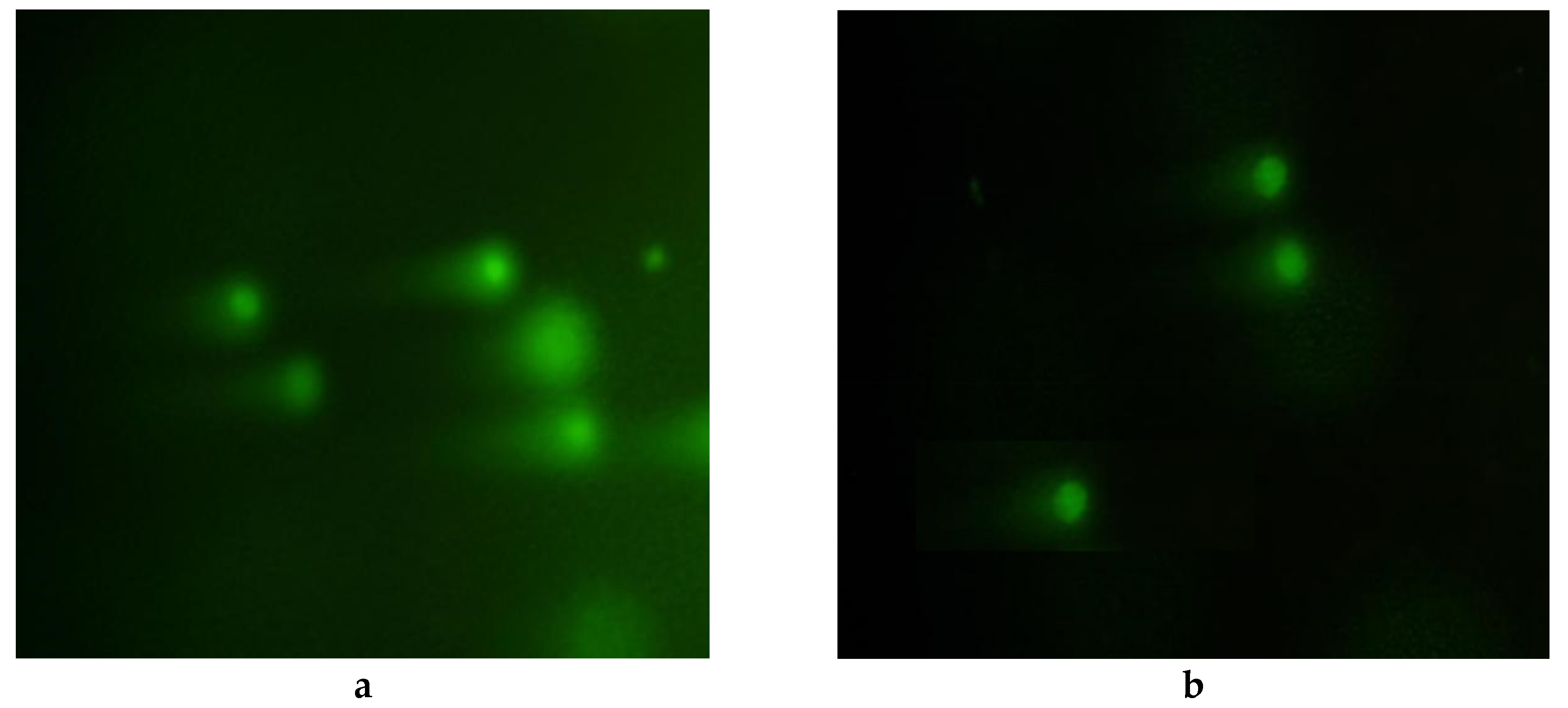
The surfaces of the tadpoles were dried with filter paper, the tails were cut, and then the tail blood was used to make a blood smear, which was dried naturally and fixed with methanol for 15 min. Wright’s stain was applied for 15–20 min, followed by washing with phosphate-buffered saline (PBS) (pH 6.8). Finally, the blood smears were dyed with the Gimsa solution for 25 min, washed with deionized water, and dried naturally for observation under a microscope. For each blood smear, 1000 cells were observed, and the number of micronuclei and abnormal red blood cells was recorded. The micronucleus cell rate (MCN) = number of cells with micronucleus/total number of observed cells * 1000‰, and the nuclear abnormal cell rate (ONA) = total number of cells with nuclear abnormalities (except micronuclei)/total number of observed cells * 1000‰. The data obtained from the experiment, the comparison between the control group and the treatment group were analyzed using the Origin 8.0 (Originlab, US) software method. All experiments were conducted in triplicate.

2.8. Comet Assay

The heads of the F. multistriata tadpoles were cut; blood cells were collected and diluted with PBS (pH 6.8) until the cell density reached 105–106 mL. These blood cells were used directly for the comet assay. The buffer containing the blood cells was transferred into a 2-mL centrifuge tube. Completely frosted microscopic slides were covered by a 90 µL thin layer of 0.7% normal melting agarose (NMA), (NMA), which was allowed to solidify at 4 °C for 30 min. Then, 75 µL of low melting point agarose (1%) containing 25 µL of blood cells was added to cover the NMA at 4 °C for 30 min. The slides were placed in a lysis solution at 4 °C (2.5 M NaCl, 100 mM Na2EDTA, 10 mM Tris-HCl, 1% Triton X-100 and 10% DMSO, pH 10) for 1.5 h in dark. Then, the slides were washed with chilled distilled water and placed on a horizontal gel electrophoresis apparatus in an alkaline buffer (300 mM NaOH, and 1 mM Na2EDTA, pH > 13) at 20 °C (20 V and 200 mA) for 30 min in the dark to avoid DNA damage. After electrophoresis, each slide was neutralized with the neutralization buffer (0.4 M Tris-HCI, pH 7.3) three times, washed with chilled distilled water, and placed in ethanol for 5 min at 4 °C. Finally, the slides were stained with Godview for 20 min in the dark and observed under a fluorescence microscope at 490 nm, with the excitation plate wavelength at 420–485 nm, and the emission plate wavelength at 515 nm [27]. Each group contained three parallel samples, and 100 cells were observed for each slide; the images of comets were analyzed using the CASP 10.0 (NIH, US) software.

2.9. Statistical Analysis

The data obtained from the experiment were analyzed and processed using the Origin 8.0 software processing system and Windows Excel and Word (2003, 2010 version) Office software.

3. Results and Discussion

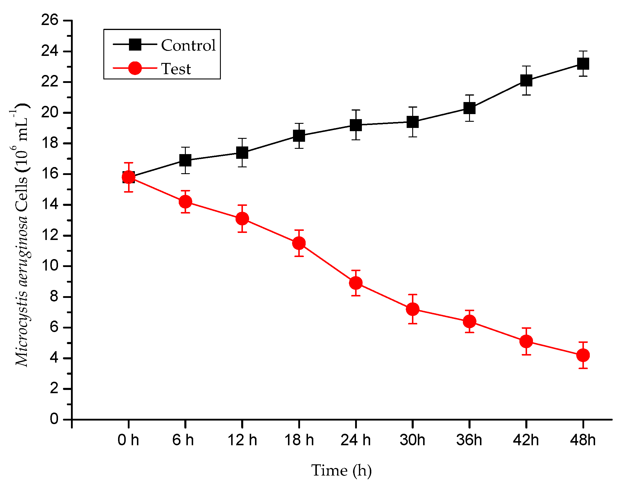
3.1. Algicidal Efficiency

The chlorophyll-a content of M. aeruginosa was evaluated after the P. chrysosporium treatment. P. chrysosporium induced reductions of 91.23% in the chlorophyll-a content of M. aeruginosa within 48 h (Figure 1), indicating P. chrysosporium limits growth effectively. This places the algicidal properties of P. chrysosporium in the reported range of algicidal bacteria (70–90%) [29], and at an increased rate. We also used cell counting to quantify the inhibitory effect of P. chrysosporium on M. aeruginosa at different time points using the growth curves of M. aeruginosa cells (Figure 2). We observed that the P. chrysosporium had a strong inhibitory effect on M. aeruginosa, and the inhibitory effect was time-dependent.
SOD is a protective antioxidant enzyme that is induced during cellular antioxidant defenses and protects cells by scavenging harmful reactive oxygen species (ROS) [30,31]. POD is an integral part of the plant antioxidant system, and plays an important role in plant stress resistance physiology, which has the effect of removes oxygen free radicals and hydrogen peroxide [32]. MDA [33], which is the final product of membrane lipid decomposition, is an important biochemical indicator of oxidative membrane damage, and can reflect the degree of injury suffered by a plant.
The reduced DHA levels (Figure 3a) in M. aeruginosa and P. chrysosporium co-cultures indicates successful degradation of DHA by P. chrysosporium, which may, in turn, inhibit chlorophyll-a synthesis and suppress algal growth. The experiments show that the activity of POD and SOD in the experimental groups was significantly higher than that in the control group but sharply declined at 24 h (Figure 3b,c), from 65 U/(min·mg) to 36 U/(min·mg) and from 5.8 U/(min·mg) to 3.7 U/(min·mg), respectively. The MDA level in M. aeruginosa co-cultured with P. chrysosporium increased to 0.92 nmol·mL−1 after 48 h (Figure 3d). These results indicate that P. chrysosporium may have caused damage to membranes and chloroplast structural integrity. Such effects suggest that it may be appropriate to use P. chrysosporium as an economically sustainable management strategy for controlling harmful algal blooms.

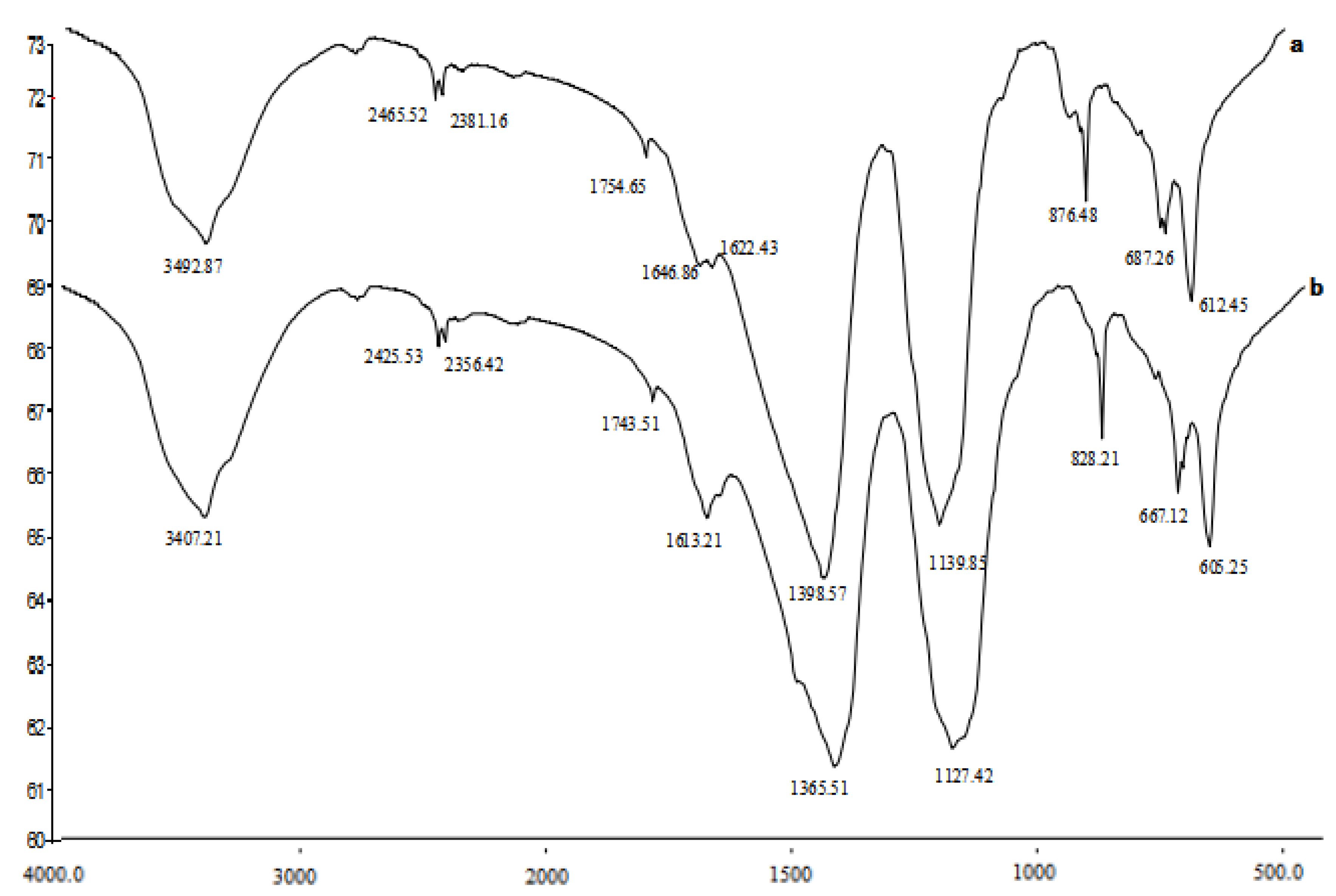
3.2. Fourier Transform Infrared Spectroscopy

Characterization of the chemical fingerprints of M. aeruginosa treated with P. chrysosporium and untreated cultures are shown in Figure 4.
The infrared spectrum of the main absorption peak position and the waveform of M. aeruginosa cells before and after the white-rot fungi treatment were approximately the same. Irrespective, there is a clear difference among the relative intensities of the peaks. The intensity of the absorption peak of the algal cells treated with white-rot fungi was weaker than that of the untreated algal cells (Figure 4). The algae capture light energy mainly through pigment-protein complexes, and the photosynthetic pigments can be divided into three categories: Chlorophyll-a (Chl), carotenoids (Car), and phycobiliprotein [34]. The absorption at 3100–3500 cm−1 was attributed to chlorophyll-a-OH, which stretches adsorbed water molecules. Treatment of M. aeruginosa with white-rot fungus altered the absorption peak from 3492.87 to 3407.21cm−1, which may be related to the polysaccharides in M. aeruginosa cells and damage to the O-H bonds in the algal cell protein components by P. chrysosporium. The absorption peak for C-H stretching that is observed at 2300–2900 cm−1 changed from the absorption peak 2465.52 to 2425.53 cm−1. This may be related to the hydrocarbon bond in the algal cell protein structure being broken by P. chrysosporium. The C = O stretching vibration peaks of C7 and C10·C = O in Chl-a is observed 1600–1700 cm−1; however, the intensity of amide C = O stretch of M. aeruginosa cells changed from 1646.86 to 1613.21 cm−1 after P. chrysosporium co-culturing, which indicated that the amide bond of the M. aeruginosa cell protein was destroyed. Several sharp small peaks appeared in the range of 600–1000 cm−1, indicating disruption of M. aeruginosa cell structure and considerable loss of the contents of the algal cells. Finally, the content of chlorophyll-a in M. aeruginosa cells decreased. Thus, P. chrysosporium showed a high algicidal ability against Chl-a in M. aeruginosa and ultimately inhibited the intensity of the algal.

3.3. Scanning Electron Microscopy

SEM can identify changes in the spatial structure of algal cells. Without the P. chrysosporium treatment, the M. aeruginosa cell structure showed retention of its integrity, strong cell growth, and no intracellular release of soluble substances occurs. Following co-culture with P. chrysosporium, the structure of M. aeruginosa cells was severely damaged, and soluble cell constituents were released in large quantities (Figure 5). However, from an ecological security perspective, the eco-toxicity of P. chrysosporium should be strictly evaluated in future studies before practical applications.

3.4. Micronuclei Assay

The algicidal efficiency of fungi on algae was first reported in 2010 [8], but the ecological effects are yet to be reported in detail. The micronucleus test could reflect the damage to and aneuploidy of the chromosome or spindle [35]. Monitoring the micronucleus is a useful technique for determining chromosome damage. Therefore, the micronucleus and frequency of nuclear anomalies can be used as an index to show the genetic toxicity in cells after the physical, chemical and biological processing [36,37].
This study is the first genotoxicity assessment for M. aeruginosa as treated with P. chrysosporium in the red blood cells of F. multistriata tadpoles. According to the results of this study, M. aeruginosa could induce micronuclear and nuclear anomalies. Figure 6 shows the visual classifications of the micronucleus. The results showed that the micronucleus and nuclear anomalies rate increased in a certain concentration range. However, the results (Table 1) indicate that the MCN and ONA increased from 1.05 ± 0.09‰ to 4.52 ± 0.07‰ and from 2.45 ± 0.06‰ to 13.21 ± 0.08‰ in the control group treated with algae after 48 h, respectively. Meanwhile, in the P. chrysosporium treatment groups, MCN and ONA increase from 1.05 ± 0.09‰ to 1.99 ± 0.05‰ and from 2.45 ± 0.06‰ to 5.83 ± 0.07‰, respectively.

3.5. Comet Assay

The effect of environmental pollution on biological genetic material has become a leading concern. The comet assay can quantitatively detect DNA damage in eukaryotic cells, such as single-strand breaks, double-strand breaks and DNA cross-linking [36]. Previous studies have reported no cleavage of DNA cells; moreover, only one circular fluorescent head was noted in a normal blood cell; however, injury to the blood cells led to migration of the fracture fragment towards the anode and formed a tail-like comet as observed the fluorescence microscope [38,39] and could be stained with Trypan blue.
The F. multistriata tadpoles were exposed to M. aeruginosa or M. aeruginosa co-cultured with P. chrysosporium for 48 h (Figure 7). Subsequent trypan blue staining showed that the survival of the cells was more than 90% blood cells survived following co-culture treatment, which was consistent with the international standards of the comet assay. For the analysis of the DNA damage in blood cells was analyzed at 0 h, 24 h, and 48 h using the comet test (Table 2).
The comet assay was performed to analyze the blood cells of F. multistriata tadpoles. This test allows the damage induced by high concentrations of algal cells or by algae co-cultured with P. chrysosporium to be analyzed. The tail length of the M. aeruginosa co-cultured with P. chrysosporium was only increased from 1.12 ± 0.21% to 21.68 ± 0.34%, and the comet length was only increased from 6.45 ± 0.09% to 36.45 ± 0.67% within 48 h. The comet rate and degree of DNA damage all decreased compared with the control. This demonstrates that P. chrysosporium may inhibit algal blooms and decrease the formation of algal toxins. Although the present study reported the genotoxicity induced by M. aeruginosa alone and by M. aeruginosa co-cultured with P. chrysosporium, much more research needs to be done to improve our understanding of the genetic toxicity of M. aeruginosa treated by P. chrysosporium in the future, such as the safety for human drinking water.

4. Conclusions

The fungus P. chrysosporium is a fungal strain that efficiently inhibits and reduces the population of M. aeruginosa. Results from the Fourier transform infrared spectroscopy and SEM results suggest that P. chrysosporium may destroy the membrane of these algal cells, reduce their photosynthetic intensity, and damage algal structure to inhibit algal blooms. We also found that the number of micronuclei, and frequencies of nuclear anomalies and DNA damage in the red blood cells of F. multistriata tadpoles were significantly reduced after treatment with P. chrysosporium. Although P. chrysosporium can decrease the formation of the algal toxins and has little genotoxicity, many factors influence its ecological safety. Thus, further research is warranted to improve our understanding of the genotoxicity of algae treated with P. chrysosporium.

Author Contributions

Conceptualization, G.Z. and M.Z.; methodology, G.Z. and M.Z.; validation, G.Z. and M.Z. and P.G.; data curation, J.W.; writing—original draft preparation, G.Z. and M.Z.; writing—review and editing, D.S.; funding acquisition, G.Z. All authors have read and agreed to the published version of the manuscript.

Funding

This research was funded by National Natural Science Foundation, grant number 51808086 and Chongqing Science and Technology Commission Project, grant number cstc2018jcyjAX0078, Scientific Research Funding Project of Chongqing University of Science and Technology grant number ckrc2019015, ckrc2019047, Water pollution control and management technology innovation project in Wenzhou grant number W20170010.

Acknowledgments

The Key Laboratory of the Three Gorges Reservoir Region’s Eco-Environment (Chongqing University, Southwest Normal University), Ministry of Education. Chongqing Key Laboratory of Energy Engineering Mechanics & Disaster Prevention and Mitigation, Chongqing, China.

Conflicts of Interest

The authors declare no conflict of interest.

References

  1. Smith, J.G.; Daniels, V. Algal blooms of the 18th and 19th centuries. Toxicon 2018, 142, 42–44. [Google Scholar] [CrossRef]
  2. Codd, G.A. Cyanobacterial toxins, the perception of water quality, and the prioritisation of eutrophication control. Ecol. Eng. 2000, 16, 51–60. [Google Scholar] [CrossRef]
  3. Anderson, D.M.; Glibert, P.M.; Burkholder, J.M. Harmful algal blooms and eutrophication: Nutrient sources, composition and consequences. Estuaries 2002, 25, 562–584. [Google Scholar] [CrossRef]
  4. Dillon, A.; Parry, J.D. Characterization of temperate cyanophages active against freshwater phycocyanin-rich Synechococcus species. Freshw. Biol. 2008, 53, 1253–1261. [Google Scholar] [CrossRef]
  5. Hare, C.E.; Demir, E.; Coyne, K.J.; Cary, S.C.; Kirchman, D.L.; Hutchins, D.A. A bacterium that inhibits the growth of Pfiesteria piscicida and other dinoflagellates. Harmful Algae 2005, 4, 221–234. [Google Scholar] [CrossRef]
  6. Dirk, W.; Irene, K.; Spiros, A. White-rot fungi and their enzymes for the treatment of industrial dye effluence. Biotechnol. Adv. 2003, 22, 161–187. [Google Scholar]
  7. Han, G.M.; Feng, X.G.; Jia, Y.; Wang, C.Y.; He, X.B.; Zhou, Q.Y.; Tian, X.J. Isolation and evaluation of terrestrial fungi with algicidal ability from Zijin Mountain, Nanjing, China. J. Microbiol. 2011, 49, 562–567. [Google Scholar] [CrossRef] [PubMed]
  8. Jia, Y.; Wang, Q.; Chen, Z.H.; Jiang, W.X.; Zhang, P.; Tian, X.J. Inhibition of phytoplankton species by co-culture with a fungus. Ecol. Eng. 2010, 36, 1389–1391. [Google Scholar] [CrossRef]
  9. Zeng, G.M.; Wang, P.; Wang, Y. Algicidal efficiency and mechanism of Phanerochaete chrysosporium against harmful algal bloom species. Algal Res. 2015, 12, 182–190. [Google Scholar] [CrossRef]
  10. Bai, Y.; Sun, Z.Z.; Zeng, B.; Long, J.Y.; Li, L.; José, V.O.; Li, C. A comparison of dimension reduction techniques for support vector machine modeling of multi-parameter manufacturing quality prediction. J. Intell. Manuf. 2019, 30, 2245–2256. [Google Scholar] [CrossRef]
  11. Zeng, G.M.; Zhang, M.L.; Wang, P.; Li, X.; Wu, P.; Sun, D. Genotoxicity effects of Phanerochaete chrysosporium against harmful algal bloom species by micronucleus test and comet assay. Chemosphere 2019, 218, 1031–1041. [Google Scholar] [CrossRef]
  12. Singh, N.P.; McCoy, M.T.; Tice, R.R.; Schneider, E.L. A simple technique for quantitation of low levels of DNA damage in individual cells. Exp. Cell Res. 1988, 175, 184–191. [Google Scholar] [CrossRef] [Green Version]
  13. Jackson, S.P.; Bartek, J. The DNA-damage response in human biology and disease. Nature 2009, 461, 1071–1078. [Google Scholar] [CrossRef] [PubMed] [Green Version]
  14. Aravind, L.; Walker, D.R.; Koonin, E.V. Conserved domains in DNA repair proteins and evolution of repair systems. Nucleic Acids Res. 1999, 27, 1223–1242. [Google Scholar] [CrossRef] [PubMed]
  15. Zhu, L.Y.; Huang, Y.; Liu, G.X. Using DNA damage to monitor water environment. Chin. J. Oceanol. Limnol. 2005, 23, 340–348. [Google Scholar]
  16. Frenzilli, G.; Nigro, M.; Lyons, B. The Comet assay for the evaluation of genotoxic impact in aquatic environments. Mutat. Res. Rev. Mutat. Res. 2009, 681, 80–92. [Google Scholar] [CrossRef]
  17. Ma, F.F.; Carbone, F.; Forti, G.C.; Buschini, A.; Poli, P.; Rossi, C.; Marabini, L.; Radice, S.; Chiesara, E.; Hrelia, P. Drinking water quality: An in vitro approach for the assessment of cytotoxic and genotoxic load in water sampled along distribution system. Environ. Int. 2009, 35, 1053–1061. [Google Scholar]
  18. Çavas, T. In vivo genotoxicity evaluation of atrazine and atrazine-based herbicide on fish Carassius auratus using the micronucleus test and the comet assay. Food Chem. Toxicol. 2011, 49, 1431–1435. [Google Scholar] [CrossRef]
  19. Crasta, K.; Ganem, N.J.; Dagher, R.; Lantermann, A.B.; Ivanova, E.V.; Pan, Y.; Nezi, L.; Protopopov, A.; Chowd-hury, D.; Pellman, D. DNA breaks and chromosome pulverization from errors in mitosis. Nature 2012, 482, 53–58. [Google Scholar] [CrossRef]
  20. Koehler, C.; Ginzkey, C.; Iriehs, C.; Hackenberg, S.; Iroelich, K.; Scherzed, A.; Burghartz, M.; Kessler, M.; Kleinsasser, N. Aspects of nitrogen dioxide toxicity in environmental urban concentrations in hunan nasal epithelium. Toxicol. Appl. Pharm. 2010, 245, 219–225. [Google Scholar] [CrossRef]
  21. Birder, L.A.; Groat, W.C. Mechanisms of disease: Involvement of the urothelium in bladder dysfunction. Nat. Clin. Pract. Urol. 2007, 4, 46–54. [Google Scholar] [CrossRef] [Green Version]
  22. Teng, J.; Wang, Z.Y.; Jarrard, D.F.; Bjorling, D.E. Roles of estrogen receptor alpha and beta in modulating urothelial cell proliferation. Endocr. Relat. Cancer 2008, 15, 351–364. [Google Scholar] [CrossRef]
  23. Lavelle, J.; Meyers, S.; Ramage, R.; Bastacky, S.; Doty, D.; Apodaca, G.; Zeidel, M.L. Bladder permeability barrier: Recovery from selective injury of surface epithelial cells. Am. J. Physiol. Renal Physiol. 2002, 283, F242–F253. [Google Scholar] [CrossRef] [PubMed] [Green Version]
  24. Zeng, G.M.; Zhou, J.; Huang, T.; Liu, S.Y.; Ji, F.F.; Wang, P. Extraction of Chlorophyll-a from Eutrophic Water by Repeated Freezing and Thawing-Extraction Method. Asian J. Chem. 2014, 26, 2289–2292. [Google Scholar] [CrossRef]
  25. Xie, J.; Hu, W.R.; Pei, H.Y.; Dun, M.; Qi, F. Detection of amount and activity of living algae in fresh water by dehydrogenase activity (DHA). Environ. Monit. Assess. 2008, 146, 473–478. [Google Scholar] [CrossRef] [PubMed]
  26. Mitsuta, K. The evaluation theory of free radical scavenging and the application to xanthine oxidase. Bull. Chem. Soc. Jpn. 2010, 83, 351–363. [Google Scholar] [CrossRef]
  27. Andrews, J.; Malone, M.; Thompson, D.S.; Ho, L.C.; Burton, K.S. Peroxidase isozyme patterns in the skin of maturing tomato fruit. Plant Cell Environ. 2000, 23, 415–422. [Google Scholar] [CrossRef]
  28. Uchimaya, M.; Mihara, M. Determination of malonaldehyde precursor in tissues by thiobarbituric acid test. Anal. Biochem. 1978, 86, 271–278. [Google Scholar] [CrossRef]
  29. Uribe, P.; Espejo, R.T. Effect of associated bacteria on the growth and toxicity of Alexandrium catenella. Appl. Environ. Microbiol. 2003, 69, 659–662. [Google Scholar] [CrossRef] [Green Version]
  30. Lovejoy, C.; Bowman, J.P.; Hallegraeff, G.M. Algicidal effects of a novel marine Pseudoalteromonas isolate (class Proteobacteria, gamma subdivision) on harmful algal bloom species of the genera Chattonella, Gymnodinium, and Heterosigma. Appl. Environ. Microbiol. 1998, 64, 2806–2813. [Google Scholar] [CrossRef] [Green Version]
  31. Shedbalkar, U.; Dhanve, R.; Jadhav, J. Biodegradation of triphenylmethane dye cotton blue by Penicillium ochrochloron MTCC 517. J. Hazard. Mater. 2008, 157, 472–479. [Google Scholar] [CrossRef]
  32. Stoica, B.A.; Bordeianu, G.; Stanescu, R.; Serban, D.N.; Nechifor, M. A new method for the quantification of superoxide dismutase mimics with an allopurinol-xanthine oxidase-lucigenin enhanced system. J. Biol. Inorg. Chem. 2011, 16, 753–761. [Google Scholar] [CrossRef] [PubMed]
  33. Oncel, L.; Yurdakulol, E.; Keles, Y.; Kurt, L.; Yildiz, A. Role of antioxidant defense system and bio-chemical adaptation on stress tolerance of high mountain and steppe plants. Acta Oecol. 2004, 26, 211–218. [Google Scholar] [CrossRef]
  34. Shi, S.Y.; Tang, D.S.; Liu, Y.D. Effects of an algicidal bacterium Pseudomonas mendocina on the growth and antioxidant system of Aphanizomenon flos-aquae. Curr. Microbiol. 2009, 59, 107–112. [Google Scholar] [CrossRef]
  35. Ren, J.; Ma, Q.W.; Huang, H.H. Oxidative degradation of microcystin-LR by combination of UV/H2O2. Fresen. Environ. Bull. 2010, 12, 3037–3044. [Google Scholar]
  36. Çelik, A.; Kanık, A. Genotoxicity of occupational exposure to wood dust: Micronucleus frequency and nuclear changes in exfoliated buccal mucosa cells. Environ. Mol. Mutagen. 2006, 47, 693–698. [Google Scholar] [CrossRef]
  37. Djomo, J.E.; Ferrier, V.; Gauthier, L.; Zoll, M.C.; Marty, J. Amphibian micronucleus test in vivo: Evaluation of the genotoxicity of some major polycyclic aromatic hydrocarbons found in crude oil. Mutagenesis 1995, 10, 223–226. [Google Scholar] [CrossRef]
  38. Lee, R.F.; Steinert, S. Use of the single cell gel electrophoresis/Comet assay for detecting DNA damage in aquatic (marine and freshwater) animals. Mutat. Res. 2003, 544, 43–64. [Google Scholar] [CrossRef]
  39. Da, S.J.; Freitas, T.R.O.; Marinho, J.R.; Speit, G.; Erdtmann, B. An alkaline single-cell gel eletrophoresis (comet) assay for environmental biomonitoring with native rodents. Genet. Mol. Biol. 2000, 23, 241–245. [Google Scholar]
Figure 1. Algicidal efficiency of Phanerochaete chrysosporium: Microcystis aeruginosa alone and M. aeruginosa treated by P. chrysosporium. Data are expressed as the mean ± SD (n = 3).
Ijerph 17 04029 g001
Figure 2. The growth curves of M. aeruginosa Cells of P. chrysosporium: M. aeruginosa alone and M. aeruginosa treated by P. chrysosporium. Data are expressed as the mean ± SD (n = 3).
Ijerph 17 04029 g002
Figure 3. Effects of P. chrysosporium on dehydrogenase (DHA) (a), superoxide dismutase (SOD) (b), peroxidase (POD) (c), and malondialdehyde (MDA) (d) content in M. aeruginosa. Data are expressed as the mean ± SD (n = 3).
Ijerph 17 04029 g003
Figure 4. FTIR on M. aeruginosa: M. aeruginosa alone and (a) M. aeruginosa treated by P. chrysosporium (b).
Ijerph 17 04029 g004
Figure 5. SEM on M. aeruginosa: M. aeruginosa alone and (a) M. aeruginosa treated by P. chrysosporium (b).
Ijerph 17 04029 g005
Figure 6. Micronuclei in blood cells of F. multistriata tadpoles treated with M. aeruginosa: M. aeruginosa (a) treated by P. chrysosporium (b).
Ijerph 17 04029 g006
Figure 7. DNA damage in blood cells of F. multistriata tadpoles treated with M. aeruginosa: M. aeruginosa (a) treated by P. chrysosporium (b).
Ijerph 17 04029 g007
Table 1. Micronucleiin blood cells of Fejervarya multistriata tadpoles treated with M. aeruginosa: M. aeruginosa (a) treated by P. chrysosporium (b).
Time0 h24 h48 h
Sample
MCN
ONA
MCN
ONA
MCN
ONA
Control1.05 ± 0.092.45 ± 0.064.31 ± 0.0810.28 ± 0.104.52 ± 0.0713.21 ± 0.08
Test1.05 ± 0.092.45 ± 0.062.13 ± 0.05 **4.64 ± 0.07 **1.99 ± 0.05 **5.83 ± 0.07 **
Compared with control group, ** p < 0.01; MCN: micronucleus cell rate; ONA: abnormal cell rate.
Table 2. DNA damage induced by M. aeruginosa (a) treated by P. chrysosporium (b) to blood cells of F. multistriata tadpoles detected by Comet assay.
Time (h)Tail Length (%)Comet Length (%)
01.12 ± 0.21 a1.12 ± 0.21 b6.45 ± 0.09 a6.45 ± 0.09 b
2418.22 ± 0.41 a10.56 ± 0.45 b **37.12 ± 0.24 a21.35 ± 0.32 b **
4836.41 ± 0.65 a21.68 ± 0.34 b **52.36 ± 0.86 a36.45 ± 0.67 b *
a M. aeruginosa (a) and b M. aeruginosa (b) treated with P. chrysosporium. Compared with control group, * p < 0.05; ** p < 0.01.

