Article

Objective Analysis of Movement in Subjects with ADHD. Multidisciplinary Control Tool for Students in the Classroom

by
Mireia Sempere-Tortosa
1,*,
Francisco Fernández-Carrasco
2,
Francisco Mora-Lizán
1 and
Carlos Rizo-Maestre
3
1
Department of Computer Science and Artificial Intelligence, University of Alicante, 03690 Alicante, Spain
2
Development Psychology and Teaching Department, University of Alicante, 03690 Alicante, Spain
3
Department of Architectural Constructions, University of Alicante, 03690 Alicante, Spain
*
Author to whom correspondence should be addressed.
Int. J. Environ. Res. Public Health 2020, 17(15), 5620; https://0-doi-org.brum.beds.ac.uk/10.3390/ijerph17155620
Submission received: 10 July 2020 / Revised: 19 July 2020 / Accepted: 21 July 2020 / Published: 4 August 2020
(This article belongs to the Special Issue Diagnosis and Treatment of ADHD in Adolescents)

Abstract

:
The term Attention Deficit Hyperactivity Disorder (ADHD) has a long history of problems behind it. The origin of all these problems lies in the lack of agreement in the assessment procedures and evaluation instruments. The diagnosis is clinical and is determined by the observation and information provided by parents and teachers. So, this is highly subjective and leads to disparate results. Therefore, on the one hand the inaccuracy of the diagnosis of ADHD, which has been based on subjective criteria, together with the fact that hyperactivity is one of the main symptoms of this disorder, implies that several studies (with limitations) have been carried out to record objective measures of movement in subjects in at least the last ten years. In order to solve some of this derived problems and limitations of previous studies, a computer program has been developed to objectively record the amount of movement of subjects. The main objective of this study is threefold: first to register the amount of movement of both experimental group and control group, then to compare them with the movement registered by observers and finally to determine the validity of the software developed as a tool to support the diagnosis of ADHD. Results show that there are significant differences in the amount of objective movement between a clinical group of subjects with ADHD and a control group, obtaining a higher average of movement the experimental group. In addition, results also demonstrate that the developed software is a valid tool for the evaluation of movement that solves the limitations of previous studies. The proposed tool is developed from different aspects to give it a multidisciplinary character.

1. Introduction

The term Attention Deficit Hyperactivity Disorder (ADHD) is used by the American Psychiatric Association in the Diagnostic and Statistical Manual of Mental Disorders [1] to describe a persistent pattern of inattention and/or hyperactivity-impulsivity that is inconsistent with developmental level, and that impacts negatively and directly on social and academic/occupational activities. We are thus situated in the last interpretation of an old problem that, although highly topical, has a long history of queries behind it.
The problems related to ADHD not only come from outside the scientific community, but have also spread within itself: multiple doubts in reference to etiology, prevalence, evaluation instruments and procedures, etc. The key to all these problems lies in the lack of agreement when considering diagnosis. Thus, in the absence of any biochemical, structural or genetic condition that unequivocally determines the existence of ADHD, the diagnosis is clinical, that is, based on the professional expertise of the doctor, and it is determined by the observation and information provided by parents and teachers [2]. This is highly subjective and leads to disparate results, largely due to the lack of agreement in the assessment procedures and evaluation instruments [3]. In addition, as it has been stated by the Spanish Association of Neuropsychiatry, “the main problem is that the clinical criteria used to diagnose this disorder are too vague. The diagnostic manual used, DSM-V, includes very broad and subjective criteria. How can be determine if a child is more or less prone to a higher degree of movement?” [4].
Consequently, the inaccuracy of the diagnosis of ADHD, based on subjective criteria [5], together with the fact that hyperactivity is one of the main symptoms of this disorder [6,7,8], means that, for more than a decade, several studies have been carried out to record objective measures of movement in the subjects. However, these studies have a number of limitations. First of all, the use of accelerometers (actigraphy and inertial measurement units), that is, that some device has to be placed in the body of the subjects, limiting their ecological validity and, secondly, the use of infrared devices that only analyse some parts of the body [9,10,11,12,13].
The present paper tries to fill the aforementioned shortcomings by using Microsoft Kinect V.2. This device is able to completely track and record 25 joints of six human bodies through an infrared system, without placing any type of sensor in the body of the subjects [14,15,16,17,18,19]. The commercialized version of this device allows developers to make their own programs by using gestures and body movements in a wide variety of applications (Ding and Chang [20]) besides, some emerging researches on the use of this device (that enable us to count limb movements in subjects with ADHD) conclude that it is a good device to measure movements [21]. Furthermore, since this is a device of small dimensions, it can be introduced into the natural environment of the subjects.
For all the above, and trying to contribute to broaden the knowledge about the behaviour of subjects with ADHD, a computer program has been developed to objectively record the amount of movement of the subjects. Subsequently, study techniques workshops have been designed as an incentive used for attendance, both for subjects with a firm diagnosis of ADHD and for control subjects.
The main objective of this study is to introduce a multidisciplinary tool developed for objective analysis of movement in subjects with ADHD. A computer program was developed to record the amount of movement of subjects. The validity of this software as a tool to support the diagnosis of ADHD has been carried out by comparing the amount of movement of the group diagnosed with ADHD and the control group, and it has also been compared with the movement registered by observers.

2. Materials and Methods

2.1. Participants

This research involved 65 children, aged between 7 and 12 years old. The children were classified into two groups: experimental group and control group. The experimental group consisted of 32 children with a firm diagnosis of ADHD: 24 boys and 8 girls. In total, 24 of the 32 children had a diagnosis of ADHD with a combined presentation and 8 with a predominantly inattentive presentation, according to the DSM-V criteria. The age range of the participants of the experimental group was 7 to 12 years old (M = 9.75 and SD = 1.50).
The control group consisted of 33 children from a standardized sample: 17 boys and 16 girls. The age range of the participants in the control group was 8 to 12 years old (M = 9.69 and SD = 1.50).

2.2. Procedure

2.2.1. Sample Recruitment

The project was presented to the Ethical Committee for Clinical Research (CEIC) of the General University Hospital of Alicante (HGUA), obtaining its approval. From that moment onwards, informed consent to participate in the study was given to the parents of children diagnosed with ADHD by the Children’s Mental Health Unit (USMI).
Once the procedure for the recruitment of the sample of the experimental group was started, a primary education school was contacted for the recruitment of the sample for the control group. The same number of boys and girls were randomly recruited as the experimental group.

2.2.2. Study Techniques Workshops

The first step for the organization of the study techniques workshops was to look for an only space, since the attendees (the sample) came from different educational centres. We searched for a classroom at the University of Alicante (UA) with writing pad chairs, because it would greatly facilitate the recording with the Kinect device.
The different sessions of the workshops were organized by grouping children on age basis and ensuring that there were children from both the control group and the experimental group in the different workshops. Only between 4 and 6 people attended to each of the workshops, due to the limitations of the Kinect device (it can only detect and track 6 bodies).
Families of children sent by the USMI that were on some type of medication, were invited to attend a new workshop to “review and expand” study techniques. Second to this workshop, they had to attend without having taken any medication. The families showed interest in collaborating, since all of them agreed, so that we could have all the subjects of the experimental group that has been prescribed some medication within two measurements of movements: with and without medication.

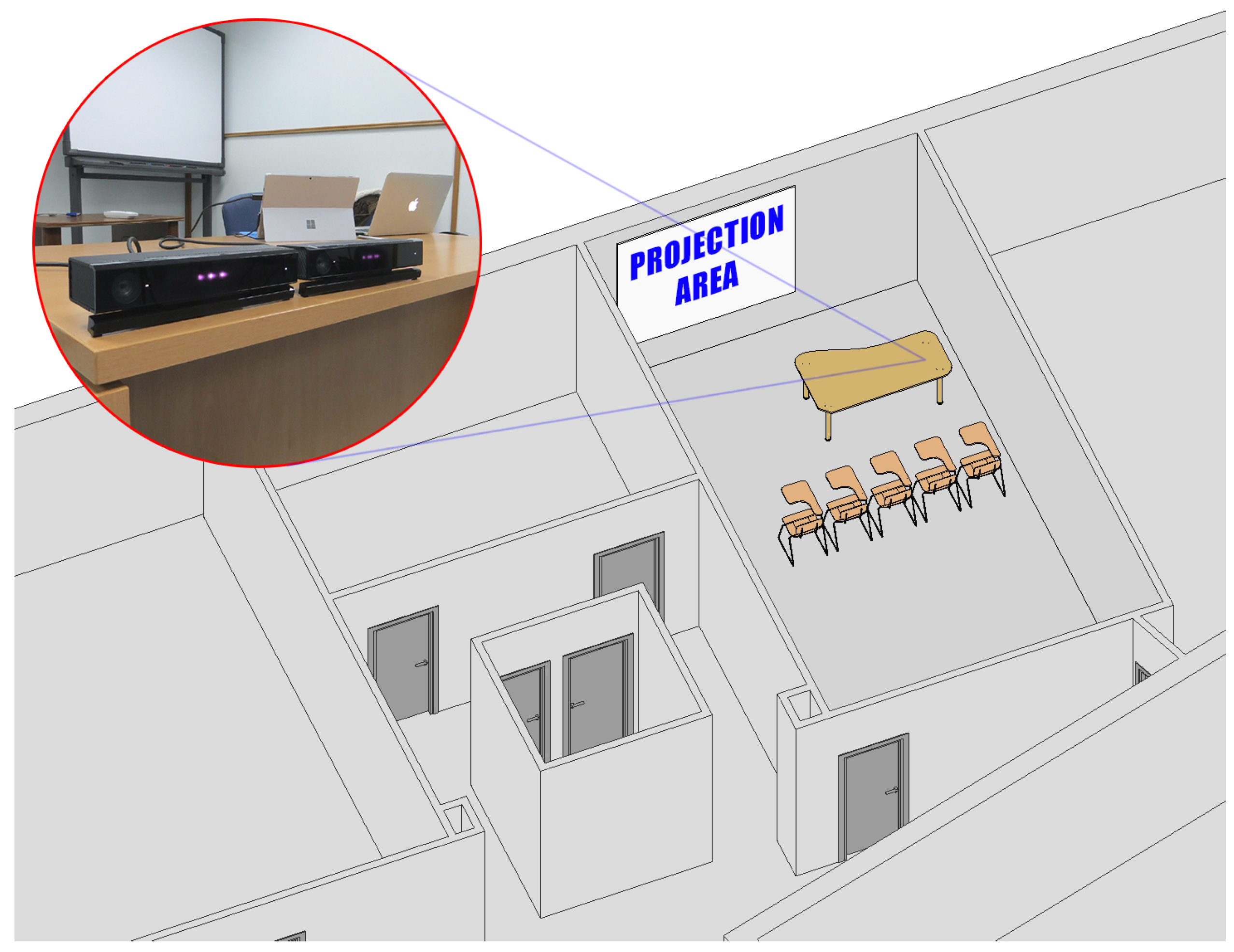
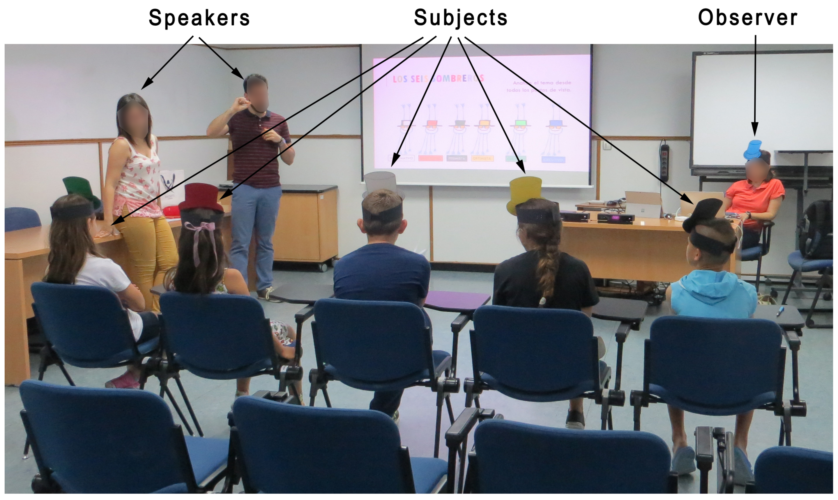
2.2.3. Classroom Layout

Two Kinect devices were used in each workshop in order to ensure the capture of movement of the subjects and prevent any type of technological incident Figure 1.
The classroom was arranged in such a way that the speakers never stood in front of the Kinect devices (Figure 2). Thus, the speakers would cross behind the chairs when it was necessary to hand out any type of material to the assistants or to answer any question.
An observer attended each workshop together with the subjects and the speakers. Her role was to take notes on the subjects’ behaviour so that, in addition to having the objective movements of the subjects, the appreciations of an independent observer would be registered. Two of the behaviours registered by the observer, along the lines of the diagnostic criteria of the DSM-V and the World Health Organization [22], were the times that the subjects left seat and squirmed in seat. These behaviours were registered in a template that had previously been arranged.

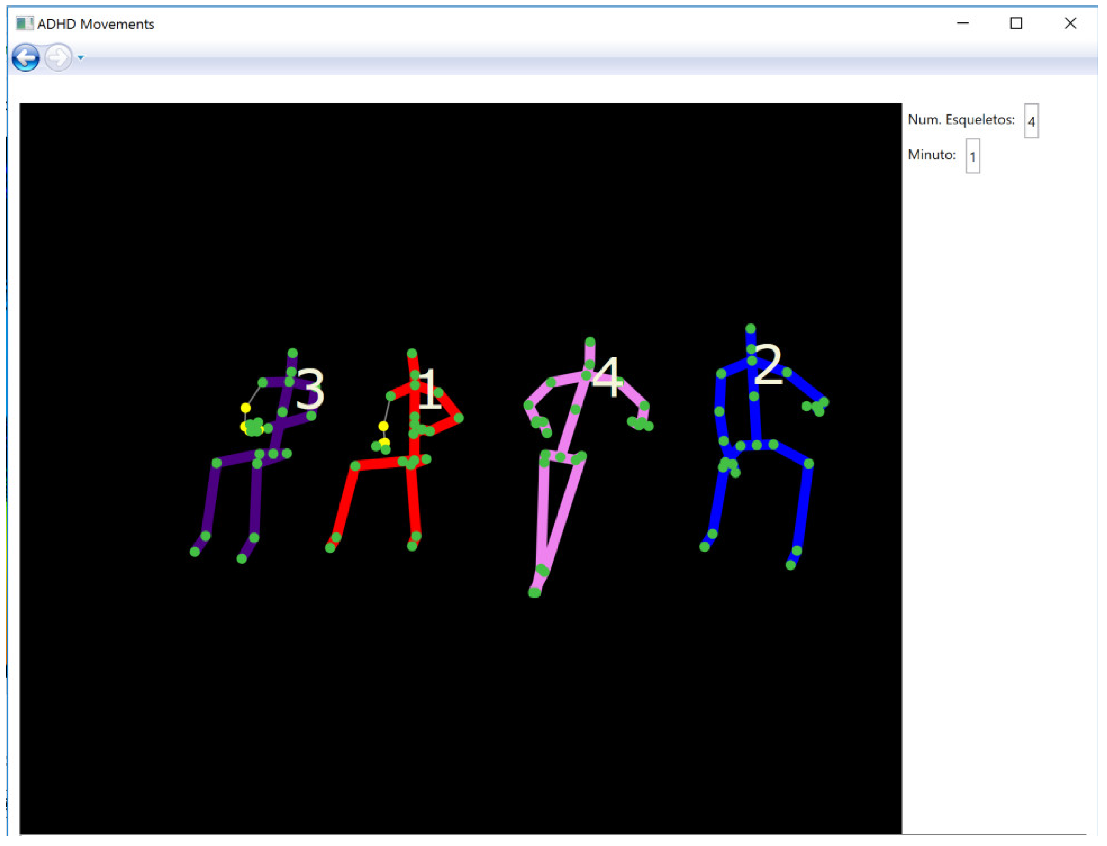
2.2.4. Computer Application ADHD Movements

The computer application ADHD Movements was developed for the analysis of the movement of several subjects using Kinect. The version of Kinect for Windows allows developers to make their own programs using gestures and body movements in a wide variety of applications, for this reason it is considered an innovative interaction interface between people and computers, in addition to its simplicity in the acquisition of data [20]. This application was developed with Microsoft Visual Studio and using the Kinect for Windows Software Development Kit (SDK) 2.0 that enables the creation of applications that support gesture recognition. For this, one of the main characteristics of this SDK is the detection of the human body that it is represented as a skeleton consisting of segments (such as forearm, hand or foot), which are connected to each other by joints (such as elbow, wrist or ankle). ADHD Movements detects the number of visible skeletons and, to allow the observer to know if the detection is correct, it shows each skeleton and each joint on the screen Figure 3.
The application numbers each skeleton (from 1 to 6) in order of detection and saves its location in the environment. In this way, if for any reason, a skeleton stops being detected (for example, a subject stands up and leaves the detection zone), when it returns to its location, the application assigns it the same number.
Data obtained by the Kinect sensor includes information of depth measures and, therefore, of the coordinates (x, y, z) of the objects present in the scene. In the case of a human body, it obtains the coordinates of each of the 25 joints that it detects for each skeleton. Each joint is identified by its name and position and it is measured with three coordinates (x, y, z) where (x, y) define the position and (z) represents the distance to the sensor, according to the coordinate space shown in Figure 4.
Using this information, the developed application estimates the distance covered by each joint of the skeleton in a three-dimensional space during each minute of the session for each of the subjects. Thus, for each joint, the distance between the different 3D points of the trajectory of the joint in the movement was calculated for each minute of the session.
As an example, Figure 5 shows the trajectory of the head joint for one minute of a subject with a firm diagnosis of ADHD (centre), the same subject without medication (right) and a subject in the control group (left).
The application obtains, for each subject, a table that shows for each joint the amount distance that it moves in each minute of the session. Sources of this computer program can be downloaded from the software repository of the research group located at https://web.ua.es/en/iamswarm/repository/sw-repository.html.

2.3. Analysis of Data

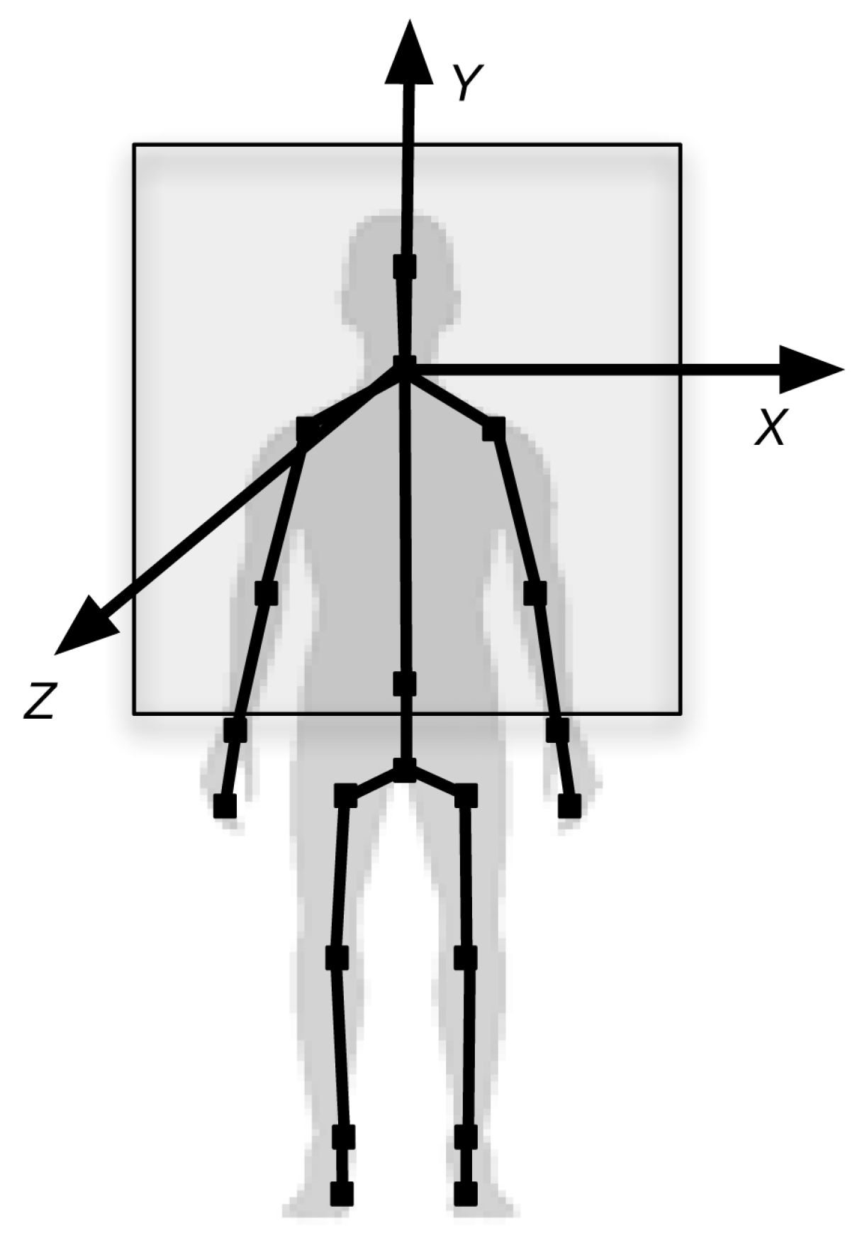
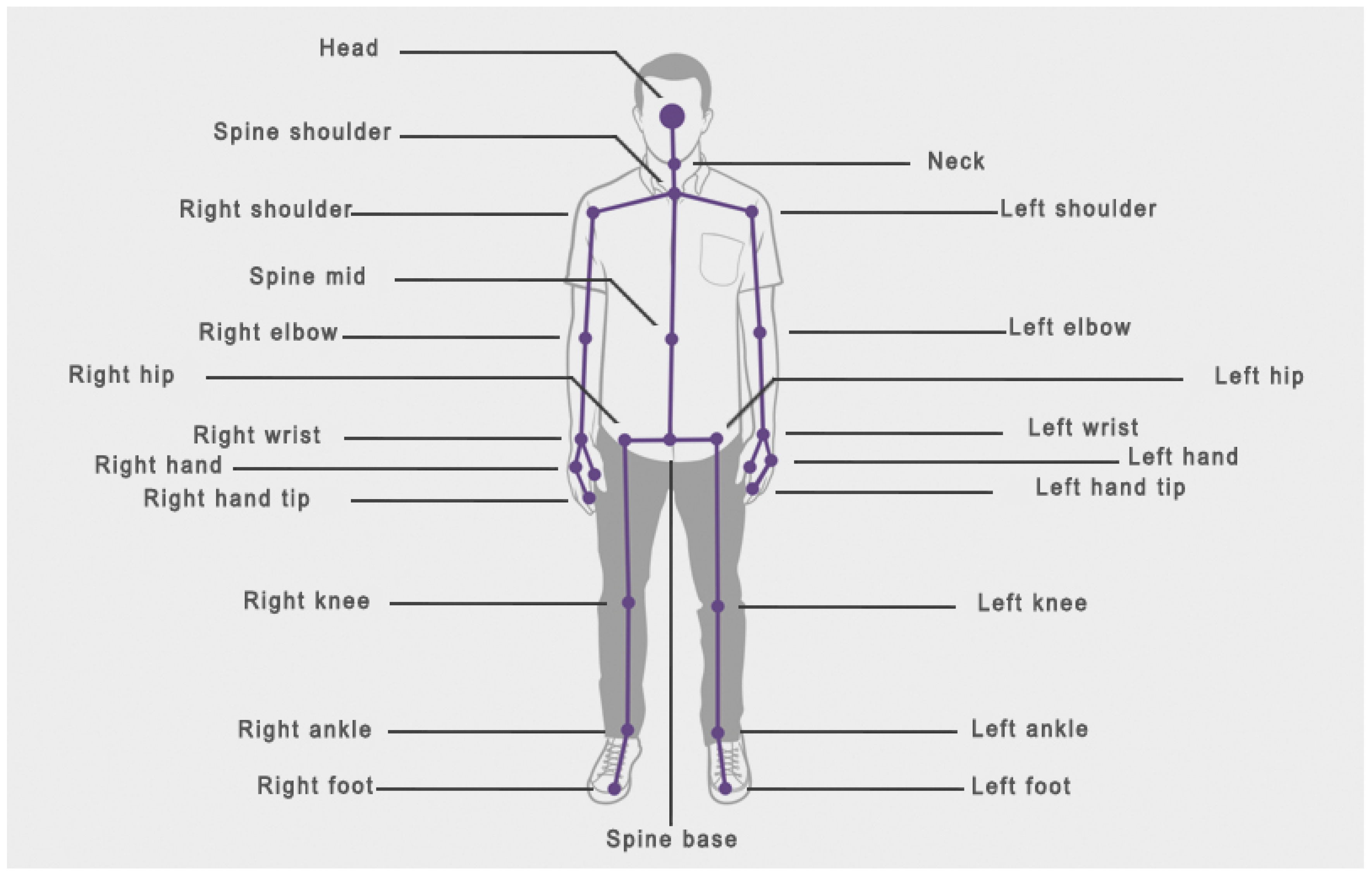
2.3.1. Data Registered by the Kinect Device

Two Kinect devices were used to record the subjects’ movement data. Each of these devices recorded the data of the complete skeletons of all participants in the session of 30 min. In order to register the information of the session for its later analysis, the application provided by Microsoft, Kinect Studio v2.0, was used. This application allows you to save all the information captured by Kinect, such as depth data, skeletons, sound, etc. for further analysis.
The computer application ADHD Movements was used for the analysis of the movement of all the subjects. The application estimates the distance covered by each joint of the skeleton during each minute of the session for each of the subjects.
As it has been previously mentioned, the Kinect device can detect up to 25 joints of 6 different bodies. Figure 6 shows the different joints detected by Kinect and their names.
The Kinect device detects 4 joints: wrist, hand, hand tip and thumb from each hand. Kinect uses these joints for the recognition of gestures in a specific situation: that a subject is standing in front of the Kinect, showing the hand to the device.
In the case of the study that has been carried out, with the subjects sitting in a natural position, that is, without clearly showing the hand to the device, it has been detected that the joint that is best recognized and registered of the four abovementioned, is the wrist. For this reason, in the results of this study, the hand, hand tip and thumb finger joints have been rejected.
Likewise, for the lower limbs, Kinect detects the ankle and foot joints. However, as shown by the research carried out by [23], this device detects skeletons in a standing position much better than in a sitting one. For this reason, after detecting that the recognition and registration of the ankle is much better than that of the foot, the foot has been rejected.
Thus, of the 25 joints registered by the Kinect device, only 17 have been analysed for the results of this study.

2.3.2. Data Registered by Observers

Once all the editions of the study techniques workshops were made, a second observer visualized the data recorded by the Kinect and the times that the subjects leave seat and squirm in seat were registered in a template previously prepared.
These data were compared with those previously collected by the observer who attended the different sessions of the workshops. For the study of the concordance of the data of the two observers, the Kappa index was used [24].

2.3.3. Statistical Analysis

In order to analyse the movement registered by observers between the experimental group and the control group, as well as to analyse the difference in movement registered by observers between the experimental group with and without taking their prescribed medication, the Student’s t-test was applied and d index proposed by Cohen [24] was included to measure the effect size of the differences found, since the t-test can erroneously detect statistically significant differences. The interpretation of this index is as follows: small effect size (0.20 ≤ d ≤ 0.50), moderate (0.51 ≤ d ≤ 0.80) and large effect size (d > 0.80).
In order to analyse the differences between averages of movement of the different joints recorded by Kinect within the experimental group with and without their prescribed medication, receiver operating characteristic curves (ROC curves) have been used in addition to the t-test.
The way of interpreting the area under the ROC curve is that a test with an area greater than 0.9 has higher accuracy, while between 0.7 and 0.9 it indicates a moderate accuracy, between 0.5 and 0.7, lower accuracy and 0.5 a chance result (Conzelmann et al. [21]).
Statistical packages SPSS 24 and MedCalc 12 were used for the statistical analysis.

3. Results

3.1. Average Differences in Movement for Every Joint in the Experimental Group and the Control Group

Results show that, except in the left ankle joint, the subjects with a firm diagnosis of ADHD obtained a higher average in the movement of the joints analysed, than those in the control group. The differences found between the two groups are significant for 14 of the 17 joints (Figure 7). The magnitude of the differences found was small in the joints: spine mid, neck, head, left shoulder, left elbow, left wrist, right shoulder, right elbow, right knee and spine shoulder, oscillating between 0.21 and 0.46 (Table 1). In the joints: spine base, right wrist, left hip, left knee and right hip, the differences found, although statistically significant, did not obtain the minimum levels required by [20] to be considered relevant.

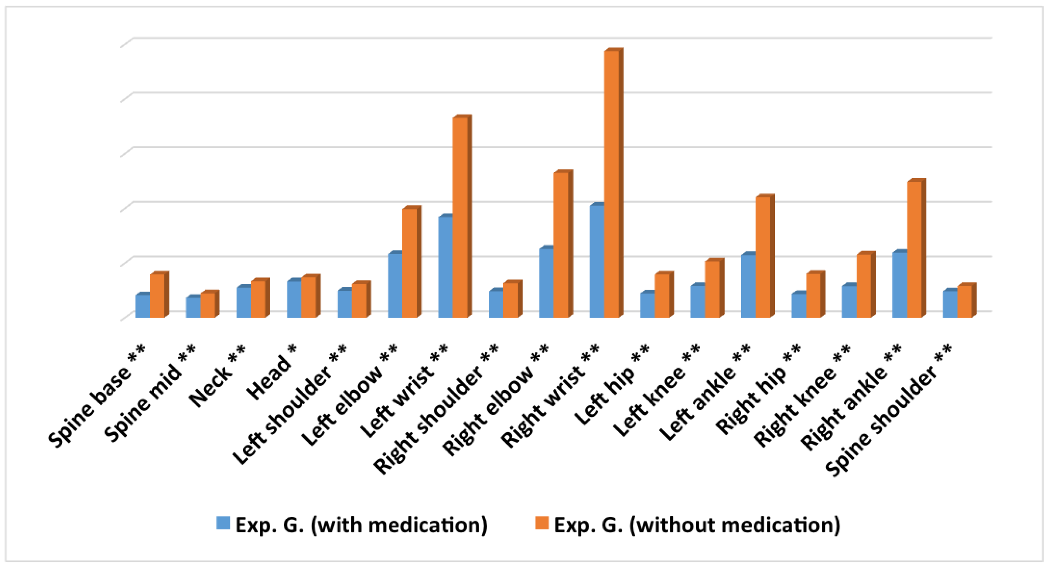
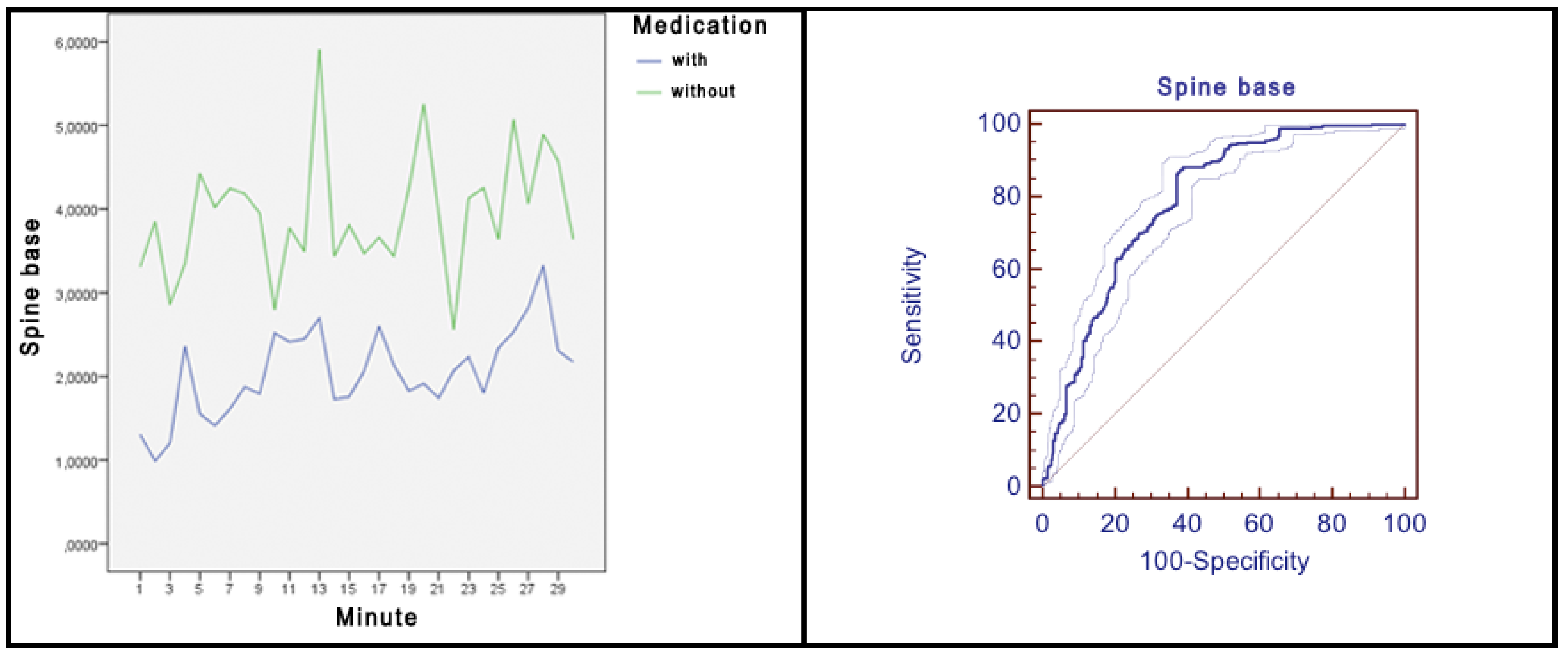
3.2. Average Differences in Movement for Every Joint within the Experimental Group with and without Medication Intake

The results revealed that there were statistically significant differences for the 17 joints analysed, being in all of them the movement average higher in the group without medication (Figure 8). The magnitude for the differences was small for the joints: spine mid, neck, left shoulder, right shoulder and spine shoulder, oscillating between 0.22 and 0.42 (Table 2). The magnitude for the differences was moderate for the joints: left elbow, left hip, left knee, left ankle, right knee and right ankle, oscillating between 0.56 and 0.79.
The magnitude of the differences was high for the joints: spine base, left wrist, right elbow, right wrist, and right hip, oscillating between 0.83 and 1.12.
In the head joint, although the difference found between both groups was statistically significant, it did not obtain the minimum levels required by Cohen to be considered relevant [25].
In the joints spine base (Figure 9), left elbow, left wrist, right elbow, right wrist, left hip, right hip and right ankle, as shown in respective graphs, the differences in movement were constant in time and maintained over time. In addition, in most of the joints, there were large peaks in which, obviously, the amount of movement of the subjects without medication intake was much higher.
All of the above implies an area under the ROC curve with values of 0.79 for the base spine, 0.73 for the left elbow, 0.81 for the left wrist, 0.77 for the right elbow, 0.83 for the right wrist, 0.77 for the left hip, 0.79 for the right hip and 0.72 for the right ankle. For the rest of the joints analysed, except for head, neck and spine shoulder joints, the differences in movement were remarkable in practically all the minutes, but they did not remain constant over time. For the joints head, neck and spine shoulder, although there were peaks in which the amount of movement of the subjects without medication was much higher, at different times the group of subjects with medication recorded more movements than the group without taking medication.

3.3. Average Differences in Movement for Every Joint in the Experimental Group, According to Sex

Results showed that girls obtained a higher average in movement in comparison with boys for 14 of the 17 joints analysed (Figure 10). Thus, the only joints in which boys obtained a higher average are spine base, right elbow and right wrist.
The differences found between the two groups were significant for 11 of the 17 joints (Table 3).

3.4. Movement Difference Registered by Observers

This section offers an analysis of the difference in movement registered by observers for the movements left seat and squirmed in their seat. To evaluate the agreement between observers, Cohen’s Kappa index was calculated [24], obtaining a result of κ = 0.93. The results showed that for the two movements registered in the experimental group and in the control group (Table 4), the highest average corresponded to the experimental group, although only in the squirm in seat movement the average difference was significant. In this case, the magnitude of the difference found was moderate: d = 0.69.
On the other hand, results revealed that for the two movements registered in the experimental group with and without the medication intake, the highest average corresponded to the group without prescribed medication (Table 5), although only in the movement squirm in seat the difference of averages was significant, with a high magnitude: d = 0.80.

4. Discussion

The identification of movement, posture, or a gesture made by a human body in real time is a difficult challenge because it has been found that the human body can perform a great deal of movement and it can have different size etc. [26]. Notwithstanding the above, after having carried out a series of tests with different technologies, the analysis of various technological devices, and the confirmation that Kinect has been previously used successfully in a wide range of research fields [27,28,29,30,31,32,33,34,35,36,37], it has been concluded that the Kinect V.2 is a suitable device to recognize and capture the movement of subjects in a teaching/learning situation, and thus achieve the objectives of this research.
Therefore, after having analysed existing technology, the computer application ADHD Movements was developed to detect the number of visible skeletons and, from each of them, analyse and draw on the screen each of the joints. From each joint that makes up all the skeletons, the application estimates the distance it covers in the 3D space during each minute. So, for each joint, the distance between the different 3D points of the trajectory of the joint in the movement has been calculated for each minute of the session.
Once the computer application was developed and verified, the movement of the subjects was captured.
Results show that there were significant differences in the amount of objective movement between a clinical group of subjects with ADHD and a control group, obtaining a higher average of movement in the experimental group in all the analysed joints (except in the left ankle joint). In addition, the differences found between the two groups are statistically significant in 14 of the 17 joints analysed.
In the left ankle joint, the control group obtained a higher movement average than the experimental group, although there was no statistically significant difference. This may be due to the dominance or preference for the use of the right or left leg of the subjects that have been part of the research groups.
From the revision of the literature it is concluded that this is the first study that counts the amount of movement in different joints with Kinect in order to find differences between a group of subjects with a firm diagnosis of ADHD and a control group. However, given the importance of hyperactivity in ADHD [7,38], objective measures of movement have been studied for more than a decade, although motion capture has almost always been done with accelerometers (actigraphy and inertial measurement units), with infrared systems placing some device in the body of the subjects or analysing only a part of the body [6,14,15,16,17,18,19].
In all the studies reviewed, children with ADHD showed more body movement than those without ADHD [6,14,39,40] so the results of the study coincide. As for the objective movement difference estimated in the group of subjects with ADHD with and without medication intake, the results show that there are statistically significant differences for the 17 joints analysed, being in all of them the average of movement higher in the group without taking medication. The results we have obtained are similar to those of previous studies that indicate that medicated ADHD children show significantly less motor activity than non-medicated ADHD children [41,42,43].
Along the lines of a recent study in which the effect of methylphenidate was studied in children with ADHD [44], and in which head movements were not found to be significantly different between the groups (medicated ADHD, non-medicated ADHD and control children), in the study, in the head joint, although the difference found between both groups is statistically significant, it does not obtain the minimum levels required by Cohen to be considered relevant [25].
The non-specificity of some diagnostic criteria defined by the DSM-V and the WHO for ADHD, such as squirm in their seat, means that although applications have been developed specifically to estimate the quantity of movement [45], the estimation of this depends on what the observer associates with the gesture. For this reason, both the movement to leave the seat, as well as the one previously mentioned, in the study, were registered by different observers.

Limitations, Strengths, and Future Directions

It is necessary to mention that this work is not exempt from limitations that must be considered when interpreting the results and their implications, as well as in the preparation of future studies in order to solve them and increase the understanding of the findings.
Firstly, the number of participants in the experimental group (32) may restrict the ability to detect significant differences between the groups. This limitation must be taken into account nevertheless minimized if we consider that other studies have presented a lower sample size [46,47]. In addition, the experimental group consisted of 24 boys and 8 girls, that is, 75% of the sample of participants with a firm diagnosis of ADHD were male. This data is in accordance with current epidemiological studies that indicate that the prevalence rate is greater in male [48]. However, it limits the extrapolation of the results analysed according to the sex of the participants. For this reason, in order to check whether the results are scalable, it would be interesting for future research to use large samples of subjects.
Besides, related to the sample size is the fact that experimental group consisted of 24 subjects with a diagnosis of ADHD with a combined presentation and eight with a predominantly inattentive presentation, according to the DSM-V criteria. In this study we have only analysed hyperactivity as a core feature of ADHD [7,8], together with the conditions of compliance with the diagnostic criteria in order to classify a predominantly inattentive presentation in ADHD that excludes those related to hyperactivity. Therefore, it would be interesting that in the future the objective movement tests carried out in this study could be carried out also in ADHD subjects with a predominant hyperactive/impulsive presentation.
Secondly, in this study the Kinect V.2 sensor was used to record the movement of subjects due to its simplicity and effectiveness [20]. In addition, different studies carried out with the Kinect device in several fields show that, although this is not as accurate as some traditional measurement technologies in research laboratories, it provides a good quality relationship for motion tracking systems with respect to the length of the body segments, joint angles and the displacement of the joints in the different body gestures [23]. All the above is not an obstacle to emphasize that the device has a series of limitations that must be considered. Thus, the Kinect device shows excellent results in wide movements such as sitting or standing up, but it shows poor precision in fine or small movements such as closing a hand [49]. This fact has led us to dismiss the results of the hand, hand tip, thumb and foot joints. On the other hand, the Kinect device detects and records the human body better in standing position and in the study the data has been captured in real education/learning situations, where the subjects are sitting. For this reason, it is possible that in certain positions of the subjects (for example, crossing the legs) occlusions have occurred. In these cases, the skeleton detection software of the device interpolates the positions of the undetected joints, being able to produce slight deviations between the real position of a joint and the interpolated position.

5. Conclusions

The results indicate that for the two registered movements, the highest average corresponds to the experimental group with respect to the control group, although only in the movement squirm in seat the difference in averages is statistically significant.
In the same way, when comparing the movements registered by the observers between the experimental group with and without the medication, the highest average corresponds to the group without medication. Only in the movement squirm in seat the difference of averages is significant, with a high magnitude (d = 0.80).
The absence of significant differences in the movement leave their seat when comparing the experimental group with the control group, may be due to both the “novelty” of the teaching/learning situation (it was the first time they were in that classroom and with those speakers) or to the effect of the medication. However, the results of the comparison of the group with and without medication cannot be due to this reason, since the assistants already knew the speakers and the classroom of the study techniques workshop.
In any case, in the absence of previous studies in which a register of this type of movements associated with the diagnostic criteria of DSM-V and WHO for ADHD in a teaching/learning situation, we cannot compare results.
The above results lead to a series of conclusions:
  • The software developed (ADHD Movements) for the Microsoft Kinect V.2 device is valid to capture the movement of 17 joints of up to 6 subjects in a teaching/learning situation.
  • Students with ADHD present more movement and squirm more in their seat, than students without ADHD.
  • Students with a firm diagnosis of ADHD without the prescribed medication present more movement and squirm more in their seat than ADHD students with the prescribed medication.
  • ADHD students with and without taking their prescribed medication present a similar amount of movement in the head joint.
  • Girls with ADHD present more movement than boys with ADHD.

Author Contributions

The work presented here was developed in collaboration amongst all authors. All authors have contributed to, seen and approved the manuscript. All authors have read and agreed to the published version of the manuscript.

Funding

This work has been supported by the Ministerio de Ciencia, Innovación y Universidades (Spain), project RTI2018-096219-B-I00. Project cofinanced with FEDER funds.

Acknowledgments

The authors of this paper thank the University of Alicante for the grant that allowed part of this research.

Conflicts of Interest

The authors declare no conflict of interest.

References

  1. American Psychiatric Association. Diagnostic and Statistical Manual of Mental Disorders (DSM–5); American Psychiatric Association: Washington, DC, USA, 2013. [Google Scholar]
  2. Alós, C.; Ruiz, C. Las otras verdades del TDAH. Aula Infant. 2013, 70, 1–52. [Google Scholar]
  3. Barkley, R. Avances en el diagnóstico y la subclasificación del trastorno por déficit de atención/hiperactividad: Qué puede pasar en el futuro respecto al DSM-V. Rev. Neurol. 2009, 48, 101–106. [Google Scholar] [CrossRef]
  4. Alberola, P. Los criterios de diagnóstico son vagos, ¿cómo se determina que un niño es más o menos movido? D. Inf. 2015, 5, 82–98. [Google Scholar]
  5. Artigas-Pallarés, J. Comorbilidad en el trastorno por déficit de atención/hiperactividad. Rev. Neurol. 2003, 36, 68–78. [Google Scholar] [CrossRef]
  6. Gawrilow, C.; Kühnhausen, J.; Schmid, J.; Stadler, G. Hyperactivity and Motoric Activity in ADHD: Characterization, Assessment, and Intervention. Front. Psychiatry 2014, 5, 171. [Google Scholar] [CrossRef] [PubMed] [Green Version]
  7. Ohasi, K.; Vitaliano, G.; Polcari, A.; Teicher, M. Unraveling the nature of hyperactivity in children with attention-deficit/hyperactivity disorder. Arch. Gen. Psychiatry 2010, 67, 388–396. [Google Scholar] [CrossRef] [PubMed] [Green Version]
  8. Teicher, M.; Polcari, A.; McGreenery, C. Utility of Objective Measures of Activity and Attention in the Assessment of Therapeutic Response to Stimulants in Children with Attention-Deficit/Hyperactivity Disorder. J. Child Adolesc. Psychopharmacol. 2008, 18, 265–270. [Google Scholar] [CrossRef] [PubMed] [Green Version]
  9. Conrad, P. The discovery of hiperkinesis: Notes on the medicalization of deviant behavior. Soc. Probl. 1975, 23, 12–21. [Google Scholar] [CrossRef] [PubMed]
  10. Fominaya, C. El TDAH no existe, y la medicación no es un tratamiento, sino un dopaje. ABC Padres e hijos, 20 October 2014. [Google Scholar]
  11. Schrag, P.; Divoky, D. The Myht of the Hiperactive Child and Othher Means of Child Control; Pantheon Books: New York, NY, USA, 1975. [Google Scholar]
  12. Timimi, S.; Moncrieff, J.; Jureidini, J.; Leo, J.; Cohen, D.; Whitfield, C.; Double, D.; Bindman, J.; Asen, E.; Bracken, P.; et al. A critique of the international consensus statement on ADHD. Clin. Child Fam. Psychol. Rev. 2004, 7, 59–63. [Google Scholar]
  13. De Vinuesa, F.G.; González, H.; Pérez, M. Volviendo a la Normalidad: La Invención del TDAH y del Trastorno Bipolar Infantil; Alianza Editorial: Madrid, Spain, 2014. [Google Scholar]
  14. García, L.; Cortese, S.; Anderson, D.; Martino, A.D.; Castellanos, F. Locomotor activity measures in the diagnosis of attention deficit hyperactivity disorder: Meta-analyses and new findings. J. Neurosci. Methods 2015, 252, 14–26. [Google Scholar] [CrossRef] [PubMed] [Green Version]
  15. Hall, C.; Valentine, A.; Groom, M.; Walker, G.; Sayal, K.; Hollis, C. The clinical utility of the continuous performance test and objective measures of activity for diagnosing and monitoring ADHD in children: A systematic review. Eur. Child Adolesc. Psychiatry 2016, 25, 677–699. [Google Scholar] [CrossRef] [PubMed] [Green Version]
  16. Jaiswal, S.; Valstar, M.; Gillott, A.; Daley, D. Automatic detecion of ADHD and ASD from Expressive Behaviour in RGBD Data. arXiv 2016, arXiv:1612.02374. [Google Scholar]
  17. Li, F.; Zheng, Y.; Smith, S.; Shic, F.; Moore, C.; Zheng, X.; Qi, Y.; Liu, Z.; Leckman, J. A preliminary study of movement intensity during a Go/No-Go task and its association with ADHD outcomes and symptom severity. Child Adolesc. Psychiatry Ment. Health 2016, 10, 47. [Google Scholar] [CrossRef] [Green Version]
  18. O’mahony, N.; Florentino-Liano, B.; Carballo, J.; Baca-García, E.; Artés, A. Objective diagnosis of ADHD using IMUs. Med. Eng. Phys. 2014, 36, 922–926. [Google Scholar] [CrossRef]
  19. Wehmeier, P.; Schacht, A.; Wolff, C.; Otto, W.; Dittmann, L.; Banaschewski, T. Neuropsychological outcomes across the day in children with attention-deficit/hyperactivity disorder treated with atomoxetine: Results from a placebo-controlled study using a computer-based continuous performance test combined with an infra-red motion-tracking device. J. Child Adolesc. Psychopharmacol. 2011, 21, 433–444. [Google Scholar]
  20. Ding, I.; Chang, C. An eigenspace-based method with a user adaptation scheme for human gesture recognition by using Kinect 3D data. Appl. Math. Model. 2015, 39, 5769–5777. [Google Scholar] [CrossRef]
  21. Conzelmann, A.; Pusch, A.; Hahn, N.; Overfeld, J.; Nehfischer, M.; Wieser, M.; Warnke, A.; Renner, T.; Taurines, R.; Romanos, M.; et al. Movement and activity assessed with the Kinect in sitting children, and the influence of hyperactivity. J. Cyberther. Rehabil. 2012, 17, 1–2. [Google Scholar]
  22. Xu, X.; McGorry, R. The validity of the first and second generation Microsoft KinectTM for identifying joint center locations during static postures. Appl. Ergon. 2015, 49, 47–54. [Google Scholar] [CrossRef]
  23. Cohen, J. A coefficient of Agreement for Nominal Scales. Educ. Psychol. Meas. 1960, 20, 37–46. [Google Scholar] [CrossRef]
  24. Cohen, J. Statistical Power Analysis for the Behavioral Sciences; Academic Press: New York, NY, USA, 1988. [Google Scholar]
  25. Akobeng, A. Understanding diagnostic tests 3: Receiver operating characteristic curves. Acta Paediatr. 2007, 96, 644–647. [Google Scholar] [CrossRef]
  26. Alabbassi, H.; Gradinaru, A.; Moldoveanu, F.; Moldoveanu, A. Human Motion Tracking & Evaluation using Kinect V2 Sensor. In Proceedings of the 5th IEEE International Conference on E-Health and Bioengineering, Lasi, Romania, 19–21 November 2015; pp. 1–4. [Google Scholar]
  27. Jana, A. Kinect for Windows SDK: Programming Guide; Packt Publishing: Birmingham, UK, 2012. [Google Scholar]
  28. Boutsika, E. Kinect in Education: A Proposal for Children with Autism. Procedia Comput. Sci. 2014, 27, 123–129. [Google Scholar] [CrossRef] [Green Version]
  29. Clark, R.; Pua, Y.; Bryant, A.; Hunt, M. Validity of the Microsoft Kinect for providing lateral trunk lean feedback during gait retraining. Gait Posture 2013, 38, 1064–1066. [Google Scholar] [CrossRef] [PubMed]
  30. OMS. CIE-10. Décima Revisión de la Clasificación Internacional de las Enfermedades. Trastornos Mentales y del Comportamiento; Descripción Clínicas y Pautas para el Diagnóstico, Méditor ed.m Madrid; Organizacion Mondial de la Salud: Madrid, Spain, 1992. [Google Scholar]
  31. Dutta, T. Evaluation of the Kinect sensor for 3-D kinematic measurement in the workplace. Appl. Ergon. 2011, 43, 645–649. [Google Scholar] [CrossRef] [PubMed]
  32. Frati, V.; Prattichizzo, D. Using kinect for hand tracking and rendering in wearable haptics. In Proceedings of the IEEE World Haptics Conference, Istanbul, Turkey, 22–24 June 2011; pp. 317–321. [Google Scholar]
  33. Izadi, S.; Kim, D.; Hilliges, O.; Molyneaux, D.; Newcombe, R.; Kohli, P.; Shotton, J.; Hodges, S.; Freeman, D.; Davison, A.; et al. KinectFusion: Real-Time 3D Reconstruction and Interaction Using a Moving Depth; Association for Computing Machinery: New York, NY, USA, 2011; pp. 559–568. [Google Scholar]
  34. Noonan, P.; Cootes, T.; Hallett, W.; Hinz, R. The design and initial calibration of an optical tracking system using the microsoft kinect. In Proceedings of the IEEE Nuclear Science Symposium and Medical Imaging Conference, Valencia, Spain, 23–29 October 2011; pp. 3614–3617. [Google Scholar]
  35. Seer, S.; Brändle, N.; Ratti, C. Kinects and human kinetics: A new approach for studying pedestrian behavior. Trans. Res. Part C Emerg. Technol. 2014, 48, 212–228. [Google Scholar] [CrossRef]
  36. Diest, M.V.; Stegenga, J.; Wörtche, H.; Postema, K.; Verkerke, G.J.; Lamoth, C. Suitability of Kinect for measuring whole body movement patterns during exergaming. J. Biomech. 2014, 47, 2925–2932. [Google Scholar] [CrossRef] [PubMed]
  37. Weber, I.; Koch, J.; Meskemper, J.; Friedl, K.; Heinrich, K.; Hartmann, K. Is the MS Kinect suitable for motion analysis? Biomed. Eng. 2012, 57. [Google Scholar] [CrossRef] [Green Version]
  38. Teicher, M.; Ito, Y.; Glod, C.; Barber, N. Objective Measurement of Hyperactivity and Attentional Problems in ADHD. J. Am. Acad. Child Adolesc. Psychiatry 1996, 35, 334–342. [Google Scholar] [CrossRef]
  39. Parsons, T.; Bowerly, T.; Buckwalter, J.; Rizzo, A.A. A controlled clinical comparison of attention performance in children with ADHD in a virtual reality classroom compared to standard neuropsychological methods. Child Neuropsychol. J. Norm. Abnorm. Dev. Child. Adolesc. 2007, 13, 363–381. [Google Scholar] [CrossRef]
  40. Crescenzo, F.D.; Armando, M.; Mazzone, L.; Ciliberto, M.; Sciannamea, M.; Figueroa, C.; Janiri, L.; Quested, D.; Vicari, S. The use of actigraphy in the monitoring of methylphenidate versus placebo in ADHD: A meta-analysis. ADHD Atten. Deficit Hyperact. Disord. 2014, 6, 49–58. [Google Scholar] [CrossRef]
  41. Diaz-Orueta, U.; Iriarte, Y.; Climent, G.; Banterla, F. AULA: An ecological virtual reality test with distractors for evaluating attention in children and adolescents. Virtual Real. Sci. Vis. J. 2012, 5, 1–20. [Google Scholar]
  42. Hughes, C.; Pickering, J.; Baker, K.; Bolanos, G.; Silver, C. Differentiating ADHD from oral language difficulties in children: Role of movements and effects of stimulant medication. BMC Psychiatry 2014, 14, 370. [Google Scholar] [CrossRef] [PubMed] [Green Version]
  43. Mühlberger, A.; Jekel, K.; Probst, T.; Schecklmann, M.; Conzelmann, A.; Andreatta, M.; Rizzo, A.; Pauli, P.; Romanos, M. The Influence of Methylphenidate on Hyperactivity and Attention Deficits in Children with ADHD. J. Atten. Disord. 2016, 1, 277–289. [Google Scholar]
  44. Fernández, F.; González, C.; Sempere, M.; Navarro, I. Assessment of the techology to measure movements in the detection of students with attention deficit hyperactivity disorder. In Proceedings of the 9th International Technology, Education and Development Conference (INTED2015), Madrid, Spain, 2–3 March 2015. [Google Scholar]
  45. Chhabildas, N.; Pennington, B.; Willcutt, E. A comparison of the neuropsychological profiles of the DSM-IV subtypes of ADHD. J. Abnorm. Child Psychol. 2001, 29, 529–540. [Google Scholar] [CrossRef] [PubMed]
  46. Geurts, H.; Verté, S.; Oosterlaan, J.; Roeyers, H.; Sergeant, J. ADHD subtypes: Do they differ in their executive functioning profile? Arch. Clin. Neuropsychol. 2005, 20, 457–477. [Google Scholar] [CrossRef] [PubMed] [Green Version]
  47. Merikangas, K.; He, J.; Burstein, M.; Swendsen, J.; Avenevoli, S.; Case, B.; Georgiades, K.; Heaton, L.; Swanson, S.; Olfson, M. Service utilization for lifetime mental disorders in U.S. adolescents: Results of the National Comorbidity Survey-Adolescent Supplement (NCS-A). J. Am. Acad. Child Adolesc. Psychiatry 2011, 50, 32–45. [Google Scholar] [CrossRef] [Green Version]
  48. Galna, B.; Barry, G.; Jackson, D.; Mhiripiri, D.; Olivier, P.; Rochester, L. Accuracy of the Microsoft Kinect sensor for measuring movement in people with Parkinson’s disease. Gait Posture 2014, 39, 1062–1068. [Google Scholar] [CrossRef] [Green Version]
  49. Clark, R.; Pua, Y.; Fortin, K.; Ritchie, C.; Webster, K.; Denehy, L.; Bryant, A. Validity of the Microsoft Kinect for assessment of postural control. Gait Posture 2012, 36, 372–377. [Google Scholar] [CrossRef]
Figure 1. Arrangement of Kinect devices in the classroom. Space previously analyzed with BIM (Building Information Modelling) tools.
Ijerph 17 05620 g001
Figure 2. Classroom layout in the study techniques workshops.
Ijerph 17 05620 g002
Figure 3. Screenshot of the application ADHD movements.
Ijerph 17 05620 g003
Figure 4. Coordinate system with respect to the skeleton detected.
Ijerph 17 05620 g004
Figure 5. Trajectory of head joint for a control subject (left), an ADHD subject with medication (centre) and the same ADHD subject without medication (right).
Ijerph 17 05620 g005
Figure 6. Joint map detected by Kinect.
Ijerph 17 05620 g006
Figure 7. Average differences in movement for every joint in the experimental group and the control group. ** = p < 0.001; * = p < 0.05.
Ijerph 17 05620 g007
Figure 8. Average differences in movement for every joint within the experimental group with and without medication intake. ** = p < 0.001; * = p < 0.05.
Ijerph 17 05620 g008
Figure 9. Spine base: movements average/minute and ROC curve.
Ijerph 17 05620 g009
Figure 10. Average differences in movement for every joint in the experimental group, according to sex. ** = p < 0.001; * = p < 0.05.
Ijerph 17 05620 g010
Table 1. Average differences in movement for every joint in the experimental group and the control rgroup.
Kinect JointLevene TestExperimental
Group
Control
Group
Statistical Significance and
Magnitude Differences
FpMDEMDEtg.l.pd
Spine base45.42<0.0012.922.412.531.824.732512.24<0.0010.18
Spine mid74.18<0.0012.141.761.621.059.592665.48<0.0010.34
Neck64.04<0.0013.012.362.261.609.702612.67<0.0010.36
Head102.41<0.0013.482.522.441.6712.792628.94<0.0010.46
Left shoulder46.76<0.0012.762.182.111.668.622496.40<0.0010.32
Left elbow46.53<0.0017.726.066.544.795.502448.92<0.0010.21
Left wrist53.83<0.00113.4110.3112.847.871.622497.420.105-
Right shoulder111.21<0.0012.762.252.001.3310.972665.93<0.0010.39
Right elbow15.46<0.0019.507.857.927.005.382265.50<0.0010.21
Right wrist92.18<0.00116.7914.0815.3810.053.002571.630.0030.11
Left hip27.87<0.0013.022.352.721.933.572393.97<0.0010.14
Left knee4.350.0373.954.003.214.494.261890.72<0.0010.18
Left ankle10.130.0018.167.728.238.70−0.2051883.480.838-
Right hip25.15<0.0013.022.342.721.973.472356.940.0010.14
Right knee24.75<0.0014.234.793.143.476.762554.91<0.0010.25
Right ankle0.6350.4268.958.548.569.541.0826660.279-
Spine shoulder73.26<0.0012.652.211.971.409.612650.59<0.0010.35
Table 2. Average differences in movement for every joint within the experimental group with and without medication intake.
Kinect JointLevene TestExperimental Group
(with Medication)
Control Group
(with Medication)
Statistical Significance and
Magnitude Differences
FpMDEMDEtg.l.pd
Spine base32.30<0.0012.051.833.942.6016.921370.59<0.0010.83
Spine mid0.0010.9741.801.572.531.898.611676<0.0010.42
Neck<0.0010.9962.752.183.322.514.931676<0.0010.24
Head4.630.0313.312.453.682.593.021609.820.0020.15
Left shoulder0.3810.5372.482.043.082.295.691676<0.0010.28
Left elbow26.33<0.0015.815.239.946.2014.611526.61<0.0010.72
Left wrist15.68<0.0019.218.6018.289.9919.751543.72<0.0010.97
Right shoulder0.4900.4842.432.043.152.416.601676<0.0010.32
Right elbow186.14<0.0016.284.8713.238.9119.391163.32<0.0010.95
Right wrist147.05<0.00110.249.4024.3714.7722.941281.24<0.0011.12
Left hip13.56<0.0012.221.963.942.4315.701492.09<0.0010.77
Left knee102.38<0.0012.912.915.154.7111.501256.51<0.0010.56
Left ankle276.86<0.0015.714.6111.019.4414.251090.70<0.0010.70
Right hip11.560.0012.171.944.002.3916.951494.15<0.0010.83
Right knee212.30<0.0012.902.805.766.0012.131064.31<0.0010.60
Right ankle266.49<0.0015.935.3712.4310.0716.121146.82<0.0010.79
Spine shoulder0.0740.7862.421.992.912.414.491676<0.0010.22
Table 3. Average differences in movement for every joint in the experimental group, according to sex.
Kinect JointBoysGirlsStatistical Significance and
Magnitude Differences
MDEMDEFpd
Spine base2.952.452.862.322.150.643-
Spine mid2.031.672.411.9462.34<0.0010.22
Neck2.772.153.622.72152.60<0.0010.37
Head3.382.423.742.73127.52<0.0010.14
Left shoulder2.562.023.262.471.140.286-
Left elbow7.446.038.436.0627.58<0.0010.16
Left wrist13.1310.6614.119.3613.38<0.0010.10
Right shoulder2.572.083.242.57129.93<0.0010.30
Right elbow9.898.408.536.152.260.132-
Right wrist17.5915.2914.7910.193.450.063-
Left hip3.002.313.062.464.370.081-
Left knee3.613.674.804.6441.07<0.0010.30
Left ankle7.457.699.957.5226.14<0.0010.33
Right hip2.992.283.092.494.290.075-
Right knee3.864.545.145.255.500.0190.30
Right ankle8.127.9811.029.5014.16<0.0010.34
Spine shoulder2.432.053.182.48143.90<0.0010.34
Table 4. Average differences in movement registered by observers in experimental group and control group.
MovementLevene TestExperimental
Group
Control
Group
Statistical Significance and
Magnitude Differences
FpMDEMDEtg.l.pd
Squirm0.820.3678.504.715.274.652.77630.0070.69
Leave sit0.250.6141.353.591.123.380.268620.790-
Table 5. Average differences in movement for every joint within the experimental group with and without intake of prescribed medication.
MovementLevene TestExperimental Group
(without Medication)
Control Group
(without Medication)
Statistical Significance and
Magnitude Differences
FpMDEMDEtg.l.pd
Squirm0.790.3248.924.8816.5212.543.34240.0030.80
Leave sit0.240.6081.643.955.2412.97−1.62240.118-

