Article

Cardiorespiratory Fitness Mediates Cognitive Performance in Chronic Heart Failure Patients and Heart Transplant Recipients

1
Preventive Medicine and Physical Activity Centre and Research Center, Montreal Heart Institute, Montreal, QC H1T 1N6, Canada
2
Department of Medicine, Université de Montréal, Montreal, QC H3T 1J4, Canada
3
Research Centre, Institut Universitaire de Gériatrie de Montréal, Montreal, QC H3W 1W5, Canada
4
Research Centre, University of Montreal Hospital Research Centre, Montreal, QC H2X 0A9, Canada
5
Montreal Health Innovations Coordinating Center, Montreal Heart Institute, Montreal, QC H1T 1C8, Canada
6
Sport Medicine Unit, Division of Physical Medicine and Rehabilitation, Swiss Olympic Medical Center, Lausanne University Hospital, 1011 Lausanne, Switzerland
*
Author to whom correspondence should be addressed.
Int. J. Environ. Res. Public Health 2020, 17(22), 8591; https://0-doi-org.brum.beds.ac.uk/10.3390/ijerph17228591
Submission received: 2 October 2020 / Revised: 11 November 2020 / Accepted: 13 November 2020 / Published: 19 November 2020
(This article belongs to the Section Health Behavior, Chronic Disease and Health Promotion)

Abstract

:
We compared cognitive profiles in chronic heart failure patients (HF), heart transplant recipients (HT) and healthy controls (HC) and examined the relationship between cardiorespiratory fitness ( V ˙ O2peak), peak cardiac output (COpeak) and cognitive performance. Stable HT patients (n = 11), HF patients (n = 11) and HC (n = 13) (61.5 ± 8.5 years) were recruited. Four cognitive composite scores targeting different cognitive functions were computed from neuropsychological tests: working memory, processing speed, executive functions and verbal memory. Processing speed and executive function scores were higher, which indicates lower performances in HF and HT compared to HC (p < 0.05). V ˙ O2peak and first ventilatory threshold (VT1) were lower in HF and HT vs. HC (p < 0.01). COpeak was lower in HF vs. HT and HC (p < 0.01). Processing speed, executive function and verbal memory performances were correlated with V ˙ O2peak, VT1 and peak cardiac hemodynamics (p < 0.05). Mediation analyses showed that V ˙ O2peak and VT1 mediated the relationship between group and processing speed and executive function performances in HF and HT. COpeak fully mediated executive function and processing speed performances in HF only. V ˙ O2peak and COpeak were related to cognitive performance in the entire sample. In addition, V ˙ O2peak and VT1 fully mediated the relationship between group and executive function and processing speed performances.

Graphical Abstract

1. Introduction

Cognitive impairment (CI) affects up to 50% of patients with heart failure (HF) [1]. It is independently associated with mortality [2], poor quality of life, reduced functional capacity [3] and an overwhelming economic burden in Western countries [4,5]. HF-related CI is correlated with the severity of the disease [6] and mainly affects episodic memory, executive functions and processing speed [7]. Previous studies have shown that interventions durably treating HF, such as heart transplantation (HT), can improve cognition, for instance, memory, mental flexibility and attention [8,9,10]. Nevertheless, around 45% of HT recipients remain cognitively impaired [11,12,13], which is not trivial given that CI negatively affects therapeutic observance [14]. According to the “vascular dysfunction hypothesis” [7,15], one of the first mechanisms leading to CI is inadequate cerebral oxygenation and perfusion at rest, secondary to impaired cardiac output in HF and HT patients [9,16,17]. In line with this hypothesis, we previously showed that HT recipients had impaired cardiorespiratory fitness ( V ˙ O2peak), V ˙ O2 at first ventilatory threshold (VT1), peak cardiac output (COpeak) and reduced cerebral oxygenation/perfusion during exercise compared to healthy controls [18]. In cardiac populations, V ˙ O2 at VT1 corresponds to a moderate intensity, with aerobic metabolism as the main energy source. Importantly, VT1 is related to quality of life and daily physical activity without fatigue and/or dyspnea in cardiac patients. Moreover, in coronary heart disease (CHD) patients, we also showed that cognitive performance was related to V ˙ O2peak, COpeak and reduced cerebral oxygenation/perfusion [19]. In HT and HF patients, daily physical activity and/or functional capacity are related to cognitive performance [20,21,22,23]. Therefore, V ˙ O2peak, VT1 and COpeak, reflecting cardiorespiratory fitness and functional capacity, could be important contributors/mediators of cognitive performance in these cardiac patients (HF–HT). To date, the relationship between cardiorespiratory fitness, COpeak and cognitive performance in HF and HT has been poorly studied. Furthermore, it remains unclear whether these variables mediate cognitive performance in HF patients and HT recipients. In healthy older adults, our group showed that improvement in cardiorespiratory fitness mediates improvement in cognitive performance, with partial mediation observed in younger-old adults and full mediation in older-old adults aged over 75 years [24]. This suggests that cardiorespiratory fitness plays a crucial role in maintaining and improving cognitive functions in healthy older adults. This remains understudied in cardiac populations. The aims of this study are: (1) to compare cognitive profiles in HF patients, HT recipients and healthy controls (HC), and (2) to examine the relationship between cardiorespiratory fitness, peak cardiac output and their contribution (mediation) to cognitive performance in HF and HT patients compared to HC.

2. Materials and Methods
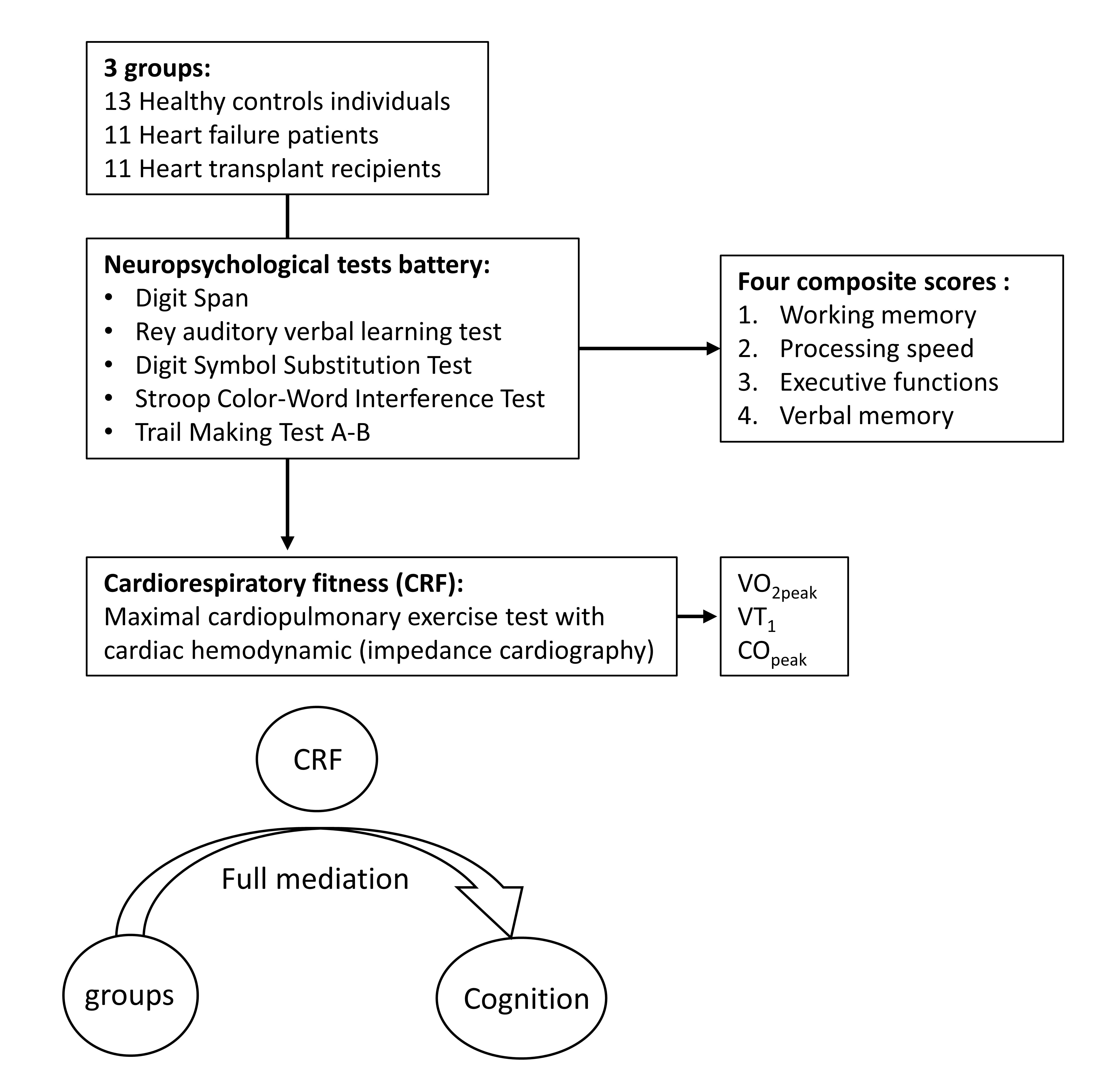
2.1. Participants

A total of 35 adults were recruited from the Cardiovascular Prevention and Rehabilitation Centre of the Montreal Heart Institute, including 11 chronic HF patients, 11 HT recipients and 13 HC. This study is part of a project conducted at the Montreal Heart Institute and approved by its research ethics board (ClinicalTrials.gov Identifier: NCT03018561). All participants provided written informed consent prior to inclusion [18]. Only subjects who completed the neurocognitive evaluation were included in the sample. Detailed inclusion and exclusion criteria for HC, HT recipients and HF patients are shown in Supplementary Materials (Table S1).

2.2. Study Design

All participants underwent a baseline evaluation that included a medical history and physical examination with measurements of height and body mass. Main components of cognition were assessed (working memory, executive functions, processing speed and verbal memory) using a comprehensive neuropsychological test battery. Participants performed a maximal cardiopulmonary exercise test with cardiac hemodynamics (impedance cardiography) and ECG measurements (see below for further details) [18,25].

2.3. Maximal Cardiopulmonary Exercise Test (CPET)

CPET was performed on an ergocycle (Ergoline 800S, Bitz, Germany), with an individualized protocol that included a 3-min warm up at 20 watts, followed by a power increase of 10 to 20 watts/min until exhaustion at a pedaling speed of >60 rpm [25]. Gas exchange [dioxygen ( V ˙ O2) and carbon dioxide ( V ˙ CO2)] was measured breath-by-breath continuously at rest, during exercise and at recovery using a metabolic gas analyzer system (Oxycon Pro, Jaegger, Germany) and then was averaged every 15 s for analysis. V ˙ O2 at first ventilatory threshold (VT1) was also calculated as previously published [18]. The highest V ˙ O2 value (15 s averaged) reached during the exercise phase was considered as the V ˙ O2peak, and peak power output (PPO) was defined as the workload reached at the last fully completed stage [18]. The electrocardiogram (ECG) was continuously monitored (Marquette, case 12, GE Healthcare) during the test. Blood pressure (manual sphygmomanometer: Welch Allyn Inc., Chicago, IL, USA) and rate of perceived exertion (RPE) were measured every 2 min throughout the test. During CPET, cardiac hemodynamics (cardiac output: CO, cardiac index: CI, and left cardiac work indexed: LCWi) were measured continuously at rest, during exercise and a recovery using an impedance cardiography device as previously described (PhysioFlow®, Enduro model, Manatec, France) [18,25].

2.4. Cognitive Evaluation

The following neuropsychological tests were administered in a fixed order. Digit Span (DS) is administered by the recall of forward and backward digit sequences. The forward span assesses auditory short-term memory, whereas the backward span targets working memory. The Rey auditory verbal learning test (RAVLT) is used to assess episodic memory in auditory and verbal domains and learning [26] through 5 learning trials of a 15-word list. In this test, participants must recall as many words as possible immediately after each trial of the learning phase, after an interfering list, as well as following a 30-min delay. The Digit Symbol Substitution Test (DSST) assesses processing speed. Participants have to associate symbols with numbers referring to a response key as fast as possible for 120 s. The Stroop Color-Word Interference Test (SCWIT) contains 4 different conditions (naming, reading, inhibition, switching). In the naming condition, participants name the color of rectangles. In the reading condition, participants read color words printed in black ink. In the inhibition condition, participants inhibit reading in order to name the incongruent ink colors in which the words are printed (e.g., RED printed in green ink). In the switching condition, participants are asked to alternate between inhibition and word reading. The first two conditions tap processing speed. The third condition assesses inhibition, and the fourth, flexibility and switching, both mechanisms of executive functioning [26]. The Trail Making Test (TMT) includes part A (TMT A), which measures processing speed and visuospatial abilities, and part B (TMT B), which assesses attentional control and cognitive flexibility. In part A, participants must link numbers 1–25 in ascending order as fast as possible. In part B, participants must alternate between letters and numbers, linking number–letter sequences in an ascending and alphabetic order, here again as fast as possible (e.g., 1-A-2-B-3-C, etc.). All cognitive scores were first transformed into standardized Z scores (Z score = (value – mean value of all the subjects)/standard deviation). Then, four composite cognitive scores were calculated using raw Z scores as follows [27]: (1) working memory = ((DS forward + DS backward scores)/2); (2) processing speed = ((DSST+ TMT A+ Stroop 1+ Stroop 2 scores)/4); (3) executive functioning = ((Trail B+ Stroop 3+ Stroop 4 scores)/3); and (4) verbal memory/episodic memory (immediate recall + delayed recall + total words scores recalled during the 5 learning trials from the RAVLT test/3). Cronbach’s alphas (α) were used to verify the internal consistency between all measures included in a composite score, considering a Cronbach α of > 0.7 as acceptable (see Results section) [27].

2.5. Statistical Analysis

Data were summarized by mean ± standard deviation (SD). CPET, cardiac hemodynamics at peak exercise and cognitive performances were compared between groups using one-way ANOVAs. In the case of a significant main group effect, pairwise comparisons were used to determine which group differences were significant. Correlations between cognitive composite scores, V ˙ O2peak, VT1 and cardiac hemodynamics at peak exercise were assessed with a Pearson coefficient (R). Mediation analyses were then conducted according to the methodology developed by Hayes et al. [28]. Mediation analyses were used to determine if V ˙ O2peak, VT1 and COpeak significantly influenced the relationship between group (HT, HF, HC) and cognitive performance. The direct effect of group on cognition and the indirect effect of group running through V ˙ O2peak, VT1 and peak cardiac output were analyzed. The direct path an represents the regression coefficient for the dummy-coded independent variable (IV) when the mediator variable (MV) is regressed on the independent variable (IV), while b is the direct path coefficient for the MV when the DV is regressed on the MV and IV. The product of the coefficient method was used to compute the indirect effect. This method determines the indirect effect by multiplying the regression coefficients: an × b = anb. The total effect (c) is the direct effect + the indirect effect. In other words, the relationship between the IV and the DV is decomposed into a direct link and an indirect link. A direct effect (c’) refers to the relationship between the IV and the DV after controlling for M. V ˙ O2peak was included as the MV between the categorical IV “group” (HC, HF, HT, where HF and HT will be compared to HC) and the DV composite cognitive scores. The same procedure was applied with VT1 and peak cardiac output. The significance of the indirect effects was tested using bias-corrected bootstrap confidence intervals (based on 5000 replications). Confidence intervals that did not contain zero represented significant effects. Partial mediation was denoted if the direct effect and the indirect effect were significant. Full mediation was denoted if a non-significant direct effect was associated with a significant indirect effect. All statistical tests were two-sided and conducted at a 0.05 significance level. Statistical analyses were performed with the use of SAS software, version 9.4 (SAS Institute) except for the mediation analyses that were conducted using Stata SE 15.1 (StataCorp LP, College Station, TX, USA) according to the methodology developed by Hayes et al. [28].

3. Results

3.1. Group Comparisons

Baseline clinical and sociodemographic characteristics were similar for the three groups (HC, HF, HT) and are presented in Table 1.
For CPET parameters, compared to HC, HF and HT had significant lower values for all of the parameters (Table 2). V ˙ O2peak in HF and HT was significantly reduced compared to HC (17.6 vs. 26.5 vs. 37.7 mL/min/kg, respectively, p < 0.001). At peak exercise, cardiac output (COpeak), cardiac index (CIpeak) and left cardiac work index (LCWIpeak) significantly differed according to group (Table 2), with lower values seen in HF vs. HT and HC but with no statistical difference between HT and HC.
Table 3 shows the neuropsychological tests and the composite cognitive scores for participants in the three groups. Cronbach’s alphas (α) were 0.640 for working memory, 0.787 for processing speed, 0.789 for executive functioning and 0.918 for verbal memory. The HF and HT groups demonstrated significantly higher composite z scores, suggesting reduced performances for processing speed and executive function when compared to HC participants (p < 0.05). Working memory performance was similar in the three groups (p = 0.425). The verbal memory composite score was greater in HC compared to HF (p < 0.001) but was similar compared to HT (p = 0.178) (Table 3).

3.2. Univariate Correlations

Univariate correlations are detailed in Supplementary Materials (Table S2). Executive functioning, processing speed and verbal memory were correlated with V ˙ O2peak, VT1, peak power output, oxygen pulse, HRpeak, COpeak, CIpeak and LWCipeak (p < 0.05) in the whole sample.

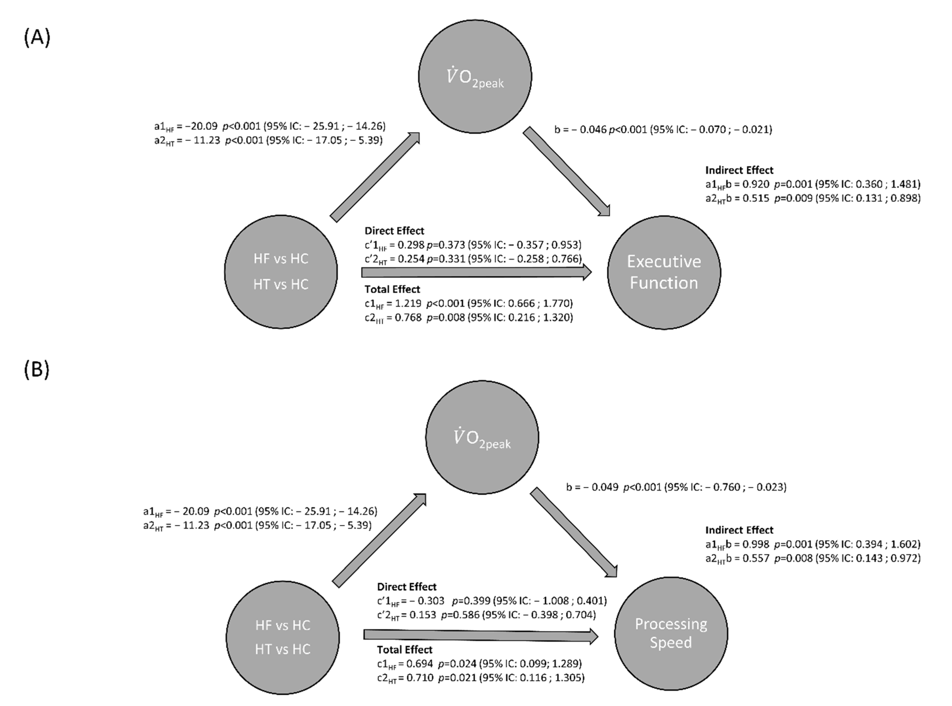
3.3. Mediation Analyses

Figure 1 presents the results of the mediation analyses. The direct effect of group predicts V ˙ O2peak (a1 path coefficient for HF = −20.1, p < 0.001, and a2 path coefficient for HT = −11.2, p < 0.001). Higher V ˙ O2peak was significantly associated with faster responses (reflected by a lower value of executive functioning Z score) (b = −0.046, p < 0.001). Each of the indirect effects of group on executive functioning through V ˙ O2peak was significant (a1b = 0.92, p = 0.001, and a2b = 0.51, p = 0.009) as well as the total indirect effect (a1b +a2b = 1.43; p = 0.002; 95% IC: 0.541; 2.329). In contrast to the control group (HC), HF and HT had executive functioning scores that were increased by 0.92 and 0.51 units, respectively (unfavorable because of slower responses). A lower V ˙ O2peak (from the sign of a1 and ab) increased the executive functioning Z score (from the sign of b), meaning that the time to complete the task increased.
The direct effect (c′ = c′1 + c′2) of group on executive functioning is 0.55 (p = 0.310) and the proportion of total effect that is mediated is 0.72 (anb/cn), meaning that 72% of the relationship between group and executive functioning performances is indirect via V ˙ O2peak. This is consistent with a full mediation of the relationship between group and executive functioning through V ˙ O2peak, where V ˙ O2peak is responsible for 72% of the effect of group difference on executive function performance. Mediation analyses showed that V ˙ O2peak and V ˙ O2 at first ventilatory threshold (VT1: Supplementary Materials Figure S1) fully mediate the relationship between group and executive function and processing speed performances, but not verbal memory performance. In addition, COpeak fully predicts executive function and processing speed performances in HF only (Supplementary Materials Figure S2).

4. Discussion

In this cross-sectional study, we first compared cognitive profiles of HF and HT patients to those of healthy controls. We also examined the relationship between cardiorespiratory fitness, peak cardiac output and their mediation on cognitive performance in HF and HT patients compared to HC. The main results of our study can be summarized as follows: (1) In addition to a reduced V ˙ O2peak, VT1 and COpeak, HF and HT have reduced cognitive performance (for processing speed and executive function composite scores) compared to HC. (2) Cardiorespiratory fitness and cardiac hemodynamics at peak exercise were associated with cognitive function (univariate correlation). (3) Cardiorespiratory fitness ( V ˙ O2peak, VT1) fully mediates the relationship between group and cognitive performance in HF and HT patients and peak cardiac output fully mediates cognitive performance in HF patients only.

4.1. Cognition in Chronic Heart Failure and in Heart Transplant Recipients

Cognitive impairment (CI) has been documented in chronic HF patients shortly (four months) after heart transplantation and in the long term in stable HT patients (one to 16 years) after their transplantation [13,16,29].
The cognitive domains affected in cardiac populations are mainly executive function, processing speed and memory [12,13,30,31,32,33]. Interestingly, our results showed lower scores for executive functions, verbal memory and processing speed composite scores in HF compared to HC. HT recipients obtained higher scores compared to HF in some cognitive functions, namely verbal memory, executive function and processing speed, but still performed lower compared to HC for the two later domains. Indeed, verbal memory performance was similar between HC and HT groups. Overall, our results suggest that multiple cognitive domains are not totally recovered even in stable HT recipients. Importantly, executive functions, which remained lower in HT recipients compared to HC, are related to medication adherence and instrumental activities of daily living, such as housework, preparing meals and engaging in physical activities. Consequently, deficits in executive functions can lead to difficulty in performing health self-management [20,21].

4.2. Related Mechanisms of Cognitive Impairment

HT recipients are more likely to present CI as a consequence of previous micro- and macrovascular disease [34], chronotropic incompetence [35], immunosuppressant therapy that could be linked to brain function [36] and even micro-cerebrovascular events (e.g., embolism) or lower hemoglobin concentration [37]. Moreover, most of the HT recipients were previously end-stage HF patients, who are at high risk of developing CI. Indeed, CI in HF is hypothesized to be due, at least in part, to a chronic reduction in cerebrovascular perfusion and oxygenation, and to oxidative stress and a pro-inflammatory status, which would then lead to structural changes in the brain [23,38,39,40]. Importantly, CI in HF patients has been related to the poorest NYHA (New York Heart Association) class, time of HF diagnostic, left ventricular ejection fraction (LVEF) under 30% and resting cardiac function [9,16], but there is less evidence exploring CI in long-term HT survivors [13].
In a pre- and post-heart transplant study with end-stage HF patients, cognitive scores (memory and dexterity/coordination and memory) were correlated with resting cardiac index (r = 0.32 and 0.52, respectively) [9]. The effect of HT on cognition remains unclear and needs more follow-up evaluations to elucidate the underlying mechanisms, as well as the effects of immunosuppressive treatments [29]. For instance, Grimm et al. observed that after HT, global cognitive performance was improved at four months, compared to prior HT, but then declined at 12 months. Cumulative cyclosporine dosage (immunosuppressive drug) was the only independent predictor of individual cognitive brain function after transplantation, meaning that this treatment could have a long-term negative impact on cognition [29].

4.3. Cardiorespiratory Fitness in Chronic Heart Failure and in Heart Transplant Recipients

Our results are in line with previous studies assessing cardiorespiratory fitness and CO at peak exercise with cardiac bioimpedance in HT and HF [18,41]. In chronic HF, exercise intolerance is multifactorial. The “systemic” pathophysiology involves central cardiac dysfunction but also peripheral abnormalities in the skeletal muscle, neuro-hormonal, endothelial and biochemical functions (oxidative stress and exacerbated pro-inflammatory status), aggravating myopathy and deconditioning [42]. In HT recipients, impaired cardiorespiratory fitness and cardiac hemodynamics at peak exercise are the result of both central (cardiac denervation, diastolic dysfunction) and peripheral abnormalities (vascular dysfunction, reduced skeletal muscle oxidative fibers, enzymes, capillarity) that limit O2 delivery and extraction by the exercising skeletal muscles [43]. Cardiorespiratory fitness in HT recipients is a strong predictor of long-term post-transplantation survival [44]. Accordingly, both for HF and HT, exercise training in order to improve V ˙ O2peak is part of cardiac rehabilitation guidelines [45].

4.4. Relationship between V ˙ O2peak, COpeak and Cognitive Performance

Resting cardiac hemodynamic function, such as cardiac output, is also related to cognitive functions in HF and HT patients [9,17,46]. Cardiorespiratory fitness is also well correlated with cognitive performance (measured at rest) in healthy adults [15]. A CPET test allows an integrated analysis of respiratory, cardiac, vascular and muscular adaptations (Fick equation: V ˙ O2 = CO × (A-V) O2). Our study demonstrated that cardiorespiratory fitness ( V ˙ O2peak) and V ˙ O2 at first ventilatory threshold correlated and fully mediated the relationship with processing speed and executive function performance, but not with verbal memory scores. It is well described in the literature that cardiorespiratory fitness is related to performances in global cognition, executive function, processing speed, attention and driving performance in HF patients [15,22,27,47], as well as with white matter structure in elderly adults [48]. This association is mediated, in part, by increases in brain perfusion and the ability of cerebral blood vessels to respond to demand [7]. Compared to HC, HT recipients had reduced V ˙ O2peak, cardiac function and cerebral oxygenation–perfusion during exercise [18]. However, cognition was not measured in this study. In CHD patients, V ˙ O2peak and CImax were both related to exercise-related cerebral oxygenation–perfusion and cognitive function (p < 0.005) [19]. In line with the “vascular dysfunction hypothesis” [49], CI may be due to a dysfunction of the mechanism involved in the regulation of cerebral blood flow and responsible for pathophysiological consequences of cerebral microvascular dysregulation (cerebral autoregulation/myogenic constriction, endothelium-dependent vasomotor function, neurovascular coupling responses). Abnormal cerebral autoregulation and cerebral vasoreactivity, potentiating the risk of impaired cerebral perfusion, therefore increasing the occurrence of silent brain damage and microvascular injury, are frequent in HF [50], but remain under discussion in HT patients [51,52]. These cerebrovascular phenomena can lead to the development of premature CI [49]. Regular physical activity improves cardiorespiratory fitness in both HF and HT and can have beneficial impacts on neurovascular functions and brain autoregulation in HF [53,54]. However, there are no data available regarding HT recipients. These benefits on cerebrovascular function may explain the positive impact of exercise on cognition, including benefits on learning, memory, attention and executive functions in healthy adults [55,56] and HF patients [38,57,58]. The positive beneficial effect of exercise training on cognitive function is likely due to its pro-neurogenic effects [59]. Some evidence also revealed that regular exercise may increase angiogenesis, neurogenesis, synaptogenesis and neurotransmitter synthesis in cerebral structures, as well as increasing grey and white matter volume [35,55]. The increased cerebral flow, especially after transplant [60], could stimulate neurobiological mechanisms leading to an improvement in cognitive function. Longitudinal studies exploring the effects of exercise training on cardiorespiratory fitness, cognition and cerebrovascular function in end-stage HF and HT are thus needed.

4.5. Study limitations and Research Perspectives

Our study has limitations that should be highlighted. First, the small number of participants in each group and the fact that they were recruited in a single center induces a potential recruitment bias. Larger cross-sectional and longitudinal studies before/after heart transplant with medium to long-term follow-ups are needed, as well as groups involved in exercise training programs. Furthermore, larger groups with different etiologies of HF leading to HT need to be taken into account in the analyses. Moreover, physical activity/sports and level of activities of daily living were not evaluated in a structured way in the study protocol, yet cognitive performances could be impacted by physical activity level, independently of cardiorespiratory fitness [21]. Furthermore, the potential positive effects of medication such as angiotensin II receptor blockers and angiotensin-converting enzyme inhibitors on cognitive performances in HF and HT patients cannot be excluded [61]. Moreover, because of the very low proportion of women in our sample, we were unable to discuss sex-related differences. Yet according to Lee et al. [62], older women with HF had higher CI (15%) and more inadequate health literacy (56.7%) compared to men. In their study, cognitive function was the strongest predictor of health literacy in men (β = 3.668, p < 0.001) and women (β = 2.926, p = 0.004). Future studies investigating sex-related differences in cardiac disease-related CI and the underlying pathophysiological mechanisms are needed. Finally, we did not differentiate in our analyses HT patients presenting evidence of cardiac reinnervation (according to heart rate responses to exercise testing and recovery). Partial reinnervation is found in some HT recipients and involves the myocardial muscle, the sinoatrial node and the coronary vessels, but remains incomplete and regionally limited many years post-transplant. Restoration of cardiac innervation can improve cardiorespiratory fitness as well as blood flow regulation in the coronary arteries, and may positively affect the heart–brain axis, the regulation of cerebral circulation and thus of cognitive functions. However, further studies are needed to investigate this hypothesis.

5. Conclusions

Our results demonstrate that cognitive performance is reduced in HF and HT patients compared to HC, for processing speed and executive function domains. For verbal memory, HT participants perform better than HF, and perform comparably to HC. Moreover, V ˙ O2peak, VT1 and cardiac output at peak exercise were related to cognitive performances (processing speed, executive function and verbal memory). The relationship between group (HF and HT vs. HC), executive functioning and processing speed was fully mediated by cardiorespiratory fitness ( V ˙ O2peak) and V ˙ O2 at VT1. Finally, peak cardiac output fully mediated processing speed and executive performances in HF only.

Supplementary Materials

The following are available online at https://0-www-mdpi-com.brum.beds.ac.uk/1660-4601/17/22/8591/s1, Figure S1: The mediating effect of V ˙ O2 at VT1 on the relationship between group and executive function (A) and processing speed (B), Figure S2: The mediating effect of peak cardiac output (COpeak) on the relationship between group and executive function (A) and processing speed (B), Table S1: Exclusion and inclusion criteria for HC, HT recipients and HF patients, Table S2: Univariate correlation coefficient between CPET, cardiac hemodynamic and composite Z scores for cognitive functions.

Author Contributions

M.G., A.N., M.J., P.A.B.R., M.W., V.G., L.B. (Louis Bherer), participated in the conception and in the design of the study. M.G., A.N., M.J., P.A.B.R., M.W., V.G., L.B. (Louis Bherer), contributed to data acquisition. F.B., B.B., C.G., M.O., P.A.B.R., M.G., A.N., M.J., L.B. (Louis Bherer), M.W., V.G., L.B. (Lucie Blondeau), participated in the data analysis. All authors participated in the data interpretation and the preparation of the manuscript. All authors have read and agreed to the published version of the manuscript.

Funding

Mirella and Lino Saputo Research Chair in Cardiovascular diseases and the prevention of cognitive decline from Université de Montréal at the Montreal Heart Institute. The ÉPIC Foundation and the Montreal Heart Institute Foundation.

Conflicts of Interest

The authors have no actual or potential conflicts of interest related to this manuscript.

References

  1. Cannon, J.A.; Moffitt, P.; Perez-Moreno, A.C.; Walters, M.R.; Broomfield, N.M.; McMurray, J.J.V.; Quinn, T.J. Cognitive Impairment and Heart Failure: Systematic Review and Meta-Analysis. J. Card. Fail. 2017, 23, 464–475. [Google Scholar] [CrossRef] [Green Version]
  2. Zuccala, G.; Pedone, C.; Cesari, M.; Onder, G.; Pahor, M.; Marzetti, E.; Lo Monaco, M.R.; Cocchi, A.; Carbonin, P.; Bernabei, R. The effects of cognitive impairment on mortality among hospitalized patients with heart failure. Am. J. Med. 2003, 115, 97–103. [Google Scholar] [CrossRef]
  3. Cameron, J.; Gallagher, R.; Pressler, S.J. Detecting and Managing Cognitive Impairment to Improve Engagement in Heart Failure Self-Care. Curr. Heart Fail. Rep. 2017, 14, 13–22. [Google Scholar] [CrossRef] [PubMed]
  4. Agarwal, K.S.; Kazim, R.; Xu, J.; Borson, S.; Taffet, G.E. Unrecognized Cognitive Impairment and Its Effect on Heart Failure Readmissions of Elderly Adults. J. Am. Geriatr. Soc. 2016, 64, 2296–2301. [Google Scholar] [CrossRef] [PubMed]
  5. Cook, C.; Cole, G.; Asaria, P.; Jabbour, R.; Francis, D.P. The annual global economic burden of heart failure. Int. J. Cardiol. 2014, 171, 368–376. [Google Scholar] [CrossRef]
  6. Trojano, L.; Antonelli Incalzi, R.; Acanfora, D.; Picone, C.; Mecocci, P.; Rengo, F.; Congestive Heart Failure Italian Study Investigators. Cognitive impairment: A key feature of congestive heart failure in the elderly. J. Neurol. 2003, 250, 1456–1463. [Google Scholar] [CrossRef]
  7. Abete, P.; Della-Morte, D.; Gargiulo, G.; Basile, C.; Langellotto, A.; Galizia, G.; Testa, G.; Canonico, V.; Bonaduce, D.; Cacciatore, F. Cognitive impairment and cardiovascular diseases in the elderly. A heart-brain continuum hypothesis. Ageing Res. Rev. 2014, 18, 41–52. [Google Scholar] [CrossRef]
  8. Deshields, T.L.; McDonough, E.M.; Mannen, R.K.; Miller, L.W. Psychological and cognitive status before and after heart transplantation. Gen. Hosp. Psychiatry 1996, 18, 62S–69S. [Google Scholar] [CrossRef]
  9. Bornstein, R.A.; Starling, R.C.; Myerowitz, P.D.; Haas, G.J. Neuropsychological function in patients with end-stage heart failure before and after cardiac transplantation. Acta Neurol. Scand. 1995, 91, 260–265. [Google Scholar] [CrossRef]
  10. Schall, R.R.; Petrucci, R.J.; Brozena, S.C.; Cavarocchi, N.C.; Jessup, M. Cognitive function in patients with symptomatic dilated cardiomyopathy before and after cardiac transplantation. J. Am. Coll Cardiol. 1989, 14, 1666–1672. [Google Scholar] [CrossRef] [Green Version]
  11. Roman, D.D.; Kubo, S.H.; Ormaza, S.; Francis, G.S.; Bank, A.J.; Shumway, S.J. Memory improvement following cardiac transplantation. J. Clin. Exp. Neuropsychol. 1997, 19, 692–697. [Google Scholar] [CrossRef] [PubMed]
  12. Cupples, S.A.; Stilley, C.S. Cognitive function in adult cardiothoracic transplant candidates and recipients. J. Cardiovasc. Nurs. 2005, 20, S74–S87. [Google Scholar] [CrossRef] [PubMed]
  13. Burker, B.S.; Gude, E.; Gullestad, L.; Grov, I.; Relbo Authen, A.; Andreassen, A.K.; Havik, O.E.; Dew, M.A.; Fiane, A.E.; Haraldsen, I.R.; et al. Cognitive function among long-term survivors of heart transplantation. Clin. Transpl. 2017, 31. [Google Scholar] [CrossRef] [PubMed]
  14. Alosco, M.L.; Spitznagel, M.B.; van Dulmen, M.; Raz, N.; Cohen, R.; Sweet, L.H.; Colbert, L.H.; Josephson, R.; Hughes, J.; Rosneck, J.; et al. Cognitive function and treatment adherence in older adults with heart failure. Psychosom. Med. 2012, 74, 965–973. [Google Scholar] [CrossRef] [PubMed] [Green Version]
  15. Davenport, M.H.; Hogan, D.B.; Eskes, G.A.; Longman, R.S.; Poulin, M.J. Cerebrovascular reserve: The link between fitness and cognitive function? Exerc. Sport Sci. Rev. 2012, 40, 153–158. [Google Scholar] [CrossRef]
  16. Hajduk, A.M.; Kiefe, C.I.; Person, S.D.; Gore, J.G.; Saczynski, J.S. Cognitive change in heart failure: A systematic review. Circ. Cardiovasc. Qual. Outcomes 2013, 6, 451–460. [Google Scholar] [CrossRef] [Green Version]
  17. Putzke, J.D.; Williams, M.A.; Rayburn, B.K.; Kirklin, J.K.; Boll, T.J. The relationship between cardiac function and neuropsychological status among heart transplant candidates. J. Card. Fail. 1998, 4, 295–303. [Google Scholar] [CrossRef]
  18. Gayda, M.; Desjardins, A.; Lapierre, G.; Dupuy, O.; Fraser, S.; Bherer, L.; Juneau, M.; White, M.; Gremeaux, V.; Labelle, V.; et al. Cerebral Hemodynamics During Exercise and Recovery in Heart Transplant Recipients. Can. J. Cardiol. 2016, 32, 539–546. [Google Scholar] [CrossRef]
  19. Gayda, M.; Gremeaux, V.; Bherer, L.; Juneau, M.; Drigny, J.; Dupuy, O.; Lapierre, G.; Labelle, V.; Fortier, A.; Nigam, A. Cognitive function in patients with stable coronary heart disease: Related cerebrovascular and cardiovascular responses. PLoS ONE 2017, 12, e0183791. [Google Scholar] [CrossRef] [Green Version]
  20. Putzke, J.D.; Williams, M.A.; Daniel, F.J.; Bourge, R.C.; Boll, T.J. Activities of daily living among heart transplant candidates: Neuropsychological and cardiac function predictors. J. Heart Lung Transpl. 2000, 19, 995–1006. [Google Scholar] [CrossRef]
  21. Alosco, M.L.; Spitznagel, M.B.; Cohen, R.; Sweet, L.H.; Hayes, S.M.; Josephson, R.; Hughes, J.; Gunstad, J. Decreases in daily physical activity predict acute decline in attention and executive function in heart failure. J. Card. Fail. 2015, 21, 339–346. [Google Scholar] [CrossRef] [PubMed] [Green Version]
  22. Alosco, M.L.; Penn, M.S.; Spitznagel, M.B.; Cleveland, M.J.; Ott, B.R.; Gunstad, J. Reduced Physical Fitness in Patients with Heart Failure as a Possible Risk Factor for Impaired Driving Performance. Am. J. Occup. 2015, 69, 6902260010p1–6902260010p8. [Google Scholar] [CrossRef] [PubMed] [Green Version]
  23. Alosco, M.L.; Spitznagel, M.B.; Sweet, L.H.; Josephson, R.; Hughes, J.; Gunstad, J. Cognitive dysfunction mediates the effects of poor physical fitness on decreased functional independence in heart failure. Geriatr. Gerontol. Int. 2015, 15, 174–181. [Google Scholar] [CrossRef] [PubMed]
  24. Bherer, L.; Langeard, A.; Kaushal, N.; Vrinceanu, T.; Desjardins-Crepeau, L.; Langlois, F.; Kramer, A.F. Physical exercise training effect and mediation through cardiorespiratory fitness on dual-task performances differ in younger-old and older-old adults. J. Gerontol B Psychol. Sci. Soc. Sci. 2019. [Google Scholar] [CrossRef]
  25. Gayda, M.; Normandin, E.; Meyer, P.; Juneau, M.; Haykowsky, M.; Nigam, A. Central hemodynamic responses during acute high-intensity interval exercise and moderate continuous exercise in patients with heart failure. Appl. Physiol. Nutr. Metab. 2012, 37, 1171–1178. [Google Scholar] [CrossRef]
  26. Lezak, M.D. Neuropsychological Assessment; Oxford University Press: Oxford, UK, 2004. [Google Scholar]
  27. Desjardins-Crepeau, L.; Berryman, N.; Vu, T.T.; Villalpando, J.M.; Kergoat, M.J.; Li, K.Z.; Bosquet, L.; Bherer, L. Physical functioning is associated with processing speed and executive functions in community-dwelling older adults. J. Gerontol. B Psychol. Sci. Soc. Sci. 2014, 69, 837–844. [Google Scholar] [CrossRef]
  28. Hayes, A.F.; Preacher, K.J. Statistical mediation analysis with a multicategorical independent variable. Br. J. Math Stat. Psychol. 2014, 67, 451–470. [Google Scholar] [CrossRef]
  29. Grimm, M.; Yeganehfar, W.; Laufer, G.; Madl, C.; Kramer, L.; Eisenhuber, E.; Simon, P.; Kupilik, N.; Schreiner, W.; Pacher, R.; et al. Cyclosporine may affect improvement of cognitive brain function after successful cardiac transplantation. Circulation 1996, 94, 1339–1345. [Google Scholar] [CrossRef]
  30. Jha, S.R.; Hannu, M.K.; Chang, S.; Montgomery, E.; Harkess, M.; Wilhelm, K.; Hayward, C.S.; Jabbour, A.; Spratt, P.M.; Newton, P.; et al. The Prevalence and Prognostic Significance of Frailty in Patients with Advanced Heart Failure Referred for Heart Transplantation. Transplantation 2016, 100, 429–436. [Google Scholar] [CrossRef]
  31. Mapelli, D.; Bardi, L.; Mojoli, M.; Volpe, B.; Gerosa, G.; Amodio, P.; Daliento, L. Neuropsychological profile in a large group of heart transplant candidates. PLoS ONE 2011, 6, e28313. [Google Scholar] [CrossRef] [Green Version]
  32. Roman, D.D.; Holker, E.G.; Missov, E.; Colvin, M.M.; Menk, J. Neuropsychological functioning in heart transplant candidates. Clin. Neuropsychol. 2017, 31, 118–137. [Google Scholar] [CrossRef] [PubMed]
  33. Putzke, J.D.; Williams, M.A.; Daniel, J.F.; Foley, B.A.; Kirklin, J.K.; Boll, T.J. Neuropsychological functioning among heart transplant candidates: A case control study. J. Clin. Exp. Neuropsychol. 2000, 22, 95–103. [Google Scholar] [CrossRef]
  34. Kanbay, M.; Sanchez-Lozada, L.G.; Franco, M.; Madero, M.; Solak, Y.; Rodriguez-Iturbe, B.; Covic, A.; Johnson, R.J. Microvascular disease and its role in the brain and cardiovascular system: A potential role for uric acid as a cardiorenal toxin. Nephrol. Dial. Transplant. Off. Publ. Eur. Dial. Transpl. Assoc. Eur. Ren. Assoc. 2011, 26, 430–437. [Google Scholar] [CrossRef] [PubMed] [Green Version]
  35. Paillard, T. Preventive effects of regular physical exercise against cognitive decline and the risk of dementia with age advancement. Sports Med. Open 2015, 1, 4. [Google Scholar] [CrossRef] [PubMed] [Green Version]
  36. Cox, E.P.; O’Dwyer, N.; Cook, R.; Vetter, M.; Cheng, H.L.; Rooney, K.; O’Connor, H. Relationship between physical activity and cognitive function in apparently healthy young to middle-aged adults: A systematic review. J. Sci. Med. Sport Sports Med. Aust. 2015. [Google Scholar] [CrossRef] [PubMed]
  37. Thomas, C.E.; Jichici, D.; Petrucci, R.; Urrutia, V.C.; Schwartzman, R.J. Neurologic complications of the Novacor left ventricular assist device. Ann. Thorac. Surg. 2001, 72, 1311–1315. [Google Scholar] [CrossRef]
  38. Gary, R.A.; Brunn, K. Aerobic exercise as an adjunct therapy for improving cognitive function in heart failure. Cardiol. Res. Pract. 2014, 2014, 157508. [Google Scholar] [CrossRef]
  39. Ogren, J.A.; Fonarow, G.C.; Woo, M.A. Cerebral impairment in heart failure. Curr. Heart Fail. Rep. 2014, 11, 321–329. [Google Scholar] [CrossRef]
  40. Almeida, O.P.; Garrido, G.J.; Beer, C.; Lautenschlager, N.T.; Arnolda, L.; Flicker, L. Cognitive and brain changes associated with ischaemic heart disease and heart failure. Eur. Heart J. 2012, 33, 1769–1776. [Google Scholar] [CrossRef] [Green Version]
  41. Myers, J.; Christle, J.W.; Tun, A.; Yilmaz, B.; Moneghetti, K.J.; Yuen, E.; Soofi, M.; Ashley, E. Cardiopulmonary Exercise Testing, Impedance Cardiography, and Reclassification of Risk in Patients Referred for Heart Failure Evaluation. J. Card. Fail. 2019, 25, 961–968. [Google Scholar] [CrossRef]
  42. Conraads, V.M.; Van Craenenbroeck, E.M.; De Maeyer, C.; Van Berendoncks, A.M.; Beckers, P.J.; Vrints, C.J. Unraveling new mechanisms of exercise intolerance in chronic heart failure: Role of exercise training. Heart Fail. Rev. 2013, 18, 65–77. [Google Scholar] [CrossRef] [PubMed]
  43. Tucker, W.J.; Beaudry, R.I.; Samuel, T.J.; Nelson, M.D.; Halle, M.; Baggish, A.L.; Haykowsky, M.J. Performance Limitations in Heart Transplant Recipients. Exerc. Sport Sci. Rev. 2018, 46, 144–151. [Google Scholar] [CrossRef] [PubMed]
  44. Yardley, M.; Havik, O.E.; Grov, I.; Relbo, A.; Gullestad, L.; Nytroen, K. Peak oxygen uptake and self-reported physical health are strong predictors of long-term survival after heart transplantation. Clin. Transpl. 2016, 30, 161–169. [Google Scholar] [CrossRef] [PubMed]
  45. Mezzani, A.; Hamm, L.F.; Jones, A.M.; McBride, P.E.; Moholdt, T.; Stone, J.A.; Urhausen, A.; Williams, M.A. Aerobic exercise intensity assessment and prescription in cardiac rehabilitation: A joint position statement of the European Association for Cardiovascular Prevention and Rehabilitation, the American Association of Cardiovascular and Pulmonary Rehabilitation and the Canadian Association of Cardiac Rehabilitation. Eur. J. Prev. Cardiol. 2013, 20, 442–467. [Google Scholar] [CrossRef]
  46. Temple, R.O.; Putzke, J.D.; Boll, T.J. Neuropsychological performance as a function of cardiac status among heart transplant candidates: A replication. Percept. Mot. Ski. 2000, 91, 821–825. [Google Scholar] [CrossRef]
  47. Kelly, M.E.; Loughrey, D.; Lawlor, B.A.; Robertson, I.H.; Walsh, C.; Brennan, S. The impact of exercise on the cognitive functioning of healthy older adults: A systematic review and meta-analysis. Ageing Res. Rev. 2014, 16, 12–31. [Google Scholar] [CrossRef]
  48. Sexton, C.E.; Betts, J.F.; Demnitz, N.; Dawes, H.; Ebmeier, K.P.; Johansen-Berg, H. A systematic review of MRI studies examining the relationship between physical fitness and activity and the white matter of the ageing brain. NeuroImage 2015. [Google Scholar] [CrossRef] [Green Version]
  49. Toth, P.; Tarantini, S.; Csiszar, A.; Ungvari, Z. Functional vascular contributions to cognitive impairment and dementia: Mechanisms and consequences of cerebral autoregulatory dysfunction, endothelial impairment, and neurovascular uncoupling in aging. Am. J. Physiol. Heart Circ. Physiol. 2017, 312, H1–H20. [Google Scholar] [CrossRef] [Green Version]
  50. Caldas, J.R.; Panerai, R.B.; Haunton, V.J.; Almeida, J.P.; Ferreira, G.S.; Camara, L.; Nogueira, R.C.; Bor-Seng-Shu, E.; Oliveira, M.L.; Groehs, R.R.; et al. Cerebral blood flow autoregulation in ischemic heart failure. Am. J. Physiol. Regul. Integr. Comp. Physiol. 2017, 312, R108–R113. [Google Scholar] [CrossRef] [Green Version]
  51. Smirl, J.D.; Haykowsky, M.J.; Nelson, M.D.; Tzeng, Y.C.; Marsden, K.R.; Jones, H.; Ainslie, P.N. Relationship between cerebral blood flow and blood pressure in long-term heart transplant recipients. Hypertension 2014, 64, 1314–1320. [Google Scholar] [CrossRef]
  52. Massaro, A.R.; Dutra, A.P.; Almeida, D.R.; Diniz, R.V.; Malheiros, S.M. Transcranial Doppler assessment of cerebral blood flow: Effect of cardiac transplantation. Neurology 2006, 66, 124–126. [Google Scholar] [CrossRef] [PubMed]
  53. Nobre, T.S.; Antunes-Correa, L.M.; Groehs, R.V.; Alves, M.J.; Sarmento, A.O.; Bacurau, A.V.; Urias, U.; Alves, G.B.; Rondon, M.U.; Brum, P.C.; et al. Exercise training improves neurovascular control and calcium cycling gene expression in patients with heart failure with cardiac resynchronization therapy. Am. J. Physiol. Heart Circ. Physiol. 2016, 311, H1180–H1188. [Google Scholar] [CrossRef] [PubMed] [Green Version]
  54. Fu, T.C.; Wang, C.H.; Lin, P.S.; Hsu, C.C.; Cherng, W.J.; Huang, S.C.; Liu, M.H.; Chiang, C.L.; Wang, J.S. Aerobic interval training improves oxygen uptake efficiency by enhancing cerebral and muscular hemodynamics in patients with heart failure. Int. J. Cardiol. 2013, 167, 41–50. [Google Scholar] [CrossRef]
  55. Chen, F.T.; Hopman, R.J.; Huang, C.J.; Chu, C.H.; Hillman, C.H.; Hung, T.M.; Chang, Y.K. The Effect of Exercise Training on Brain Structure and Function in Older Adults: A Systematic Review Based on Evidence from Randomized Control Trials. J. Clin. Med. 2020, 9, 914. [Google Scholar] [CrossRef] [PubMed] [Green Version]
  56. Herold, F.; Muller, P.; Gronwald, T.; Muller, N.G. Dose-Response Matters! A Perspective on the Exercise Prescription in Exercise-Cognition Research. Front. Psychol. 2019, 10, 2338. [Google Scholar] [CrossRef] [PubMed] [Green Version]
  57. Alosco, M.L.; Spitznagel, M.B.; Cohen, R.; Sweet, L.H.; Josephson, R.; Hughes, J.; Rosneck, J.; Gunstad, J. Cardiac rehabilitation is associated with lasting improvements in cognitive function in older adults with heart failure. Acta Cardiol. 2014, 69, 407–414. [Google Scholar] [CrossRef]
  58. Tanne, D.; Freimark, D.; Poreh, A.; Merzeliak, O.; Bruck, B.; Schwammenthal, Y.; Schwammenthal, E.; Motro, M.; Adler, Y. Cognitive functions in severe congestive heart failure before and after an exercise training program. Int. J. Cardiol. 2005, 103, 145–149. [Google Scholar] [CrossRef] [PubMed]
  59. Walsh, E.I.; Smith, L.; Northey, J.; Rattray, B.; Cherbuin, N. Towards an understanding of the physical activity-BDNF-cognition triumvirate: A review of associations and dosage. Ageing Res. Rev. 2020, 60, 101044. [Google Scholar] [CrossRef]
  60. Smirl, J.D.; Haykowsky, M.J.; Nelson, M.D.; Altamirano-Diaz, L.A.; Ainslie, P.N. Resting and exercise cerebral blood flow in long-term heart transplant recipients. J. Heart Lung Transplant. Off. Publ. Int. Soc. Heart Transplant. 2012, 31, 906–908. [Google Scholar] [CrossRef]
  61. Rouch, L.; Cestac, P.; Hanon, O.; Cool, C.; Helmer, C.; Bouhanick, B.; Chamontin, B.; Dartigues, J.F.; Vellas, B.; Andrieu, S. Antihypertensive drugs, prevention of cognitive decline and dementia: A systematic review of observational studies, randomized controlled trials and meta-analyses, with discussion of potential mechanisms. CNS Drugs 2015, 29, 113–130. [Google Scholar] [CrossRef]
  62. Lee, J.K.; Son, Y.J. Gender Differences in the Impact of Cognitive Function on Health Literacy among Older Adults with Heart Failure. Int. J. Environ. Res. Public Health 2018, 15, 2711. [Google Scholar] [CrossRef] [PubMed] [Green Version]
Figure 1. The mediating effect of cardiorespiratory fitness ( V ˙ O2peak) on the relationship between group and executive function (A) and processing speed (B). The categorical variable group has three levels: healthy controls are coded as 0, heart failure as 1 and heart transplant as 2. The healthy control group is the reference group. an path is the effect of group on V ˙ O2peak and b path is the effect of V ˙ O2peak on executive functioning. cn is the total effect of group on executive functioning and c’n is the direct effect of group on executive functioning.
Ijerph 17 08591 g001
Table 1. Baseline clinical and sociodemographic characteristics in healthy controls (HC), heart failure patients (HF) and heart transplant recipient (HT) participants.
HF n = 11HT n = 11HC n = 13p Value
Mean ± SDMean ± SDMean ± SD
Age (years)66.18 ± 7.8858.55 ± 7.9260.15 ± 8.490.081
Sex (F/M)1/101/101/12-
Heigh (cm)172.45 ± 8.26169.73 ± 7.36172.69 ± 8.280.621
Body mass (kg)77.38 ± 10.9178.07 ± 15.8573.53 ± 8.090.532
BMI (kg/m2)25.97 ± 2.8927.04 ± 4.9524.65 ± 2.030.236
Body surface (m2)1.92 ± 0.171.91 ± 0.211.88 ± 0.140.796
Level of education (years)14.36 ± 3.8311.82 ± 3.0914.67 ± 2.670.086
Time from transplantation (years)-7.40 ± 5.40--
Medication
Immunosuppressive drugs * 11 (100%)
Beta blockers11 (100%)3 (27%)
ACE inhibitors6 (55%)0
ARBs6 (55%)6 (55%)
Diuretics9 (82%)
BMI: body mass index, ACE: angiotensin I-converting enzyme, ARBs: angiotensin receptor blockers, * Rapamycine, Myfortic, Prograf, Cyclosporine, Cellecept, Tacrolimus.
Table 2. Cardiopulmonary exercise test and cardiac hemodynamic variables in HC, HF and HT participants.
Cardiopulmonary and
Hemodynamic Variables
HF n = 11HT n = 11HC n = 13p ValueHF vs. HTHF vs. HCHT vs. HC
Mean ± SDMean ± SDMean ± SD
Peak power output (watts)87.27 ± 26.96134.09 ± 66.96225.77 ± 54.42<0.00010.051<0.00010.002
V ˙ O2peak (mL/min)1346 ± 3161981 ± 6262780 ± 606<0.00010.009<0.00010.005
V ˙ O2peak (mL/min/kg)17.60 ± 4.3126.46 ± 10.1237.69 ± 7.27<0.00010.019<0.00010.007
RER1.14 ± 0.091.12 ± 0.091.16 ± 0.080.480---
HRpeak (bpm)113.91 ± 20.81138.27 ± 21.28157.15 ± 11.47<0.00010.013<0.00010.019
O2 pulse (mLO2/bpm)12.02 ± 2.4014.17 ± 3.1517.70 ± 3.720.0010.122<0.0010.011
SBPpeak (mmHg)141.4 ± 19.7176.8 ± 22.6184.8 ± 23.8<0.00010.001<0.00010.384
DBPpeak (mmHg)68.5 ± 6.374.1 ± 7.778.6 ± 11.60.0320.0760.0140.267
CIpeak (L/min/m2)5.35 ± 1.506.97 ± 1.527.98 ± 1.05<0.0010.008<0.00010.080
COpeak (L/min)10.29 ± 2.8913.11 ± 2.5714.51 ± 2.130.0010.013<0.0010.185
LCWipeak (kg.m/m2)6.50 ± 2.559.85 ± 2.4211.38 ± 2.28<0.00010.003<0.00010.131
V ˙ O2 at VT1 (mL/min)918 ± 1701309 ± 2842108 ± 528<0.00010.002<0.0001<0.001
Power at VT1 (watts)51.36 ± 17.6269.90 ± 27.20162.08 ± 47.79<0.00010.086<0.0001<0.0001
VT1: first ventilatory threshold. RER: respiratory exchange ratio. HRpeak: heart rate at peak exercise. SBP/DBP: systolic and diastolic blood pressure. CI: cardiac index. CO: cardiac output. LCWi: left cardiac workout index.
Table 3. Neuropsychological variables in HC, HF and HT participants.
HF n = 11HT n = 11HC n = 13p ValueHF vs. HTHF vs. HCHT vs. HC
Neuropsychological TestsMean ± SDMean ± SDMean ± SD
MMSE26.82 ± 1.0827.09 ± 2.4728.85 ± 0.90<0.0010.742<0.00010.045
Geriatric Depression Scale9.18 ± 8.614.36 ± 2.253.33 ± 3.280.140---
Forward8.91 ± 2.399.55 ± 1.7510.00 ± 1.210.396---
Backward6.45 ± 2.386.36 ± 1.757.17 ± 2.290.622---
DSST52.45 ± 12.1451.64 ± 14.4069.62 ± 12.150.0020.8830.0030.002
TMT A (s)47.53 ± 15.5438.86 ± 10.8136.34 ± 12.590.114---
TMT B (s)112.10 ± 54.5385.21 ± 34.3067.46 ± 17.980.0430.1900.0240.161
TMT (B-A)/A1.33 ± 0.921.16 ± 0.640.95 ± 0.440.397---
Stroop Test
Stroop 1 (s)31.19 ± 4.0933.89 ± 5.6729.41 ± 4.960.135---
Stroop 2 (s)22.63 ± 4.5723.39 ± 4.3320.10 ± 3.350.144---
Stroop 3 (s)72.39 ± 9.6063.28 ± 9.1952.05 ± 11.02<0.0010.041<0.00010.011
Stroop 4 (s)74.21 ± 13.5173.22 ± 17.7655.86 ± 16.180.0140.8840.0100.014
RAVL Test
Immediate recall6.55 ± 2.258.18 ± 4.2910.38 ± 3.040.0090.2800.0020.171
Delayed recall6.40 ± 2.329.45 ± 3.4210.00 ± 2.370.0130.0170.0060.647
Rey 1–5 total words35.18 ± 5.4646.82 ± 8.6551.62 ± 8.11<0.00010.001<0.00010.132
Composite Z scores
Working memory−0.21 ± 1.11−0.06 ± 0.820.25 ± 0.600.425---
Processing speed0.26 ± 0.610.28 ± 0.78−0.43 ± 0.730.0290.9580.0240.021
Executive functioning0.59 ± 0.630.14 ± 0.75−0.63 ± 0.60<0.0010.120<0.00010.008
Verbal memory−0.78 ± 0.530.12 ± 0.990.56 ± 0.780.0010.012<0.0010.178
MMSE: Mini-Mental State Examination. DSST: Digit Symbol Substitution Test. TMT: Trail Making Test. RAVL: Rey auditory verbal learning test.
Publisher’s Note: MDPI stays neutral with regard to jurisdictional claims in published maps and institutional affiliations.

