Article

Effects of In-Hospital Physical Therapy on Activities of Daily Living in Patients with Hepatocellular Carcinoma

1
Department of Rehabilitation, Yame General Hospital, Yame 834-0034, Japan
2
Division of Rehabilitation, Kurume University Hospital, Kurume 830-0011, Japan
3
Department of Rehabilitation, Saga Central Hospital, Saga 849-8522, Japan
4
Department of Nursing, Yame General Hospital, Yame 834-0034, Japan
5
Division of Gastroenterology, Department of Medicine, Kurume University School of Medicine, Kurume 830-0011, Japan
6
Department of Orthopedics, Kurume University School of Medicine, Kurume 830-0011, Japan
7
Department of Gastroenterology, Juntendo University, Tokyo 113-8421, Japan
*
Author to whom correspondence should be addressed.
Int. J. Environ. Res. Public Health 2020, 17(23), 9098; https://0-doi-org.brum.beds.ac.uk/10.3390/ijerph17239098
Submission received: 11 October 2020 / Revised: 4 December 2020 / Accepted: 5 December 2020 / Published: 6 December 2020
(This article belongs to the Special Issue New Frontiers in Rehabilitation)

Abstract

:
Activities of daily living (ADL) are frequently impaired in patients with hepatocellular carcinoma (HCC). In this retrospective study, we aimed to investigate the effects of physical therapy on ADLs in patients with HCC during hospitalization for cancer treatment. Nineteen patients with HCC were enrolled. During hospitalization, patients performed a combination of resistance training, stretching, and aerobic exercise (20–60 min/day). ADLs were assessed using the functional independence measure (FIM). Changes in FIM were evaluated by before–after analysis. No significant difference was seen in Child–Pugh class before and after physical therapy. The bilateral knee extension strength and chair stand test were significantly increased after physical therapy compared with before physical therapy (p = 0.001 and p = 0.008, respectively). The total FIM score was significantly increased after physical therapy compared with that before physical therapy (p = 0.0156). Among the 18 indexes of FIM, the stairs index was significantly improved after physical therapy compared with that before physical therapy (5.9 vs. 6.4 points, p = 0.0241). We demonstrated that physical therapy improved muscle strength without worsening liver function. Furthermore, physical therapy improved FIM, especially in the stairs index, in patients with HCC. Thus, physical therapy may be beneficial in patients with HCC during cancer treatment.

1. Introduction

Activities of daily living (ADLs) are frequently impaired in patients with cancer [1]. Impairment of ADLs is associated with a poor response to cancer treatment, mortality, and quality of life [1]. ADLs can be impaired by muscle atrophy and physical dysfunction [2]. In patients with hepatocellular carcinoma (HCC), sarcopenia is frequently seen. The muscle mass significantly decreases, and ADLs are impaired during hospitalization in patients with HCC [1,3,4]. One of the causative factors is that patients with HCC are generally instructed to be on bed rest during hospitalization [5].
The functional independence measure (FIM), reported by Granger et al. in 1983 [6], explores an individual’s physical, psychological, and social function, thus evaluating ADLs [7]. FIM consists of 18 physical/cognitive items, which are graded on a scale of 1–7 based on the level of independence [7]; a higher FIM score indicates more independence with ADLs [8]. The FIM is also used to evaluate the functional outcomes of medical rehabilitation [9]. FIM has been used to evaluate ADLs in patients with various cancers, including colorectal cancer [10]. FIM is associated with cognitive function, mortality, and quality of life in patients with various cancers [11,12,13].
Rehabilitation is effective at maintaining muscle mass, physical function, and cognitive function, resulting in an improvement of ADLs [13,14]. We developed a physical therapy program for patients with HCC, which is a combination of stretching, strength training, balance practice, and endurance training [15]. This physical therapy does not worsen liver function in patients with HCC [15]. In addition, physical therapy increased skeletal muscle mass in patients with HCC, even if they were receiving chemotherapy [14,16]. Physical therapy also improves cardiopulmonary function and muscle cramps during hospitalization for HCC treatment [15]. However, it remains unclear if physical therapy improves FIM scores in patients with HCC during hospitalization for cancer treatment.
In this study, we aimed to examine the impact of in-hospital physical therapy on FIM scores in patients with HCC.

2. Materials and Methods

2.1. Study Design

This is a retrospective chart review study to evaluate the impact of physical therapy on ADLs in patients with HCC during hospitalization for cancer treatment.

2.2. Ethics

The study protocol conformed to the ethical guidelines of the Declaration of Helsinki and was approved by the institutional review board of Public Yame General Hospital (approval #16-001). Informed consent from patients was obtained through an opt-out approach. Personal information was protected throughout the study. None of the patients were institutionalized.
Physical therapy for in-hospital patients with cancer is an approved healthcare service covered by the Ministry of Health, Labor, and Welfare of Japan’s health insurance. Therefore, a randomized controlled trial with an intentional setting of a non-physical therapy group is contrary to medical ethics.

2.3. Subjects

We enrolled 19 consecutive patients with HCC from January 2017 to September 2017. Inclusion criteria were hospitalized patients with HCC who (1) were 20 years of age or older, (2) had a performance status of grade 0 to 1 as defined by the Eastern Cooperative Oncology Group [17], (3) were hospitalized for more than 1 week and had more than 4 days of physical therapy, and (4) agreed to exercise. Exclusion criteria were as follows: (1) risk of HCC rupture, (2) grade 2–4 hepatic encephalopathy according to the West Haven Criteria [18], (3) risk of esophageal varices rupture, (4) heart failure, (5) respiratory failure, (6) fever of ≥38 °C, (7) leg vein thrombus, and (8) orthopedic disease within the past year resulting in the patient being unable to walk.

2.4. Protocol for Physical Therapy

This physical therapy for patients with HCC has not been used before in our facility. During hospitalization for HCC treatment, patients were treated with exercise (20–60 min/day), as instructed by physical therapists who were certified in the rehabilitation of patients with cancer. The exercise consisted of three types of training according to the guidelines of the American College of Sports Medicine (Table 1) [19]. The exercise was initiated the day after HCC treatment, as previously described [15]. Patients were instructed to continue the exercises after discharge. In our study, patients received only physical therapy, but not occupational therapy and speech–language–hearing therapy.

2.4.1. Resistance Training

Patients underwent strength training for 10–20 min, targeting the bilateral quadriceps femoris muscles, gastrocnemius, and iliopsoas muscle. Bilateral quadriceps femoris muscle strength training was performed at 60% of the maximum voluntary contraction (MVC; moderate to high intensity) using a Combit CB-1 (Minato Medical Science Co., Ltd., Osaka, Japan) [20]. The quadriceps, gastrocnemius, and iliopsoas muscles were trained using the patient’s weight. One set consisted of 10 repetitions, and a maximum of four sets were performed [15,19].

2.4.2. Stretching

Patients performed stretches for 3–5 min, targeting the quadriceps femoris muscles, the iliopsoas muscle, hip adductor muscles, and gastrocnemius muscles. Stretches were statically held for 20 s at the point of feeling tightness or slight discomfort [15,19].

2.4.3. Aerobic Exercise

Patients practiced voluntary walking with a goal of 10–30 min, depending on their motor skills. The intensity of exercise was adjusted to maintain a subjective rating of perceived exertion of 11–13 points on the Borg scale [15,19].

2.5. Diagnosis, Tumor Node Metastasis (TNM) Staging, and Treatment of HCC

HCC was diagnosed using a tumor biopsy or a combination of tests for serum tumor markers and imaging procedures, such as ultrasonography, computed tomography (CT), magnetic resonance imaging, and/or angiography. The clinical stage of HCC was evaluated by TNM staging based on the Liver Cancer Study Group of Japan criteria [21]. Treatment for HCC was selected based on the evidence-based clinical practice guidelines for HCC of The Japan Society of Hepatology [22].

2.6. Measurement of Psoas Muscle Index (PMI) and Skeletal Muscle Index (SMI)

PMI and SMI were evaluated using CT images obtained before and after physical therapy. The CT images were taken for the assessment of HCC in the outpatient department. A standard landmark for measuring the psoas muscle mass and skeletal muscle mass was the lower border of the third lumbar vertebra (L3). We measured the psoas muscle mass and skeletal muscle mass by manual tracings on CT images using Image-J software (National Institutes of Health, Bethesda, Maryland, USA, version Windows 10), and their sum was calculated [23]. Skeletal muscle mass and psoas muscle mass were normalized by the square of the height, and the data were expressed as PMI and SMI [24].

2.7. Measurement of Muscle Strength

2.7.1. Maximum Muscle Strength Assessment of Knee Extension Strength

The maximum knee extension strength was measured during isometric contraction using Combit CB-1 according to the manufacturer’s instructions. To prevent injury, several knee extension exercises were performed before the measurement. The measurement angle was set at 90° of the knee joint flexion. The measurement posture was set to start with the trunk and both thighs secured with a belt while sitting on the seat of Combit CB-1, and the lower leg of the measurement side was secured to the attachment. During the measurement of maximum knee extension strength, a mechanical voice played from the Combit CB-1 asking the patient to apply force in the direction of knee extension. In addition, the measurer also asked the patient to put more force in the direction of knee extension. Each side of the knee extension strength was measured once. The measured value was displayed as the maximum knee extension strength on the Combit CB-1 monitor.

2.7.2. Grip Strength

An analog grip strength meter (Takei Machine Industry, Niigata, Japan) was used to measure grip strength. Left and right grip strength were measured in a standing position with the upper limbs hanging down to the side of the body. The test was completed twice each in both the left and right arms. A resting interval of at least 30 s was allowed between each measurement. The grip strength was defined as the average value of the higher grip strength of the left and right arms [25,26].

2.7.3. 30-Second Chair–Stand Test (CS-30)

We employed the CS-30 to evaluate lower extremity muscle strength with a high retest reliability [27]. CS-30 was measured according to previous reports [28]. The patients were asked to sit on a chair that was 40 cm high. They were asked to position their legs shoulder width apart and to cross their arms in front of their chest as the starting posture. A single attempt was given before the measurement of CS-30. The test lasted 30 s. The subject was instructed to repeatedly stand and sit as many times as possible with their arms crossed.

2.8. Measurement of Walking Speed

Walking speed in meters per second (m/s) was measured by a 10-m walking test (10MWT). The 10MWT was modified from a previous report [29]. A stopwatch was used to measure the steady-state walking speed. To record the steady state walking speed during the 10MWT, 3 m acceleration and deceleration zones were added at the beginning and end of the course. Thus, the total walking distance was 16 m. The watch was started and stopped when the patient’s toe passed over the lines marking the start and end of the 10-m course, respectively [26]. We conducted the 10MWT twice, and the average walking speed was used for the analysis. Two subjects used walking aids.

2.9. Measurement of FIM

FIM is an assessment tool comprising 18 items, including eating, grooming, bathing, dressing the upper body, dressing the lower body, toileting, bladder management, bowel management, bed/chair/wheelchair, toilet, tub/shower, walk/wheelchair, stairs, comprehension, expression, social interaction, problem solving, and memory. Thirteen items belong to the motor subscale, and five items belong to the cognitive subscale, including comprehension, expression, social interaction, problem solving, and memory [7]. All of the items were scored as follows: 1 (total assist), 2 (maximal assist), 3 (moderate assist), 4 (minimal assist), 5 (supervision), 6 (modified independence), and 7 (complete independence). The FIM item scores were added together for a total score ranging between 18 and 126, or a total motor score ranging between 13 and 91 and a total cognitive score ranging between 5 and 35 [30]. These assessments were evaluated by physical therapists qualified in cancer rehabilitation with more than 13 years of experience.

2.10. Biochemical Tests

The following blood biochemical tests were performed: serum levels of aminotransferase (AST), alanine aminotransferase (ALT), lactate dehydrogenase (LD), alkaline phosphatase (ALP), gamma-glutamyl transpeptidase (GGT), albumin, total bilirubin, blood urea nitrogen (BUN), creatinine, estimated glomerular filtration rate (eGFR), sodium, and potassium, which were measured using a BM-6070 (JEOL Ltd., Tokyo, Japan). Hemoglobin A1c (HbA1c) was measured using HA8181 (ARKRAY Inc. Kyoto, Japan). The prothrombin activity was measured using a CS2100 (Sysmex, Kobe, Japan). Alpha-fetoprotein and des-γ-carboxy prothrombin were measured using the Lumipulse G1200 (Fujirevio, Tokyo, Japan). The platelet count was measured using a Sysmex XE-5000. Ammonia was measured using Pocket Chem BA (ARKRAY Inc).

2.11. Statistical Analysis

Data are expressed as median (interquartile range (IQR)), range, or number. Changes in variables between before and after physical therapy were evaluated using Wilcoxon signed-rank tests. The level of statistical significance was set at p < 0.05. These analyses were performed using JMP Pro® 14 (SAS Institute Inc., Cary, NC, USA).

3. Results

3.1. Patient Characteristics

The patient characteristics are summarized in Table 2. The median age of the patients was 78 years, and 31.6% were women. The median BMI was 21.2 kg/m2. All of the patients had a performance status of grade 0 or 1, and the median FIM score was 126 points.
More than 80% of patients had stage II HCC. The median Child–Pugh score was 5, and 78.9% of patients were Child–Pugh class A. The prevalence of sarcopenia was 31.6%. The median hospitalization time was 10 days, and the median duration of exercise therapy was 5 days. The median right knee extension strength and left knee extension strength were 25.2 kgf and 24.2 kgf, respectively. The implementation rate was 66.7% for resistance training, stretching, standing stepping, calf raise, and half squat. The implementation rate for aerobic exercise was 75% (Table 2).

3.2. Changes in Liver and Renal Function after Physical Therapy

No significant differences were observed in the serum levels of AST, ALT, LD, ALP, and GGT (Table 3). There was no significant difference in the Child–Pugh score and Child–Pugh class before and after physical therapy (Table 3). No significant difference was seen in eGFR (Table 3).

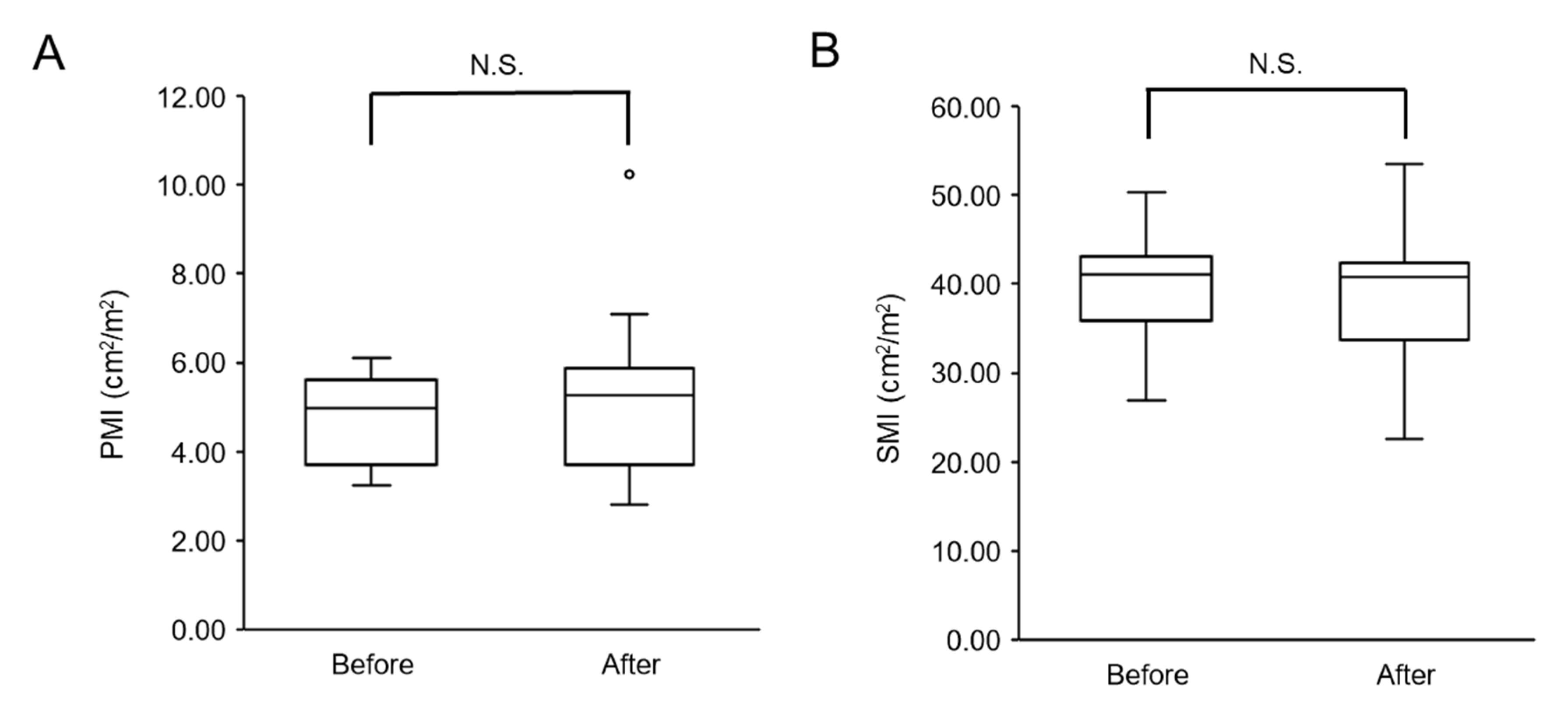
3.2.1. Changes in Skeletal Muscle Mass after Physical Therapy

There was no significant decrease in PMI or SMI after physical therapy compared with before physical therapy (Figure 1A,B).

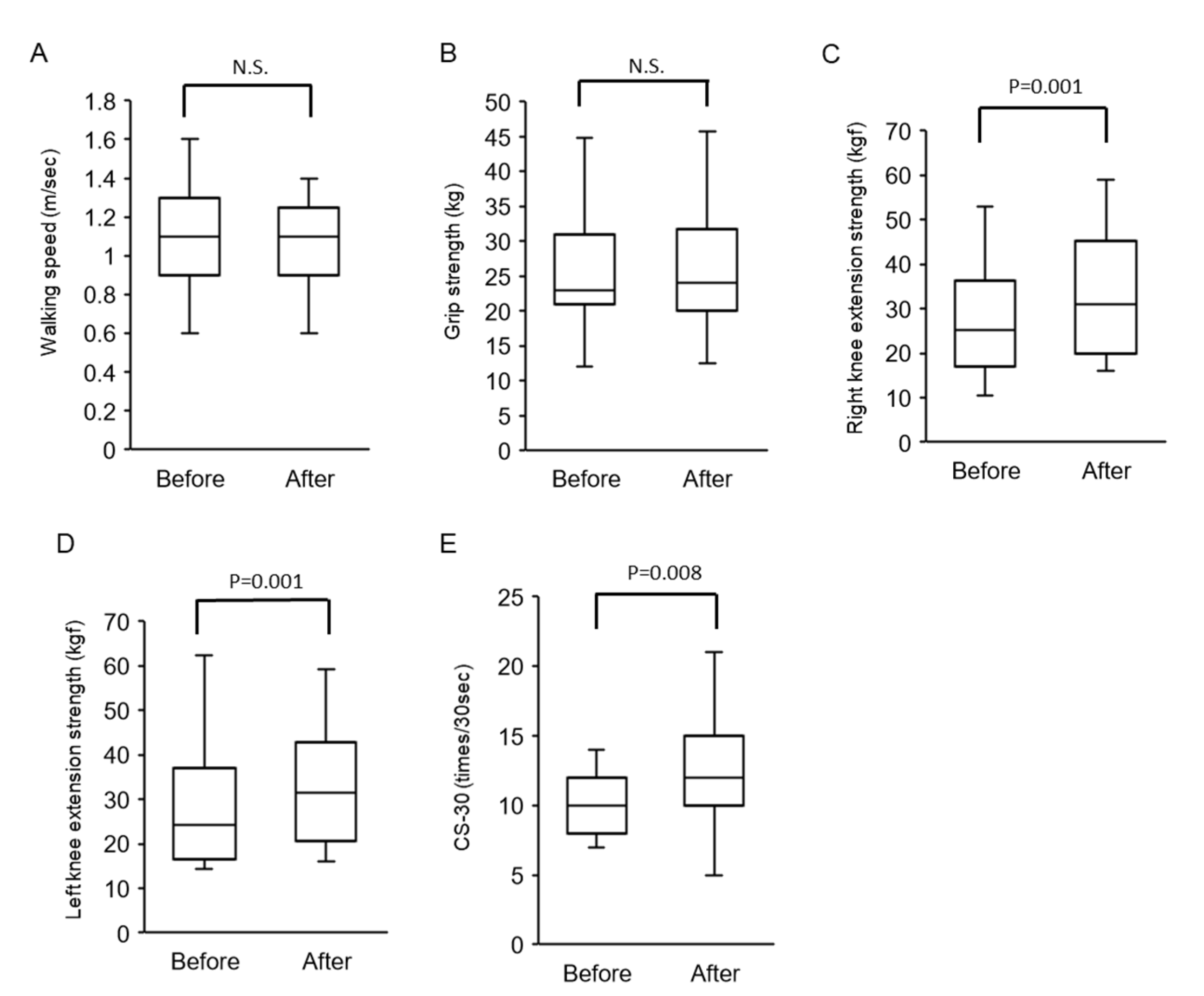
3.2.2. Changes in Walking Speed and Muscle Strength after Physical Therapy

No significant difference was seen in walking speed before and after physical therapy (Figure 2A). There was no significant difference in grip strength before and after physical therapy (Figure 2B). There was a significant increase in right and left knee extension strength after physical therapy compared with before physical therapy (Figure 2C,D). CS-30 was significantly improved after physical therapy compared with that before physical therapy (Figure 2E).

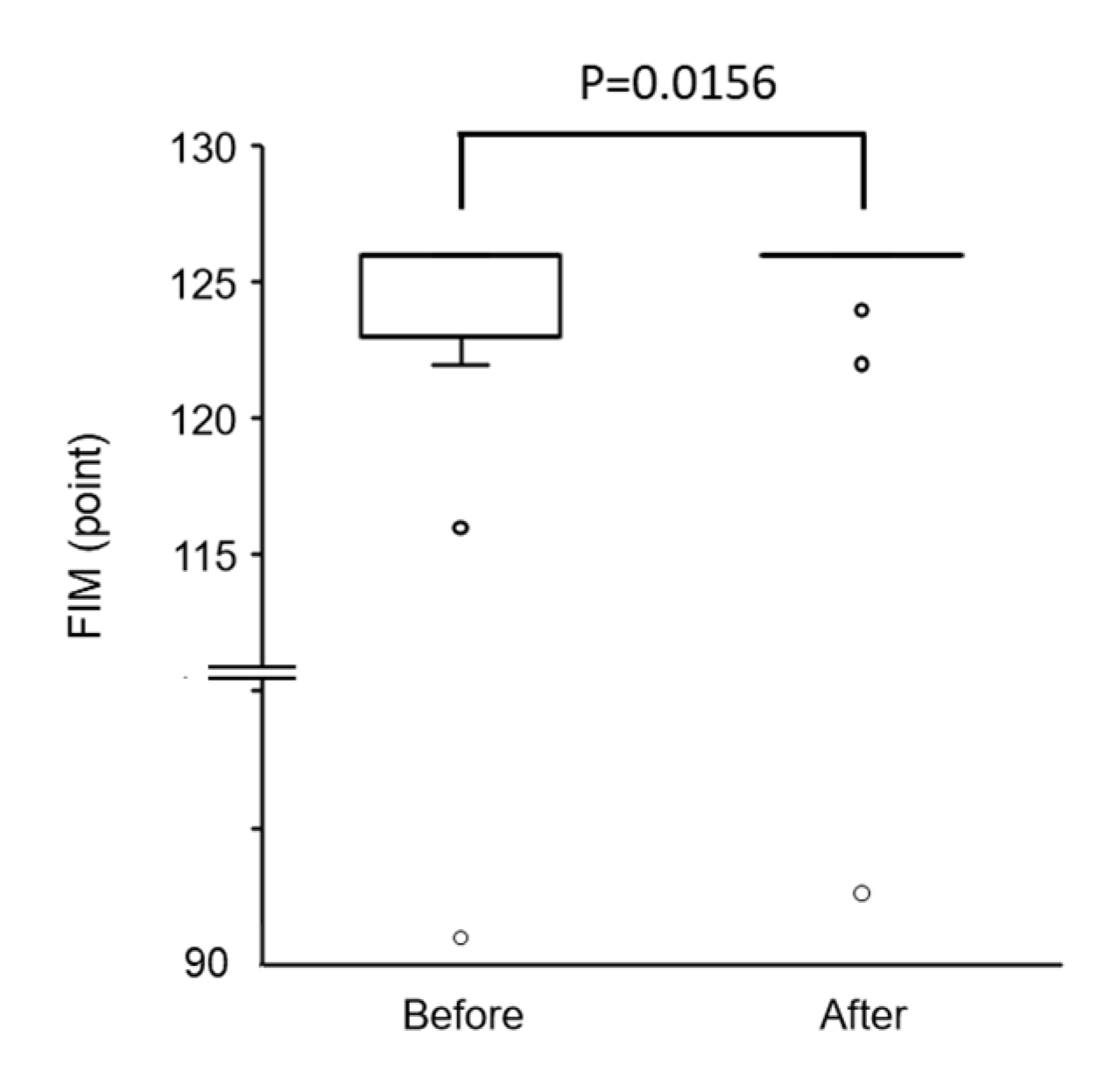
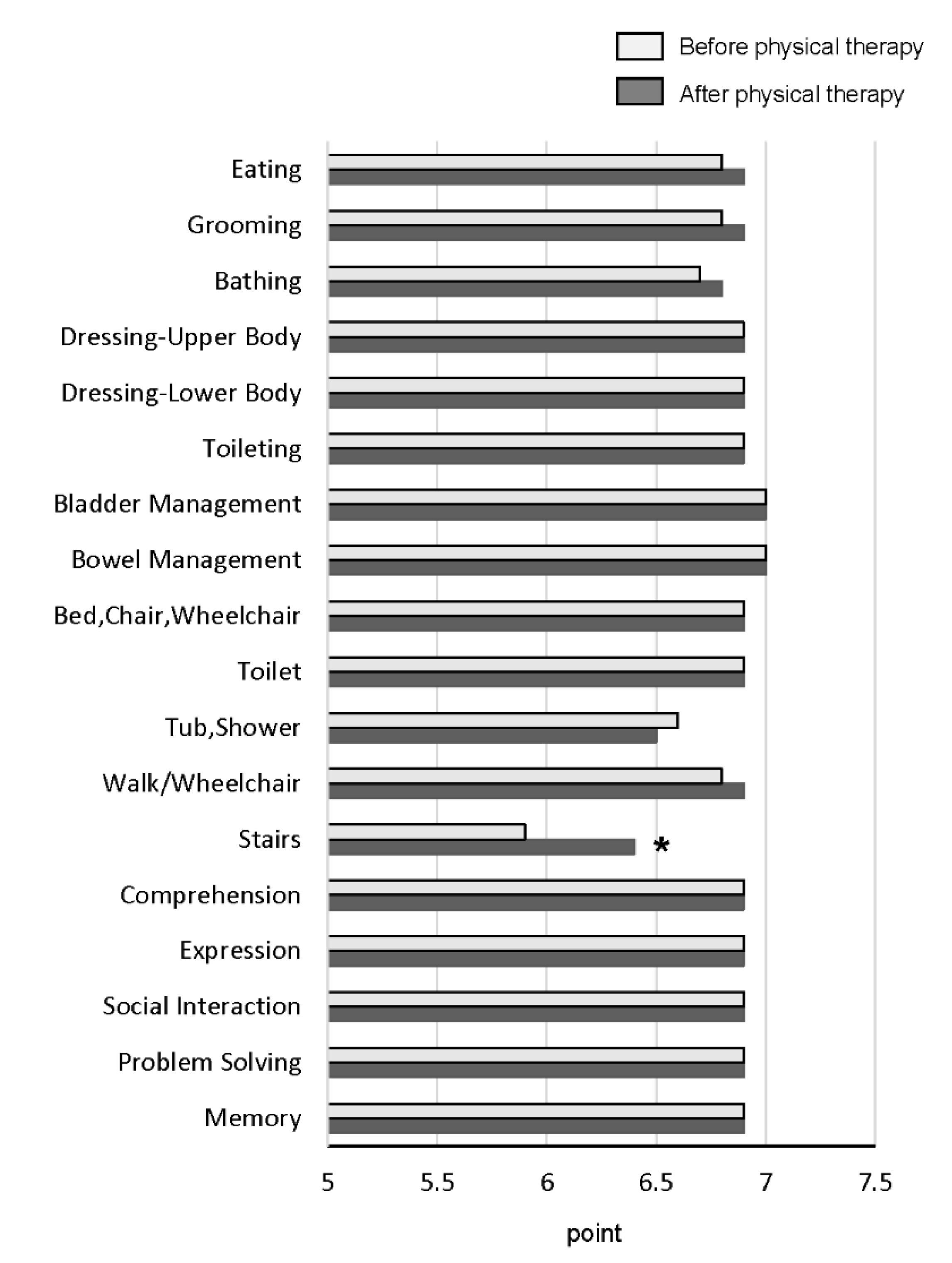
3.2.3. Changes in FIM Score and All Items of FIM after Physical Therapy

The total FIM score was significantly increased after physical therapy compared with before physical therapy (Figure 3). The stairs index is a motor index of FIM, and was the only item that was significantly improved after physical therapy (Figure 4). There were no significant changes in the scores of the other motor and cognitive indexes between before and after physical therapy (Figure 4).

4. Discussion

We demonstrated the safety and beneficial effects of in-hospital physical therapy, including resistance exercise, for patients with HCC. Liver and renal function did not worsen after physical therapy. Physical therapy with personalized intensity improved muscle strength with no reduction in muscle mass during hospitalization for HCC treatment. In addition, physical therapy improved FIM, especially in the stairs index, in patients with HCC during hospitalization for cancer treatment.
In our study, liver and renal function were not worsened by physical therapy. On the other hand, García-Pagàn et al. reported that moderate exercise increased the risk of variceal bleeding in patients with esophageal varices [31]. Saló et al. reported that moderate physical exercise may cause marked impairment in renal function in patients with liver cirrhosis [32]. Although it remains unclear why the liver function did not worsen with physical therapy, a possible reason is a difference in the type of exercises used in our study compared with that of previous studies. In our study, we mainly employed resistance training. However, previous studies employed aerobic exercise such as cycloergometric exercise [31,32]. The intensity of aerobic exercise is generally higher than that of resistance exercise [33], and cardiopulmonary function is known to be impaired in patients with HCC [34]. Therefore, aerobic exercise is a cardiopulmonary burden in patients with HCC. Our results may indicate that resistance exercise is more suitable than aerobic exercise for patients with HCC.
In this study, our physical therapy program was able to maintain muscle mass in patients with HCC. Our results were in good agreement with a previous report of HCC patients [14]. In addition, we demonstrated that our physical therapy program improved the muscle strength of the lower limbs. Although the reason the physical therapy program improved the muscle strength of the lower limbs remains unclear, it may be because resistance exercises were performed at a personalized intensity using a machine such as COMBIT CB-1. COMBIT CB-1 is able to precisely assess the maximal muscle strength of the lower limbs and allows the operator to set the appropriate intensity of exercise [20], which may result in increased muscle strength in the lower limbs. In fact, Nadia Schott et al. reported that exercise using machine training significantly increased the muscle strength of the lower limbs [35].
We first demonstrated that physical therapy increased the FIM score in patients with HCC. In previous studies, physical therapy has been reported to increase the FIM score in patients with various cancers, including colorectal cancer [10] and pancreatic cancer [36]. Thus, our results are in good agreement with these previous reports. Saotome et al. reported that an increase in FIM score by cancer rehabilitation was associated with prolonged survival in patients with cancer [37]. Moreover, an increase in FIM score through physical therapy has been reported to improve cognitive function in patients with cancer [13,38]. Furthermore, an increase in FIM score has been reported to be associated with an improved quality of life in patients with cancer. Therefore, evaluation of FIM seems to be important for the management of patients with HCC.
In this study, our physical therapy program increased the FIM score in patients with HCC. A possible reason for an increase in the FIM score is that we performed machine training with appropriate exercise intensity using COMBIT CB-1. Furthermore, a feature of our physical therapy program was that we mainly focused on exercising the lower-limb muscles, which is associated with physical performance [39]. As a result, physical therapy improved CS-30, an index for the strength of lower limb muscles in this study. In fact, among all 18 factors consisting of the FIM score, the ability to climb stairs was the only factor that was improved by physical therapy in this study. Matsufuji et al. also reported that chair stand exercises improved ADLs in patients on hemodialysis aged >60 years in a randomized controlled trial [40]. Yoshimura et al. also reported that an in-hospital chair–stand exercise was independently associated with an improvement in FIM-motor scores [41]. These findings indicate that physical therapy improved lower limb strength and subsequently the ability to climb stairs, leading to an increase in FIM score in this study.
This study has several limitations. First, this was a single-center study with a small sample size. Second, this was a retrospective chart review with no control group, which cannot prove the efficacy of physical therapy. Third, ADLs were assessed using only the FIM score and not by other methods such as the cancer functional assessment set [42]. Fourth, we could not perform a multivariate analysis because of the small sample size, and it remains unclear what type of physical therapy was associated with an improvement of FIM. Thus, the usefulness of physical therapy should be evaluated by a multi-center prospective study with a large sample size by using multiple assessments for ADLs. Fifth, Saotome et al. have reported that survival was significantly improved in patients with a total FIM score ≥80 after cancer rehabilitation [37]. In our study, all patients showed a total FIM score ≥ 80, even at the admission of the hospital. Therefore, it remains unclear if physical therapy had an impact on prognosis in our study. It also remains unclear which subscale of the FIM is important for survival. Thus, further studies should be focused on an association between an increase in FIM scores/the subscale of the FIM and an improvement in survival.

5. Conclusions

In this study, we performed in-hospital physical therapy, including resistance exercises, at a personalized intensity for patients with HCC. We demonstrated that in-hospital physical therapy did not worsen hepatic function. We also showed that exercise improved muscle strength with no reduction in muscle mass. Furthermore, we also showed that exercise improved the stair index of FIM in patients with HCC. Thus, in-hospital physical therapy may improve the ability to climb stairs in patients with HCC. Future research is needed to more clearly assess the benefits of physical therapy for ADL performance.

Author Contributions

Conceptualization, H.N. (Hayato Narao), K.H., S.K., M.T., T.K., and H.N. (Hiroaki Nagamatu); methodology, H.N. (Hayato Narao), K.H., and S.K.; validation, M.T., T.K., and H.N. (Hiroaki Nagamatu); formal analysis, D.N. and R.H.; investigation, Y.M., S.O., N.N., T.T. (Tsubasa Tsutsumi), and R.H.; resources, H.N. (Hayato Narao); data curation, Y.M., S.O., and N.N.; writing—original draft preparation, H.N. (Hayato Narao), K.H., S.K., M.T., and T.K.; writing—review and editing, H.M. and T.T. (Takuji Torimura); visualization, H.N. (Hayato Narao) and K.H.; supervision, H.M. and T.T. (Takuji Torimura); project administration, H.N. (Hayato Narao); funding acquisition, T.K. All authors have read and agreed to the published version of the manuscript.

Funding

This research was funded by the Research Program on Hepatitis from the Japan Agency for Medical Research and Development, AMED under JP20fk0210045.

Conflicts of Interest

Takumi Kawaguchi received lecture fees from Mitsubishi Tanabe Pharma Corporation and Otsuka Pharmaceutical Co., Ltd. The other authors declare no conflict of interest. The funders had no role in the design of the study; in the collection, analyses, or interpretation of data; in the writing of the manuscript; or in the decision to publish the results.

References

  1. Kenis, C.; DeCoster, L.; Bastin, J.; Bode, H.; Van Puyvelde, K.; De Grève, J.; Conings, G.; Fagard, K.; Flamaing, J.; Milisen, K.; et al. Functional decline in older patients with cancer receiving chemotherapy: A multicenter prospective study. J. Geriatr. Oncol. 2017, 8, 196–205. [Google Scholar] [CrossRef] [PubMed]
  2. Duchowny, K.A.; Clarke, P.; Peterson, M.D. Muscle Weakness and Physical Disability in Older Americans: Longitudinal Findings from the U.S. Health and Retirement Study. J. Nutr. Health Aging 2018, 22, 501–507. [Google Scholar] [CrossRef] [PubMed]
  3. Van Ancum, J.M.; Scheerman, K.; Jonkman, N.H.; Smeenk, H.E.; Kruizinga, R.C.; Meskers, C.; Maier, A.B. Change in muscle strength and muscle mass in older hospitalized patients: A systematic review and meta-analysis. Exp. Gerontol. 2017, 92, 34–41. [Google Scholar] [CrossRef] [PubMed]
  4. Hirota, K.; Kawaguchi, T.; Hashida, R.; Koya, S.; Bekki, M.; Goshima, N.; Yoshiyama, T.; Otsuka, T.; Nozoe, R.; Nakano, D.; et al. Profiles Associated with Sarcopenia in Hepatoma Patients Underwent Transcatheter Arterial Chemoembolization: A Data-Mining Analysis. JCSM Clin. Rep. 2018, 3, 1–17. [Google Scholar] [CrossRef] [Green Version]
  5. Franken, F.H. Bed rest, diet and working capability in liver disease (author’s transl). Leber. Magen Darm. 1977, 7, 300–305. [Google Scholar]
  6. Glenny, C.; Stolee, P. Comparing the Functional Independence Measure and the interRAI/MDS for use in the functional assessment of older adults: A review of the literature. BMC Geriatr. 2009, 9, 52. [Google Scholar] [CrossRef] [Green Version]
  7. Maritz, R.; Tennant, A.; Fellinghauer, C.; Stucki, G.; Prodinger, B. The Functional Independence Measure 18-item version can be reported as a unidimensional interval-scaled metric: Internal construct validity revisited. J. Rehabil. Med. 2019, 51, 193–200. [Google Scholar] [CrossRef] [Green Version]
  8. Bender, A.; Bauch, S.; Grill, E. Efficacy of a post-acute interval inpatient neurorehabilitation programme for severe brain injury. Brain Inj. 2013, 28, 44–50. [Google Scholar] [CrossRef]
  9. Hamilton, B.B.; A Laughlin, J.; Fiedler, R.C.; Granger, C.V. Interrater reliability of the 7-level functional independence measure (FIM). Scand. J. Rehabil. Med. 1994, 26, 115–119. [Google Scholar]
  10. McEwen, S.; Elmi, S.; Waldman, M.; Bishev, M. Inpatient oncology rehabilitation in Toronto: A descriptive 18-month retrospective record review. Support. Care Cancer 2011, 20, 1541–1547. [Google Scholar] [CrossRef]
  11. Wang, D.X.; Yao, J.; Zirek, Y.; Reijnierse, E.M.; Maier, A. Muscle mass, strength, and physical performance predicting activities of daily living: A meta-analysis. J. Cachex- Sarcopenia Muscle 2019, 11, 3–25. [Google Scholar] [CrossRef] [PubMed] [Green Version]
  12. Nelson, J.A.; Tecci, M.G.; Lanni, M.A.; Fischer, J.P.; Fosnot, J.; Selber, J.C.; Wu, L.C.; Serletti, J.M. Function and Strength after Free Abdominally Based Breast Reconstruction: A 10-Year Follow-Up. Plast Reconstr. Surg. 2019, 143, 22e–31e. [Google Scholar] [CrossRef] [PubMed]
  13. Cole, R.P.; Scialla, S.J.; Bednarz, L. Functional recovery in cancer rehabilitation. Arch. Phys. Med. Rehabil. 2000, 81, 623–627. [Google Scholar] [CrossRef]
  14. Koya, S.; Kawaguchi, T.; Hashida, R.; Hirota, K.; Bekki, M.; Goto, E.; Yamada, M.; Sugimoto, M.; Hayashi, S.; Goshima, N.; et al. Effects of in-hospital exercise on sarcopenia in hepatoma patients who underwent transcatheter arterial chemoembolization. J. Gastroenterol. Hepatol. 2018, 34, 580–588. [Google Scholar] [CrossRef] [PubMed]
  15. Koya, S.; Kawaguchi, T.; Hashida, R.; Goto, E.; Matsuse, H.; Saito, H.; Hirota, K.; Taira, R.; Matsushita, Y.; Imanaga, M.; et al. Effects of In-hospital Exercise on Liver Function, Physical Ability, and Muscle Mass during Treatment of Hepatoma in Patients with Chronic Liver Disease. Hepatol. Res. 2016, 47, 22. [Google Scholar] [CrossRef] [PubMed] [Green Version]
  16. Hashida, R.; Kawaguchi, T.; Koya, S.; Hirota, K.; Goshima, N.; Yoshiyama, T.; Otsuka, T.; Bekki, M.; Iwanaga, S.; Nakano, D.; et al. Impact of cancer rehabilitation on the prognosis of patients with hepatocellular carcinoma. Oncol. Lett. 2020, 19, 2355–2367. [Google Scholar] [CrossRef] [PubMed]
  17. Oken, M.M.; Creech, R.H.; Tormey, D.C.; Horton, J.; E Davis, T.; McFadden, E.T.; Carbone, P.P. Toxicity and response criteria of the Eastern Cooperative Oncology Group. Am. J. Clin. Oncol. 1982, 5, 649–655. [Google Scholar] [CrossRef]
  18. Citro, V.; Milan, G.; Tripodi, F.S.; Gennari, A.; Sorrentino, P.; Gallotta, G.; Postiglione, A.; Tarantino, G. Mental status impairment in patients with West Haven grade zero hepatic encephalopathy: The role of HCV infection. J. Gastroenterol. 2007, 42, 79–82. [Google Scholar] [CrossRef] [Green Version]
  19. Garber, C.E.; Blissmer, B.; Deschenes, M.R.; Franklin, B.A.; Lamonte, M.J.; Lee, I.M.; Nieman, D.C.; Swain, D.P. American College of Sports, M. American College of Sports Medicine position stand. Quantity and quality of exercise for developing and maintaining cardiorespiratory, musculoskeletal, and neuromotor fitness in apparently healthy adults: Guidance for prescribing exercise. Med. Sci. Sports Exerc. 2011, 43, 1334–1359. [Google Scholar]
  20. Miyatake, N.; Saito, T.; Miyachi, M.; Tabata, I.; Numata, T. Evaluation of muscle strength and its relation to exercise habits in Japanese. Acta Med. Okayama 2009, 63, 151–155. [Google Scholar]
  21. Kudo, M.; Chung, H.; Osaki, Y. Prognostic staging system for hepatocellular carcinoma (CLIP score): Its value and limitations, and a proposal for a new staging system, the Japan Integrated Staging Score (JIS score). J. Gastroenterol. 2003, 38, 207–215. [Google Scholar] [CrossRef] [PubMed]
  22. Arii, S.; Sata, M.; Sakamoto, M.; Shimada, M.; Kumada, T.; Shiina, S.; Yamashita, T.; Kokudo, N.; Tanaka, M.; Takayama, T.; et al. Management of hepatocellular carcinoma: Report of Consensus Meeting in the 45th Annual Meeting of the Japan Society of Hepatology (2009). Hepatol. Res. 2010, 40, 667–685. [Google Scholar] [CrossRef] [PubMed]
  23. Schneider, C.A.; Rasband, W.S.; Eliceiri, K.W. NIH Image to ImageJ: 25 years of image analysis. Nat. Methods 2012, 9, 671–675. [Google Scholar] [CrossRef]
  24. Ohara, M.; Suda, G.; Kimura, M.; Maehara, O.; Shimazaki, T.; Shigesawa, T.; Suzuki, K.; Nakamura, A.; Kawagishi, N.; Nakai, M.; et al. Analysis of the optimal psoas muscle mass index cut-off values, as measured by computed tomography, for the diagnosis of loss of skeletal muscle mass in Japanese people. Hepatol. Res. 2020, 50, 715–725. [Google Scholar] [CrossRef] [PubMed]
  25. Hirota, K.; Kawaguchi, T.; Koya, S.; Nagamatsu, A.; Tomita, M.; Hashida, R.; Nakano, D.; Niizeki, T.; Matsuse, H.; Shiba, N.; et al. Clinical utility of the Liver Frailty Index for predicting muscle atrophy in chronic liver disease patients with hepatocellular carcinoma. Hepatol. Res. 2019, 50, 330–341. [Google Scholar] [CrossRef]
  26. Nagamatsu, A.; Kawaguchi, T.; Hirota, K.; Koya, S.; Tomita, M.; Hashida, R.; Kida, Y.; Narao, H.; Manako, Y.; Tanaka, D.; et al. Slow walking speed overlapped with low handgrip strength in chronic liver disease patients with hepatocellular carcinoma. Hepatol. Res. 2019, 49, 1427–1440. [Google Scholar] [CrossRef] [PubMed]
  27. Lord, S.R.; Murray, S.M.; Chapman, K.; Munro, B.; Tiedemann, A. Sit-to-Stand Performance Depends on Sensation, Speed, Balance, and Psychological Status in Addition to Strength in Older People. Journals Gerontol. Ser. A Boil. Sci. Med Sci. 2002, 57, M539–M543. [Google Scholar] [CrossRef]
  28. Jones, C.J.; Rikli, R.E.; Beam, W.C. A 30-s Chair-Stand Test as a Measure of Lower Body Strength in Community-Residing Older Adults. Res. Q. Exerc. Sport 1999, 70, 113–119. [Google Scholar] [CrossRef]
  29. Murray, M.P. Gait as a total pattern of movement. Am. J. Phys. Med. 1967, 46, 290–333. [Google Scholar]
  30. Keith, R.A.; Granger, C.V.; Hamilton, B.B.; Sherwin, F.S. The functional independence measure: A new tool for rehabilitation. Adv. Clin. Rehabil. 1987, 1, 6–18. [Google Scholar]
  31. García-Pagàn, J.C.; Santos, C.; A Barberá, J.; Luca, A.; Roca, J.; Rodriguez-Roisin, R.; Bosch, J.; Rodes, J. Physical exercise increases portal pressure in patients with cirrhosis and portal hypertension. Gastroenterology 1996, 111, 1300–1306. [Google Scholar] [CrossRef] [PubMed]
  32. Salo, J.; Guevara, M.; Fernandez-Esparrach, G.; Bataller, R.; Gines, A.; Jiménez, W.; Ginès, P.; Rivera, F.; Arroyo, V.; Rodés, J. Impairment of renal function during moderate physical exercise in cirrhotic patients with ascites: Relationship with the activity of neurohormonal systems. Hepatology 1997, 25, 1338–1342. [Google Scholar] [CrossRef] [PubMed]
  33. Hashida, R.; Kawaguchi, T.; Bekki, M.; Omoto, M.; Matsuse, H.; Nago, T.; Takano, Y.; Ueno, T.; Koga, H.; George, J.; et al. Aerobic vs. resistance exercise in non-alcoholic fatty liver disease: A systematic review. J. Hepatol. 2017, 66, 142–152. [Google Scholar] [CrossRef]
  34. Muderrisoglu, H.; Yilmaz, K.C.; Karacaglar, E.; Bal, U.; Aydinalp, A.; Moray, G.; Haberal, M. Preoperative Cardiac Risk Assessment in Patients Undergoing Liver Transplant Due to Hepatocellular Carcinoma: Should It Be Different? Exp. Clin. Transplant. 2017, 15, 65–68. [Google Scholar] [PubMed]
  35. Schott, N.; Johnen, B.; Holfelder, B. Effects of free weights and machine training on muscular strength in high-functioning older adults. Exp. Gerontol. 2019, 122, 15–24. [Google Scholar] [CrossRef]
  36. Mikami, Y.; Kouda, K.; Kawasaki, S.; Okada, K.-I.; Kawai, M.; Kitahata, Y.; Miyazawa, M.; Hirono, S.; Unno, M.; Tajima, F.; et al. Preoperative In-Hospital Rehabilitation Improves Physical Function in Patients with Pancreatic Cancer Scheduled for Surgery. Tohoku J. Exp. Med. 2020, 251, 279–285. [Google Scholar] [CrossRef]
  37. Saotome, T.; Klein, L.A.; Faux, S.G. Cancer rehabilitation: A barometer for survival? Support. Care Cancer 2015, 23, 3033–3041. [Google Scholar] [CrossRef]
  38. Ng, A.; Gupta, E.; Fontillas, R.C.; Bansal, S.; Williams, J.L.; Park, M.; Liu, D.; Fu, J.B.; Yadav, R.; Bruera, E. Patient-Reported Usefulness of Acute Cancer Rehabilitation. PM R 2017, 9, 1135–1143. [Google Scholar] [CrossRef]
  39. Jung, H.; Yamasaki, M. Association of lower extremity range of motion and muscle strength with physical performance of community-dwelling older women. J. Physiol. Anthr. 2016, 35, 1–9. [Google Scholar] [CrossRef] [Green Version]
  40. Matsufuji, S.; Shoji, T.; Yano, Y.; Tsujimoto, Y.; Kishimoto, H.; Tabata, T.; Emoto, M.; Inaba, M. Effect of Chair Stand Exercise on Activity of Daily Living: A Randomized Controlled Trial in Hemodialysis Patients. J. Ren. Nutr. 2015, 25, 17–24. [Google Scholar] [CrossRef]
  41. Yoshimura, Y.; Wakabayashi, H.; Nagano, F.; Bise, T.; Shimazu, S.; Shiraishi, A. Chair-stand exercise improves post-stroke dysphagia. Geriatr. Gerontol. Int 2020. [Google Scholar] [CrossRef]
  42. Miyata, C.; Tsuji, T.; Tanuma, A.; Ishikawa, A.; Honaga, K.; Liu, M. Cancer Functional Assessment Set: A new tool for functional evaluation in cancer. Am. J. Phys. Med. Rehabil. 2014, 93, 656–664. [Google Scholar] [CrossRef] [PubMed]
Figure 1. Changes in skeletal muscle mass after physical therapy: (A) psoas muscle index (PMI) and (B) skeletal muscle index (SMI). N.S.—not significant.
Ijerph 17 09098 g001
Figure 2. Changes in walking speed and muscle strength after physical therapy. (A) Walking speed, (B) grip strength, (C) right knee extension strength, (D) left knee extension strength, and (E) CS-30. Abbreviations: N.S.—not significant; CS-30—30-s chair–stand test.
Ijerph 17 09098 g002
Figure 3. Changes in functional independence measure (FIM) score after physical therapy.
Ijerph 17 09098 g003
Figure 4. Changes in all items of FIM after physical therapy *, p < 0.01.
Ijerph 17 09098 g004
Table 1. A protocol of physical therapy for patients with hepatocellular carcinoma (HCC).
Type of ExercisePhysical TherapyExercise IntensityFrequency
1. Resistance trainingKnee extension
(Used Combit CB-1)
60% of MVC2–3 days/week
Standing steppingBodyweight5 days/week
Calf raiseBodyweight5 days/week
Half squatBodyweight5 days/week
2. StretchingLower limb stretchingPainless strength3 days/week
3. Aerobic exerciseWalkingBorg scale 11–13
(Fairly light to somewhat hard)
Every day
Table 2. Patient characteristics.
Reference ValueMedian (IQR)Range
(Min–Max)
NN/A19N/A
Age (years)N/A78.0 (73.0–82.0)60–86
Sex (female/male)N/A6/13N/A
Body mass index (kg/m2)18.5–24.921.2 (20.7–26.5)16.8–31.5
Performance status (0/1/2/3/4)N/A73.7%/26.3%/0%/0%/0% (14/5/0/0/0)N/A
FIM score (points)N/A126 (123–126)92–126
Walking assistance devise (yes/no)N/A10.5%/89.5%
(2/17)
N/A
HCC stage (I/II/III/IV)N/A5.3%/84.1%/5.3%/5.3% (1/16/1/1)N/A
HCV/HBV/othersN/A78.9%/15.8%/5.3%
(15/3/1)
N/A
Hospitalization (days)N/A10.0 (10.0–13.0)7.0–28.0
Physical therapy (days)N/A5.0 (5.0–6.0)4.0–15.0
Implementation rate of knee extension (%)N/A66.7 (51.8–77.5)33.0–100.0
Implementation rate of standing stepping (%)N/A66.7 (51.8–77.5)33.0–100.0
Implementation rate of calf raise (%)N/A66.7 (51.8–77.5)33.0–100.0
Implementation rate of half squat (%)N/A66.7 (51.8–77.5)33.0–100.0
Implementation rate of stretching (%)N/A66.7 (51.8–77.5)33.0–100.0
Implementation rate of aerobic exercise (%)N/A75.0 (56.0–90.0)0.0–100.0
Hepatic encephalopathy (Yes/No)N/A10.5%/89.5%
(2/17)
N/A
Ascites (Yes/No)N/A10.5%/89.5%
(2/17)
N/A
Walking speed (m/sec)N/A1.1 (0.9–1.3)0.6–1.6
Six-minute walk test (m)N/A300 (262–340)150.5–465.0
Grip strength (women/men) (kg)N/A17.1 (13.8–21.7)
/28.0 (24.3–31.3)
12.0–22.3
/21.0–44.8
Grip strength (low/normal)N/A42.1%/57.9% (8/11)N/A
Right knee extension strength (kgf)N/A25.2 (17.0–36.3)10.5–53.0
Left knee extension strength (kgf)N/A24.2 (16.5–36.9)14.3–62.3
SMI (women/men) (cm2/m2)N/A39.9 (35.8–42.7)
/41.1 (39.3–42.2)
26.9–50.2
/27.4–50.2
SMI (muscle atrophy/non-muscle atrophy)N/A26.3%/73.7% (5/14)N/A
PMI (women/men) (cm2/m2)N/A3.6 (3.4–4.6)
/5.0 (4.9–5.6)
3.25–6.11
/3.48–6.07
Presence of sarcopenia (Yes/No) N/A31.6%/68.4% (6/13)N/A
CS-30 (times/30 s)N/A10.0 (8.0–12.0)7–22
Child-Pugh score (points)N/A5 (5–6)5–12
Child-Pugh class (A/B/C)N/A78.9%/15.8%/5.3%
(15/3/1)
N/A
Anti-diabetic medication (Yes/No)N/A26.3%/73.7%
(5/14)
N/A
BCAA supplementation (Yes/No)N/A31.6%/68.4%
6/13
N/A
Biochemical examinations
Platelet count (× 104/μL)13.3–36.911.0 (7.4–17.6)4.8–26.7
AST (U/L)10–4033.0 (29.0–52.0)16.0–79.0
ALT (U/L)5–4026.0 (18.0–43.0)13.0–70.0
Lactate dehydrogenase (U/L)115–245213.0 (186.0–255.0)166.0–355.0
ALP (U/L)115–359377.0 (276.0–522.0)192.0–870.0
GGT (U/L)0–3037.0 (23.0–69.0)15.0–187.0
Albumin (g/dL)4.0–5.03.7 (3.0–4.1)2.6–4.6
Prothrombin activity (%)70–13079.2 (67.1–86.8)55.0–102.0
Total bilirubin (mg/dL)0.30–1.200.8 (0.6–0.9)0.3–3.1
AFP (ng/mL)0–1013.3 (5.8–40.2)3.5–1216.7
des-γ-carboxy prothrombin (mAU/mL)0–39107 (21.0–352.0)15–618
BUN (mg/dL)8.0–22.015.5 (13.5–17.8)6.4–28.1
Creatinine (mg/dL)0.61–1.040.8 (0.6–1.0)0.5–1.36
eGFR (mL/min/1.73 m2)>90.065.1 (55.0–82.9)39.1–90.0
Sodium (mmol/L)138–145139 (138.0–141.0)134–142
Potassium (mmol/L)3.6–4.84.0 (3.7–4.2)2.9–5.2
HbA1c (%)4.3–5.85.6 (5.4–6.8)4.5–7.8
Ammonia (μg/dL)13–8679.0 (52.7–134.2)40.0–156.0
Note: Data are expressed as median (IQR), range, or number. IQR—interquartile range; N/A—not applicable; FIM—functional independence measure; HCC—hepatocellular carcinoma; HCV—hepatitis C virus; HBV—hepatitis B virus; SMI—skeletal muscle index; PMI—psoas muscle index; CS-30, 30-s chair–stand test; ALBI—albumin–bilirubin; BCAA, branched chain amino acids; AST—aspartate aminotransferase; U/L—unit/litter; ALT—alanine aminotransferase; ALP—alkaline phosphatase; GGT—gamma-glutamyl transpeptidase; AFP—alpha-fetoprotein; BUN—blood urea nitrogen; eGFR—estimated glomerular filtration rate; HbA1c—hemoglobin A1c.
Table 3. Changes in liver and renal functions after physical therapy.
BeforeAfter
Median (IQR)Range
(Min–Max)
Median (IQR)Range
(Min–Max)
p
Child-Pugh score (point)5 (5–6)5–126 (6–7)5–100.112
Child-Pugh class (A/B/C)78.9%/15.8%/5.3%
(15/3/1)
N/A63.1%/31.6%/5.3%
(12/6/1)
N/A0.180
AST (U/L)33.0 (29.0–52.0)16.0–79.037.0 (28.0–49.0)21.0–114.00.7210
ALT (U/L)26.0 (18.0–43.0)13.0–70.037.0 (23.0–56.0)15.0–99.00.2579
Lactate dehydrogenase (U/L)213.0 (186.0–255.0)166.0–355.0198.0 (171.0–246.0)148.0–392.00.3225
ALP (U/L)377.0 (276.0–522.0)192.0–870.0382.0 (305.0–479.0)158.0–511.00.1703
GGT (U/L)37.0 (23.0–69.0)15.0–187.053.0 (29.0–82.0)14.0–203.00.4247
eGFR (mL/min/1.73 m2)65.1 (52.8–84.3)39.1–90.065.8 (57.9–84.6)34.0–99.90.2293
Note: Data are expressed as median (IQR), range, or number. IQR—interquartile range; N/A—not applicable; AST—aspartate aminotransferase; U/L—unit/litter; ALT—alanine aminotransferase; ALP—alkaline phosphatase; GGT—gamma-glutamyl transpeptidase; eGFR—estimated glomerular filtration rate.
Publisher’s Note: MDPI stays neutral with regard to jurisdictional claims in published maps and institutional affiliations.

