Epileptic Seizures Detection Using Deep Learning Techniques: A Review

 , , , , and
Abstract
:1. Introduction
- Providing information on available EEG datasets;
- Reviewing works done using various DL models for automated detection of epileptic seizures with various modality signals;
- Introducing future challenges on the detection of epileptic seizures;
- Analyzing the best performing model for various modalities of data.
2. Epileptic Seizures Detection Based on DL Techniques
2.1. Dataset
2.1.1. Fribourg
2.1.2. CHB-MIT
2.1.3. Kaggle
2.1.4. Bonn
2.1.5. Flint-Hills
2.1.6. Bern Barcelona
2.1.7. Hauz Khas
2.1.8. Zenodo
2.2. Preprocessing
2.3. Review of Deep Learning Techniques
2.3.1. Convolutional Neural Networks (CNNs)
A. 2D Convolutional Neural Networks (2D-CNNs)
B. AlexNet
C. VGG
D. GoogleNet
E. ResNet
F. 1D—Convolutional Neural Network (1D-CNN)
2.3.2. Recurrent Neural Networks (RNNs)
A. Long Short-Term Memory (LSTM)
B. Gated Recurrent Unit (GRU)
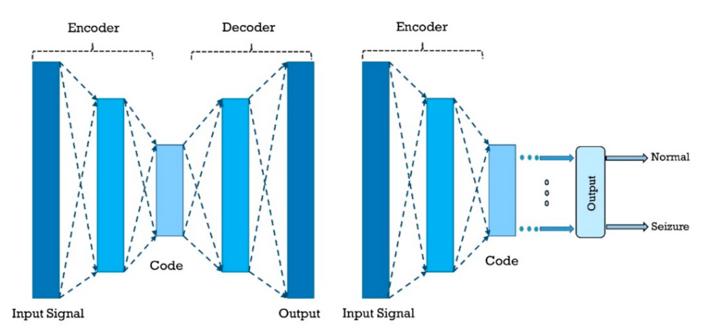
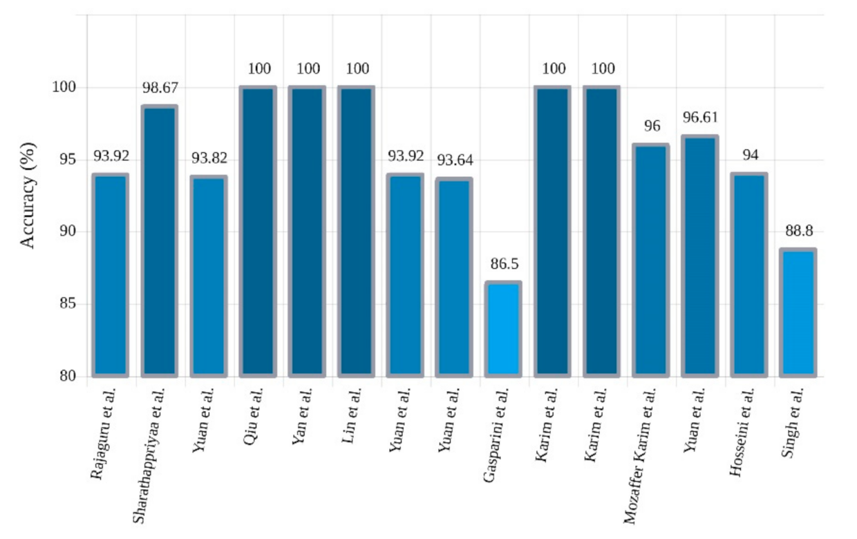
2.3.3. Autoencoders (AEs)
A. Other Types of AEs
2.3.4. Deep Belief Networks (DBNs)
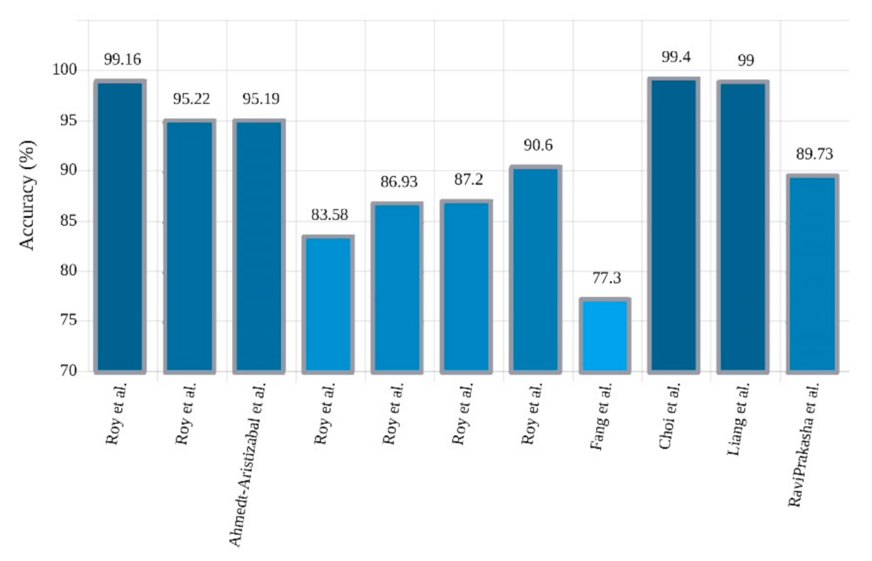
2.3.5. Convolutional Recurrent Neural Networks (CNN-RNNs)
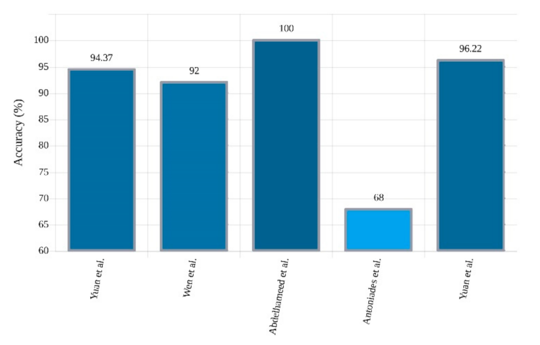
2.3.6. Convolutional Autoencoders (CNN-AEs)
3. Non-EEG-Based Epileptic Seizures Detection
3.1. Medical Imaging
3.2. Other Neuroimaging Modalities
4. Rehabilitation Systems for Epileptic Seizures Detection
5. Discussion
6. Challenges
7. Conclusion and Future Works
Author Contributions
Funding
Informed Consent Statement
Data Availability Statement
Conflicts of Interest
Appendix A

| Work | Dataset | Preprocessing | DL Toolbox | DL Network | K-Fold | Classifier | Accuracy (%) |
|---|---|---|---|---|---|---|---|
| [50] | Clinical | Down-Sampling, Normalization, Data Augmentation | Keras | SeizNet | -- | -- | -- |
| [51] | CHB-MIT | Visualization | PyTorch | 2D-CNN | -- | Softmax | 98.05 |
| [52] | Clinical | Filtering, Normalization, Visualization | NA | 2D-CNN | 10 | Softmax | NA |
| [53] | TUH | DivSpec | PyTorch | SeizureNet | 5 | Softmax | NA |
| [54] | Clinical | STFT | NA | TGCN | -- | Sigmoid | NA |
| [55] | CHB-MIT | Spatial Representation | NA | 2D-CNN | -- | Softmax | 99.48 |
| [61] | Clinical | Filtering, Visualization | Chainer | 2D-CNN | -- | Softmax | NA |
| [64] | Clinical | Spectrogram | NA | 2D-CNN | -- | LR | 87.51 |
| [65] | Clinical | Normalization | Matlab | 2D-CNN | -- | Softmax | NA |
| [66] | Clinical | Filtering | NA | 1D-CNN with 2D-CNN | -- | Sigmoid | 90.50 |
| CHB-MIT | 85.60 | ||||||
| [67] | Clinical | Filtering, Down-Sampling | Octave | 2D-CNN | -- | Softmax | NA |
| Keras | |||||||
| Theano | |||||||
| [68] | TUH | Filtering | NA | CNN-RNN | -- | Different Methods | NA |
| Clinical | |||||||
| [69] | TUH | Different Methods | NA | 1D-CNN-GRU | -- | Softmax | 99.16 |
| [70] | Clinical | Normalization, STFT | PyTorch | 1D-CNN | -- | Softmax | -- |
| 2D-CNN | |||||||
| [71] | TUH | Feature Extraction | TensorFlow | 2D-CNN | 10 | Softmax | 74.00 |
| [72] | Clinical | Filtering, EMD, DWT, Fourier | Octave | 2D-CNN | 4 | Sigmoid | 99.50 |
| Bern Barcelona | Keras | Softmax | |||||
| [73] | Bern Barcelona | Normalization, STFT | TensorFlow | 2D-CNN | 10 | Softmax | 91.80 |
| [74] | Bonn | DWT | NA | 2D-CNN | 10 | Softmax | 100 |
| [75] | Bonn | CWT | Keras | 2D-CNN | 10 | Softmax | 100 |
| [76] | Bonn | Filtering | Matlab | 2D-CNN | -- | Softmax | 99.60 |
| 90.10 | |||||||
| [77] | CHB-MIT | FFT, WPD | TensorFlow | 2D-CNN | 5 | MV-TSK-FS | 98.35 |
| Matlab | 3D-CNN | ||||||
| [78] | Clinical | Different Methods | Matlab | 2D-CNN | 10 | Sigmoid | NA |
| RF | |||||||
| [79] | CHB-MIT | MAS | NA | 2D-CNN | 5 | KELM | 99.33 |
| Clinical | |||||||
| [80] | Clinical | Filtering, Down-Sampling | TensorFlow | 1D-CNN | 4 | Softmax | 83.86 |
| SVM | |||||||
| [60] | Clinical | Different Techniques | Caffe | FRCNN with 2D-CNN | 5 | SVM | 95.19 |
| Keras | |||||||
| FRCNN with 2D-CNN-LSTM | |||||||
| Theano | Sigmoid | ||||||
| [57] | Bern Barcelona | NA | Caffe | Pre-Train Methods | -- | Softmax | 100 |
| [58] | UCI | Signal2Image | PyTorch | 1D-CNN | -- | DenseNet | 85.30 |
| [86] | Bonn | DA | TensorFlow | P-1D-CNN | 10 | Majority Voting | 99.10 |
| [87] | Bonn | Normalization | Matlab | 1D-CNN | 10 | Softmax | 86.67 |
| [88] | CHB-MIT | Filtering, DA | NA | MPCNN | -- | Softmax | NA |
| [89] | Clinical | Down-Sampling, Filtering | Keras | 1D-FCNN | 5 | Softmax | NA |
| [91] | TUH | Normalization | Keras | 1D-CNN | -- | Softmax | 79.34 |
| [90] | Clinical | Filtering | Theano | 1D-CNN | -- | Binary LR | NA |
| Lasagne | |||||||
| [81] | CHB-MIT | DWT, Feature Extraction, Normalization | NA | 1D-CNN | 10 | -- | 99.07 |
| [92] | Bonn | DWT, Normalization | NA | 1D-CNN | 5 | Sigmoid | 97.27 |
| [85] | Bonn | Normalization | NA | 1D-TCNN | NA | NA | 100 |
| [82] | Bonn | EMD, MPF | NA | 1D-CNN | 10 | Softmax | 98.60 |
| [93] | CHB-MIT | Windowing | NA | IndRNN | 10 | NA | 87.00 |
| [94] | Bern Barcelona | Filtering, Normalization | TensorFlow | 1D-CNN | -- | Softmax | 91.80 |
| 99.00 | |||||||
| Bonn | |||||||
| [83] | CHB-MIT | Filtering | PyTorch | 1D-PCM-CNN | 5 | Softmax | NA |
| Clinical | |||||||
| [95] | CHB-MIT | MIDS, WGAN | NA | 1D-CNN | -- | Softmax | 84.00 |
| [96] | Clinical | Down-Sampling, PSD, FFT | NA | 1D-CNN | 4 | Sigmoid | 86.29 |
| [97] | CHB-MIT | Filtering | TensorFlow | 1D-CNN | 4 | Softmax | NA |
| [99] | Bern Barcelona | Filtering, DA | NA | 1D-CNN | 10 | NA | 89.28 |
| [100] | Bonn | Normalization | Keras | 1D-CNN | 10 | Softmax | 98.67 |
| TensorFlow | |||||||
| [101] | Clinical | Filtering, Normalization, Segmentation, resampling strategies | NA | Deep ConvNet | 10 | Softmax | 80.00 |
| [84] | Clinical | Down-Sampling, Filtering, DA | Keras | CNN-BP | 5 | Sigmoid | NA |
| TensorFlow | |||||||
| Matlab | |||||||
| [98] | Clinical | Filtering, DWT | NA | 1D-CNN | -- | Sigmoid | NA |
| LSTM | RF | ||||||
| GRU | SVM | ||||||
| [105] | CHB-MIT | Filtering, Montage Mapping | Matlab | DRNN | -- | MLP | NA |
| [110] | Bonn | Filtering | NA | LSTM | -- | Softmax | 100 |
| [106] | Bonn | Filtering | Keras | LSTM | 3 | Softmax | 100 |
| TensorFlow | 5 | ||||||
| Matlab | 10 | ||||||
| [107] | Bonn | Windowing | Keras | LSTM | 10 | Sigmoid | 91.25 |
| [108] | Bonn | Filtering | Keras | LSTM | 3 | Softmax | 100 |
| TensorFlow | 5 | ||||||
| Matlab | 10 | ||||||
| [109] | Freiburg | Filtering, Normalization | NA | LSTM | 5 | Softmax | 97.75 |
| [102] | CHB-MIT | Windowing | NA | ADIndRNN | 10 | NA | 88.70 |
| Bonn | |||||||
| [103] | Bonn | Autocorrelation | Keras | GRU | -- | LR | 98.00 |
| [111] | Bonn | DWT | Keras | RNN | -- | LR | 98.50 |
| [112] | Freiburg | Segmentation, DA, Stockwell Transform | Matlab | Bi-LSTM | -- | Softmax | 98.91 |
| TensorFlow | |||||||
| [104] | TUH | TCP | NA | ChronoNet | -- | Softmax | 90.60 |
| [113] | Clinical | Windowing | NA | AE with EM-PCA | -- | GA | 93.92 |
| [114] | Bonn | Filtering, HWPT, FD | Matlab | AE | -- | Softmax | 98.67 |
| [120] | Clinical | Down-Sampling, Filtering, Normalization | TensorFlow | AE | -- | Sigmoid | NA |
| [121] | CHB-MIT | STFT | NA | SSDA | -- | Softmax | 93.82 |
| [115] | Bonn | Normalization | Matlab | DSAE | -- | LR | 100 |
| [116] | TUH | Different Methods | Toolkits | SDA | -- | LR | NA |
| Theano | |||||||
| [117] | Bonn | Filtering | NA | SAE | -- | SVM | 100 |
| [122] | Bonn | Normalization | NA | SSAE | -- | Softmax | 100 |
| [118] | CHB-MIT | Scalogram | Theano | Wave2Vec | -- | Softmax | 93.92 |
| [136] | CHB-MIT | DA, STFT | PyTorch | CNN-AE | 5 | Softmax | 94.37 |
| [123] | Clinical | Filtering, CWT, Feature Extraction | NA | SAE | -- | Softmax | 86.50 |
| [124] | Bonn | Taguchi Method | NA | SSAE | -- | Softmax | 100 |
| [125] | Clinical | Dimension Reduction, ESD | NA | DeSAE | -- | Softmax | 100 |
| [126] | Bonn | DWT | NA | SAE | -- | Softmax | 96.00 |
| [119] | CHB-MIT | Different Methods | NA | mSSDA | -- | Softmax | 96.61 |
| [127] | Clinical | PCA, I-ICA | Matlab | SSAE | -- | Softmax | 94.00 |
| [128] | Bonn | Windowing | Matlab | SAE | -- | Softmax | 88.80 |
| [129] | Clinical | DWT | Matlab | DBN | -- | -- | 96.87 |
| [130] | Clinical | Normalization, Feature Extraction | Theano | DBN | -- | LR | NA |
| SVM | |||||||
| KNN | |||||||
| [133] | CHB-MIT | Image Based Representation | NA | 2D-CNN-LSTM | -- | -- | -- |
| [131] | Clinical | Filtering | TensorFlow | ST-GRU ConNets | -- | -- | 77.30 |
| [132] | CHB-MIT | STFT, 2D-Mapping | NA | 3D-CNN with Bi GRU | -- | -- | 99.40 |
| Clinical | |||||||
| [134] | CHB-MIT | Visualization | NA | 2D-CNN-LSTM | -- | Softmax | 99.00 |
| [135] | Clinical ECoG | Filtering | NA | 1D-CNN-LSTM | 5 | Sigmoid | 89.73 |
| [138] | CHB-MIT | Channel Selection | NA | CNN-AE | 5 | Different Methods | 92.00 |
| Bonn | 10 | ||||||
| [139] | Bonn | Windowing | NA | 1D-CNN with Bi LSTM | -- | Softmax | 99.33 |
| Sigmoid | 100 | ||||||
| [140] | Clinical | Mapping | Theano | ASAE-CNN | -- | LR | 68.00 |
| AAE-CNN | |||||||
| [137] | CHB-MIT | STFT | PyTorch | CNN-AE | 5 | Softmax | 96.22 |
| [141] | SCTIMST | Noise reduction with BM3D, Skull stripping, Segmentation, | Keras | 2D-CNN | 5 | Sigmoid | NA |
| TensorFlow | |||||||
| [142] | Clinical MRI | Different Techniques | NA | 2D-CNN | 5 | Softmax | NA |
| [143] | Clinical MRI | Filtering, ICA, BCG, GLM, MCS | NA | ResNet | -- | Softmax | NA |
| Triplet | |||||||
| [144] | Clinical Datasets | Different Methods | NA | 2D-CNN | -- | SVM | NA |
| [145] | Clinical MRI | Scaling Down | NA | 3D-CNN | 5 | Softmax | 89.80 |
| [146] | Clinical MRI | Connectivity Feature extraction | NA | 2D-CNN | -- | -- | -- |
| [147] | Kaggle | ROI, Normalization, AAL, CNNI, Down-sampling, NNI (3D images) | TensorFlow | 2D-ResNet | -- | Sigmoid | 98.22 |
| 2D-VGG | |||||||
| Clinical MRI | 2D-Inception V3 | ||||||
| 3D-SVGG-C3D | |||||||
| [148] | Clinical MRI | OSEM, DA | TensorFlow | DAC | -- | Tanh | NA |
References
- Ghassemi, N.; Shoeibi, A.; Rouhani, M.; Hosseini-Nejad, H. Epileptic seizures detection in EEG signals using TQWT and ensemble learning. In Proceedings of the 2019 9th International Conference on Computer and Knowledge Engineering (ICCKE), Mashhad, Iran, 24–25 October 2019; pp. 403–408. [Google Scholar]
- Shoeibi, A.; Ghassemi, N.; Alizadehsani, R.; Rouhani, M.; Hosseini-Nejad, H.; Khosravi, A.; Panahiazar, M.; Nahavandi, S. A comprehensive comparison of handcrafted features and convolutional autoencoders for epileptic seizures detection in EEG signals. Expert Syst. Appl. 2021, 163, 113788. [Google Scholar] [CrossRef]
- Bhattacharyya, A.; Pachori, R.B.; Upadhyay, A.; Acharya, U.R. Tunable-Q wavelet transform based multiscale entropy measure for automated classification of epileptic EEG signals. Appl. Sci. 2017, 7, 385. [Google Scholar] [CrossRef] [Green Version]
- Kulaseharan, S.; Aminpour, A.; Ebrahimi, M.; Widjaja, E. Identifying lesions in paediatric epilepsy using morphometric and textural analysis of magnetic resonance images. Clin. NeuroImage 2019, 21, 101663. [Google Scholar] [CrossRef]
- Zazzaro, G.; Cuomo, S.; Martone, A.; Montaquila, R.V.; Toraldo, G.; Pavone, L. Eeg signal analysis for epileptic seizures detection by applying data mining techniques. Internet Things 2019, 100048. [Google Scholar] [CrossRef]
- van Klink, N.; Mooij, A.; Huiskamp, G.; Ferrier, C.; Braun, K.; Hillebrand, A.; Zijlmans, M. Simultaneous MEG and EEG to detect ripples in people with focal epilepsy. Clin. Neurophysiol. 2019, 130, 1175–1183. [Google Scholar] [CrossRef] [PubMed]
- Pianou, N.; Chatziioannou, S. Imaging with PET/CT in Patients with Epilepsy. In Epilepsy Surgery and Intrinsic Brain Tumor Surgery; Springer: Cham, Switzerland, 2019; pp. 45–50. [Google Scholar]
- Subasi, A.; Kevric, J.; Canbaz, M.A. Epileptic seizure detection using hybrid machine learning methods. Neural Comput. Appl. 2019, 31, 317–325. [Google Scholar] [CrossRef]
- Acharya, U.R.; Oh, S.L.; Hagiwara, Y.; Tan, J.H.; Adeli, H.; Subha, D.P. Automated EEG-based screening of depression using deep convolutional neural network. Comput. Methods Programs Biomed. 2018, 161, 103–113. [Google Scholar] [CrossRef]
- Lauretani, F.; Longobucco, Y.; Ravazzoni, G.; Gallini, E.; Salvi, M.; Maggio, M. Imaging the Functional Neuroanatomy of Parkinson’s Disease: Clinical Applications and Future Directions. Int. J. Environ. Res. Public Health 2021, 18, 2356. [Google Scholar] [CrossRef]
- Carbó-Carreté, M.; Cañete-Massé, C.; Figueroa-Jiménez, M.D.; Peró-Cebollero, M.; Guàrdia-Olmos, J. Relationship between Quality of Life and the Complexity of Default Mode Network in Resting State Functional Magnetic Resonance Image in Down Syndrome. Int. J. Environ. Res. Public Health 2020, 17, 7127. [Google Scholar] [CrossRef]
- Morales Chacón, L.M.; González González, J.; Ríos Castillo, M.; Berrillo Batista, S.; Batista García-Ramo, K.; Santos Santos, A.; Cordero Quintanal, N.; Zaldívar Bermúdez, M.; Garbey Fernández, R.; Estupiñan Díaz, B.; et al. Surgical Outcome in Extratemporal Epilepsies Based on Multimodal Pre-Surgical Evaluation and Sequential Intraoperative Electrocorticography. Behav. Sci. 2021, 11, 30. [Google Scholar] [CrossRef]
- Takagi, S.; Sakuma, S.; Morita, I.; Sugimoto, E.; Yamaguchi, Y.; Higuchi, N.; Inamoto, K.; Ariji, Y.; Ariji, E.; Murakami, H. Application of Deep Learning in the Identification of Cerebral Hemodynamics Data Obtained from Functional Near-Infrared Spectroscopy: A Preliminary Study of Pre-and Post-Tooth Clenching Assessment. J. Clin. Med. 2020, 9, 3475. [Google Scholar] [CrossRef]
- Ronan, L.; Alhusaini, S.; Scanlon, C.; Doherty, C.P.; Delanty, N.; Fitzsimons, M. Widespread cortical morphologic changes in juvenile myoclonic epilepsy: Evidence from structural MRI. Epilepsia 2012, 53, 651–658. [Google Scholar] [CrossRef]
- Sharma, R.; Pachori, R.B. Classification of epileptic seizures in EEG signals based on phase space representation of intrinsic mode functions. Expert Syst. Appl. 2015, 42, 1106–1117. [Google Scholar] [CrossRef]
- Sheoran, M.; Kumar, S.; Chawla, S. Methods of denoising of electroencephalogram signal: A review. Int. J. Biomed. Eng. Technol. 2015, 18, 385. [Google Scholar] [CrossRef]
- Romaine, J.; Martín, M.P.; Ortiz, J.S.; Crespo, J.M. EEG—Single-Channel Envelope Synchronisation and Classification for Seizure Detection and Prediction. Brain Sci. 2021, 11, 516. [Google Scholar] [CrossRef]
- Perez-Sanchez, A.V.; Perez-Ramirez, C.A.; Valtierra-Rodriguez, M.; Dominguez-Gonzalez, A.; Amezquita-Sanchez, J.P. Wavelet Transform-Statistical Time Features-Based Methodology for Epileptic Seizure Prediction Using Electrocardiogram Signals. Mathematics 2020, 8, 2125. [Google Scholar] [CrossRef]
- Raschka, S.; Mirjalili, V. Python Machine Learning: Machine Learning and Deep Learning with Python. In Scikit-Learn and TensorFlow, 2nd ed.; Packt Publishing Ltd.: Birmingham, UK, 2017. [Google Scholar]
- Bonaccorso, G. Machine Learning Algorithms; Packt Publishing Ltd.: Birmingham, UK, 2017. [Google Scholar]
- Gulli, A.; Pal, S. Deep Learning with KERAS; Packt Publishing Ltd.: Birmingham, UK, 2017. [Google Scholar]
- Tang, X.; Zhang, X. Conditional adversarial domain adaptation neural network for motor imagery EEG decoding. Entropy 2020, 22, 96. [Google Scholar] [CrossRef] [Green Version]
- Alickovic, E.; Kevric, J.; Subasi, A. Performance evaluation of empirical mode decomposition, discrete wavelet transform, and wavelet packed decomposition for automated epileptic seizure detection and prediction. Biomed. Signal Process. Control 2018, 39, 94–102. [Google Scholar] [CrossRef]
- Sharma, M.; Bhurane, A.A.; Acharya, U.R. MMSFL-OWFB: A novel class of orthogonal wavelet filters for epileptic seizure detection. Knowl. Based Syst. 2018, 160, 265–277. [Google Scholar] [CrossRef]
- Mohammadpoor, M.; Shoeibi, A.; Shojaee, H. A hierarchical classification method for breast tumor detection. Iran. J. Med. Phys. 2016, 13, 261–268. [Google Scholar]
- Assi, E.B.; Nguyen, D.K.; Rihana, S.; Sawan, M. Towards accurate prediction of epileptic seizures: A review. Biomed. Signal Process. Control. 2017, 34, 144–157. [Google Scholar] [CrossRef]
- Khodatars, M.; Shoeibi, A.; Ghassemi, N.; Jafari, M.; Khadem, A.; Sadeghi, D.; Moridian, P.; Hussain, S.; Alizadehsani, R.; ZARE, A.; et al. Deep Learning for Neuroimaging-based Diagnosis and Rehabilitation of Autism Spectrum Disorder: A Review. arXiv 2020, arXiv:2007.01285. [Google Scholar]
- Sadeghi, D.; Shoeibi, A.; Ghassemi, N.; Moridian, P.; Khadem, A.; Alizadehsani, R.; Teshnehlab, M.; Gorriz, J.M.; Nahavandi, S. An Overview on Artificial Intelligence Techniques for Diagnosis of Schizophrenia Based on Magnetic Resonance Imaging Modalities: Methods, Challenges, and Future Works. arXiv 2021, arXiv:2103.03081. [Google Scholar]
- Craik, A.; He, Y.; Contreras-Vidal, J.L. Deep learning for electroencephalogram (EEG) classification tasks: A review. J. Neural Eng. 2019, 16, 031001. [Google Scholar] [CrossRef]
- Ghassemi, N.; Shoeibi, A.; Khodatars, M.; Heras, J.; Rahimi, A.; Zare, A.; Pachori, R.B.; Gorriz, J.M. Automatic Diagnosis of COVID-19 from CT Images using CycleGAN and Transfer Learning. arXiv 2021, arXiv:2104.11949. [Google Scholar]
- Sharifrazi, D.; Alizadehsani, R.; Hassannataj Joloudari, J.; Shamshirband, S.; Hussain, S.; Alizadeh Sani, Z.; Hasanzadeh, F.; Shoaibi, A.; Dehzangi, A.; Alinejad-Rokny, H. CNN-KCL: Automatic Myocarditis Diagnosis using Convolutional Neural Network Combined with K-means Clustering. Preprints 2020, 2020070650. [Google Scholar] [CrossRef]
- Srivastava, N.; Salakhutdinov, R. Multimodal Learning with Deep Boltzmann Machines. NIPS 2012, 1, 2. [Google Scholar]
- Yu, D.; Deng, L. Deep Learning and Its Applications to Signal and Information Processing Exploratory DSP. IEEE Signal Process. Mag. 2011, 28, 145–154. [Google Scholar] [CrossRef]
- Ihle, M.; Feldwisch-Drentrup, H.; Teixeira, C.A.; Witon, A.; Schelter, B.; Timmer, J.; Schulze-Bonhage, A. EPILEPSIAE–A European epilepsy database. Comput. Methods Programs Biomed. 2012, 106, 127–138. [Google Scholar] [CrossRef]
- Shoeb, A.H. Application of Machine Learning to Epileptic Seizure onset Detection and Treatment; Massachusetts Institute of Technology: Cambridge, MA, USA, 2009. [Google Scholar]
- Seizure Prediction Challenge. Available online: https://www.kaggle.com/c/seizure-prediction (accessed on 15 May 2021).
- Andrzejak, R.G.; Lehnertz, K.; Mormann, F.; Rieke, C.; David, P.; Elger, C.E. Indications of nonlinear deterministic and finite-dimensional structures in time series of brain electrical activity: Dependence on recording region and brain state. Phys. Rev. E 2001, 64, 061907. [Google Scholar] [CrossRef] [Green Version]
- Andrzejak, R.G.; Schindler, K.; Rummel, C. Nonrandomness, nonlinear dependence, and nonstationarity of elec-troencephalographic recordings from epilepsy patients. Phys. Rev. E 2012, 86, 046206. [Google Scholar] [CrossRef] [PubMed] [Green Version]
- Stevenson, N.J.; Tapani, K.; Lauronen, L.; Vanhatalo, S. A dataset of neonatal EEG recordings with seizure annotations. Sci. Data 2019, 6, 1–8. [Google Scholar] [CrossRef] [PubMed] [Green Version]
- Sharma, R.; Sircar, P.; Pachori, R.B. Computer-aided diagnosis of epilepsy using bispectrum of EEG signals. In Application of Biomedical Engineering in Neuroscience; Springer: Singapore, 2019; pp. 197–220. [Google Scholar]
- Goodfellow, I.; Bengio, Y.; Courville, A.; Bengio, Y. Deep Learning; MIT Press: Cambridge, UK, 2016; Volume 1, p. 2. [Google Scholar]
- Faust, O.; Hagiwara, Y.; Hong, T.J.; Lih, O.S.; Acharya, U.R. Deep learning for healthcare applications based on physiological signals: A review. Comput. Methods Programs Biomed. 2018, 161, 1–13. [Google Scholar] [CrossRef] [PubMed]
- Yildirim, O.; Talo, M.; Ay, B.; Baloglu, U.B.; Aydin, G.; Acharya, U.R. Automated detection of diabetic subject using pre-trained 2D-CNN models with frequency spectrum images extracted from heart rate signals. Comput. Biol. Med. 2019, 113, 103387. [Google Scholar] [CrossRef]
- Martis, R.J.; Acharya, U.R.; Lim, C.M.; Mandana, K.M.; Ray, A.K.; Chakraborty, C. Application of higher order cumulant features for cardiac health diagnosis using ECG signals. Int. J. Neural Syst. 2013, 23, 1350014. [Google Scholar] [CrossRef]
- Pham, T.-H.; Vicnesh, J.; Wei, J.K.E.; Oh, S.L.; Arunkumar, N.; Abdulhay, E.W.; Ciaccio, E.J.; Acharya, U.R. Autism Spectrum Disorder Diagnostic System Using HOS Bispectrum with EEG Signals. Int. J. Environ. Res. Public Health 2020, 17, 971. [Google Scholar] [CrossRef] [Green Version]
- Alizadehsani, R.; Roshanzamir, M.; Hussain, S.; Khosravi, A.; Koohestani, A.; Zangooei, M.H.; Abdar, M.; Beykikhoshk, A.; Shoeibi, A.; Zare, A.; et al. Handling of uncertainty in medical data using machine learning and probability theory techniques: A review of 30 years (1991–2020). Ann. Oper. Res. 2021, 1–42. [Google Scholar] [CrossRef]
- Alizadehsani, R.; Sharifrazi, D.; Izadi, N.H.; Joloudari, J.H.; Shoeibi, A.; Gorriz, J.M.; Hussain, S.; Arco, J.E.; Sani, Z.A.; Khozeimeh, F.; et al. Uncertainty-Aware Semi-supervised Method using Large Unlabelled and Limited Labeled COVID-19 Data. arXiv 2021, arXiv:2102.06388. [Google Scholar]
- Shoeibi, A.; Khodatars, M.; Alizadehsani, R.; Ghassemi, N.; Jafari, M.; Moridian, P.; Khadem, A.; Sadehi, D.; Hussain, S.; Zare, A.; et al. Automated detection and forecasting of covid-19 using deep learning techniques: A review. arXiv 2020, arXiv:2007.10785. [Google Scholar]
- Krizhevsky, A.; Sutskever, I.; Hinton, G.E. Imagenet classification with deep convolutional neural networks. Commun. ACM 2012, 60, 1097–1105. [Google Scholar] [CrossRef]
- Avcu, M.T.; Zhang, Z.; Chan, D.W.S. Seizure detection using least eeg channels by deep convolutional neural network. In Proceedings of the ICASSP 2019-2019 IEEE International Conference on Acoustics, Speech and Signal Processing, Brighton, UK, 12–17 May 2019; pp. 1120–1124. [Google Scholar]
- Hossain, M.S.; Amin, S.U.; Alsulaiman, M.; Muhammad, G. Applying Deep Learning for Epilepsy Seizure Detection and Brain Mapping Visualization. ACM Trans. Multim. Comput. Commun. Appl. 2019, 15, 1–17. [Google Scholar]
- Zuo, R.; Wei, J.; Li, X.; Li, C.; Zhao, C.; Ren, Z.; Liang, Y.; Geng, X.; Jiang, C.; Yang, X.; et al. Automated Detection of High-Frequency Oscillations in Epilepsy Based on a Convolutional Neural Network. Front. Comput. Neurosci. 2019, 13, 6. [Google Scholar] [CrossRef] [Green Version]
- Asif, U.; Roy, S.; Tang, J.; Harrer, S. SeizureNet: Multi-Spectral Deep Feature Learning for Seizure Type Classification. In Machine Learning in Clinical Neuroimaging and Radiogenomics in Neuro-Oncology; Springer International Publishing: Cham, Switzerland, 2020; pp. 77–87. [Google Scholar]
- Covert, I.C.; Krishnan, B.; Najm, I.; Zhan, J.; Shore, M.; Hixson, J.; Po, M.J. Temporal graph convolutional networks for automatic seizure detection. In Proceedings of the Machine Learning for Healthcare Conference, Online, 6–7 August 2021; pp. 160–180. [Google Scholar]
- Bouaziz, B.; Chaari, L.; Batatia, H.; Quintero-Rincón, A. Epileptic seizure detection using a convolutional neural network. In Digital Health Approach for Predictive, Preventive, Personalised and Participatory Medicine; Springer: Cham, Switzerland, 2019; pp. 79–86. [Google Scholar]
- Deng, J.; Dong, W.; Socher, R.; Li, L.J.; Li, K.; Li, F.F. Imagenet: A Large-Scale Hierarchical Image Database. In Proceedings of the 2009 IEEE Conference on Computer Vision and Pattern Recognition, Miami, FL, USA, 20–25 June 2009; pp. 248–255. [Google Scholar]
- Taqi, A.M.; Al-Azzo, F.; Mariofanna, M.; Al-Saadi, J.M. Classification and discrimination of focal and non-focal EEG signals based on deep neural network. In Proceedings of the 2017 International Conference on Current Research in Computer Science and Information Technology (ICCIT), Sulaymaniyah, Iraq, 26–27 April 2017; pp. 86–92. [Google Scholar]
- Bizopoulos, P.; Lambrou, G.I.; Koutsouris, D. Signal2image modules in deep neural networks for eeg classification. In Proceedings of the 2019 41st Annual International Conference of the IEEE Engineering in Medicine and Biology Society (EMBC), Berlin, Germany, 23–27 July 2019; pp. 702–705. [Google Scholar]
- Simonyan, K.; Zisserman, A. Very deep convolutional networks for large-scale image recognition. arXiv 2014, arXiv:1409.1556. [Google Scholar]
- Ahmedt-Aristizabal, D.; Fookes, C.; Nguyen, K.; Denman, S.; Sridharan, S.; Dionisio, S. Deep facial analysis: A new phase I epilepsy evaluation using computer vision. Epilepsy Behav. 2018, 82, 17–24. [Google Scholar] [CrossRef]
- Emami, A.; Kunii, N.; Matsuo, T.; Shinozaki, T.; Kawai, K.; Takahashi, H. Seizure detection by convolutional neural network-based analysis of scalp electroencephalography plot images. NeuroImage Clin. 2019, 22, 101684. [Google Scholar] [CrossRef]
- Szegedy, C.; Liu, W.; Jia, Y.; Sermanet, P.; Reed, S.; Anguelov, D.; Erhan, D.; Vanhoucke, V.; Rabinovich, A. Going deeper with convolutions. In Proceedings of the IEEE Conference on Computer Vision and Pattern Recognition, Boston, MA, USA, 7–15 June 2015; pp. 1–9. [Google Scholar]
- Ayoobi, N.; Sharifrazi, D.; Alizadehsani, R.; Shoeibi, A.; Gorriz, J.M.; Moosaei, H.; Khosravi, H.; Nahavandi, S.; Chofreh, A.G.; Goni, F.A. Time Series Forecasting of New Cases and New Deaths Rate for COVID-19 using Deep Learning Methods. arXiv 2021, arXiv:2104.15007. [Google Scholar]
- Antoniades, A.; Spyrou, L.; Took, C.C.; Sanei, S. Deep learning for epileptic intracranial EEG data. In Proceedings of the 2016 IEEE 26th International Workshop on Machine Learning for Signal Processing (MLSP), Vietri sul Mare, Italy, 13–16 September 2016; pp. 1–6. [Google Scholar]
- Achilles, F.; Tombari, F.; Belagiannis, V.; Loesch, A.M.; Noachtar, S.; Navab, N. Convolutional neural networks for real-time epileptic seizure detection. Comput. Methods Biomech. Biomed. Eng. Imaging Vis. 2016, 6, 264–269. [Google Scholar] [CrossRef]
- Park, C.; Choi, G.; Kim, J.; Kim, S.; Kim, T.J.; Min, K.; Jung, K.-Y.; Chong, J. Epileptic seizure detection for multi-channel EEG with deep convolutional neural network. In Proceedings of the 2018 International Conference on Electronics, Information, and Communication (ICEIC), Honolulu, HI, USA, 24–27 January 2018; pp. 1–5. [Google Scholar]
- Tjepkema-Cloostermans, M.C.; de Carvalho, R.C.; van Putten, M.J. Deep learning for detection of focal epileptiform discharges from scalp EEG recordings. Clin. Neurophysiol. 2018, 129, 2191–2196. [Google Scholar] [CrossRef]
- Golmohammadi, M.; Ziyabari, S.; Shah, V.; de Diego, S.L.; Obeid, I.; Picone, J. Deep architectures for automated seizure detection in scalp EEGs. arXiv 2017, arXiv:1712.09776. [Google Scholar]
- Roy, S.; Kiral-Kornek, I.; Harrer, S. Deep learning enabled automatic abnormal EEG identification. In Proceedings of the 2018 40th Annual International Conference of the IEEE Engineering in Medicine and Biology Society (EMBC), Honolulu, HI, USA, 18–21 July 2018; pp. 2756–2759. [Google Scholar]
- Nejedly, P.; Kremen, V.; Sladky, V.; Nasseri, M.; Guragain, H.; Klimes, P.; Cimbalnik, J.; Varatharajah, Y.; Brinkmann, B.H.; Worrell, G.A. Deep-learning for seizure forecasting in canines with epilepsy. J. Neural Eng. 2019, 16, 036031. [Google Scholar] [CrossRef]
- Iešmantas, T.; Alzbutas, R. Convolutional neural network for detection and classification of seizures in clinical data. Med. Biol. Eng. Comput. 2020, 58, 1919–1932. [Google Scholar] [CrossRef]
- San-Segundo, R.; Gil-Martín, M.; D’Haro-Enríquez, L.F.; Pardo, J.M. Classification of epileptic EEG recordings using signal transforms and convolutional neural networks. Comput. Biol. Med. 2019, 109, 148–158. [Google Scholar] [CrossRef]
- Sui, L.; Zhao, X.; Zhao, Q.; Tanaka, T.; Cao, J. Localization of Epileptic Foci by Using Convolutional Neural Network Based on iEEG. In IFIP International Conference on Artificial Intelligence Applications and Innovations; Springer: Cham, Switzerland, 2019; pp. 331–339. [Google Scholar]
- Akut, R. Wavelet based deep learning approach for epilepsy detection. Health Inf. Sci. Syst. 2019, 7, 1–9. [Google Scholar] [CrossRef]
- Türk, Ö.; Özerdem, M.S. Epilepsy Detection by Using Scalogram Based Convolutional Neural Network from EEG Signals. Brain Sci. 2019, 9, 115. [Google Scholar] [CrossRef] [Green Version]
- Liu, J.; Woodson, B. Deep learning classification for epilepsy detection using a single channel electroencephalography (EEG). In Proceedings of the 2019 3rd International Conference on Deep Learning Technologies, Xiamen, China, 5–7 July 2019; pp. 23–26. [Google Scholar]
- Tian, X.; Deng, Z.; Ying, W.; Choi, K.-S.; Wu, D.; Qin, B.; Wang, J.; Shen, H.; Wang, S. Deep Multi-View Feature Learning for EEG-Based Epileptic Seizure Detection. IEEE Trans. Neural Syst. Rehabil. Eng. 2019, 27, 1962–1972. [Google Scholar] [CrossRef]
- Ansari, A.H.; Cherian, P.J.; Caicedo, A.; Naulaers, G.; De Vos, M.; Van Huffel, S. Neonatal Seizure Detection Using Deep Convolutional Neural Networks. Int. J. Neural Syst. 2019, 29, 1850011. [Google Scholar] [CrossRef] [Green Version]
- Cao, J.; Zhu, J.; Hu, W.; Kummert, A. Epileptic Signal Classification with Deep EEG Features by Stacked CNNs. IEEE Trans. Cogn. Dev. Syst. 2020, 12, 709–722. [Google Scholar] [CrossRef]
- Thomas, J.; Comoretto, L.; Jin, J.; Dauwels, J.; Cash, S.S.; Westover, M.B. EEG CLassification Via Convolutional Neural Network-Based Interictal Epileptiform Event Detection. In Proceedings of the 2018 40th Annual International Conference of the IEEE Engineering in Medicine and Biology Society (EMBC), Honolulu, HI, USA, 18–21 July 2018; Institute of Electrical and Electronics Engineers (IEEE): New York, NY, USA; pp. 3148–3151.
- Boonyakitanont, P.; Lek-uthai, A.; Chomtho, K.; Songsiri, J. A Comparison of Deep Neural Networks for Seizure Detection in EEG Signals. bioRxiv 2019, 702654. [Google Scholar] [CrossRef]
- Daoud, H.G.; Abdelhameed, A.M.; Bayoumi, M. Automatic epileptic seizure detection based on empirical mode decomposition and deep neural network. In Proceedings of the 2018 IEEE 14th International Colloquium on Signal Processing & Its Applications (CSPA); Penang, Malaysia, 9–10 March 2018, Institute of Electrical and Electronics Engineers (IEEE): Los Alamitos, CA, USA, 2018; pp. 182–186. [Google Scholar]
- Craley, J.; Johnson, E.; Venkataraman, A. Integrating convolutional neural networks and probabilistic graphical modeling for epileptic seizure detection in multichannel EEG. In International Conference on Information Processing in Medical Imaging; Springer: Cham, Switzerland, 2019; pp. 291–303. [Google Scholar]
- Jaoude, M.A.; Jing, J.; Sun, H.; Jacobs, C.S.; Pellerin, K.R.; Westover, M.B.; Cash, S.S.; Lam, A.D. Detection of mesial temporal lobe epileptiform discharges on intracranial electrodes using deep learning. Clin. Neurophysiol. 2020, 131, 133–141. [Google Scholar] [CrossRef]
- Zhang, J.; Wu, H.; Su, W.; Wang, X.; Yang, M.; Wu, J. A New Approach for Classification of Epilepsy EEG Signals Based on Temporal Convolutional Neural Networks. In Proceedings of the 2018 11th International Symposium on Computational Intelligence and Design (ISCID), Hangzhou, China, 8–9 December 2018; Institute of Electrical and Electronics Engineers (IEEE): New York, NY, USA; Volume 2, pp. 80–84.
- Ullah, I.; Hussain, M.; Qazi, E.-U.-H.; Aboalsamh, H. An automated system for epilepsy detection using EEG brain signals based on deep learning approach. Expert Syst. Appl. 2018, 107, 61–71. [Google Scholar] [CrossRef] [Green Version]
- Acharya, U.R.; Oh, S.L.; Hagiwara, Y.; Tan, J.H.; Adeli, H. Deep convolutional neural network for the automated detection and diagnosis of seizure using EEG signals. Comput. Biol. Med. 2018, 100, 270–278. [Google Scholar] [CrossRef] [PubMed]
- Page, A.; Shea, C.; Mohsenin, T. Wearable seizure detection using convolutional neural networks with transfer learning. In Proceedings of the 2016 IEEE International Symposium on Circuits and Systems (ISCAS), Montreal, QC, Canada, 22–25 May 2016; pp. 1086–1089. [Google Scholar]
- O’Shea, A.; Lightbody, G.; Boylan, G.; Temko, A. Neonatal seizure detection using convolutional neural networks. In Proceedings of the 2017 IEEE 27th International Workshop on Machine Learning for Signal Processing (MLSP), Tokyo, Japan, 25–28 September 2017; pp. 1–6. [Google Scholar]
- Johansen, A.R.; Jin, J.; Maszczyk, T.; Dauwels, J.; Cash, S.S.; Westover, M.B. Epileptiform spike detection via convolutional neural networks. In Proceedings of the 2016 IEEE International Conference on Acoustics, Speech and Signal Processing (ICASSP), Shanghai, China, 20–25 March 2016; pp. 754–758. [Google Scholar]
- Yıldırım, Ö.; Baloglu, U.B.; Acharya, U.R. A deep convolutional neural network model for automated identification of abnormal EEG signals. Neural Comput. Appl. 2018, 32, 1–12. [Google Scholar] [CrossRef]
- Chen, X.; Ji, J.; Ji, T.; Li, P. Cost-Sensitive Deep Active Learning for Epileptic Seizure Detection. In Proceedings of the 2018 ACM International Conference on Bioinformatics, Computational Biology and Health Informatics, Washington, DC, USA, 2–4 May 2018; Association for Computing Machinery (ACM): New York, NY, USA; pp. 226–235.
- Yao, X.; Cheng, Q.; Zhang, G.Q. A novel independent RNN approach to classification of seizures against non-seizures. arXiv 2019, arXiv:1903.09326. [Google Scholar]
- Lu, D.; Triesch, J. Residual deep convolutional neural network for eeg signal classification in epilepsy. arXiv 2019, arXiv:1903.08100. [Google Scholar]
- Wei, Z.; Zou, J.; Zhang, J.; Xu, J. Automatic epileptic EEG detection using convolutional neural network with improvements in time-domain. Biomed. Signal Process. Control 2019, 53, 101551. [Google Scholar] [CrossRef]
- Meisel, C.; Atrache, R.E.; Jackson, M.; Schubach, S.; Ufongene, C.; Loddenkemper, T. Deep learning from wristband sensor data: Towards wearable, non-invasive seizure forecasting. arXiv 2019, arXiv:1906.00511. [Google Scholar]
- Yuvaraj, R.; Thomas, J.; Kluge, T.; Dauwels, J. A deep Learning Scheme for Automatic Seizure Detection from Long-Term Scalp EEG. In Proceedings of the 2018 52nd Asilomar Conference on Signals, Systems, and Computers, Pacific Grove, CA, USA, 28–31 October 2018; pp. 368–372. [Google Scholar]
- Fukumori, K.; Nguyen, H.T.T.; Yoshida, N.; Tanaka, T. Fully Data-driven Convolutional Filters with Deep Learning Models for Epileptic Spike Detection. In Proceedings of the ICASSP 2019-2019 IEEE International Conference on Acoustics, Speech and Signal Processing (ICASSP), Brighton, UK, 12–17 May 2019; pp. 2772–2776. [Google Scholar]
- Zhao, X.; Solé-Casals, J.; Li, B.; Huang, Z.; Wang, A.; Cao, J.; Tanaka, T.; Zhao, Q. Classification of Epileptic IEEG Signals by CNN and Data Augmentation. In Proceedings of the ICASSP 2020-2020 IEEE International Conference on Acoustics, Speech and Signal Processing (ICASSP), Barcelona, Spain, 4–8 May 2020; pp. 926–930. [Google Scholar]
- Abiyev, R.; Arslan, M.; Idoko, J.B.; Sekeroglu, B.; Ilhan, A. Identification of epileptic eeg signals using convolutional neural networks. Appl. Sci. 2020, 10, 4089. [Google Scholar] [CrossRef]
- Lin, L.-C.; Ouyang, C.-S.; Wu, R.-C.; Yang, R.-C.; Chiang, C.-T. Alternative Diagnosis of Epilepsy in Children without Epileptiform Discharges Using Deep Convolutional Neural Networks. Int. J. Neural Syst. 2019, 30, 1850060. [Google Scholar] [CrossRef]
- Yao, X.; Cheng, Q.; Zhang, G.Q. Automated Classification of Seizures against Nonseizures: A Deep Learning Approach. arXiv 2019, arXiv:1906.02745. [Google Scholar]
- Talathi, S.S. Deep Recurrent Neural Networks for seizure detection and early seizure detection systems. arXiv 2017, arXiv:1706.03283. [Google Scholar]
- Roy, S.; Kiral-Kornek, I.; Harrer, S. ChronoNet: A deep recurrent neural network for abnormal EEG identification. In Conference on Artificial Intelligence in Medicine in Europe; Springer: Cham, Switzerland, 2019; pp. 47–56. [Google Scholar]
- Vidyaratne, L.; Glandon, A.; Alam, M.; Iftekharuddin, K.M. Deep recurrent neural network for seizure detection. In Proceedings of the 2016 International Joint Conference on Neural Networks (IJCNN), Vancouver, BC, Canada, 24–29 July 2016; pp. 1202–1207. [Google Scholar]
- Hussein, R.; Palangi, H.; Ward, R.; Wang, Z.J. Epileptic seizure detection: A deep learning approach. arXiv 2018, arXiv:1803.09848. [Google Scholar]
- Ahmedt-Aristizabal, D.; Fookes, C.; Nguyen, K.; Sridharan, S. Deep Classification of Epileptic Signals. In Proceedings of the 2018 40th Annual International Conference of the IEEE Engineering in Medicine and Biology Society (EMBC), Honolulu, HI, USA, 18–21 July 2018; pp. 332–335. [Google Scholar]
- Hussein, R.; Palangi, H.; Ward, R.K.; Wang, Z.J. Optimized deep neural network architecture for robust detection of epileptic seizures using EEG signals. Clin. Neurophysiol. 2019, 130, 25–37. [Google Scholar] [CrossRef]
- Jaafar, S.T.; Mohammadi, M. Epileptic Seizure Detection using Deep Learning Approach. UHD J. Sci. Technol. 2019, 3, 41–50. [Google Scholar] [CrossRef] [Green Version]
- Hussein, R.; Palangi, H.; Wang, Z.J.; Ward, R. Robust detection of epileptic seizures using deep neural networks. In Proceedings of the 2018 IEEE International Conference on Acoustics, Speech and Signal Processing (ICASSP), Calgary, AB, Canada, 15–20 April 2018; pp. 2546–2550. [Google Scholar]
- Verma, A.; Janghel, R.R. Epileptic Seizure Detection Using Deep Recurrent Neural Networks in EEG Signals. In Advances in Biomedical Engineering and Technology; Springer: Singapore, 2021; pp. 189–198. [Google Scholar]
- Geng, M.; Zhou, W.; Liu, G.; Li, C.; Zhang, Y. Epileptic Seizure Detection Based on Stockwell Transform and Bidirectional Long Short-Term Memory. IEEE Trans. Neural Syst. Rehabil. Eng. 2020, 28, 573–580. [Google Scholar] [CrossRef]
- Rajaguru, H.; Prabhakar, S.K. Multilayer Autoencoders and EM-PCA with Genetic Algorithm for Epilepsy Classification from EEG. In Proceedings of the 2018 Second International Conference on Electronics, Communication and Aerospace Technology (ICECA), Coimbatore, India, 29–31 March 2018; Institute of Electrical and Electronics Engineers (IEEE): New York, NY, USA, 2018; pp. 353–358. [Google Scholar]
- Sharathappriyaa, V.; Gautham, S.; Lavanya, R. Auto-encoder Based Automated Epilepsy Diagnosis. In Proceedings of the 2018 International Conference on Advances in Computing, Communications and Informatics (ICACCI), Bangalore, India, 19–22 September 2018; Institute of Electrical and Electronics Engineers (IEEE): New York, NY, USA, 2018; pp. 976–982. [Google Scholar]
- Qiu, Y.; Zhou, W.; Yu, N.; Du, P. Denoising Sparse Autoencoder Based Ictal EEG Classification. IEEE Trans. Neural Syst. Rehabil. Eng. 2018, 26, 1717–1726. [Google Scholar] [CrossRef]
- Golmohammadi, M.; Torbati, A.H.H.N.; De Diego, S.L.; Obeid, I.; Picone, J. Automatic Analysis of EEGs Using Big Data and Hybrid Deep Learning Architectures. Front. Hum. Neurosci. 2019, 13, 76. [Google Scholar] [CrossRef] [Green Version]
- Yan, B.; Wang, Y.; Li, Y.; Gong, Y.; Guan, L.; Yu, S. An EEG signal classification method based on sparse auto-encoders and support vector machine. In Proceedings of the 2016 IEEE/CIC International Conference on Communications in China (ICCC), Chengdu, China, 27–29 July 2016; pp. 1–6. [Google Scholar]
- Yuan, Y.; Xun, G.; Suo, Q.; Jia, K.; Zhang, A. Wave2Vec: Deep representation learning for clinical temporal data. Neurocomputing 2019, 324, 31–42. [Google Scholar] [CrossRef]
- Yuan, Y.; Xun, G.; Ma, F.; Suo, Q.; Xue, H.; Jia, K.; Zhang, A. A novel channel-aware attention framework for multi-channel EEG seizure detection via multi-view deep learning. In Proceedings of the 2018 IEEE EMBS International Conference on Biomedical & Health Informatics, Las Vegas, NV, USA, 4–7 March 2018; Institute of Electrical and Electronics Engineers (IEEE): New York, NY, USA, 2018; pp. 206–209. [Google Scholar]
- Emami, A.; Kunii, N.; Matsuo, T.; Shinozaki, T.; Kawai, K.; Takahashi, H. Autoencoding of long-term scalp electroencephalogram to detect epileptic seizure for diagnosis support system. Comput. Biol. Med. 2019, 110, 227–233. [Google Scholar] [CrossRef]
- Yuan, Y.; Xun, G.; Jia, K.; Zhang, A. A multi-view deep learning method for epileptic seizure detection using short-time fourier transform. In Proceedings of the 8th ACM International Conference on Bioinformatics, Computational Biology and Health Informatics, Boston, MA, USA, 20–23 August 2017; pp. 213–222. [Google Scholar]
- Lin, Q.; Ye, S.Q.; Huang, X.M.; Li, S.Y.; Zhang, M.Z.; Xue, Y.; Chen, W.S. Classification of epileptic EEG signals with stacked sparse autoencoder based on deep learning. In International Conference on Intelligent Computing; Springer: Cham, Switzerland, 2016; pp. 802–810. [Google Scholar]
- Gasparini, S.; Campolo, M.; Ieracitano, C.; Mammone, N.; Ferlazzo, E.; Sueri, C.; Tripodi, G.G.; Aguglia, U.; Morabito, F.C. Information Theoretic-Based Interpretation of a Deep Neural Network Approach in Diagnosing Psychogenic Non-Epileptic Seizures. Entropy 2018, 20, 43. [Google Scholar] [CrossRef] [Green Version]
- Karim, A.M.; Güzel, M.S.; Tolun, M.R.; Kaya, H.; Çelebi, F.V. A new generalized deep learning framework combining sparse autoencoder and Taguchi method for novel data classification and processing. Math. Probl. Eng. 2018. [Google Scholar] [CrossRef]
- Karim, A.M.; Güzel, M.S.; Tolun, M.R.; Kaya, H.; Çelebi, F.V. A new framework using deep auto-encoder and energy spectral density for medical waveform data classification and processing. Biocybern. Biomed. Eng. 2019, 39, 148–159. [Google Scholar] [CrossRef]
- Karim, A.M.; Karal, Ö.; Çelebi, F.V. A new automatic epilepsy serious detection method by using deep learning based on discrete wavelet transform. 2018; Volume 4, 15–18. [Google Scholar]
- Hosseini, M.P.; Soltanian-Zadeh, H.; Elisevich, K.; Pompili, D. Cloud-based deep learning of big EEG data for epileptic seizure prediction. In Proceedings of the 2016 IEEE Global Conference on Signal and Information Processing (GlobalSIP), Washington, DC, USA, 7–9 December 2016; pp. 1151–1155. [Google Scholar]
- Singh, K.; Malhotra, J. Stacked autoencoders based deep learning approach for automatic epileptic seizure detection. In Proceedings of the 2018 First International Conference on Secure Cyber Computing and Communication (ICSCCC), Jalandhar, India, 15–17 December 2018; pp. 249–254. [Google Scholar]
- Le, T.X.; Le, T.T.; Dinh, V.V.; Tran, Q.L.; Nguyen, L.T.; Nguyen, D.T. Deep learning for epileptic spike detection. VNU J. Sci. Comput. Sci. Commun. Eng. 2018, 33, 1–13. [Google Scholar]
- Turner, J.T.; Page, A.; Mohsenin, T.; Oates, T. Deep belief networks used on high resolution multichannel electroencephalography data for seizure detection. arXiv 2017, arXiv:1708.08430. [Google Scholar]
- Fang, Z.; Leung, H.; Choy, C.S. Spatial temporal GRU convnets for vision-based real time epileptic seizure detection. In Proceedings of the 2018 IEEE 15th International Symposium on Biomedical Imaging (ISBI 2018), Washington, DC, USA, 4–7 April 2018; pp. 1026–1029. [Google Scholar]
- Choi, G.; Park, C.; Kim, J.; Cho, K.; Kim, T.-J.; Bae, H.; Min, K.; Jung, K.-Y.; Chong, J. A Novel Multi-scale 3D CNN with Deep Neural Network for Epileptic Seizure Detection. In Proceedings of the 2019 IEEE International Conference on Consumer Electronics (ICCE), Las Vegas, NV, USA, 11–13 January 2019; pp. 1–2. [Google Scholar]
- Thodoroff, P.; Pineau, J.; Lim, A. Learning robust features using deep learning for automatic seizure detection. Mach. Learn. Healthc. Conf. 2016, 56, 178–190. [Google Scholar]
- Liang, W.; Pei, H.; Cai, Q.; Wang, Y. Scalp EEG epileptogenic zone recognition and localization based on long-term recurrent convolutional network. Neurocomputing 2020, 396, 569–576. [Google Scholar] [CrossRef]
- RaviPrakash, H.; Korostenskaja, M.; Castillo, E.M.; Lee, K.H.; Salinas, C.M.; Baumgartner, J.; Anwar, S.M.; Spampinato, C.; Bagci, U. Deep Learning provides exceptional accuracy to ECoG-based Functional Language Mapping for epilepsy surgery. Front. Neurosci. 2020, 14, 409. [Google Scholar] [CrossRef]
- Yuan, Y.; Xun, G.; Jia, K.; Zhang, A. A Multi-View Deep Learning Framework for EEG Seizure Detection. IEEE J. Biomed. Health Inform. 2018, 23, 83–94. [Google Scholar] [CrossRef]
- Yuan, Y.; Jia, K. FusionAtt: Deep Fusional Attention Networks for Multi-Channel Biomedical Signals. Sensors 2019, 19, 2429. [Google Scholar] [CrossRef] [Green Version]
- Wen, T.; Zhang, Z. Deep Convolution Neural Network and Autoencoders-Based Unsupervised Feature Learning of EEG Signals. IEEE Access 2018, 6, 25399–25410. [Google Scholar] [CrossRef]
- Abdelhameed, A.M.; Daoud, H.G.; Bayoumi, M. Epileptic Seizure Detection using Deep Convolutional Autoencoder. In Proceedings of the 2018 IEEE International Workshop on Signal Processing Systems (SiPS), Los Alamitos, CA, USA, 21–24 October 2018; Institute of Electrical and Electronics Engineers (IEEE): New York, NY, USA, 2018; pp. 223–228. [Google Scholar]
- Antoniades, A.; Spyrou, L.; Martin-Lopez, D.; Valentin, A.; Alarcon, G.; Sanei, S.; Took, C.C. Deep neural architectures for mapping scalp to intracranial EEG. Int. J. Neural Syst. 2018, 28, 1850009. [Google Scholar] [CrossRef]
- Dev, K.B.; Jogi, P.S.; Niyas, S.; Vinayagamani, S.; Kesavadas, C.; Rajan, J. Automatic detection and localization of Focal Cortical Dysplasia lesions in MRI using fully convolutional neural network. Biomed. Signal Process. Control. 2019, 52, 218–225. [Google Scholar]
- Gill, R.S.; Hong, S.J.; Fadaie, F.; Caldairou, B.; Bernhardt, B.C.; Barba, C.; Brandt, A.; Coelho, V.C.; d’Incerti, L.; Lenge, M. Deep convolutional networks for automated detection of epileptogenic brain malformations. In Proceedings of the International Conference on Medical Image Computing and Computer-Assisted Intervention, Granada, Spain, 16–20 September 2018; pp. 490–497. [Google Scholar]
- Hao, Y.; Khoo, H.M.; von Ellenrieder, N.; Zazubovits, N.; Gotman, J. DeepIED: An epileptic discharge detector for EEG-fMRI based on deep learning. NeuroImage Clin. 2018, 17, 962–975. [Google Scholar] [CrossRef] [PubMed]
- Hosseini, M.P.; Tran, T.X.; Pompili, D.; Elisevich, K.; Soltanian-Zadeh, H. Deep learning with edge computing for localization of epileptogenicity using multimodal rs-fMRI and EEG big data. In Proceedings of the 2017 IEEE international conference on autonomic computing (ICAC), Columbus, OH, USA, 17–21 July 2017; pp. 83–92. [Google Scholar]
- Yan, M.; Liu, L.; Chen, S.; Pan, Y. A deep learning method for prediction of benign epilepsy with centrotemporal spikes. In International Symposium on Bioinformatics Research and Applications; Springer: Cham, Switzerland, 2018; pp. 253–258. [Google Scholar]
- Gleichgerrcht, E.; Munsell, B.; Bhatia, S.; Vandergrift, W.A., III; Rorden, C.; McDonald, C.; Edwards, J.; Kuzniecy, R.; Bonilha, L. Deep learning applied to whole-brain connectome to determine seizure control after epilepsy surgery. Epilepsia 2018, 59, 1643–1654. [Google Scholar] [CrossRef] [PubMed] [Green Version]
- Jiang, H.; Gao, F.; Duan, X.; Bai, Z.; Wang, Z.; Ma, X.; Chen, Y.W. Transfer Learning and Fusion Model for Classification of Epileptic PET Images. In Innovation in Medicine and Healthcare Systems, and Multimedia; Springer: Singapore, 2019; pp. 71–79. [Google Scholar]
- Shiri, I.; Ghafarian, P.; Geramifar, P.; Leung, K.H.-Y.; Ghelichoghli, M.; Oveisi, M.; Rahmim, A.; Ay, M.R. Direct attenuation correction of brain PET images using only emission data via a deep convolutional encoder-decoder (Deep-DAC). Eur. Radiol. 2019, 29, 6867–6879. [Google Scholar] [CrossRef]
- Rosas-Romero, R.; Guevara, E.; Peng, K.; Nguyen, D.K.; Lesage, F.; Pouliot, P.; Lima-Saad, W.E. Prediction of epileptic seizures with convolutional neural networks and functional near-infrared spectroscopy signals. Comput. Biol. Med. 2019, 111, 103355. [Google Scholar] [CrossRef]
- Kiral-Kornek, I.; Roy, S.; Nurse, E.; Mashford, B.; Karoly, P.; Carroll, T.; Payne, D.; Saha, S.; Baldassano, S.; O’Brien, T.; et al. Epileptic Seizure Prediction Using Big Data and Deep Learning: Toward a Mobile System. EBioMedicine 2018, 27, 103–111. [Google Scholar] [CrossRef] [Green Version]
- Alizadehsani, R.; Khosravi, A.; Roshanzamir, M.; Abdar, M.; Sarrafzadegan, N.; Shafie, D.; Khozeimeh, F.; Shoeibi, A.; Nahavandi, S.; Panahiazar, M.; et al. Coronary Artery Disease Detection Using Artificial Intelligence Techniques: A Survey of Trends, Geographical Differences and Diagnostic Features 1991–2020. Comput. Biol. Med. 2020, 128, 104095. [Google Scholar] [CrossRef]
- Khozeimeh, F.; Sharifrazi, D.; Izadi, N.H.; Joloudari, J.H.; Shoeibi, A.; Alizadehsani, R.; Gorriz, J.M.; Hussain, S.; Sani, Z.A.; Moosaei, H. CNN AE: Convolution Neural Network combined with Autoencoder approach to detect survival chance of COVID 19 patients. arXiv 2021, arXiv:2104.08954. [Google Scholar]
- Ghassemi, N.; Shoeibi, A.; Rouhani, M. Deep neural network with generative adversarial networks pre-training for brain tumor classification based on MR images. Biomed. Signal Process. Control 2020, 57, 101678. [Google Scholar] [CrossRef]
- Ghassemi, N.; Mahami, H.; Darbandi, M.T.; Shoeibi, A.; Hussain, S.; Nasirzadeh, F.; Alizadehsani, R.; Nahabandi, D.; Khosravi, A.; Nahavandi, S. Material Recognition for Automated Progress Monitoring using Deep Learning Methods. arXiv 2020, arXiv:2006.16344. [Google Scholar]
- Yildirim, O.; Baloglu, U.B.; Acharya, U.R. A Deep Learning Model for Automated Sleep Stages Classification Using PSG Signals. Int. J. Environ. Res. Public Health 2019, 16, 599. [Google Scholar] [CrossRef] [Green Version]
- Kim, S.; Kim, J.; Chun, H.-W. Wave2Vec: Vectorizing Electroencephalography Bio-Signal for Prediction of Brain Disease. Int. J. Environ. Res. Public Health 2018, 15, 1750. [Google Scholar] [CrossRef] [Green Version]
- Sarić, R.; Jokić, D.; Beganović, N.; Pokvić, L.G.; Badnjević, A. FPGA-based real-time epileptic seizure classification using Artificial Neural Network. Biomed. Signal Process. Control 2020, 62, 102106. [Google Scholar] [CrossRef]
- Saidi, A.; Othman, S.B.; Kacem, W.; Saoud, S.B. FPGA Implementation of EEG Signal Analysis System for the Detection of epileptic seizure. In Proceedings of the 2018 International Conference on Advanced Systems and Electric Technologies (IC_ASET), Hammamet, Tunisia, 22–25 March 2018; pp. 415–420. [Google Scholar]
- Feng, L.; Li, Z.; Wang, Y. VLSI Design of SVM-Based Seizure Detection System with On-Chip Learning Capability. IEEE Trans. Biomed. Circuits Syst. 2017, 12, 171–181. [Google Scholar] [CrossRef]
- Craley, J.; Johnson, E.; Jouny, C.; Venkataraman, A. Automated inter-patient seizure detection using multichannel Convolutional and Recurrent Neural Networks. Biomed. Signal Process. Control 2021, 64, 102360. [Google Scholar] [CrossRef]
- Martínez-Rodrigo, A.; García-Martínez, B.; Huerta, Álvaro; Alcaraz, R. Detection of Negative Stress through Spectral Features of Electroencephalographic Recordings and a Convolutional Neural Network. Sensors 2021, 21, 3050. [Google Scholar] [CrossRef]
- Moore, J.L.; Carvalho, D.Z.; Louis, E.K.S.; Bazil, C. Sleep and epilepsy: A focused review of pathophysiology, clinical syndromes, co-morbidities and therapy. Neurotherapeutics 2021, 18, 1–11. [Google Scholar]
- Shoeibi, A.; Khodatars, M.; Jafari, M.; Moridian, P.; Rezaei, M.; Alizadehsani, R.; Khozeimeh, F.; Gorriz, J.M.; Heras, J.; Acharya, U.R.; et al. Applications of Deep Learning Techniques for Automated Multiple Sclerosis Detection Using Magnetic Resonance Imaging: A Review. arXiv 2021, arXiv:2105.04881. [Google Scholar]
- Rim, B.; Sung, N.J.; Min, S.; Hong, M. Deep learning in physiological signal data: A survey. Sensors 2020, 20, 969. [Google Scholar] [CrossRef] [Green Version]
- LeCun, Y. 1.1 deep learning hardware: Past present and future. In Proceedings of the 2019 IEEE International Solid-State Circuits Conference-(ISSCC), San Francisco, CA, USA, 17–21 February 2019; pp. 12–19. [Google Scholar]
- Haensch, W.; Gokmen, T.; Puri, R. The next generation of deep learning hardware: Analog computing. Proc. IEEE 2018, 107, 108–122. [Google Scholar] [CrossRef]
- Tzallas, A.T.; Tsipouras, M.G.; Tsalikakis, D.G.; Karvounis, E.C.; Astrakas, L.; Konitsiotis, S.; Tzaphlidou, M. Automated epileptic seizure detection methods: A review study. Epilepsy-Histol. Electroencephalogr. Psychol. Asp. 2012, 4, 75–98. [Google Scholar]
- Paul, Y. Various epileptic seizure detection techniques using biomedical signals: A review. Brain Inform. 2018, 5, 1–19. [Google Scholar] [CrossRef] [PubMed] [Green Version]
- Siddiqui, M.K.; Morales-Menendez, R.; Huang, X.; Hussain, N. A review of epileptic seizure detection using machine learning classifiers. Brain Inform. 2020, 7, 1–18. [Google Scholar] [CrossRef] [PubMed]
- Boonyakitanont, P.; Lek-Uthai, A.; Chomtho, K.; Songsiri, J. A review of feature extraction and performance evaluation in epileptic seizure detection using EEG. Biomed. Signal Process. Control 2020, 57, 101702. [Google Scholar] [CrossRef] [Green Version]
- Chakrabarti, S.; Swetapadma, A.; Pattnaik, P.K. A review on epileptic seizure detection and prediction using soft computing techniques. In Smart Techniques for a Smarter Planet; Springer: Cham, Switzerland, 2019; pp. 37–51. [Google Scholar]
- Rajendran, T.; Sridhar, K.P. An overview of EEG seizure detection units and identifying their complexity-A review. Curr. Signal Transduct. Ther. 2020, 15, 234–242. [Google Scholar] [CrossRef]
- Rasheed, K.; Qayyum, A.; Qadir, J.; Sivathamboo, S.; Kwan, P.; Kuhlmann, L.; O’Brien, T.; Razi, A. Machine learning for predicting epileptic seizures using eeg signals: A review. IEEE Rev. Biomed. Eng. 2020, 14, 139–155. [Google Scholar] [CrossRef]
- Abbasi, B.; Goldenholz, D.M. Machine learning applications in epilepsy. Epilepsia 2019, 60, 2037–2047. [Google Scholar] [CrossRef]
- Acharya, U.R.; Hagiwara, Y.; Adeli, H. Automated seizure prediction. Epilepsy Behav. 2018, 88, 251–261. [Google Scholar] [CrossRef]


| Dataset | Number of Patients | Number of Seizures | Recording | Times | Sampling Frequency |
|---|---|---|---|---|---|
| Flint-Hills [26] | 10 | 59 | Continues intracranial ling term ECoG | 1419 | 249 |
| Hauz Khas [26] | 10 | NA | Scalp EEG | NA | 200 |
| Freiburg [34] | 21 | 87 | IEEG | 708 | 256 |
| CHB-MIT [35] | 22 | 163 | Scalp EEG | 844 | 256 |
| Kaggle [36] | 5 dogs | 48 | IEEG | 627 | 400 |
| 2 patients | 5 KHz | ||||
| Bonn [37] | 10 | NA | Surface and IEEG | 39 m | 173.61 |
| Bern Barcelona [38] | 5 | 3750 | IEEG | 83 | 512 |
| Zenodo [39] | 79 neonatal | 460 | Sclap EEG | 74 m | 256 |
| Works | Networks | Number of Layers | Classifier | Accuracy (%) |
|---|---|---|---|---|
| [50] | SeizNet | 16 | NA | NA |
| [51] | 2D-CNN | 9 | Softmax | 98.05 |
| [52] | 2D-CNN | 16 | Softmax | NA |
| [53] | SeizureNet | 133 | Softmax | NA |
| [54] | TGCN | 14 | Sigmoid | NA |
| 18 | ||||
| 22 | ||||
| 22 | ||||
| 26 | ||||
| [55] | 2D-CNN | 8 | Softmax | 99.48 |
| [57] | GoogleNet | Standard Networks | Softmax | 100 |
| AlexNet | ||||
| LeNet | ||||
| [58] | Different PreTrain Networks | Standard Networks | Softmax | 85.30 |
| [60] | 2D-CNN | VGG-16 | SVM | 95.19 |
| VGG-8 | ||||
| [64] | 2D-CNN | 3 | Logistic Regression (LR) | 87.51 |
| 4 | ||||
| [65] | 2D-CNN | 9 | Softmax | NA |
| [66] | Combination 1DCNN and 2D-CNN | 11 | Sigmoid | 90.58 |
| [67] | 2D-CNN | 18 | Softmax | NA |
| [68] | 2D-CNN/MLP hybrid | 11 | Sigmoid | NA |
| [69] | 2D-CNN | 9 | Softmax | 86.31 |
| [70] | 2D-CNN with 1D-CNN | 12 | Softmax | NA |
| [71] | 2D-CNN | 6 | Softmax | 74.00 |
| [72] | 2D-CNN | 12 | Softmax and Sigmoid | 99.50 |
| [73] | 2D-CNN | 16 | 91.80 | |
| [74] | 2D-CNN | 23 | Softmax | 100 |
| [75] | 2D-CNN | 5 | Softmax | 100 |
| [76] | 2D-CNN | 14 | Softmax | 98.30 |
| [77] | 2D-CNN | 7 | MV-TSK-FS | 98.33 |
| 5 | ||||
| 3D-CNN | 8 | |||
| [78] | 2D-CNN | 23 | Sigmoid | NA |
| 18 | RF | |||
| [79] | 2D-CNN | 7 | KELM | 99.33 |
| [61] | 2D-CNN | VGG-16 | Softmax | NA |
| Works | Networks | Number of Layers | Classifier | Accuracy (%) |
|---|---|---|---|---|
| [58] | 1D-CNN | VGG-16, 19 | Standard PreTrain Nets | 83.30 |
| DenseNet 161 | ||||
| [69] | 1D-CNN | 7 | Softmax | 82.04 |
| [80] | 1D-CNN | 5 | Softmax, SVM | 86.86 |
| [81] | 1D-CNN | 33 | NA | 99.07 |
| [82] | 1D-CNN | 12 | Softmax | 98.60 |
| [83] | PGM-CNN | 10 | Softmax | NA |
| [84] | 1D-CNN-BP | 14 | Sigmoid | NA |
| [85] | 1D-TCNN | NA | NA | 100 |
| [86] | P-1D-CNN | 14 | Softmax | 99.10 |
| [87] | 1D-CNN | 13 | Softmax | 88.67 |
| [88] | MPCNN | 11 | Softmax | NA |
| [89] | 1D-FCNN | 11 | Softmax | NA |
| [90] | 1D-CNN | 5 | Binary LR | NA |
| [91] | 1D-CNN | 23 | Softmax | 79.34 |
| [92] | 1D-CNN | 4 | Sigmoid | 97.27 |
| [93] | 1D-CNN | 13 | NA | 82.90 |
| [94] | 1D-CNN with residual connections | 17 | Softmax | 99.00 |
| 91.80 | ||||
| [95] | 1D-CNN | 15 | Softmax | 84.00 |
| [96] | 1D-CNN | 10 | Sigmoid | 86.29 |
| [97] | 1D-CNN | 13 | Softmax | NA |
| [98] | 1D-CNN | 9 | Sigmoid | NA |
| [99] | 1D-CNN | 8 | NA | 99.28 |
| [100] | 1D-CNN | 15 | Softmax | 98.67 |
| [101] | Deep ConvNet | 14 | Softmax | 80.00 |
| Works | Networks | Number of Layers | Classifier | Accuracy (%) |
|---|---|---|---|---|
| [68] | LSTM | 3 | Sigmoid | NA |
| 4 | ||||
| [92] | LSTM | 3 | Sigmoid | 96.67 |
| GRU | 96.82 | |||
| [93] | IndRNN | 48 | NA | 87.00 |
| LSTM | 4 | 84.35 | ||
| [98] | LSTM | 6 | Sigmoid | NA |
| GRU | ||||
| [102] | ADIndRNN | 31 | NA | 88.70 |
| [103] | GRU | 4 | LR | 98.00 |
| [104] | GRU | 5 | Softmax | NA |
| [105] | RNN | NA | MLP | NA |
| [106] | LSTM | 4 | Softmax | 100 |
| [107] | LSTM | 2 | Sigmoid | 95.54 |
| 5 | ||||
| [108] | LSTM | 4 | Softmax | 100 |
| [109] | LSTM | 3 | Softmax | 97.75 |
| [110] | LSTM | 4 | Softmax | 100 |
| [111] | GRU | 3 | LR | 98.50 |
| [112] | Bi LSTM | One Bi LSTM | Softmax | 98.91 |
| Works | Networks | Number of Layers | Classifier | Accuracy (%) |
|---|---|---|---|---|
| [68] | SDAE | 3 | NA | NA |
| [113] | MAE | NA | GA | 93.92 |
| [114] | AE | 3 | Softmax | 98.67 |
| [115] | DSpAE | 3 | LR | 100 |
| [116] | SPSW-SDA | Each Model has 3 hidden layers | LR | NA |
| 6W-SDA | ||||
| EYEM-SDA | ||||
| [117] | SpAE | Single-Layer SpAE | SVM | 100 |
| [118] | Wave2Vec | NA | Softmax | 93.92 |
| SSpDAE | 2 | 93.64 | ||
| [119] | SAE | 3 | Softmax | 96.10 |
| [120] | AE | One Layer | Sigmoid | NA |
| [121] | SSpDAE | 8 | Softmax | 93.82 |
| [122] | SSpAE | 3 | Softmax | 100 |
| [123] | SAE | 3 | Softmax | 86.50 |
| [124] | SSpAE | 3 | Softmax | 100.00 |
| [125] | SpAE | 3 | Softmax | 100.00 |
| [126] | SAE | 3 | Softmax | 96.00 |
| [127] | SSpAE | 3 | Softmax | 94.00 |
| [128] | SAE | 3 | Softmax | 88.80 |
| Works | Networks | Number of Layers | Classifier | Accuracy (%) |
|---|---|---|---|---|
| [60] | 2D CNN-LSTM | VGG-16 | Sigmoid | 95.19 |
| [68] | 2D-CNN BiLSTM | 13 | Sigmoid | NA |
| [69] | 1D CNN-GRU | 7 | Softmax | 99.16 |
| TCNN-RNN | 10 | 95.22 | ||
| [104] | C-RNN | 8 | Softmax | 83.58 |
| IC-RNN | 14 | 86.90 | ||
| C-DRNN | 8 | 87.20 | ||
| ChronoNet | 14 | 90.60 | ||
| [131] | ST-GRU ConvNets | Inception-V3 + GRU | NA | 77.30 |
| [132] | 3D-CNN BiGRU | NA | NA | 99.40 |
| [133] | 2D CNN-LSTM | 8 | NA | NA |
| [134] | 2D CNN-LSTM | 18 | Softmax | 99.00 |
| [135] | 1D CNN-LSTM | 7 | Sigmoid | 89.73 |
| 8 |
| Works | Networks | Number of Layers | Classifier | Accuracy (%) |
|---|---|---|---|---|
| [136] | CNN-AE | 10 | Softmax | 94.37 |
| [137] | CNN-AE | NA | Softmax | 96.22 |
| [138] | CNN-AE | 15 | Different Classifiers | 92.00 |
| [139] | 1D-CNN-AE | 16 | Sigmoid | 100 |
| [140] | CNN-ASAE | 8 | LR | 66.00 |
| CNN-AAE | 7 | 68.00 |
| Works | Networks | Number of Layers | Classifier | Accuracy (%) |
|---|---|---|---|---|
| [141] | 2D-CNN | 30 | sigmoid | 82.50 |
| [142] | 2D-CNN | 11 | Softmax | NA |
| [143] | ResNet | 31 | Softmax | NA |
| Triplet | ||||
| [144] | 2D-CNN | NA | SVM | NA |
| [145] | 2D-CNN | 11 | Softmax | 89.80 |
| 3D-CNN | 82.50 | |||
| [146] | 2D-CNN | NA | NA | NA |
| [147] | ResNet | 14 | sigmoid | 98.22 |
| VGGNet | ||||
| Inception-V3 | ||||
| SVGG-C3D | ||||
| [148] | Deep Direct Attenuation Correction (Deep-DAC) | 44 | Tanh | NA |
Publisher’s Note: MDPI stays neutral with regard to jurisdictional claims in published maps and institutional affiliations. |
© 2021 by the authors. Licensee MDPI, Basel, Switzerland. This article is an open access article distributed under the terms and conditions of the Creative Commons Attribution (CC BY) license (https://creativecommons.org/licenses/by/4.0/).
Share and Cite
Shoeibi, A.; Khodatars, M.; Ghassemi, N.; Jafari, M.; Moridian, P.; Alizadehsani, R.; Panahiazar, M.; Khozeimeh, F.; Zare, A.; Hosseini-Nejad, H.; et al. Epileptic Seizures Detection Using Deep Learning Techniques: A Review. Int. J. Environ. Res. Public Health 2021, 18, 5780. https://0-doi-org.brum.beds.ac.uk/10.3390/ijerph18115780
Shoeibi A, Khodatars M, Ghassemi N, Jafari M, Moridian P, Alizadehsani R, Panahiazar M, Khozeimeh F, Zare A, Hosseini-Nejad H, et al. Epileptic Seizures Detection Using Deep Learning Techniques: A Review. International Journal of Environmental Research and Public Health. 2021; 18(11):5780. https://0-doi-org.brum.beds.ac.uk/10.3390/ijerph18115780
Chicago/Turabian StyleShoeibi, Afshin, Marjane Khodatars, Navid Ghassemi, Mahboobeh Jafari, Parisa Moridian, Roohallah Alizadehsani, Maryam Panahiazar, Fahime Khozeimeh, Assef Zare, Hossein Hosseini-Nejad, and et al. 2021. "Epileptic Seizures Detection Using Deep Learning Techniques: A Review" International Journal of Environmental Research and Public Health 18, no. 11: 5780. https://0-doi-org.brum.beds.ac.uk/10.3390/ijerph18115780