
Effect of Compensatory Mechanisms on Postural Disturbances and Musculoskeletal Pain in Elite Sitting Volleyball Players: Preparation of a Compensatory Intervention


Abstract
:1. Introduction
2. Materials and Methods
2.1. Participants
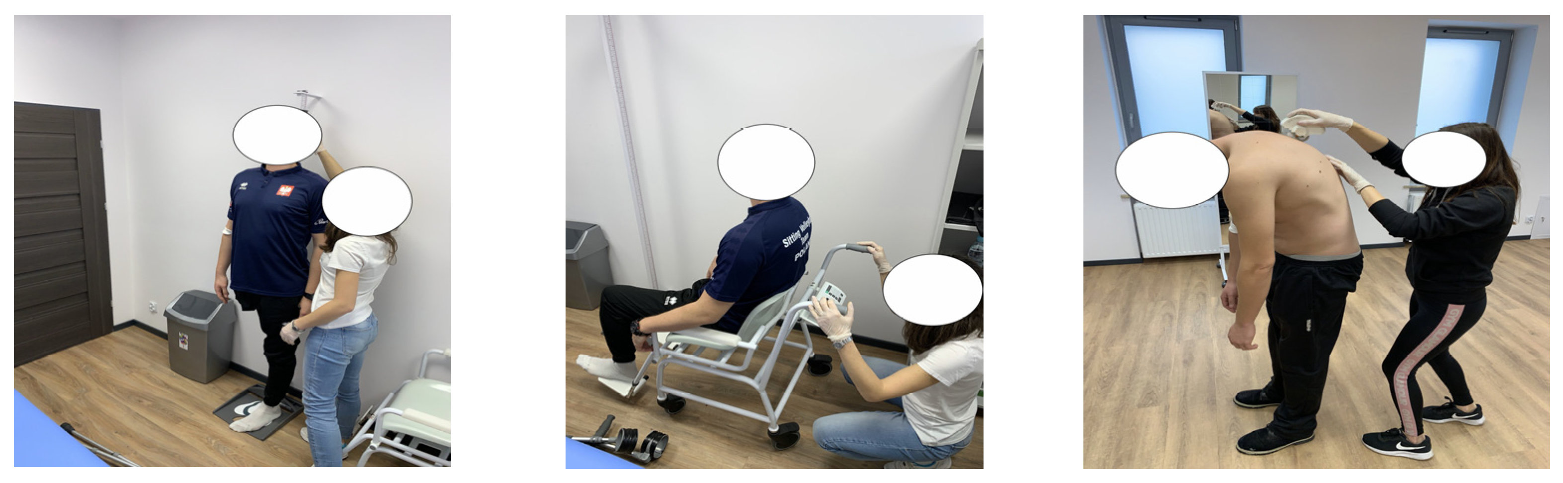
2.2. Methods and Measurements
2.3. Statistical Analysis
3. Results
4. Discussion
Limitations
5. Conclusions
- Internal and external compensation have an effect on the prevalence of deformities of spinal curvature in the sagittal plane, with thoracic hyperkyphosis (38%) and lumbar hyperlordosis (33%) being the most common.
- The neck, lower back (43%), and upper back (38%) were the most frequent painful areas in sitting volleyball players. More severe LBP and upper back pain were correlated with a greater angle of thoracic kyphosis and lumbar lordosis in the habitual position.
- The findings of the study have inspired the programming of an adapted compensatory training program to decrease and prevent the abovementioned spinal deformities and musculoskeletal pain.
Author Contributions
Funding
Institutional Review Board Statement
Informed Consent Statement
Data Availability Statement
Acknowledgments
Conflicts of Interest
Appendix A

| Exercise Number | The Kind of Exercise | Compensatory Influence | Initial Number of Series and Repetitions | Exercise Process–Version A (Easy) | Exercise Process–Version B (Difficult) | Comments |
|---|---|---|---|---|---|---|
| 1. | Mobilization & breathing exercise (thoracic segment) | - Strengthening breathing muscles (inspiratory/expiratory) - Stretching chest muscles - Thoracic spine mobilization | 2 × 10 | I.P. 90/90 sit, arms behind the neck, elbows inside. Movement: 1–4. Deep breath through the nose with progressive backward trunk bending and side elbow abduction. 5–8. Frontal trunk bending with deep exhale through the mouth and inside elbow adduction. E.P. = I.P. | I.P. 90/90 sit, arms crossed on the chest Movement: 1–4. Deep breath through the nose while going up with progressive side arm abduction and backward trunk bending. 5–8. Frontal trunk bending with a deep exhale through the mouth and crossing the arms across the chest while going down to the initial position. E.P. = I.P. | - The inspiratory and expiratory phase should last 4 seconds. |
| 2. | Mobilization & breathing exercise (upper limbs) | - Enhancement of the range of motion in the humeral joint - Stretching chest muscles - Strengthening breathing muscles (inspiratory/expiratory) | The side without a disfunction 2 × 10 The side with a disfunction 2 × 15 | I.P. Lying sideways (left side), legs bent at the knee joints, arms in the front, hands together. Movement: 1–4. Side move of the right arm from the front to the right side while turning the head to the right and taking a deep breath through the nose. 5–8. Side move of the right arm from the right side to the front while turning the head to the left and exhaling deeply through the mouth. E.P. = I.P. | I.P. Lying sideways (left side), legs bent at the knee joints, arms in the front, hands together. Movement: 1–4. Side move of the right arm from the front to the back with a hand rotation to the dorsal position while turning the head to the right and taking a deep breath through the nose. 5–8. Side move of the right arm from the back to the front with a hand rotation to the areal position while turning the head to the left and exhaling deeply through the mouth. E.P. = I.P. | -Versions A and B are performed on both sides. - The inspiratory and expiratory phase should last 4 seconds. |
| 3. | Mobilization exercise (lower limbs) | - Enhancement of the range of motion in the hip joints - Stretching the iliolumbar and quadriceps muscles | The side without a disfunction 2 × 10 The side with a disfunction 2 × 15 | I.P. Seated frontal bend, arms on the floor. Movement: 1. Right leg abduction to the floor. 2. Right leg adduction to the initial position. 3. Left leg abduction to the floor. 4. Left leg adduction to the initial position. E.P. = I.P. | I.P. Seated frontal bend, arms to the side. Movement: 1. Right and left leg abduction to the floor (movement to the right). 2. Leg adduction to the initial position. 3. Left and right leg abduction to the floor. (movement to the left) 4. Leg adduction to the initial position. E.P. = I.P. | |
| 4. | Activation exercise (upper limbs) | - Rotator cuff activation - Balance the shoulder blade rhythm - Thoracic spine activation | Version A 3 × 10 Version B 2 × 8 | I.P. Lying on the front, arms overhead, hands vertical, forehead on the floor. Movement: 1. Raising the arms upwards. 2. Lowering the arms downwards while bending the elbow joints and rotating the hands to the dorsal position. 3. Raising the arms upwards while extending the elbow joints and rotating the hands to the initial position. 4. Lowering arms downwards. E.P. = I.P. | I.P. Lying on the side, arms overhead, hands vertical, forehead on the floor. Movement: 1. Raising the arms upwards. 2. Adduction of the arms sideways. 3. Internal hand rotation to the dorsal position 4. Bending arms at the elbow joints with a side move to the thoracic spine. 5. Lowering the elbows downwards. 6. Raising the elbows upwards with an arm extension to the side (hands in internal rotation). 7. Lifting the arms upwards from the front while rotating the hands to the initial position 8. Lowering arms downwards. E.P. = I.P. | - Before each repetition–retraction and depression of the scapula. |
| 5. | Activation exercise (lower limbs) | - Gluteus muscle activation - Central stability | The side without a disfunction 3 × 6 The side with a disfunction 3 × 10 | I.P. 90/90 sit, front foot in the dorsal position, arms between the knee joint. Movement: 1. Raising the front leg (bent at the knee joint) upwards. 2. Lowering the front leg to the initial position. E.P. = I.P. | I.P. Four-point kneeling Movement: 1. Moving the left leg backward. 2. Holding. 3. Moving the left leg sideways. 4. Lowering the left leg downward. 5. Moving the left leg sideways. 6. Moving the left leg backward. 7. Holding. 8. Lowering the left leg to the initial position. E.P. = I.P. | - Before each repetition–scapula protraction. - Versions A and B are performed on both sides. -Version B–lumbar spine and pelvis without extreme rotation. |
| 6. | Activation exercise (trunk) | - Abdominal muscles activation | Version A 2 × 8 Version B The side without a disfunction 2 × 8 The side with a disfunction 2 × 12 | I.P. Lying on the front, arms crossed on the chest. Movement: 1. Raising the trunk upwards. 2–3. Holding. 4. Lowering the trunk downwards. 5. Raising and turning the trunk to the left side. 6. Lowering the trunk to the initial position. 7. Raising and turning the trunk to the right side. 8. Lowering the trunk to the initial position. E.P. = I.P. | I.P. Lying on the back with the legs bent at the knee joints (feet in a dorsal position), left arm upwards, right arm on the left knee joint– pushing slightly. Movement: 1. Lowering the left arm and the right leg downwards (to the straight body level). 2. Raising the left arm and the right leg upwards to the initial position. E.P. = I.P. | - Verison A: raising and lowering the trunk, vertebra by vertebra. - Version B: the exercise is performed on both sides, and the lumbar spine should globally touch the floor during the entire motor activity. |
| 7. | Directional exercise (thoracic segment) | - Stretching chest muscles - Strengthening the latissimus dorsi muscle and teres major muscle - Strengthening the rotator cuff - Balance the shoulder blade rhythm | 3 × 10 | I.P. Kneeling sit, arms downwards, hands are holding a resistance band (hips widthways). Movement: 1. Moving the right arm from the front to the back. 2. Moving the right arm from the back to the front. E.P. = I.P. | I.P. Kneeling sit, arms downwards, hands are holding a resistance band (hips widthways). Movement: 1. Moving the arms from the front to the back. 2. Moving the arms from the back to the front. E.P. = I.P. | - Before each repetition–retraction and depression of the scapula. - During the exercise, there should not be any compensation with trunk arcuation in the lumbar spine - Version A: the exercise is performed on both sides. During the entire movement activity, the band should be maintained in a slight tension. - Version B: during the entire movement activity, the band should be maintained with the same tension. |
| 8. | Directional exercise (thoracic segment) | - Stretching chest muscles - Strengthening the quadratus lumborum muscle, rhomboid muscle, and latissimus dorsi muscle - Strengthening the rotator cuff - Balance the shoulder-blade rhythm | 3 × 10 | I.P. 90/90 sit, arms in the front (head level), hands are holding the resistance band (shoulders widthways). Movement: 1. Pulling the band from the front to the side. 2. Returning to the initial position. E.P. = I.P. | I.P. 90/90 sit, arms upwards, hands are holding the resistance band (shoulders widthways). Movement: 1. Pulling the band downwards. 2. Returning to the initial position. E.P. = I.P. | - Before each repetition–scapulas retraction and depression. - During the entire movement activity, the band should be kept with a slight tension. |
| 9. | Directional exercise (lumbar segment) | - Strengthening gluteus muscles - Strengthening serratus anterior muscle, superior and external oblique muscles - Central stability | The side without a disfunction 3 × 10 The side with a disfunction 3 × 15 | I.P. Lying sideways (left side) with bent legs, the left arm bent at the elbow joint and lying on the forearm, the right arm bent at the elbow joint (on the trunk level), Movement: 1. Raising trunk upwards while raising the right leg upwards. 2. Holding the trunk with a right arm abduction and while turning the trunk to the right. 3. Holding the trunk with a right arm adduction and a trunk flexion to the body level. 4. Lowering the trunk and legs to the initial position. E.P. = I.P. | I.P. Lying sideways (left side) with bent legs, the left arm bent at the elbow joint and lying on the forearm, the right arm upwards. Movement: 1. Raising the trunk and right leg upwards. 2. Holding the trunk while lowering the right leg. 3. Holding the trunk while raising the right leg upwards. 4. Lowering the trunk and legs to the initial position. E.P. = I.P. | - Before each repetition–retraction and depression of the scapula. - During the motor activity, the trunk should be stabilized. |
| 10. | Directional exercise (lumbar segment) | - Strengthening the rectus abdominis muscle (version A) and superior and external oblique muscles (version B) - Stretching the quadratus lumborum muscle | 3 × 30 s. | I.P. Lying on the back with raised legs bent at the knee joints, arms upwards. Movement: 1. Raising the trunk while moving the arms downwards (knee joints level, hands vertical). 2. Holding (30 s). 3. Lowering the legs to the initial position. 4. Moving the arms upwards. E.P. = I.P. | I.P. Lying on the back with raised legs bent at the knee joints, arms upwards. Movement: 1. Raising the trunk while moving the arms downwards (knee joints level, hands in areal position). Alternative 2–3 (30 s.) 2. Trunk flexion to the left side. 3. Trunk flexion to the left side. 4. Lowering the legs to the initial position while moving the arms upwards. E.P. = I.P. | - During the exercise, the lumbar spine should globally touch the floor. When a lumbar lordosis accentuation can be seen, the exercise should be stopped. |
References
- Grabara, M. A comparison of the posture between young female handball players and non-training peers. J. Back Musculoskelet. Rehabil. 2014, 27, 85–92. [Google Scholar] [CrossRef]
- Roussouly, P.; Pinheiro-Franco, J.L. Biomechanical analysis of the spino-pelvic organization and adaptation in pathology. Eur. Spine J. 2011, 20 (Suppl. S5), 609–618. [Google Scholar] [CrossRef] [Green Version]
- Zwierzchowska, A.; Tuz, J. Evaluation of the impact of sagittal spinal curvatures on musculoskeletal disorders in young people. Med. Pr. 2018, 69, 29–36. [Google Scholar] [CrossRef]
- Grabara, M. Comparison of posture among adolescent male volleyball players and non-athletes. Biol. Sport 2015, 32, 79–85. [Google Scholar] [CrossRef] [PubMed] [Green Version]
- Zaina, F.; Donzelli, S.; Lusini, M.; Minnella, S.; Negrini, S. Swimming and spinal deformities: A cross-sectional study. J. Pediatr. 2015, 166, 163–167. [Google Scholar] [CrossRef] [PubMed]
- Challoumas, D.; Artemiou, A.; Dimitrakakis, G. Dominant vs. non-dominant shoulder morphology in volleyball players and associations with shoulder pain and spike speed. J. Sports Sci. 2017, 35, 65–73. [Google Scholar] [CrossRef] [PubMed] [Green Version]
- Hainline, B.; Turner, J.A.; Caneiro, J.P.; Stewart, M.; Lorimer Moseley, G. Pain in elite athletes-neurophysiological, biomechanical and psychosocial considerations: A narrative review. Br. J. Sports Med. 2017, 51, 1259–1264. [Google Scholar] [CrossRef]
- Ortega-Santiago, R.; González-Aguado, Á.J.; Fernández-de-Las-Peñas, C.; Cleland, J.A.; de-la-Llave-Rincón, A.I.; Kobylarz, M.D.; Plaza-Manzano, G. Pressure pain hypersensitivity and referred pain from muscle trigger points in elite male wheelchair basketball players. Braz. J. Phys. Ther. 2020, 24, 333–341. [Google Scholar] [CrossRef]
- Jeoung, B. Relationship between sitting volleyball performance and field fitness of sitting volleyball players in Korea. J. Exerc. Rehabil. 2017, 13, 647–652. [Google Scholar] [CrossRef] [Green Version]
- Hendershot, B.D.; Nussbaum, M.A. Persons with lower-limb amputation have impaired trunk postural control while maintaining seated balance. Gait Posture 2013, 38, 438–442. [Google Scholar] [CrossRef] [PubMed]
- Ahmadi, S.; Gutierrez, G.L.; Uchida, M.C. Asymmetry in glenohumeral muscle strength of sitting volleyball players: An isokinetic profile of shoulder rotations strength. J. Sports Med. Phys. Fit. 2020, 60, 395–401. [Google Scholar] [CrossRef] [PubMed]
- Kuorinka, I.; Jonsson, B.; Kilbom, A.; Vinterberg, H.; Biering-Sørensen, F.; Andersson, G.; Jørgensen, K. Standardised Nordic questionnaires for the analysis of musculoskeletal symptoms. Appl. Ergon. 1987, 18, 233–237. [Google Scholar] [CrossRef]
- Zwierzchowska, A.; Głowacz, M.; Batko-Szwaczka, A.; Dudzińska-Griszek, J.; Mostowik, A.; Drozd, M.; Szewieczek, J. The Body Mass Index and Waist Circumference as Predictors of Body Composition in Post CSCI Wheelchair Rugby Players (Preliminary Investigations). J. Hum. Kinet. 2014, 43, 191–198. [Google Scholar] [CrossRef] [Green Version]
- Külling, F.A.; Florianz, H.; Reepschläger, B.; Gasser, J.; Jost, B.; Lajtai, G. High Prevalence of Disc Degeneration and Spondylolysis in the Lumbar Spine of Professional Beach Volleyball Players. Orthop. J. Sports Med. 2014, 2. [Google Scholar] [CrossRef]
- Triki, M.; Koubaa, A.; Masmoudi, L.; Fellmann, N.; Tabka, Z. Prevalence and risk factors of low back pain among undergraduate students of a sports and physical education institute in Tunisia. Libyan J. Med. 2015, 10, 26802. [Google Scholar] [CrossRef] [Green Version]
- Yang, C.; Lee, E.; Hwang, E.H.; Kwon, O.; Lee, J.H. Management of Sport Injuries with Korean Medicine: A Survey of Korean National Volleyball Team. Evid.-Based Complement. Altern. Med. 2016, 2016, 8639492. [Google Scholar] [CrossRef]
- Noormohammadpour, P.; Rostami, M.; Mansournia, M.A.; Farahbakhsh, F.; Pourgharib Shahi, M.H.; Kordi, R. Low back pain status of female university students in relation to different sport activities. Eur. Spine J. 2016, 25, 1196–1203. [Google Scholar] [CrossRef]
- Farahbakhsh, F.; Akbari-Fakhrabadi, M.; Shariat, A.; Cleland, J.A.; Farahbakhsh, F.; Seif-Barghi, T.; Mansournia, M.A.; Rostami, M.; Kordi, R. Neck pain and low back pain in relation to functional disability in different sport activities. J. Exerc. Rehabil. 2018, 14, 509–515. [Google Scholar] [CrossRef]
- Movahed, M.; Salavati, M.; Sheikhhoseini, R.; Arab, A.M.; O’Sullivan, K. Single leg landing kinematics in volleyball athletes: A comparison between athletes with and without active extension low back pain. J. Bodyw. Mov. Ther. 2019, 23, 924–929. [Google Scholar] [CrossRef]
- Eshraghi, A.; Safaeepour, Z.; Geil, M.D.; Andrysek, J. Walking and balance in children and adolescents with lower-limb amputation: A review of literature. Clin. Biomech. 2018, 59, 181–198. [Google Scholar] [CrossRef] [PubMed]
- Grabara, M. Anteroposterior curvatures of the spine in adolescent athletes. J. Back Musculoskelet. Rehabil. 2014, 27, 513–519. [Google Scholar] [CrossRef]
- Šarčević, Z.; Tepavčević, A. Association between adolescent idiopathic scoliosis and sacroiliac joint dysfunction in young athletes: A case control study. Medicine (Baltimore) 2019, 98, e15161. [Google Scholar] [CrossRef]
- Fett, D.; Trompeter, K.; Platen, P. Prevalence of back pain in a group of elite athletes exposed to repetitive overhead activity. PLoS ONE 2019, 14, e0210429. [Google Scholar] [CrossRef] [PubMed] [Green Version]
- Molik, B.; Morgulec-Adamowicz, N.; Marszałek, J.; Kosmol, A.; Rutkowska, I.; Jakubicka, A.; Kaliszewska, E.; Kozłowski, R.; Kurowska, M.; Ploch, E.; et al. Evaluation of Game Performance in Elite Male Sitting Volleyball Players. Adapt. Phys. Act. Q. 2017, 34, 104–124. [Google Scholar] [CrossRef]
- Forthomme, B.; Wieczorek, V.; Frisch, A.; Crielaard, J.M.; Croisier, J.L. Shoulder pain among high-level volleyball players and preseason features. Med. Sci. Sports Exerc. 2013, 45, 1852–1860. [Google Scholar] [CrossRef] [PubMed]
- Martelli, G.; Ciccarone, G.; Grazzini, G.; Signorini, M.; Urgelli, S. Isometric evaluation of rotator cuff muscles in volleyball athletes. J. Sports Med. Phys. Fit. 2013, 53, 283–288. [Google Scholar]
- Mizoguchi, Y.; Akasaka, K.; Otsudo, T.; Hall, T. Factors associated with low back pain in elite high school volleyball players. J. Phys. Ther. Sci. 2019, 31, 675–681. [Google Scholar] [CrossRef]
- Paravolley Poland. Available online: Paravolleypoland.eu/historia/ (accessed on 20 August 2021).

| Characteristics (n = 21; nW = 6, nM = 15) | Mean ± SD or Percentage |
|---|---|
| Age (years) | 34.1 ± 7.5 |
| Body mass (kg) | 77.9 ± 16.0 |
| Body height * (cm) | 178.6 ± 0.1 |
| Hip circumference (cm) | 103.3 ± 10.0 |
| Waist circumference (cm) | 89.3 ± 11.1 |
| BMI with a limb deficiency (n = 16) | 23.7 ± 4.9 |
| BMI without a limb deficiency (n = 5) | 24.9 ± 1.9 |
| BAI* (%) | 24.8 ± 3.8 |
| Disability time (years) | 20.2 ± 11.1 |
| Experience in sitting volleyball training (years) | 8.1 ± 7.6 |
| Medical Classification | |
| Amputees in general | 62% |
| Amputees–A1 | 5% |
| Amputees–A2 | 28.5% |
| Amputees–A4 | 28.5% |
| Les Autres in general | 38% |
| Les Autres–LA5 | 33% |
| Les Autres–LA6 | 5% |
| Spinal Curvature Measurements: Sagittal Plane (n = 21; nW = 6, nM = 15) | Mean ± SD (°) | Body Parts (NMQ = 7) | (n = %) |
|---|---|---|---|
| TK–sagittal standing | 37.1 ± 18.8 | Neck | 43% |
| Physiological values | 38.8 ± 18.9 | ||
| Difference | 1.7 ± 2.6 | ||
| TK–sagittal standing flexion | 49.6 ± 23.7 | Shoulders | 19% |
| Physiological values | 51.2 ± 24.9 | ||
| Difference | 3.7 ± 5.0 | ||
| TK–sagittal standing extension | 30.4 ± 15.3 | Upper back | 38% |
| Physiological values | 30.2 ± 16.2 | ||
| Difference | 2.5 ± 3.3 | ||
| LL–sagittal standing | 18.9 ± 13.5 | Elbows | 19% |
| Physiological values | 20.5 ± 14.2 | ||
| Difference | 1.6 ± 2.7 | ||
| LL–sagittal standing flexion | 21.8 ± 13.5 | Wrists | 24% |
| Physiological values | 20.0 ± 12.2 | ||
| Difference | 3.7 ± 5.0 | ||
| LL–sagittal standing extension | 29.8 ± 16.4 | Lower back | 43% |
| Physiological values | 29.6 ± 15.5 | ||
| Difference | 2.6 ± 3.3 | ||
| Hips/ties | 24% | ||
| Knees * | 29% | ||
| Ankles/feet * | 19% | ||
| Body Parts (NMQ = 7) | Sagittal Standing | Sagittal Standing Flexion | Sagittal Standing Extension | |||||||||
|---|---|---|---|---|---|---|---|---|---|---|---|---|
| TH | ± | LL | ± | TH | ± | LL | ± | TH | ± | LL | ± | |
| Neck | 0.3 | 0.3 | SI | SI | 0.4 | SI | −0.09 | SI | 0.4 | 0.2 | 0.2 | SI |
| Arms | 0.3 | SI | SI | SI | 0.3 | SI | SI | SI | SI | SI | SI | SI |
| Upper back | SI | 0.4 | 0.3 | 0.3 | 0.16 | SI | 0.15 | −0.1 | 0.15 | SI | 0.2 | SI |
| Low back | 0.4 | −0.2 | 0.5 | −0.2 | 0.2 | SI | SI | −0.1 | 0.4 | −0.2 | 0.4 | −0.2 |
Publisher’s Note: MDPI stays neutral with regard to jurisdictional claims in published maps and institutional affiliations. |
© 2021 by the authors. Licensee MDPI, Basel, Switzerland. This article is an open access article distributed under the terms and conditions of the Creative Commons Attribution (CC BY) license (https://creativecommons.org/licenses/by/4.0/).
Share and Cite
Gaweł, E.; Zwierzchowska, A. Effect of Compensatory Mechanisms on Postural Disturbances and Musculoskeletal Pain in Elite Sitting Volleyball Players: Preparation of a Compensatory Intervention. Int. J. Environ. Res. Public Health 2021, 18, 10105. https://0-doi-org.brum.beds.ac.uk/10.3390/ijerph181910105
Gaweł E, Zwierzchowska A. Effect of Compensatory Mechanisms on Postural Disturbances and Musculoskeletal Pain in Elite Sitting Volleyball Players: Preparation of a Compensatory Intervention. International Journal of Environmental Research and Public Health. 2021; 18(19):10105. https://0-doi-org.brum.beds.ac.uk/10.3390/ijerph181910105
Chicago/Turabian StyleGaweł, Eliza, and Anna Zwierzchowska. 2021. "Effect of Compensatory Mechanisms on Postural Disturbances and Musculoskeletal Pain in Elite Sitting Volleyball Players: Preparation of a Compensatory Intervention" International Journal of Environmental Research and Public Health 18, no. 19: 10105. https://0-doi-org.brum.beds.ac.uk/10.3390/ijerph181910105