Review

A Review of the Role of Endo/Sarcoplasmic Reticulum-Mitochondria Ca2+ Transport in Diseases and Skeletal Muscle Function

1
School of Sport Science, Beijing Sport University, Beijing 100084, China
2
Faculty of Health, Southern Cross University, East Lismore, NSW 2480, Australia
*
Author to whom correspondence should be addressed.
Int. J. Environ. Res. Public Health 2021, 18(8), 3874; https://0-doi-org.brum.beds.ac.uk/10.3390/ijerph18083874
Submission received: 21 February 2021 / Revised: 23 March 2021 / Accepted: 24 March 2021 / Published: 7 April 2021
(This article belongs to the Special Issue Neuromuscular Responses and Adaptations in Exercise, Sport and Health)

Abstract

:
The physical contact site between a mitochondrion and endoplasmic reticulum (ER), named the mitochondria-associated membrane (MAM), has emerged as a fundamental platform for regulating the functions of the two organelles and several cellular processes. This includes Ca2+ transport from the ER to mitochondria, mitochondrial dynamics, autophagy, apoptosis signalling, ER stress signalling, redox reaction, and membrane structure maintenance. Consequently, the MAM is suggested to be involved in, and as a possible therapeutic target for, some common diseases and impairment in skeletal muscle function, such as insulin resistance and diabetes, obesity, neurodegenerative diseases, Duchenne muscular dystrophy, age-related muscle atrophy, and exercise-induced muscle damage. In the past decade, evidence suggests that alterations in Ca2+ transport from the ER to mitochondria, mediated by the macromolecular complex formed by IP3R, Grp75, and VDAC1, may be a universal mechanism for how ER-mitochondria cross-talk is involved in different physiological/pathological conditions mentioned above. A better understanding of the ER (or sarcoplasmic reticulum in muscle)-mitochondria Ca2+ transport system may provide a new perspective for exploring the mechanism of how the MAM is involved in the pathology of diseases and skeletal muscle dysfunction. This review provides a summary of recent research findings in this area.

1. Introduction

Mitochondria and endoplasmic reticulum (ER) are two organelles in a cell that play essential roles in cellular survival and stress responses. The mitochondrion is the powerhouse of the cell. The ER is involved in protein synthesis, modification, secretion, lipid and steroid synthesis, and modulation of Ca2+ signalling. Sarcoplasmic reticulum (SR) is a special form of ER in striated muscle; it is specialised in the lipid synthesis, Ca2+ modulation, and excitation–contraction coupling. Structurally and functionally, these two organelles are connected via the mitochondria-associated membrane (MAM), a contact site that acts as a platform for inter-organelle communication [1]. In particular, the MAM is involved in a multitude of cellular processes [2], among which the Ca2+ transport from the ER to the mitochondrion (ER-mitochondria Ca2+ transport) is the focus of this review. This process is mediated by the molecular complex inositol 1,4,5-triphosphate receptor (IP3R)–glucose-regulated protein 75 (Grp75)–voltage-dependent anion channel 1 (VDAC1), which works as a hub for cellular survival and death based on its role in regulating mitochondrial physiology and cellular Ca2+ homeostasis [3,4]. During the past decade, alterations of ER-mitochondria Ca2+ transport have been observed in diabetes, obesity, cancer, neurodegenerative diseases like Alzheimer’s disease and Parkinson’s disease, and ischemia/reperfusion injuries in which the integrity or number of MAMs are changed [2,5].
Skeletal muscle is a high energy-consuming organ in which adequate energy supply is essential for muscle performance. Changes in mitochondrial Ca2+ homeostasis could cause Ca2+-dependent damage to mitochondrial structure and function. Conjecture exists that these changes contribute to the age- and disease-related attenuation in muscle performance [6]. There are also some indications that these changes may contribute to exercise-induced muscle damage [7,8,9]. The [Ca2+] variations on either the outer mitochondrial membrane (OMM) surface or within mitochondria ([Ca2+]mt), in response to stimulation of the IP3R, are frequently measured for exploring the Ca2+ communication between the ER and mitochondria in the liver and brain [10,11,12]. However, few studies have been undertaken on skeletal muscle in this regard. The MAM’s role concerning impaired skeletal muscle function has been confirmed in muscular dystrophy, ageing, and insulin resistance, with the evidence that there is a reduction in the number of MAMs in these conditions [13,14,15]. A reduced MAM number has also been implicated in heavy-load endurance exercise that may be caused by SR stress and an increased mitochondrial division rate [16,17,18]. Furthermore, according to the studies investigating MAM and ER-mitochondria Ca2+ transport in response to ER stress [13], mitochondrial dynamics [19], and insulin resistance [20], speculation has arisen that this transport system plays an essential role in mitochondrial Ca2+ overload in response to intensive exercise (Figure 1).
The aims of this review are (1) to summarise the current literature on the function of the MAM and Ca2+ transport from the ER (or SR) to mitochondria, mediated by the IP3R-Grp75-VDAC1 complex, in regulating mitochondrial Ca2+ homeostasis; and (2) to discuss the changes in the MAM and ER/SR-mitochondria Ca2+ transport concerning diseases and skeletal muscle function.

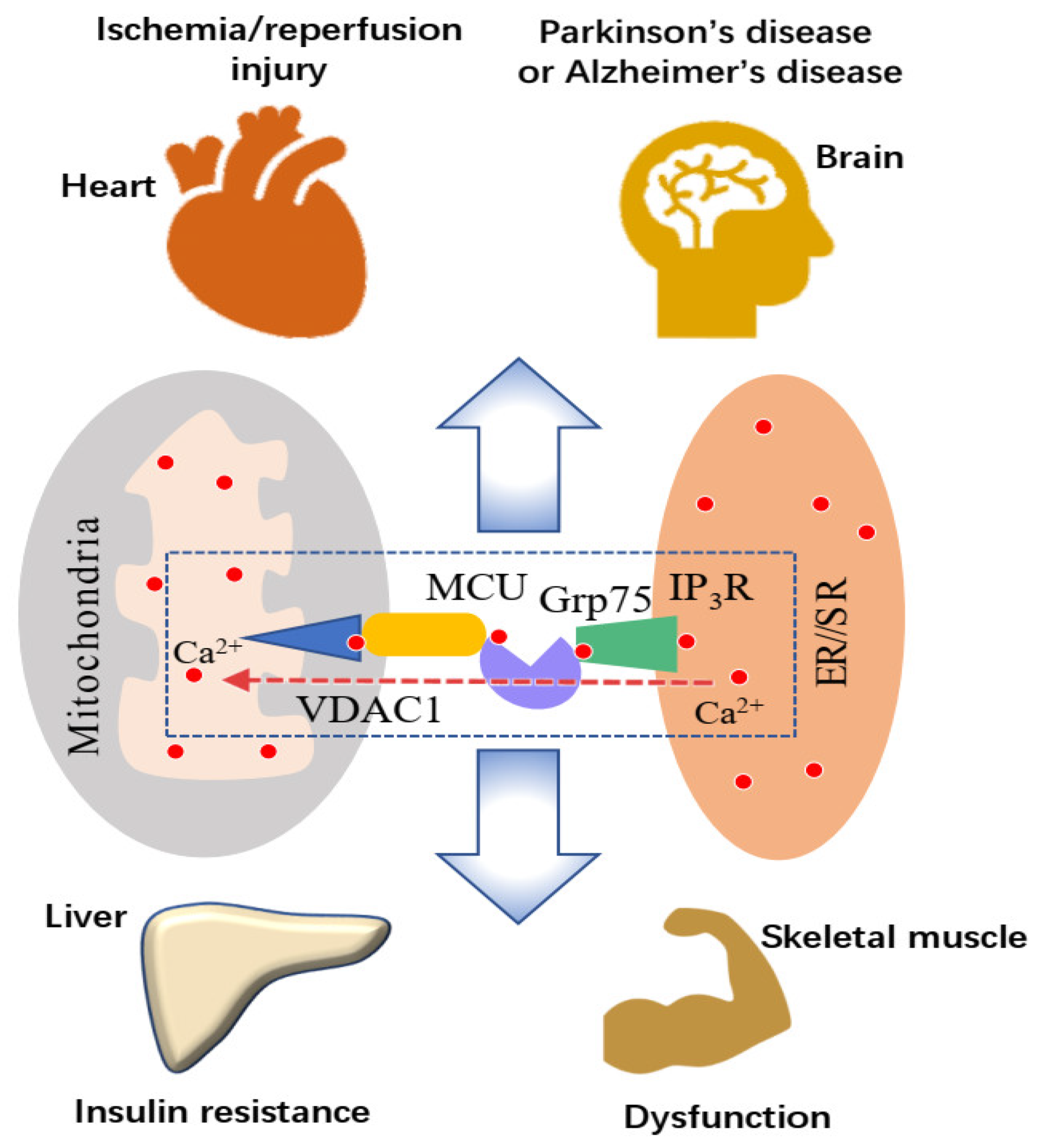
2. Mitochondria-Associated Membrane: A Platform for ER-Mitochondria Ca2+ Transport

The calcium ion (Ca2+) in the mitochondrial matrix is required to stimulate oxidative metabolism by regulating three rate-limiting enzymes in the tricarboxylic acid cycle (TCA): isocitrate dehydrogenase, α-ketoglutarate dehydrogenase, and pyruvate dehydrogenase. It works as a booster in the synthesis of the reduced oxidative phosphorylation substrates (NADH and FADH2) [21]. Physiologically, mitochondrial Ca2+ accumulation is required for enhancing mitochondrial bioenergetics and cellular processes in response to cell stress. However, an excessively high [Ca2+]mt, i.e., mitochondrial Ca2+ overload, could cause a constant opening of the mitochondrial permeability transition pore (mPTP). This may subsequently lead to mitochondrial swelling, rupture of the OMM, and the collapse of the mitochondrial membrane potential, resulting in structural and functional damage to mitochondria [22].

2.1. High Ca2+ Microdomain between the ER and Mitochondria

Under resting conditions, the OMM [23] is permeable to ions and small proteins. Predominantly this is due to the facilitation by the voltage-dependent anion channel 1 (VDAC1) for the diffusion of succinate, pyruvate, ATP, etc., across the OMM [24]. The voltage-dependent anion channel is also permeable to some ions, e.g., K+, Na+, Ca2+, and it has higher permeability to Ca2+ in a high potential state (so-called “closed” state) than in a low potential state (so-called “opened” state). It has been reported that the VDAC1 possesses Ca2+-binding sites, and its closure promotes Ca2+ influx into mitochondria, which causes the opening of the mitochondrial permeability transition pore. Consequently, closure of the VDAC1 is recognised as a pro-apoptotic signal [25]. At the physiological state, [Ca2+] in the mitochondrial intermembrane space is indistinguishable from that in the cytoplasm ([Ca2+]c) (around 100 nM) [26]. Mitochondrial Ca2+ uptake is driven by the electrochemical gradient across the inner mitochondrial membrane (IMM) (−150 to −180 mV, negative inside) [27], while the mitochondrial calcium uniporter (MCU) on the IMM is responsible for mitochondrial Ca2+ uptake. The MCU is characterised by a low Ca2+ affinity (KD = 20–30 μM), which makes it hard to understand how mitochondria uptake Ca2+ at such a low level of [Ca2+] under resting conditions or even in response to physiological stimulation (approximately 1 μM) [28]. This paradox is explained by the widely accepted “Ca2+ microdomain” hypothesis, in which mitochondrial Ca2+ accumulation is effectively favoured by the high Ca2+ microdomain close to the ER Ca2+ release channels [29]. The [Ca2+] at the ER–mitochondrial interface is much higher than that in bulk cytoplasm. This unique [Ca2+] localisation has been directly demonstrated by monitoring the local [Ca2+] at the ER–mitochondrial interface by fluorescent interorganelle linkers [10], or GFP-based Ca2+ probe localised on the cytosolic surface of the OMM [29]. Upon the Ca2+ mobilisation from the ER, the [Ca2+] in local regions near the mitochondrial surface is 5–10-fold higher than that in bulk cytoplasm [30]. The local [Ca2+] at the ER–mitochondrial interface may then rise to several tens of micromolar during the activation of the IP3R. There are two important types of Ca2+ release channels at the ER membrane, ryanodine receptor (RyR) and IP3R. While IP3R is mainly for forming the high Ca2+ microdomain [11,12,31], the RyR has a major role in the excitation–contraction coupling processe in skeletal muscle [32].

2.2. Endoplasmic Reticulum-Mitochondrial Ca2+ Transport

The IP3R is predominantly localised on the ER membrane and is one of the important Ca2+ release channels of ER. There are three IP3R isoforms, IP3R1, IP3R2, and IP3R3 in mammalian cells. The ion permeability of the IP3R could be regulated by inositol 1,4,5-triphosphate (IP3), cytosolic Ca2+, and ATP. Upon receiving a stimulus, IP3 is produced by the cleavage of phosphatidylinositol 4,5-bisphosphate (PIP2). In response to the presence of IP3, the IP3R releases Ca2+ from the ER [33]. The ER is a major organelle for storage of intracellular Ca2+. The efficiency of mitochondrial Ca2+ uptake depends on the close localisation of mitochondria to Ca2+ release channels at the ER membrane that is essential to Ca2+ diffusion from the ER to mitochondria (ER-mitochondria Ca2+ transport) [29]. The IP3R is required for increasing [Ca2+]mt, and at times activation of this protein can result in [Ca2+]mt 20 times higher than the global increase in [Ca2+]c [31]. Additionally, the activation of IP3R may evoke a large [Ca2+] change at the ER–mitochondrial interface even when the increase in [Ca2+]c is suppressed in bulk by the ethylene glycol-bis(β-aminoethyl ether)-N,N,N’,N’-tetraacetic acid (EGTA)/Ca2+ buffer [10]. These findings suggest that IP3R plays a vital role in mediating ER-mitochondria Ca2+ transport. The VDAC1 protein is also required for the control of ER-mitochondria Ca2+ transport. Recombinant expression of the VDAC1 could enhance the transfer of Ca2+ from the microdomains to mitochondria [34]. Moreover, a recombinant expression of IP3R-LBD224–605 increases the ER-mitochondria Ca2+ transport, and this phenomenon can be inhibited by the downregulation of Grp75 [35]. The Grp75 is a 75 kDa glucose-regulated protein and molecular chaperone that belongs to the heat shock protein 70 (Hsp70) family. Predominantly, Grp75 is localised in the mitochondrial matrix, while it is also found in cytoplasm or other subcellular compartments [35]. This protein is demonstrated to tether the N-terminal domain of the IP3R to VDAC1 and enhance mitochondrial Ca2+ accumulation by stabilising the conformation or coupling of IP3R and VDAC1 [36].
The physical link between the ER and mitochondria is necessary for normal ER-mitochondrial Ca2+ transport, with the [Ca2+]mt being increased by moving the ER closer to the mitochondria [37]. Additionally, the ER-mitochondria Ca2+ transport may depend on an appropriate distance between the ER and mitochondria. One study reported that a distance of ~20 nm or shorter between the two organelles is required for proper ER-mitochondria Ca2+ transport [30]. Although the optimal or average width of the MAM that coordinates efficient ER-mitochondria Ca2+ transport is still unknown, it is hypothesised that the width can impact the Ca2+ transfer by regulating the assembly of the IP3R-Grp75-VDAC1 complex [30].
During the past decade, the function of the IP3R-Grp75-VDAC1 complex in transporting Ca2+ has attracted a great deal of attention [4,13,38]. Numerous proteins, like DJ-1 [39], transglutaminase type 2 (TG2) [35], FUN14 domain-containing protein 1 (FUNDC1) [19], glycogen synthase kinase-3β (GSK3β) [5], pyruvate dehydrogenase kinase 4(PDK4) [15], etc., are found to regulate the integrity of MAM, or interact with the complex IP3R-Grp75-VDAC1, influencing Ca2+ transport from ER to mitochondria. Abnormal transmission of Ca2+ between the ER and mitochondria has been implicated in several pathological conditions [2,5,40,41,42,43]. According to the current literature, maintaining the structural integrity of the MAM and ER-mitochondria Ca2+ transport within normal physiological ranges may be essential to retarding either the pathogenesis or the progression of these diseases.

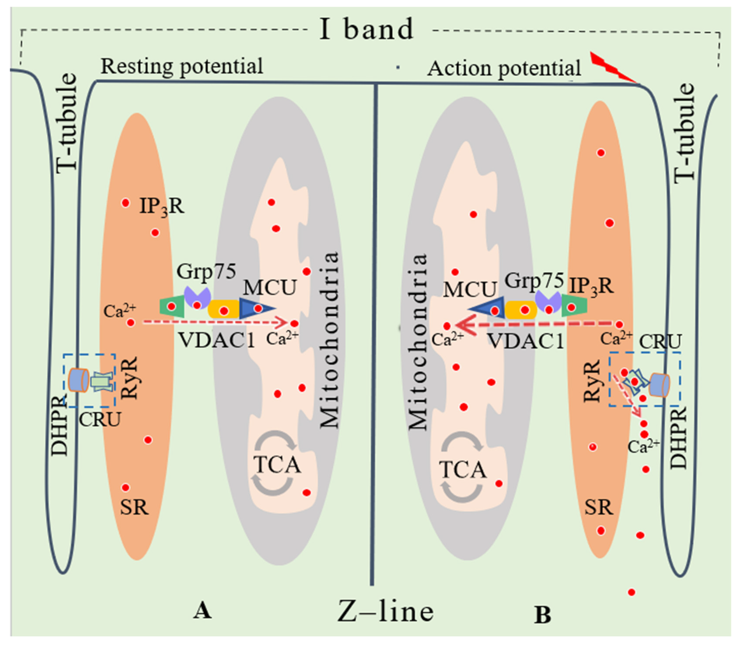
3. Effects of the MAM and SR-Mitochondria Ca2+ Transport on Skeletal Muscle Function

Over the past 40 years, the idea that mitochondrial dysfunction contributes to muscle function impairment has been hypothesised and tested [6]. It is now understood that the disruption of mitochondrial Ca2+ homeostasis is a general mechanism for mitochondrial function impairment [44]. Mitochondria-associated membranes were found in skeletal muscle about 30 years ago [45], adding to the evidence that the MAM contributes to regulating mitochondrial Ca2+ homeostasis. As the SR is a large Ca2+ store regulating intracellular and mitochondrial Ca2+ concentrations, the Ca2+ cross-talk between SR and mitochondria has attracted increased attention in the recent past [23].

3.1. Mitochondria-Associated Membrane in Cardiac and Skeletal Muscle

Close localisation between the SR and mitochondria was first demonstrated in frog skeletal muscle using scanning electron microscopy in 1987 [45]. More recently, Ainbinder et al. (2015) found that mitochondria were located close to the I-band in the flexor digitorum of mice, and that the OMM was linked to the terminal cistern of SR by MAMs. They additionally found that the MAMs in the skeletal muscle were established and maintained during postnatal maturation. At the same time, mitochondria shifted from a longitudinal cluster to a transversal orientation and moved from the H-band to the I-band [46]. Furthermore, protein analysis showed that SR membrane fractions contained proteins involved in oxidative phosphorylation and the TCA [47]. These findings indicate that these proteins in mitochondria are connected with proteins in the cell’s SR, and that the SR and mitochondria are also functionally connected through MAMs [47].
There is currently limited experimental research demonstrating the molecular composition of SR–mitochondria interaction and its specific functions in skeletal muscle. However, numerous studies have investigated MAM formation proteins and their functional relevance in the brain, liver, and cardiac muscle [48,49,50]. A protein analysis of the MAM in skeletal muscle has identified 459 proteins, including 101 membrane proteins, in which VDAC1, VDAC2, and VDAC3 might participate in the formation of the tethers between SR and mitochondria [51]. Among the MAM constitutive proteins, mitofusin 2 (Mfn2) is most commonly recognised as a molecule for regulating the structure of the MAMs [52,53]. However, in other studies, Mfn2 is demonstrated to be a physical tether protein [54] or a tethering antagonist [55]. In skeletal muscle, Mfn2 may work as a tether protein because its depletion leads to mitochondrial migration away from the I-band towards the A-band, resulting in reduced MAMs [46]. Moreover, proteins that are involved in Ca2+ transport from the ER to mitochondria, like IP3R and Grp75, have been attracting increased investigative attention in the recent past. Additionally, the investigations suggest these ‘emerging factors’ being directly involved in the changes of mitochondrial Ca2+ homeostasis [15,56].

3.2. Ca2+ Release by SR in Skeletal Muscle Fibres

It is well known that Ca2+ is essential in the excitation–contraction coupling process during muscle contraction [57]. Calcium ions required for skeletal muscle contraction are predominantly released from the SR via the Ca2+ release unit (CRU). The CRU consists of the L-type Ca2+ channels (dihydropyridine receptors, DHPRs) on the exterior membrane and the RyRs on the SR [58]. The major type of RyR in skeletal muscle is RyR1. The RyR1 on the SR membrane is positioned facing the voltage-dependent L-type Ca2+ channels (Cav1.1) that are located on the transverse tubules (T-tubules, Figure 2). This position is the key factor for Ca2+ being released by the RyR upon T-tubule membrane depolarisation, causing a conformational change of the DHPRs [59].
Furthermore, Ca2+ is also required by the mitochondrial matrix in the energy (ATP) supplying processes for muscle contraction [60]. These processes require Ca2+ to be under precise physiological control within the mitochondria of skeletal muscle [61]. In skeletal muscle, mitochondria are located close to the CRU [14]. This localisation might be due to the inhibitory effect of Ca2+ on mitochondria motility so that mitochondria are kept around the Ca2+ intracellular stores [62]. In addition to Ca2+ release by RyR’s from the SR to the cytosol, the cellular membrane depolarisation could also cause Ca2+ release via the IP3R protein [63]. The Ca2+ cross-talk between SR and mitochondria was demonstrated by comparing the [Ca2+]mt with the [Ca2+]c in response to caffeine when using a fast Ca2+ buffer BAPTA (1, 2-bis(2-aminophenoxy)ethane-N, N, Nʹ, Nʹ-tetraacetic acid) in the cytosol, with the mitochondrial Ca2+ concentration increasing and cytosolic Ca2+ showing little change [64]. This finding indicates that the Ca2+ could be transported from the SR to mitochondria directly, not depending on the increased cytosolic Ca2+ concentration. According to Csordás et al. (2010), Ca2+ cross-talk between the SR and mitochondria is likely caused by activation of IP3R and depends on the close location between the SR and mitochondria. It was found that mitochondrial Ca2+ concentration changes in skeletal muscle may depend on the mitochondrial location being close to the CRU’s [65]. Furthermore, overexpression of VDAC1 can increase Ca2+ transport into mitochondria from the high Ca2+ microdomain around SR [34].
It is known that Ca2+ can be transported from the ER/SR to mitochondria via the IP3R-Grp75-VDAC1 complex in cardiac muscle and other types of cells. However, whether this is a major pathway and how the mitochondrial and cellular function is affected by this pathway in skeletal muscle are currently not clear and require further investigation.

4. Sarcoplasmic Reticulum-Mitochondrial Ca2+ Transport in Relation to Skeletal Muscle Dysfunction

Mitochondria-associated membranes have been shown to affect the metabolic adaption and cellular function in response to physical environmental changes [66]. Evidence suggests that changes in the MAM and ER/SR-mitochondria Ca2+ transport are related to several health conditions and impaired skeletal muscle function, such as insulin resistance [40], Duchenne muscular dystrophy (DMD), or age-related muscle dystrophy [67]. Insulin resistance is a metabolic disorder, and the MAM has been shown to be a hub for insulin resistance in three key insulin-targeting tissues or cells: the liver [68], adipocytes [69], and skeletal muscle [70]. Reorganisation of the MAMs and mitochondrial Ca2+ overload has been shown in the liver of obese animals. A downregulation of MAMs and the ER-mitochondria Ca2+ transport has been demonstrated to improve glucose metabolism [71].
Additionally, it has been found that a reduction in ER-mitochondria Ca2+ transport may contribute to insulin resistance in liver cells and adipocytes [40,72]. The disruption of the MAMs in hepatocytes plays a central role in developing insulin resistance [72]. This was demonstrated by using sulforaphane for treating insulin resistance, with insulin resistance being improved along with a reduced MAM disruption and impaired interaction between VDAC1 and IP3R1 [56]. Moreover, in adipose tissue of obese mice and humans who were suffering from type 2 diabetes mellitus, [Ca2+]mt was found to be increased, but [Ca2+]c showed no change, with an accompanying rise in MCU expression and activity [73].
Skeletal muscle is an important regulator for whole-body glucose homeostasis and has been extensively studied concerning insulin resistance pathologies [74]. Tubbs et al. (2018 b) showed that the gastrocnemius muscle of obese mice with type 2 diabetes exhibited a significant disruption of SR–mitochondria interaction, which is compatible with experimental findings in human patients with obesity and type 2 diabetes [70]. These patients showed a reduction in VDAC1/IP3R interaction, indicating that the Ca2+ communication between the SR and mitochondria is likely to play a key role in altered glucose homeostasis in type 2 diabetes [70]. One study by Zhe et al. (2020) investigated the improvement of glucose intolerance induced by swimming. The findings suggested that decreased MAM quantity might help in understanding the mechanism of type 2 diabetes mellitus [75]. However, contrary to the findings mentioned above, there has been evidence suggesting that the increased formation of MAMs is a contributor to insulin resistance. For example, one study found that mitochondrial Ca2+ accumulation in skeletal muscle with insulin resistance may be caused by augmented MAM formation [15]. This is because insulin signalling can be improved by preventing MAM-mediated mitochondrial Ca2+ accumulation and mitochondrial dysfunction through inhibiting pyruvate dehydrogenase kinase 4 [15]. The opening of mitochondrial permeability transition pores is a necessary contributor to skeletal muscle insulin resistance because it prevents the insulin-induced translocation of glucose transporter 4 into the cytoplasm [76]. Additionally, mitochondrial Ca2+ overload is commonly regarded as a central contributor to the opening of mitochondrial permeability transition pores [6,24]. As shown above, controversy still exists regarding the relationship between insulin resistance and changes in the MAMs, SR-mitochondria Ca2+ transport, and mitochondrial Ca2+ homeostasis in skeletal muscle. It is also far from clear whether the alterations in MAM and IP3R-Grp75-VDAC1 result from or are the cause of muscular insulin resistance.
Muscle weakness and wasting have long been confirmed as pathogenic DMD symptoms [77]. It is accepted that the defective gene dystrophin is involved in the DMD pathology [78], and it has been suggested that the increase in Ca2+ release by the sarcoplasmic reticulum is involved in calcium dysregulation in DMD myotubes. In DMD foetal muscle, myopathology begins with a significant delay of foetal muscle differentiation at an early foetal stage. This phenomenon is related to the activation of the PLC/IP3R/calcium/PKCα pathway, which may be induced by the absence of dystrophy [79]. An increased IP3R activity negatively regulates autophagy and elevates the [Ca2+]c and [Ca2+]mt [80]. However, the increase in IP3R expression does not enhance mitochondrial Ca2+ uptake because of the reduction of the complex quantity of IP3R-Grp75-VDAC1 [80]. Thus, the reduced ER-mitochondria Ca2+ transport is a suspected contributor to the reduced [Ca2+]mt in DMD [13].
Disrupted SR-mitochondria Ca2+ transport may also potentially play a role in muscle dysfunction in ageing through mitochondrial Ca2+ homeostasis impairment. Further, tetanic stimulation on the flexor digitorum brevis of mice was reportedly the cause of reduced mitochondrial Ca2+ uptake, along with a decrease in the association between mitochondria and CRUs in aged mice [14]. In aged mice with knockdown of mitofusin 2, repetitive high-frequency tetanic stimulation reduced an impaired association between CRUs and mitochondria. This was caused by a shift of mitochondria away from the triadic positions, which may explain the declined muscle performance associated with ageing [14]. Gill et al. (2019) reported that the SR-mitochondria Ca2+ transport proteins IP3R1, Grp75, and VDAC were downregulated and that the IP3 gene expression reduced in the skeletal muscle of aged mice [81]. An ameliorated age-dependent skeletal muscle dysfunction was reported to be related to a reduced SR Ca2+ release and increased SR Ca2+ load [82]. Exercise-induced muscle function improvement is hypothesised to be attributed to the increase in [Ca2+]mt caused by the increased MCU expression [83], which is probably related to the improved/maintained association between CRUs and mitochondria [84]. Based on these findings, the increasing loss of Ca2+ homeostasis in the SR and mitochondria are said to cause the functional decline of skeletal muscle in ageing. The defective SR-mitochondria Ca2+ cross-talk may precede muscle function impairment in age-related dystrophy, but further studies on molecular mechanism and Ca2+ signalling are required to understand this explanation better.

5. Sarcoplasmic Reticulum-Mitochondria Ca2+ Transport and Mitochondrial Ca2+ Overload

It is commonly accepted that ER stress, which can be caused by ageing, genetic mutations, or environmental factors, can result in various diseases, including those mentioned above [85]. The increased ER Ca2+ release through Ca2+ channels is an essential cause of ER stress, and ER stress can be attenuated by the cone-specific gene deletion of IP3R1 [86]. The disturbance of mitochondrial Ca2+ homeostasis, mostly through mitochondrial Ca2+ overload, also occurs in the diseases mentioned above, and it is probably due to the increased ER-mitochondria Ca2+ transport. In the early phase of ER stress, mitochondria were found to migrate to the ER’s periphery [18,87], while in the later phase, mitochondrial Ca2+ overload was shown to be caused by mitochondrial migration to the ER surface, resulting in mitochondria-dependent apoptosis [88]. In addition, according to a recent study on macromolecular complex IP3R-Grp75-VDAC1, Ca2+ transport from the ER to the mitochondria is likely to contribute to podocyte apoptosis by facilitating mitochondrial Ca2+ overload [4]. As can be seen from the evidence discussed above, the Ca2+ transport from the ER to the mitochondria may contribute to the ER stress and mitochondrial Ca2+ overload at the same time, but this speculation needs further experimental evidence.

6. Conclusions

This review focused on the mitochondria-associated membrane’s role, particularly the IP3R-Grp75-VDAC1 pathway, in calcium ion transport from the endoplasmic/sarcoplasmic reticulum to mitochondria, in relation to normal and abnormal cellular functions. Current evidence suggests that the distance between the ER/SR and mitochondria, and the modulation of the IP3R, Grp75, and VDAC1 activities, play a critical role in regulating mitochondrial Ca2+ homeostasis. A disruption in this pathway and subsequently Ca2+ homeostasis (such as mitochondrial Ca2+ overload or insufficient mitochondrial Ca2+) is believed to be a common causative factor in some health conditions, such as neurodegenerative diseases, obesity, insulin resistance, cancer, and ischemia/reperfusion damage, as well as in age-related muscle dystrophy and intensive exercise-induced reduction of skeletal muscle function. However, the exact cause-and-effect relationship and the potential physiological/pathological mechanisms under specific conditions require further investigation to shed light on potential therapeutic targets or interventions for such conditions.

Author Contributions

S.-S.Z. and S.Z. contributed to the designing and drafting of the manuscript. Z.J.C.-M. contributed to the drafting and editing of the manuscript. J.-P.L. and R.-Y.W. assisted in the design of the manuscript. All authors have read and agreed to the published version of the manuscript.

Funding

This review was supported by the National Natural Science Foundation of China (31471133) and Fundamental Research Funds for the Central Universities (2019PT013, 2020025).

Institutional Review Board Statement

Not applicable. Ethical review and approval were waived for this study, due to it was an analysis of the published literature only.

Informed Consent Statement

Not applicable.

Data Availability Statement

Not applicable.

Acknowledgments

The first author received a stipend scholarship from the China Scholarship Council during her study at Southern Cross University, where she constructed the draft of this review.

Conflicts of Interest

The authors declare no conflict of interest.

Abbreviations

ER, endoplasmic reticulum; MAM, mitochondria-associated membrane; SR, sarcoplasmic reticulum, IP3R: inositol 1,4,5-trisphosphate receptor; Grp75, glucose regulated protein 75; VDAC1, voltage-dependent anion channel 1; [Ca2+], Ca2+ concentration; TAC, tricarboxylic acid cycle; IMM, inner mitochondrial membrane; MCU, mitochondrial calcium uniporter; [Ca2+]mt, mitochondrial Ca2+ concentration; [Ca2+]c, cytosolic Ca2+ concentration; DHPR, dihydropyridine receptor; RyR, ryanodine receptor; Tubule, transversal tubule; CRU, calcium release unit.

References

  1. Rowland, A.A.; Voeltz, G.K. Endoplasmic reticulum-mitochondria contacts: Function of the junction. Nat. Rev. Mol. Cell Biol. 2012, 13, 607–625. [Google Scholar] [CrossRef] [Green Version]
  2. Simmen, T.; Herrera-Cruz, M.S. Plastic mitochondria-endoplasmic reticulum (ER) contacts use chaperones and tethers to mould their structure and signaling. Curr. Opin. Cell Biol. 2018, 53, 61–69. [Google Scholar] [CrossRef]
  3. Rieusset, J. The role of endoplasmic reticulum-mitochondria contact sites in the control of glucose homeostasis: An update. Cell Death Dis. 2018, 9, 388. [Google Scholar] [CrossRef] [PubMed] [Green Version]
  4. Xu, H.; Guan, N.; Ren, Y.-L.; Wei, Q.-J.; Tao, Y.-H.; Yang, G.-S.; Liu, X.-Y.; Bu, D.-F.; Zhang, Y.; Zhu, S.-N. IP3R-Grp75-VDAC1-MCU calcium regulation axis antagonists protect podocytes from apoptosis and decrease proteinuria in an Adriamycin nephropathy rat model. BMC Nephrol. 2018, 19, 140. [Google Scholar] [CrossRef]
  5. Gomez, L.; Thiebaut, P.A.; Paillard, M.; Ducreux, S.; Abrial, M.; Crola Da Silva, C.; Durand, A.; Alam, M.R.; Van Coppenolle, F.; Sheu, S.S.; et al. The SR/ER-mitochondria calcium crosstalk is regulated by GSK3β during reperfusion injury. Cell Death Differ. 2015, 22, 1890. [Google Scholar] [CrossRef] [Green Version]
  6. Zulian, A.; Schiavone, M.; Giorgio, V.; Bernardi, P. Forty years later: Mitochondria as therapeutic targets in muscle diseases. Pharmacol. Res. 2016, 113 Pt A, 563–573. [Google Scholar] [CrossRef]
  7. Rattray, B.; Thompson, M.; Ruell, P.; Caillaud, C. Specific training improves skeletal muscle mitochondrial calcium homeostasis after eccentric exercise. Eur. J. Appl. Physiol. 2013, 113, 427–436. [Google Scholar] [CrossRef]
  8. Rizo-Roca, D.; Rios-Kristjansson, J.G.; Nunez-Espinosa, C.; Santos-Alves, E.; Magalhaes, J.; Ascensao, A.; Pages, T.; Viscor, G.; Torrella, J.R. Modulation of mitochondrial biomarkers by intermittent hypobaric hypoxia and aerobic exercise after eccentric exercise in trained rats. Appl. Physiol. Nutr. Metab. 2017, 42, 683–693. [Google Scholar] [CrossRef]
  9. Yoo, S.Z.; No, M.H.; Heo, J.W.; Park, D.H.; Kang, J.H.; Kim, J.H.; Seo, D.Y.; Han, J.; Jung, S.J.; Kwak, H.B. Effects of acute exercise on mitochondrial function, dynamics, and mitophagy in rat cardiac and skeletal muscles. Int. Neurourol. J. 2019, 23 (Suppl. 1), S22–S31. [Google Scholar] [CrossRef]
  10. Csordás, G.; Várnai, P.; Golenár, T.; Roy, S.; Purkins, G.; Schneider, T.G.; Balla, T.; Hajnóczky, G. Imaging interorganelle contacts and local calcium dynamics at the ER-mitochondrial interface. Mol. Cell 2010, 39, 121–132. [Google Scholar] [CrossRef]
  11. Rizzuto, R.; Brini, M.; Murgia, M.; Pozzan, T. Microdomains with high Ca2+ close to IP3-sensitive channels that are sensed by neighboring mitochondria. Science 1993, 262, 744–747. [Google Scholar] [CrossRef]
  12. Rizzuto, R.; Pinton, P.; Carrington, W.; Fay, F.S.; Fogarty, K.E.; Lifshitz, L.M.; Tuft, R.A.; Pozzan, T. Close contacts with the endoplasmic reticulum as determinants of mitochondrial Ca2+ responses. Science 1998, 280, 1763–1766. [Google Scholar] [CrossRef] [PubMed]
  13. Pauly, M.; Angebault-Prouteau, C.; Dridi, H.; Notarnicola, C.; Scheuermann, V.; Lacampagne, A.; Matecki, S.; Fauconnier, J. ER stress disturbs SR/ER-mitochondria Ca2+ transfer: Implications in Duchenne muscular dystrophy. Biochim. Biophys. Acta Mol. Basis Dis. 2017, 1863, 2229–2239. [Google Scholar] [CrossRef] [PubMed]
  14. Pietrangelo, L.; D’Incecco, A.; Ainbinder, A.; Michelucci, A.; Kern, H.; Dirksen, R.T.; Boncompagni, S.; Protasi, F. Age-dependent uncoupling of mitochondria from Ca2+ release units in skeletal muscle. Oncotarget 2015, 6, 35358–35371. [Google Scholar] [CrossRef] [Green Version]
  15. Thoudam, T.; Ha, C.-M.; Leem, J.; Chanda, D.; Park, J.-S.; Kim, H.-J.; Jeon, J.-H.; Choi, Y.-K.; Liangpunsakul, S.; Huh, Y.H.; et al. PDK4 augments ER-mitochondria contact to dampen skeletal muscle insulin signaling during obesity. Diabetes 2019, 68, 571–586. [Google Scholar] [CrossRef]
  16. Liu, X.; Yu, Y.; Wang, Y.; Yan, S.; Wang, R.; Li, J. Effect of High-load Exercise on the Mitochondrial Fusion and Fission Proteins in Rat Skeletal muscle. J. Cap. Univ. Phys. Educ. Sports 2016, 28, 167–171. [Google Scholar] [CrossRef]
  17. Shang, H.; Bai, S.; Xia, Z.; Zhou, Y.; Wang, R. Effects of acupuncture on mitochondrial structure and function of skeletal muscle in rats with heavy load exercise. Chin. J. Rehabil. Med. 2018, 33, 901–908. [Google Scholar] [CrossRef]
  18. Bravo, R.; Vicencio, J.M.; Parra, V.; Troncoso, R.; Munoz, J.P.; Bui, M.; Quiroga, C.; Rodriguez, A.E.; Verdejo, H.E.; Ferreira, J.; et al. Increased ER-mitochondrial coupling promotes mitochondrial respiration and bioenergetics during early phases of ER stress. J. Cell Sci. 2011, 124 Pt 13, 2143–2152. [Google Scholar] [CrossRef] [Green Version]
  19. Wu, S.; Lu, Q.; Wang, Q.; Ding, Y.; Ma, Z.; Mao, X.; Huang, K.; Xie, Z.; Zou, M.-H. Binding of FUN14 domain containing 1 with inositol 1,4,5-trisphosphate receptor in mitochondria-associated endoplasmic reticulum membranes maintains mitochondrial dynamics and function in hearts in vivo. Circulation 2017, 136, 2248–2266. [Google Scholar] [CrossRef]
  20. Fernström, M.; Tonkonogi, M.; Sahlin, K. Effects of acute and chronic endurance exercise on mitochondrial uncoupling in human skeletal muscle. J. Physiol. 2004, 554 Pt 3, 755–763. [Google Scholar] [CrossRef]
  21. Mammucari, C.; Raffaello, A.; Vecellio Reane, D.; Gherardi, G.; De Mario, A.; Rizzuto, R. Mitochondrial calcium uptake in organ physiology: From molecular mechanism to animal models. Pflug. Arch. Eur. J. Physiol. 2018, 470, 1165–1179. [Google Scholar] [CrossRef] [Green Version]
  22. Xue, J.; Li, R.; Zhao, X.; Ma, C.; Lv, X.; Liu, L.; Liu, P. Morusin induces paraptosis-like cell death through mitochondrial calcium overload and dysfunction in epithelial ovarian cancer. Chem. Biol. Interact. 2018, 283, 59–74. [Google Scholar] [CrossRef]
  23. Missiroli, S.; Danese, A.; Iannitti, T.; Patergnani, S.; Perrone, M.; Previati, M.; Giorgi, C.; Pinton, P. Endoplasmic reticulum-mitochondria Ca2+ crosstalk in the control of the tumor cell fate. Biochim. Biophys. Acta Mol. Cell Res. 2017, 1864, 858–864. [Google Scholar] [CrossRef]
  24. Hurst, S.; Hoek, J.; Sheu, S.-S. Mitochondrial Ca2+ and regulation of the permeability transition pore. J. Bioenerg. Biomembr. 2017, 49, 27–47. [Google Scholar] [CrossRef] [Green Version]
  25. Shoshan-Barmatz, V.; Shteinfer-Kuzmine, A.; Verma, A. VDAC1 at the Intersection of Cell Metabolism, Apoptosis, and Diseases. Biomolecules 2020, 10, 1485. [Google Scholar] [CrossRef]
  26. Shoshan-Barmatz, V.; Krelin, Y.; Shteinfer-Kuzmine, A. VDAC1 functions in Ca2+ homeostasis and cell life and death in health and disease. Cell Calcium 2018, 69, 81–100. [Google Scholar] [CrossRef]
  27. Xu, Z.; Zhang, D.; He, X.; Huang, Y.; Shao, H. Transport of calcium ions into mitochondria. Curr. Genom. 2016, 17, 215–219. [Google Scholar] [CrossRef] [PubMed] [Green Version]
  28. Penna, E.; Espino, J.; De Stefani, D.; Rizzuto, R. The MCU complex in cell death. Cell Calcium 2018, 69, 73–80. [Google Scholar] [CrossRef]
  29. Giacomello, M.; Drago, I.; Bortolozzi, M.; Scorzeto, M.; Gianelle, A.; Pizzo, P.; Pozzan, T. Ca2+ hot spots on the mitochondrial surface are generated by Ca2+ mobilization from stores, but not by activation of store-operated Ca2+ channels. Mol. Cell 2010, 38, 280–290. [Google Scholar] [CrossRef] [PubMed]
  30. Giacomello, M.; Pellegrini, L. The coming of age of the mitochondria-ER contact: A matter of thickness. Cell Death Differ. 2016, 23, 1417–1427. [Google Scholar] [CrossRef]
  31. Csordás, G.; Thomas, A.P.; Hajnóczky, G. Quasi-synaptic calcium signal transmission between endoplasmic reticulum and mitochondria. EMBO J. 1999, 18, 96–108. [Google Scholar] [CrossRef] [PubMed] [Green Version]
  32. Ríos, E.; Gillespie, D.; Franzini-Armstrong, C. The binding interactions that maintain excitation-contraction coupling junctions in skeletal muscle. J. Gen. Physiol. 2019, 151, 593–605. [Google Scholar] [CrossRef] [Green Version]
  33. Katona, M.; Alzayady, K.; Yule, D.I.; Hajnoczky, G. IP3 receptor isoform dependence of the ER-mitochondrial Ca2+-transfer in mammalian cells. Biophys. J. 2017, 112, 130a. [Google Scholar] [CrossRef] [Green Version]
  34. Rapizzi, E.; Pinton, P.; Szabadkai, G.; Wieckowski, M.R.; Vandecasteele, G.; Baird, G.; Tuft, R.A.; Fogarty, K.E.; Rizzuto, R. Recombinant expression of the voltage-dependent anion channel enhances the transfer of Ca2+ microdomains to mitochondria. J. Cell Biol. 2002, 159, 613–624. [Google Scholar] [CrossRef] [PubMed] [Green Version]
  35. D’Eletto, M.; Rossin, F.; Occhigrossi, L.; Farrace, M.G.; Faccenda, D.; Desai, R.; Marchi, S.; Refolo, G.; Falasca, L.; Antonioli, M.; et al. Transglutaminase Type 2 Regulates ER-Mitochondria Contact Sites by Interacting with GRP75. Cell Rep. 2018, 25, 3573–3581. [Google Scholar] [CrossRef] [Green Version]
  36. Szabadkai, G.; Bianchi, K.; Várnai, P.; De Stefani, D.; Wieckowski, M.R.; Cavagna, D.; Nagy, A.I.; Balla, T.; Rizzuto, R. Chaperone-mediated coupling of endoplasmic reticulum and mitochondrial Ca2+ channels. J. Cell Biol. 2006, 175, 901–911. [Google Scholar] [CrossRef] [Green Version]
  37. Csordás, G.; Renken, C.; Várnai, P.; Walter, L.; Weaver, D.; Buttle, K.F.; Balla, T.; Mannella, C.A.; Hajnóczky, G. Structural and functional features and significance of the physical linkage between ER and mitochondria. J. Cell Biol. 2006, 174, 915–921. [Google Scholar] [CrossRef] [Green Version]
  38. Van Vliet, A.R.; Verfaillie, T.; Agostinis, P. New functions of mitochondria associated membranes in cellular signaling. Biochim. Biophys. Acta 2014, 1843, 2253–2262. [Google Scholar] [CrossRef] [Green Version]
  39. Liu, Y.; Ma, X.; Fujioka, H.; Liu, J.; Chen, S.; Zhu, X. DJ-1 regulates the integrity and function of ER-mitochondria association through interaction with IP3R3-Grp75-VDAC1. Proc. Natl. Acad. Sci. USA 2019, 116, 25322–25328. [Google Scholar] [CrossRef]
  40. Cárdenas-Pérez, R.E.; Camacho, A. Roles of calcium and mitochondria-associated membranes in the development of obesity and diabetes. Med. Univ. 2016, 18, 23–33. [Google Scholar] [CrossRef] [Green Version]
  41. Bononi, A.; Giorgi, C.; Patergnani, S.; Larson, D.; Verbruggen, K.; Tanji, M.; Pellegrini, L.; Signorato, V.; Olivetto, F.; Pastorino, S.; et al. BAP1 regulates IP3R3-mediated Ca2+ flux to mitochondria suppressing cell transformation. Nature 2017, 546, 549–553. [Google Scholar] [CrossRef] [PubMed] [Green Version]
  42. Ivanova, H.; Kerkhofs, M.; La Rovere, R.M.; Bultynck, G. Endoplasmic reticulum-mitochondrial Ca2+ fluxes underlying cancer cell survival. Front. Oncol. 2017, 7, 70. [Google Scholar] [CrossRef] [PubMed] [Green Version]
  43. Area-Gomez, E.; Schon, E.A. Alzheimer disease. Adv. Exp. Med. Biol. 2017, 997, 149–156. [Google Scholar] [CrossRef]
  44. Bravo-Sagua, R.; Parra, V.; López-Crisosto, C.; Díaz, P.; Quest, A.F.G.; Lavandero, S. Calcium transport and signaling in mitochondria. Compr. Physiol. 2017, 7, 623–634. [Google Scholar] [CrossRef] [PubMed]
  45. Ogata, T.; Yamasaki, Y. High-resolution scanning electron-microscopic studies on the three-dimensional structure of mitochondria and sarcoplasmic reticulum in the different twitch muscle fibers of the frog. Cell Tissue Res. 1987, 250, 489–497. [Google Scholar] [CrossRef]
  46. Ainbinder, A.; Boncompagni, S.; Protasi, F.; Dirksen, R.T. Role of Mitofusin-2 in mitochondrial localization and calcium uptake in skeletal muscle. Cell Calcium 2015, 57, 14–24. [Google Scholar] [CrossRef] [Green Version]
  47. Liu, Z.; Du, X.; Yin, C.; Chang, Z. Shotgun proteomic analysis of sarcoplasmic reticulum preparations from rabbit skeletal muscle. Proteomics 2013, 13, 2335–2338. [Google Scholar] [CrossRef] [PubMed]
  48. Eisner, V.; Csordás, G.; Hajnóczky, G. Interactions between sarco-endoplasmic reticulum and mitochondria in cardiac and skeletal muscle-pivotal roles in Ca2+ and reactive oxygen species signaling. J. Cell Sci. 2013, 126 Pt 14, 2965–2978. [Google Scholar] [CrossRef] [Green Version]
  49. Elgass, K.D.; Smith, E.A.; LeGros, M.A.; Larabell, C.A.; Ryan, M.T. Analysis of ER-mitochondria contacts using correlative fluorescence microscopy and soft X-ray tomography of mammalian cells. J. Cell Sci. 2015, 128, 2795–2804. [Google Scholar] [CrossRef] [PubMed] [Green Version]
  50. Herrera-Cruz, M.S.; Simmen, T. Of yeast, mice and men: MAMs come in two flavors. Biol. Direct 2017, 12, 3. [Google Scholar] [CrossRef] [Green Version]
  51. Liu, Z.; Du, X.; Deng, J.; Gu, M.; Hu, H.; Gui, M.; Yin, C.-C.; Chang, Z. The interactions between mitochondria and sarcoplasmic reticulum and the proteome characterization of mitochondrion-associated membrane from rabbit skeletal muscle. Proteomics 2015, 15, 2701–2704. [Google Scholar] [CrossRef]
  52. Wu, C.X.; Liu, R.; Gao, M.; Zhao, G.; Wu, S.; Wu, C.F.; Du, G.H. Pinocembrin protects brain against ischemia/reperfusion injury by attenuating endoplasmic reticulum stress induced apoptosis. Neurosci. Lett. 2013, 546, 57–62. [Google Scholar] [CrossRef] [PubMed]
  53. Sood, A.; Jeyaraju, D.V.; Prudent, J.; Caron, A.; Lemieux, P.; McBride, H.M.; Laplante, M.; Tóth, K.; Pellegrini, L. A Mitofusin-2-dependent inactivating cleavage of Opa1 links changes in mitochondria cristae and ER contacts in the postprandial liver. Proc. Natl. Acad. Sci. USA 2014, 111, 16017–16022. [Google Scholar] [CrossRef] [PubMed] [Green Version]
  54. De Brito, O.M.; Scorrano, L. Mitofusin 2 tethers endoplasmic reticulum to mitochondria. Nature 2008, 456, 605–610. [Google Scholar] [CrossRef] [PubMed]
  55. Filadi, R.; Pozzan, T. Generation and functions of second messengers microdomains. Cell Calcium 2015, 58, 405–414. [Google Scholar] [CrossRef]
  56. Tubbs, E.; Axelsson, A.S.; Vial, G.; Wollheim, C.B.; Rieusset, J.; Rosengren, A.H. Sulforaphane improves disrupted ER-mitochondria interactions and suppresses exaggerated hepatic glucose production. Mol. Cell. Endocrinol. 2018, 461, 205–214. [Google Scholar] [CrossRef]
  57. Takekura, H.; Fujinami, N.; Nishizawa, T.; Ogasawara, H.; Kasuga, N. Eccentric exercise-induced morphological changes in the membrane systems involved in excitation-contraction coupling in rat skeletal muscle. J. Physiol. 2001, 533 Pt 2, 571–583. [Google Scholar] [CrossRef]
  58. Paolini, C.; Protasi, F.; Franzini-Armstrong, C. The relative position of RyR feet and DHPR tetrads in skeletal muscle. J. Mol. Biol. 2004, 342, 145–153. [Google Scholar] [CrossRef]
  59. Romberg, C.F.; Beqollari, D.; Meza, U.; Bannister, R.A. RGK protein-mediated impairment of slow depolarization- dependent Ca2+ entry into developing myotubes. Channels 2014, 8, 243–248. [Google Scholar] [CrossRef] [Green Version]
  60. Csordás, G.; Thomas, A.P.; Hajnóczky, G. Calcium signal transmission between ryanodine receptors and mitochondria in cardiac muscle. Trends Cardiovasc. Med. 2001, 11, 269–275. [Google Scholar] [CrossRef]
  61. Zhou, J.; Dhakal, K.; Yi, J. Mitochondrial Ca2+ uptake in skeletal muscle health and disease. Sci. China Life Sci. 2016, 59, 770–776. [Google Scholar] [CrossRef] [Green Version]
  62. Hajnóczky, G.; Robb-Gaspers, L.; Seitz, M.B.; Thomas, A. Decoding of cytosolic calcium oscillations in the mitochondria. Cell 1995, 82, 415–424. [Google Scholar] [CrossRef] [Green Version]
  63. Díaz-Vegas, A.R.; Alex, C.; Denisse, V.; Paola, L.; Cecilia, H.; Gaia, G.; Diego, D.S.; Cristina, M.; Rosario, R.; Ariel, C.-F. Mitochondrial Calcium Increase Induced by RyR1 and IP3R Channel Activation After Membrane Depolarization Regulates Skeletal Muscle Metabolism. Front. Physiol. 2018, 9, 791–805. [Google Scholar] [CrossRef] [Green Version]
  64. Shkryl, V.M.; Shirokova, N. Transfer and tunneling of Ca2+ from sarcoplasmic reticulum to mitochondria in skeletal muscle. J. Biol. Chem. 2006, 281, 1547–1554. [Google Scholar] [CrossRef] [Green Version]
  65. Simona, B.; Ann, E.R.; Micaroni, M.; Beznoussenko, G.V.; Polishchuk, R.S.; Dirksen, R.T.; Protasi, F. Mitochondria are linked to calcium stores in striated muscle by developmentally regulated tethering structures. Mol. Biol. Cell 2009, 20, 1058–1067. [Google Scholar] [CrossRef]
  66. Rieusset, J. Mitochondria-associated membranes (MAMs): An emerging platform connecting energy and immune sensing to metabolic flexibility. Biochem. Biophys. Res. Commun. 2018, 500, 35–44. [Google Scholar] [CrossRef]
  67. Del Campo, A.; Contreras-Hernández, I.; Castro-Sepúlveda, M.; Campos, C.A.; Figueroa, R.; Tevy, M.F.; Eisner, V.; Casas, M.; Enrique, J. Muscle function decline and mitochondria changes in middle age precede sarcopenia in mice. Aging 2018, 10, 34–55. [Google Scholar] [CrossRef] [PubMed] [Green Version]
  68. Theurey, P.; Tubbs, E.; Vial, G.; Jacquemetton, J.; Bendridi, N.; Chauvin, M.-A.; Alam, M.R.; Le Romancer, M.; Vidal, H.; Rieusset, J. Mitochondria-associated endoplasmic reticulum membranes allow adaptation of mitochondrial metabolism to glucose availability in the liver. J. Mol. Cell Biol. 2016, 8, 129–143. [Google Scholar] [CrossRef] [PubMed] [Green Version]
  69. Wang, C.-H.; Chen, Y.-F.; Wu, C.-Y.; Wu, P.-C.; Huang, Y.-L.; Kao, C.-H.; Lin, C.-H.; Kao, L.-S.; Tsai, T.-F.; Wei, Y.-H. Cisd2 modulates the differentiation and functioning of adipocytes by regulating intracellular Ca2+ homeostasis. Hum. Mol. Genet. 2014, 23, 4770–4785. [Google Scholar] [CrossRef] [PubMed] [Green Version]
  70. Tubbs, E.; Chanon, S.; Robert, M.; Bendridi, N.; Bidaux, G.; Chauvin, M.-A.; Ji-Cao, J.; Durand, C.; Gauvrit-Ramette, D.; Vidal, H.; et al. Disruption of mitochondria-associated endoplasmic reticulum membrane (MAM) integrity contributes to muscle insulin resistance in mice and humans. Diabetes 2018, 67, 636–650. [Google Scholar] [CrossRef] [PubMed] [Green Version]
  71. Arruda, A.P.; Pers, B.M.; Parlakgül, G.; Güney, E.; Inouye, K.; Hotamisligil, G.S. Chronic enrichment of hepatic endoplasmic reticulum-mitochondria contact leads to mitochondrial dysfunction in obesity. Nat. Med. 2014, 20, 1427–1435. [Google Scholar] [CrossRef] [Green Version]
  72. Shinjo, S.; Jiang, S.; Nameta, M.; Suzuki, T.; Kanai, M.; Nomura, Y.; Goda, N. Disruption of the mitochondria-associated ER membrane (MAM) plays a central role in palmitic acid-induced insulin resistance. Exp. Cell Res. 2017, 359, 86–93. [Google Scholar] [CrossRef] [PubMed]
  73. Wright, L.E.; Vecellio Reane, D.; Milan, G.; Terrin, A.; Di Bello, G.; Belligoli, A.; Sanna, M.; Foletto, M.; Favaretto, F.; Raffaello, A.; et al. Increased mitochondrial calcium uniporter in adipocytes underlies mitochondrial alterations associated with insulin resistance. Am. J. Physiol. Endocrinol. Metab. 2017, 313, E641–E650. [Google Scholar] [CrossRef] [PubMed] [Green Version]
  74. Zheng, L.-F.; Chen, P.-J.; Xiao, W.-H. Roles and mechanism of microRNAs in the regulation of skeletal muscle insulin resistance. Sheng Li Xue Bao [Acta Physiol. Sin.] 2019, 71, 497–504. [Google Scholar] [PubMed]
  75. Zhang, Z.; Di Cui, T.Z.; Sun, Y.; Ding, S. Swimming differentially affects T2DM-induced skeletal muscle ER stress and mitochondrial dysfunction related to MAM. Diabetes Metab. Syndr. Obes. Targets Ther. 2020, 13, 1417–1428. [Google Scholar] [CrossRef]
  76. Taddeo, E.P.; Laker, R.C.; Breen, D.S.; Akhtar, Y.N.; Kenwood, B.M.; Liao, J.A.; Zhang, M.; Fazakerley, D.J.; Tomsig, J.L.; Harris, T.E.; et al. Opening of the mitochondrial permeability transition pore links mitochondrial dysfunction to insulin resistance in skeletal muscle. Mol. Metab. 2014, 3, 124–134. [Google Scholar] [CrossRef] [PubMed]
  77. Timpani, C.A.; Hayes, A.; Rybalka, E. Revisiting the dystrophin-ATP connection: How half a century of research still implicates mitochondrial dysfunction in Duchenne Muscular Dystrophy aetiology. Med. Hypotheses 2015, 85, 1021–1033. [Google Scholar] [CrossRef]
  78. Davies, K.; Guiraud, S. Regenerative biomarkers for Duchenne muscular dystrophy. Neural Regen. Res. 2019, 14, 1317–1320. [Google Scholar] [CrossRef]
  79. Farini, A.; Sitzia, C.; Cassinelli, L.; Colleoni, F.; Parolini, D.; Giovanella, U.; Maciotta, S.; Colombo, A.; Meregalli, M.; Torrente, Y. Inositol 1,4,5-trisphosphate (IP3)-dependent Ca2+ signaling mediates delayed myogenesis in Duchenne muscular dystrophy fetal muscle. Development 2016, 143, 658–669. [Google Scholar] [CrossRef] [PubMed] [Green Version]
  80. Valladares, D.; Utreras-Mendoza, Y.; Campos, C.; Morales, C.; Diaz-Vegas, A.; Contreras-Ferrat, A.; Westermeier, F.; Jaimovich, E.; Marchi, S.; Pinton, P.; et al. IP3 receptor blockade restores autophagy and mitochondrial function in skeletal muscle fibers of dystrophic mice. Biochim. Biophys. Acta Mol. Basis Dis. 2018, 1864, 3685–3695. [Google Scholar] [CrossRef]
  81. Gill, J.F.; Delezie, J.; Santos, G.; McGuirk, S.; Schnyder, S.; Frank, S.; Rausch, M.; St-Pierre, J.; Handschin, C. Peroxisome proliferator-activated receptor gamma coactivator 1alpha regulates mitochondrial calcium homeostasis, sarcoplasmic reticulum stress, and cell death to mitigate skeletal muscle aging. Aging Cell 2019, 18, e12993. [Google Scholar] [CrossRef] [Green Version]
  82. Umanskaya, A.; Santulli, G.; Xie, W.; Andersson, D.C.; Reiken, S.R.; Marks, A.R. Genetically enhancing mitochondrial antioxidant activity improves muscle function in aging. Proc. Natl. Acad. Sci. USA 2014, 111, 15250–15255. [Google Scholar] [CrossRef] [Green Version]
  83. Zampieri, S.; Mammucari, C.; Romanello, V.; Barberi, L.; Pietrangelo, L.; Fusella, A.; Mosole, S.; Gherardi, G.; Höfer, C.; Löfler, S.; et al. Physical exercise in aging human skeletal muscle increases mitochondrial calcium uniporter expression levels and affects mitochondria dynamics. Physiol. Rep. 2016, 4. [Google Scholar] [CrossRef]
  84. Feliciano, P. Mitochondria association to Calcium Release Units is controlled by age and muscle activity. Eur. J. Transl. Myol. 2014, 25, 257–262. [Google Scholar] [CrossRef] [Green Version]
  85. Zheng, Z.; Shang, Y.; Tao, J.; Zhang, J.; Sha, B. Endoplasmic reticulum stress signaling pathways: Activation and diseases. Curr. Protein Pept. Sci. 2019. [Google Scholar] [CrossRef] [PubMed]
  86. Butler, M.R.; Ma, H.; Yang, F.; Belcher, J.; Ding, X.Q. Endoplasmic reticulum (ER) Ca2+-channel activity contributes to ER stress and cone death in cyclic nucleotide-gated channel deficiency. J. Biol. Chem. 2017, 292, 11189–11205. [Google Scholar] [CrossRef] [PubMed] [Green Version]
  87. Green, D.R.; Wang, R. Calcium and energy: Making the cake and eating it too? Cell 2010, 142, 200–202. [Google Scholar] [CrossRef] [Green Version]
  88. Chami, M.; Oulès, B.; Szabadkai, G.; Tacine, R.; Rizzuto, R.; Paterlini-Bréchot, P. Role of SERCA1 truncated isoform in the proapoptotic calcium transfer from ER to mitochondria during ER stress. Mol. Cell 2008, 32, 641–651. [Google Scholar] [CrossRef] [Green Version]
Figure 1. The changes in ER/SR-mitochondrial Ca2+ transport mediated by IP3R-Grp75-VDAC1 that are involved in diseases and skeletal muscle dysfunction. Under normal physiological conditions, parts of the mitochondrial membrane are physically connected to the ER/SR. The ER/SR-mitochondria Ca2+ transport is mediated by the macromolecular complex IP3R-Grp75-VDAC1 and supplies Ca2+ to mitochondria for ATP production. However, an increase in ER/SR-mitochondria Ca2+ transport is involved in diseases such as Alzheimer’s disease, hepatic diabetes, heart ischemia/reperfusion injury, and skeletal muscle impairment. A decrease in the ER/SR-mitochondria Ca2+ transport is also a contributing factor for Parkinson’s disease, insulin resistance in the liver, and muscle performance reduction.
Ijerph 18 03874 g001
Figure 2. SR-mitochondria Ca2+ transport in skeletal muscle. The terminals of the SR are located near the T-tubules. Mitochondria are located close to the CRU on both sides of the Z-line within the I-band. (A) At rest, RyR releases no Ca2+. (B) Upon membrane depolarization, the conformational change of RyR causes Ca2+ release from the SR to cytosol (triggering muscle contraction) and hypothetically transported to mitochondria via the IP3R-Grp75-VDAC1 pathway (required by ATP production).
Ijerph 18 03874 g002
Publisher’s Note: MDPI stays neutral with regard to jurisdictional claims in published maps and institutional affiliations.

