Article

Ozone Enhances Diesel Exhaust Particles (DEP)-Induced Interleukin-8 (IL-8) Gene Expression in Human Airway Epithelial Cells through Activation of Nuclear Factors- κB (NF-κB) and IL-6 (NF-IL6)

by
Ramzi M. Kafoury
* and
James Kelley
Center of Environmental Health and Molecular Toxicology Laboratory, College of Science, Engineering and Technology, Jackson State University, Jackson, MS 39217, USA
*
Author to whom correspondence should be addressed.
Int. J. Environ. Res. Public Health 2005, 2(3), 403-410; https://0-doi-org.brum.beds.ac.uk/10.3390/ijerph2005030004
Submission received: 11 October 2005 / Accepted: 21 December 2005 / Published: 30 December 2005

Abstract

:
Ozone, a highly reactive oxidant gas is a major component of photochemical smog. As an inhaled toxicant, ozone induces its adverse effects mainly on the lung. Inhalation of particulate matter has been reported to cause airway inflammation in humans and animals. Furthermore, epidemiological evidence has indicated that exposure to particulate matter (PM2.5-10), including diesel exhaust particles (DEP) has been correlated with increased acute and chronic respiratory morbidity and exacerbation of asthma. Previously, exposure to ozone or particulate matter and their effect on the lung have been addressed as separate environmental problems. Ozone and particulate matter may be chemically coupled in the ambient air. In the present study we determined whether ozone exposure enhances DEP effect on interleukin-8 (IL-8) gene expression in human airway epithelial cells. We report that ozone exposure (0.5 ppm x 1 hr) significantly increased DEP-induced IL-8 gene expression in A549 cells (117 ± 19 pg/ml, n = 6, p < 0.05) as compared to cultures treated with DEP (100 µg/ml x 4 hr) alone (31 ± 3 pg/ml, n = 6), or cultures exposed to purified air (24 ± 6 pg/ml, n = 6). The increased DEP-induced IL-8 gene expression following ozone exposure was attributed to ozone-induced increase in the activity of the transcription factors NF-κB and NF-IL6. The results of the present study indicate that ozone exposure enhances the toxicity of DEP in human airway epithelial cells by augmenting IL-8 gene expression, a potent chemoattractant of neutrophils in the lung.

Introduction

Ozone and particulate matter (PM2.5-10), including diesel exhaust particles (DEP) are major ambient air pollutants. The lung is the primary target for their toxic effect. Ozone, a highly reactive oxidant gas is a major component of photochemical smog. As an inhaled toxicant, ozone induces its adverse effects mainly on the lung [1]. Inhalation of particulate matter has been reported to cause airway inflammation in humans and animals [2,3,4,5,6,7,8]. Furthermore, epidemiological evidence has indicated that exposure to particulate matter (PM) in urban areas of the United States has been correlated with increased acute and chronic respiratory morbidity [9,10,11] and exacerbation of asthma [12]. Previously, exposure to ozone or particulate matter and their effect on the lung have been addressed as separate environmental problems. Ozone and particulate matter may be chemically coupled in the ambient air [13]. Ozone, because of its high reactivity will react with the airway epithelial lining fluid and cell membrane (lipids and proteins) causing phospholipids remodeling and the generation of inflammatory mediators [14,15,16]. Conversely, particulate matter will deposit and will be engulfed by the lung cells, or phagocytized by macrophages. Many studies have shown that the inflammatory response associated with ozone exposure is mediator-related. This view is supported by data demonstrating increased recovery of several soluble mediators of inflammation in bronchoalveolar lavage (BAL) fluid from humans exposed to ozone. Among the various pro-inflammatory mediators, increased levels of prostaglandin E2 (PGE2), interleukin- (IL-) 6 and IL-8 were consistently found in BAL from humans exposed to low levels of ozone [17,18,19]. Airway epithelial cells have been proposed to play an important role in the ozone-induced inflammatory process [20]. They are among the first cells to come in contact with inhaled ozone, and act as target cells for ozone-induced toxicity.
Particulate matter is released into the ambient air from the combustion of fossil products by industrial and agricultural processes, and transportation. Diesel exhaust particles generated and emitted from diesel engines are a major component of atmospheric PM. The pulmonary epithelium and resident macrophages are primary targets of inhaled particulate matter (PM2.5-10). Increased morbidity and mortality from cardiopulmonary complications have been associated with exposure to PM2.5-10. Diesel exhaust particles are of the criteria air pollutants that are implicated in inducing lung disease and injury [21]. Many studies have implicated fine particles, including DEP in airway inflammation and hyper-responsiveness [22] and in exacerbation of asthma and chronic obstructive pulmonary disease (COPD) [23,24,25]. Benzo[a]pyrene, a major aromatic hydrocarbon constituent coupled with DEP was shown to induce the release of inflammatory cytokines in human airway epithelial cells [21]. In vitro studies have indicated that airway epithelial cells and macrophages can bind and ingest various types of PM2.5-10 [26].
Over-expression of pro-inflammatory genes in the lung is regulated at the transcription level [27,28]. Many pro-inflammatory genes, for example, IL-8, IL-6, tumor necrosis factor-α (TNF-α), and granulocyte macrophage-colony stimulating factor (GM-CSF) have κB sites in their 5’-flanking regions [29]. Activation of several transcription factors, notably, NF-κB results in the expression of many of these inflammatory genes [30]. Increased activation of NF-κB has been demonstrated in airways of asthmatics and in sputum macrophages [31]. Moreover, Glucocorticoids that inhibit NF-κB activation have been shown to reduce the survival of eosinophils, a characteristic in asthma [32].
Our objectives in the present study were to determine whether, ozone exposure enhances DEP-induced IL-8 release in small airway epithelial cells. IL-8, a potent inflammatory mediator has been demonstrated to play a key role in neutrophilia, a trademark of lung inflammation and COPD.

Materials and Methods

Materials

The human airway epithelial-type II-like cell line A549, F 12K basal medium, antibiotics and supplements, and fetal bovine serum (FBS) were obtained from American Type Culture Collection (Rockville, MD). Hank’s buffered saline solution (HBSS containing 30 mM Hepes) was obtained from Clonetics (San Diego, CA). Double stranded NF-κB and NF-IL6 consensus oligonucleotides were obtained from Santa Cruz Biotechnology (Santa Cruz, CA). Lactate dehyrogenase (LDH) activity assay was obtained from Sigma Chemical (St. Louis, MO). Human recombinant sandwich enzyme-linked immunosorbent assay kits specific for IL-8 were purchased from Pierce-Endogen (Springfield, IL). Diesel exhaust particulates (DEP, SRM 2975) with a mean diameter of 0.3 µm were purchased from the National Institute of Standards and Technology (NIST, Rockville, MD).

Cell Culture

A549 cells: A549 cell line, human pulmonary type-II-like epithelial cells were utilized in the present study as an airway epithelial cell model. This cell line retains the features of type-II epithelial cells. A549 cells were cultured on the apical side of a Transwell-Col membrane system (Costar, Cambridge, MA) in F-12K medium supplemented with 10% fetal bovine serum (FBS) and 1% penicillin and streptomycin (Gibco BRL, Gaithersburg, MD). A549 cell were grown on collagen-coated Transwell-Col membranes (25 mm diameter and 0.4 µm pore size) until confluency [15]. Before exposure to ozone (0.5 ppm x 1 hr) or to DEP (10, 25, or 100 µg/ml x 4 hr), following ozone exposure, growth medium was aspirated and HBSS (with 30 mM Hepes) was added basolaterally to keep the cells hydrated. In studies that examined the effect of DEP alone, cells were grown on plastic plates (35 mm diameter).

Measurement of Lactate Dehydrogenase Levels

To determine the effect of combined ozone and PM 2.5-10 exposure on membrane integrity of A549 cells, lactate dehydrogenase levels in supernatants of exposed cultures were used as an index of membrane leakage. Confluent cultures of A549 were exposed to DEP (10, 25, or 100 µg/ml x 4 hr) and ozone (0.5 ppm x 1 hr) separately and simultaneously. Following exposure, levels of lactate dehydrogenase in supernatants were determined.

In vitro Ozone Exposure System

An in vitro ozone exposure system, previously described [33] is used in the present study. This system has been used to study the in vitro effects of ozone on various cell types, including airway epithelial cells [34,35]. Briefly, the exposure system consists of two plexiglass chambers of 2.5L capacity each, through which humidified high purity air (negligible or non-detectable levels of hydrocarbons or nitrogen dioxide) with or without ozone is delivered through Teflon-made delivery lines (with 0.1 µm particle removal filter) at 1.2L/min. Ozone is generated by passing humidified high purity air through a chamber equipped with an occluded UV pen lamp and regulated through a voltage output regulator. Ozone concentrations (0.1-1.0 ppm) were monitored continuously with an in-line ozone analyzer (Dasibi Environment, Glendale, CA). The ozone concentration was maintained within ± 10% of the desired concentration. Cultured A549 cells were exposed inside an incubator where the temperature was maintained at 37°C. The lack of CO2 supply to the incubation system does not affect the pH of the medium.

Measurement of IL-8 Protein

To measure the effect of ozone or particulate matter on IL-8 production, confluent monolayers of A549 cells, grown on Transwell-Col membranes were exposed at 37°C to either air, ozone alone, or to ozone followed by DEP. Following the various exposures, the amount of IL-8 released to the apical or basolateral compartments was determined using a recombinant human IL-8 ELISA (Endogen) according to the manufacturer’s directions. IL-8 protein levels in the culture supernatants were calculated from corresponding absorbances measured at 450 nm using a Bio-Tek EL311 autoplate reader (Bio-Tek, Winooski, VT) and standard calibration curves. The ELISA kit has a lower limit of detection of 5 pg/ml, is specific for IL-8, and does not cross react with IL-6 or other interleukins.

Cell Fractionation

Cells were processed to obtain cytosolic and nuclear fractions. Cells were pelleted and suspended again in ice-cold hypotonic buffer (10 mM Hepes, pH 7.5, 10 mM KCl, 3 mM NaCl, 3 mM MgCl2, 1 mM EDTA, 1 mM EGTA, 2 mM DTT) containing protease and phosphatase inhibitors (60 µL per 5 x 10⁶ cells). Cells were centrifuged at 500 x g for 10 minutes at 4oC to collect the supernatant. This supernatant was labeled as cytosolic extract and was aliquoted and stored at −80oC for further analysis.

Nuclear Factors -κB (NF-κB) and IL6 (NF-IL6) Activation Assays

A549 cells were cultured as described earlier in the methods section. Following the various exposure regimens to DEP alone, or DEP following ozone exposure, cells were harvested on ice and washed with phosphate buffered saline (PBS, pH 7.4) without calcium or magnesium (Kafoury et al., submitted). Cell suspensions were centrifuged at 1000 g at 4°C for 10 min. Peletted cells were suspended in lysing buffer [20 mM Hepes, pH 7.9, 125 mM NaCl, 5 mM MgCl2, 12% glycerol, 0.1% nonidet P-40, 5 mM dithiothreitol (DTT), 0.5 mM polymethyl sulphonylflouride (PMSF), 10 µg/ml leupeptin] and left to swell on ice for 10 min. Following incubation on ice, cell suspensions were subjected to sonication (35% cycle) and microcentrifuged at 12000 rpm at 4°C for 10 min. Total protein extracts were determined using the Bradford method (Bio Rad, Richmond, CA) and the extracts were stored at -80°C.

Gel Mobility Assays

The double stranded NF-κB consensus oligonucleotide (5’-AGTTGAGGGGACTTTCCCAGGC-3’) and NF-IL6 consensus oligonucleotide (5’-TGC AGA TTG CGC AAT CTG CA-3’ (Santa Cruz Biotechnology, Santa Cruz, CA) were labeled using T4 polynucleotide kinase and [γ-32P]-ATP (70,000 cpm/ng). The binding reaction mixture contained 50 mM Hepes, 36% glycerol, 10 µg bovine serum albumin (BSA), 15 mM MgCl2, 0.3% nonidet-P40, 15 mM DTT, and 1 µg of poly (dI-dC). 5 µg of nuclear extract were loaded and the DNA-protein complexes were resolved by electrophoresis on 6% polyacrylamide gels (Novex, San Diego, CA) in 0.5x Tris borate/EDTA and electrophoresed at 100 V for 1 hr. Following electrophoresis, the gels were dried and exposed to film and autoradiographed at -70°C for detection of binding reactions.

Statistical Analysis

Data are expressed as mean ± standard error of the mean (SEM). Comparisons between DEP-exposed, air-, or ozone-exposed groups and control cultures were carried out using one-way analysis of variance (ANOVA) [36].

Results

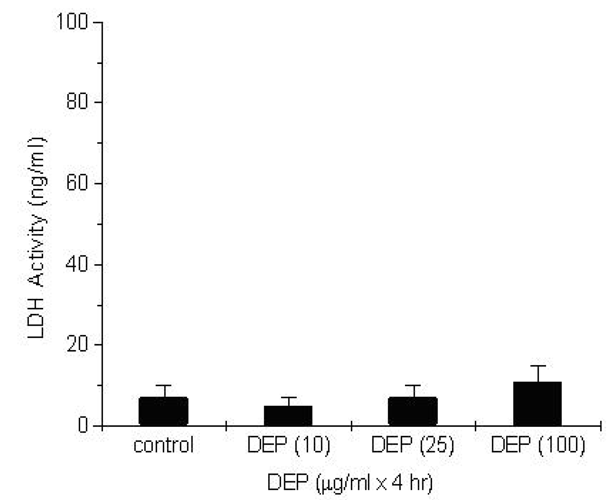
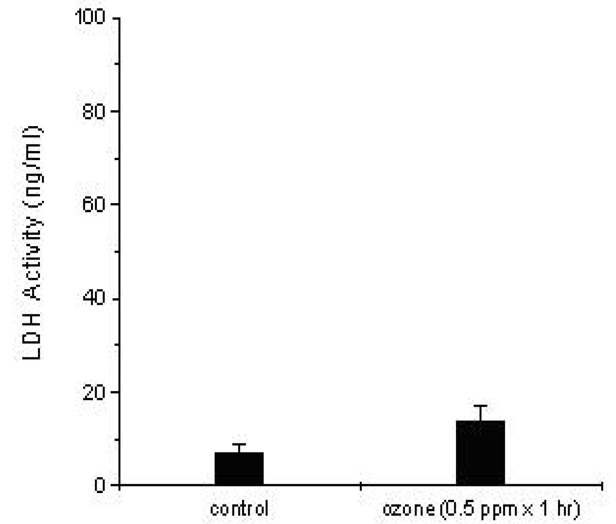
Measurement of the Cytotoxic Effect of DEP and Ozone Exposure

To determine the effect of DEP exposure on small airway epithelial cell membrane integrity, A549 cells were cultured in F12k medium, supplemented with antibiotics and 10 % fetal bovine serum (FBS). The cells were plated on plastic plates (35 mm diameter) at a density of 100 x 105 cells/well in 95% air, 5% CO2 and humid environment. The cells reached confluency in 3 days. Monolayers of A549 cells were treated with DEP at concentrations of 10, 25, and 100 µg/ml for 4 hr, and the cytotoxic effect of DEP was assessed by lactate dehydrogenase activity assay. Control cultures were treated with HBSS (containing 30 mM Hepes) alone. Figure 1 demonstrates that DEP exposure at concentrations of 10, 25, and 100 µg/ml (x 4 hr) did not induce any significant effect on A549 cell membrane integrity, as determined by lactate dehydrogenase activity (5 ng/ml ± 2 ng/ml, n = 6, 7 ± 3 ng/ml, n = 6, and 11 ± 4 ng/ml, n = 3; p > 0.05, respectively).
Figure 1. Cytotoxic Effect of DEP on A549 Cell Membrane Integrity. Confluent A549 cultures were treated with DEP at concentrations of 10, 25, and 100 µg/ml for 4 hr and lactate dehydrogenase activity (LDH) was measured as an indicator of cell membrane integrity. Data (ng/ml) of LDH are presented as mean ± SEM (n = 6, each group).
Ijerph 02 00403 g001
To determine the cytotoxic effect of ozone exposure on A 549 cells, cultures were grown on the apical side of collagen-coated Transwell-Col membranes (25 mm diameter and 0.4 µm pore size) (Costar, Cambridge, MA) until confluency [15] Cells reached confluency in 3 days. Prior to exposure to ozone, growth media were aspirated and the cells were hydrated with 2 ml HBSS (with 30 mM Hepes) in the basolateral compartment. Following ozone exposure (0.5 ppm x 1 hr), 1 ml of HBSS was added apically and LDH levels were measured. Figure 2 shows that ozone exposure (0.5 ppm x 1 hr) did not induce any significant cytotoxic effect on A 549 cells (14 ± 3 ng/ml, n = 6, p > 0.05), as compared to air control cultures (7 ± 2 ng/ml, n = 6).
Figure 2. Cytotoxic Effect of Ozone on A549 Cell Membrane Integrity. Confluent A549 cultures were exposed to ozone at a concentration of 0.5 ppm for 1 hr and lactate dehydrogenase activity (LDH) was measured as an indicator of cell membrane integrity. Data (ng/ml) of LDH are presented as mean ± SEM (n = 6, each group).
Ijerph 02 00403 g002
Figure 3. Effect of Exposure to DEP alone, Ozone, or DEP Following Ozone on IL-8 Release in A549 Cells.
Ijerph 02 00403 g003
The effect of ozone exposure on DEP-induced IL-8, an inflammatory cytokine was measured. Confluent A549 monolayers grown on Transwell-Col membranes were exposed to ozone (0.5 ppm x 1 hr) prior to DEP exposure (100 µg/ml of DEP for 4 hr). After the different treatment regiments, IL-8 was measured in the cell-free supernatants by ELISA. The data (pg/ml) are presented as the mean ± SEM (n = 6, each group). *Significant difference (p < 0.05) from control values. TSignificant difference from cultures exposed to ozone (p < 0.05).

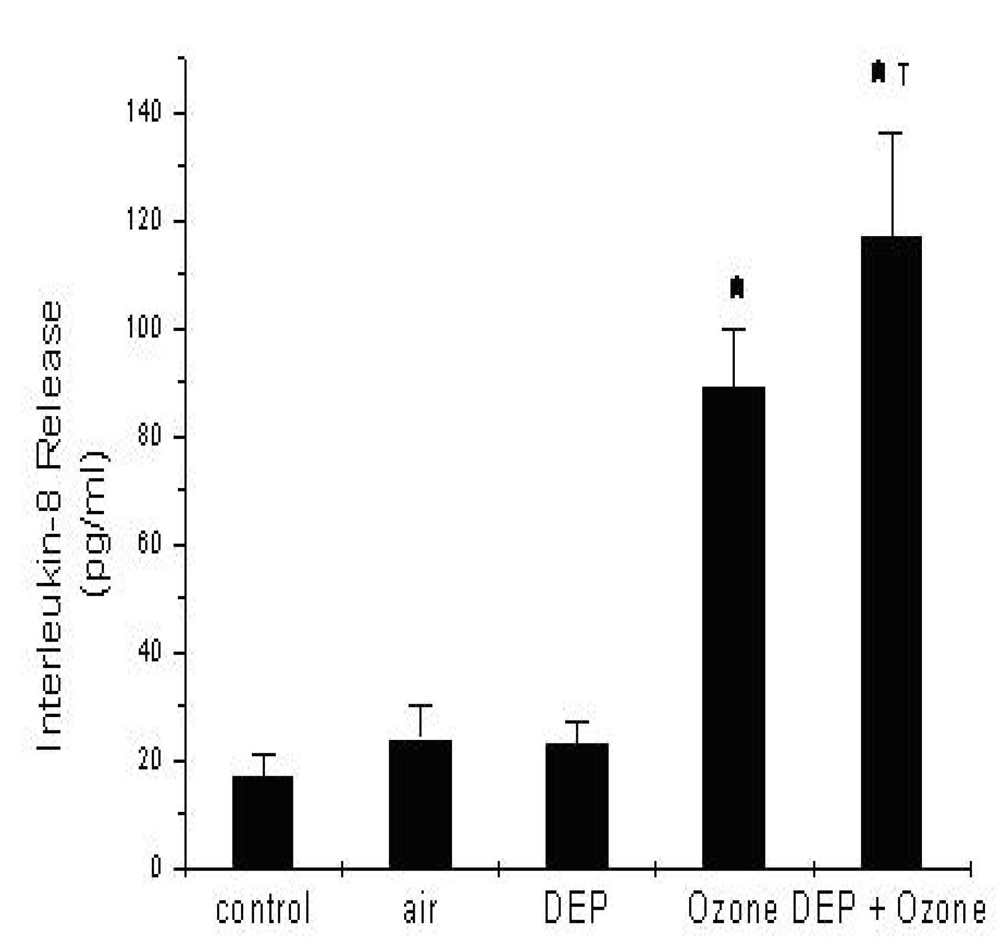
Effect of DEP on IL-8 Release

To determine the effect of DEP on IL-8 release in A549 cells, monolayers were exposed to DEP at a concentration of 100 µg/ml for 4 hr and IL-8 release was measured using human recombinant IL-8 ELISA. Figure 3 shows that exposure of A549 cultures to DEP did not induce any significant release of IL-8 (31 ± 6 pg/ml, n = 6, p > 0.05), as compared to control cultures (23 ± 4 pg/ml, n = 6).

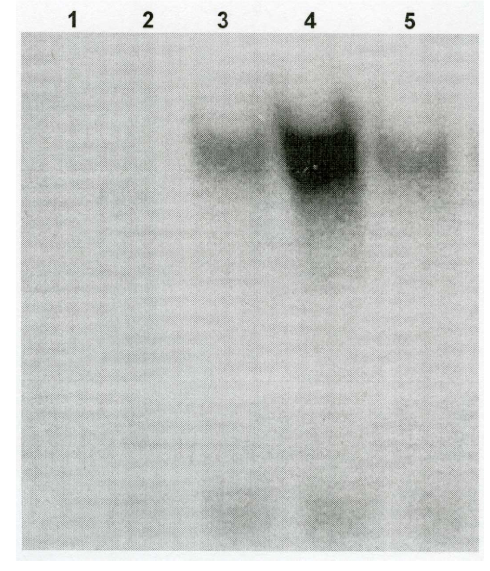
Ozone Exposure Increases DEP-Induced NF-κB Binding Activity

Exposure of human airway epithelial cells, A 549, in culture to biologically-relevant concentrations of DEP (100 µg/ml x 4 hr) following ozone exposure (0.5 ppm x 1 hr) showed that ozone induced an increase in the binding activity of NF-κB, as determined by electrophoretic mobility shift assays of crude nuclear extracts (Fig. 4). In contrast, exposure of A549 cultures to DEP alone failed to induce any significant increase in NF-κB binding activity (Fig. 4).
Figure 4. Activation of Nuclear Factor-κB (NF-κB) in A549 Cells by Ozone and DEP following Ozone Exposure. Cells were exposed to ozone (0.5 ppm x 1 hr) or purified air. Following ozone exposure, A 549 monolayers were exposed to DEP at a concentration of 100 µg/ml for 4 hr. Cells were scraped using rubber policeman, fractionated and nuclear extracts were subjected to EMSA using a labeled oligonucleotide containing an NF-κB binding site. Lanes are as follows: 1, probe; 2, excess cold oligo + ozone (0.5 ppm x 1 hr); 3, Control; 4, ozone (0.5 ppm x 1 hr); 5, DEP (100 µg/ml x 4 hr); 6, DEP (100 µg/ml x 4 hr) following ozone exposure (0.5 ppm x 1 hr).
Ijerph 02 00403 g004

Ozone Exposure Increases NF-IL6-DNA Binding Activity

Figure 5 demonstrates that exposure of A 549 cultures to ozone resulted in a significant increase in NF-IL6-DNA binding activity, as demonstrated by DNA mobility assays. DEP exposure (100 µg/ml x 4 hr) alone, however, failed to induce any significant increase in NF-IL6-DNA binding activity, as compared to control cultures (Fig. 5).
Figure 5. Activation of Nuclear Factor-IL6 (NF-IL6) in A549 Cells by Ozone Exposure. Confluent monolayers of A 549 cells were exposed to purified air (control), ozone (0.5 ppm x 1 hr), or DEP (100 µg/ml x 4 hr). Following treatment, nuclear extracts were subjected to electrophoretic mobility assays as described before. Double-stranded NF-IL-6 consensus oligonucleotide 5’-AGT TGA GGG GAC TTT CCC AGG C-3’ was used as the cognate DNA binding sequence for NFIL-6. Prior to probing the oligonucleotide, DNA was end labeled with [γ-32P]-ATP utilizing T4 polynucleotide kinase. Panel shows bands corresponding to treatment groups: probe, 1; excess cold oligonucleotide with ozone (0.5 ppm x 1 hr), 2; control, 3; ozone, 4; and DEP, 5.
Ijerph 02 00403 g005

Discussion

Ambient air pollutants, including ozone and diesel exhaust particles have been linked to increased incidence of lung inflammation and exacerbation of COPD and asthma. Recent studies have shown increased levels of IL-8 and neutrophil counts during severe lung inflammation and exacerbation of COPD [37,38]. Lung inflammation is characterized by an influx of neutrophils to sites of inflammation [39]. Furthermore, COPD has been associated with neutrophilic inflammation [40]. Neutrophil, lymphocyte, and eosinophil counts were reported to be significantly higher in humans with moderate to severe COPD than in healthy individuals [37]. Moreover, increased levels of IL-8 were observed in patients with COPD [41] and severe asthma [42]. IL-8 has been initially identified as a potent neutrophil chemo attractant in the lung [43].
Numerous stimuli and agonists have been shown to induce IL-8 gene expression in human bronchial epithelial cells, macrophages, fibroblasts, and lymphocytes [44,45,46]. IL-8-stimulated neutrophils in vitro, were reported to release granule-derived factors, including cathepsins and elastases [47]. Beta-2 agonists which are used in the treatment of COPD have been shown to inhibit IL-8 gene expression, elastase release, and migration of neutrophils in airway epithelial and endothelial cells, in vitro [48]. Exposure to DEP has been shown to significantly increase IL-8 synthesis and release in human bronchial epithelial cells [49,50]. However, limited data is available regarding the effect of DEP on IL-8 gene expression in distal small airway epithelial cells, major cells in the lower airways where DEP is likely to deposit. The results of the present study indicate that exposure to DEP did not induce any significant IL-8 gene expression in A 549 cells, type-II-like human airway epithelial cells. However, DEP treatment of A549 cells following ozone exposure induced a significant increase in IL-8 gene expression (Fig. 3). Boland et al. [51] have reported that DEP carbonaceous core failed to induce any significant increase in IL-8 or Granulocyte macrophage-colony stimulating factor (GM-CSF) in human bronchial epithelial cells. The results reported by Boland and co-workers are in agreement with our findings.
Previously, exposure to ozone or particulate matter and their effect on the lung have been addressed as separate environmental problems. Ozone and PM 2-10, including DEP may be chemically coupled in the ambient air [13]. Ozone, because of its high reactivity will react with the airway epithelial lining fluid and cell membrane (lipids and proteins). Conversely particulate matter will deposit and bind to lung cells, or become phagocytized by macrophages. The molecular and cellular events underlying the effect of ozone, DEP, or the simultaneous exposure to these two airborne pollutants remain unclear.
In the present study, ozone exposure enhanced DEP-induced IL-8 release in the human airway epithelial cell line, A549 (Fig. 3). This increase in DEP-induced IL-8 release following ozone exposure may be related to either, increased binding of DEP to A549 cell membrane, or increased uptake of DEP following ozone exposure, especially that separate or simultaneous exposure to ozone or DEP did not have any significant effect on A549 membrane integrity (Figs. 1 and 2). IL-8 has been demonstrated to serve as neutrophil chemoattractant to sites of inflammation [44]. Ozone-induced increase in DEP-induced IL-8 gene expression may explain the role of DEP in lung inflammation and aggravation of COPD. Therefore, the results of the present study may provide information regarding the cellular and molecular mechanisms underlying exacerbation of COPD and asthma in response to DEP exposure. In the present study, DEP alone failed to induce any significant release of IL-8, an important chemotactic in the lung. However, exposure of A549 cultures to ozone (0.5 ppm x 1 hr) followed by DEP treatment (100 µg/ml x 4 hr) resulted in a significant increase IL-8 release. Jaspers et al. [52] have shown that ozone exposure at 0.1 ppm induced significant release of IL-8 in A549 cells. The Increased IL-8 gene expression by ozone exposure was associated with activation of NF-κB in A549 cells [52]. Previously, we have shown that lipid ozonation products (LOP) generated from reaction of ozone with unsaturated fatty acids in airway epithelial cell induce significant release of IL-8 in cultured human bronchial epithelial cells [14]. Furthermore, co-activation of NF-κB (Fig. 4) and NF-IL6 (Fig. 5) was demonstrated to be responsible for LOP-induced IL-8 release in human bronchial epithelial cells [53].
Therefore, we propose that ozone can prime A549 cells to induce increased pro-inflammatory gene expression in response to DEP, and other PM2.5-10 by activating DNA-binding transcription factors in lung target cells. The effect of ozone on simultaneous DEP exposure may aggravate lung inflammation, and exacerbate COPD and asthma. The molecular mechanisms underlying the observed DEP-mediated IL-8 release by lung cells remain to be further elucidated. Results in the present study indicate that activation of NF-κB and its translocation to the nucleus, in addition to NF-IL6 activation appear to be responsible for regulating the release of IL-8 in response to DEP exposure in A549 cells following ozone exposure. Currently, studies to examine the signaling mechanisms responsible for activation of NF-κB and NF-IL6 in DEP-induced IL-8 gene expression following ozone exposure are being conducted.

Acknowledgments

This work was supported by NIH grant G12RR13459, and a grant from the American Lung Association. The authors wish to thank Dr. Abdul K. Mohamed and Dr. Paul B. Tchounwou for their technical support.

References

  1. U.S. Environmental Protection Agency. Air Quality Criteria for Ozone and Other Photochemical Oxidants. National Technical Information Service: Springfield, VA, Report No. EPA/600/8-84/020df; p. 1986.
  2. Lippmann, M. Health effects of ozone: a critical review. J. Air Poll. Control Assoc. 1989, 39, 672–695. [Google Scholar]
  3. Koren, H.S.; Devlin, R.B.; Graham, D.E.; Mann, R.; McGee, M.P.; Horstman, D.H.; Kozumbo, W.J.; Becker, S.; House, D.E.; McDonnell, W.F.; Bromberg, P.A. Ozone-induced inflammation in the lower airways of human subjects. Am. Rev. Respir. Dis. 1989, 139, 407–415. [Google Scholar] [CrossRef]
  4. Devlin, R.B.; McDonnell, W.F.; Becker, S.; Madden, M.C.; McGee, M.P.; Perez, R.; Hatch, G.; Hourse, D.E.; Koren, H.S. Time-dependent changes of inflammatory mediators in the lungs of humans exposed to 0.4 ppm ozone for 2 hr: a comparison of mediators found in bronchoalveolar lavage fluid 1 and 18 hr after exposure. Toxicol. Appl. Pharmacol. 1996, 138, 176–185. [Google Scholar] [CrossRef]
  5. Pino, M.V.; Levin, J.R.; Stovall, M.Y.; Hyde, D.M. Pulmonary Inflammation and epithelial injury in response to acute ozone exposure in the rat. Toxicol. Appl. Pharmacol. 1992, 112, 64–72. [Google Scholar] [CrossRef]
  6. Basha, M.A.; Gross, K.B.; Gwizdala, C.J.; Haidar, A.H.; Popovich, J., Jr. Bronchoalveolar lavage neutrophilia in asthmatics and healthy volunteers after controlled exposure to ozone and filtered purified air. Chest 1994, 106, 1757–1765. [Google Scholar] [CrossRef]
  7. Balmes, J.R.; Chen, L.L.; Scannell, C.; Tager, I.; Christian, D.; Hearne, P.Q.; Kelly, T.; Aris, R. Ozone-induced decrements in FEV1 and FVC do not correlate with measures of inflammation. Am. J. Respir. Crit. Care Med. 1996, 153, 904–909. [Google Scholar] [CrossRef]
  8. Health Effects Institute. Effects of ozone on normal and potentially sensitive human subjects. Part I: airway inflammation and responsiveness to ozone in normal and asthmatic subjects. Research Report No. 78; Cambridge, MA, 1997. [Google Scholar]
  9. Dockery, D.W.; POPE, C.A., 3rd. Acute respiratory effects of particulate air pollution. Annu. Rev. Public Health. 1994, 15, 107–132. [Google Scholar] [CrossRef]
  10. Norris, G.; Youn-Pong, S.N.; Koenig, J.Q.; Larson, T.V.; Sheppard, L.; Stout, J.W. An association between fine particles and asthma emergency department visits for children in Seattle. Environ. Health Perspect. 1999, 107, 489–493. [Google Scholar]
  11. Ghio, A.J. Biological effects of Utah Valley ambient air particles in humans: review. J. Aerosol Med. 2004, 17, 157–164. [Google Scholar] [CrossRef]
  12. Timblin, C.; Berube, K.; Churg, A.; Driscoll, K.; Gordon, T.; Hemenway, D.; Walsh, E.; Cummins, A.B.; Vacek, P.; Mossman, B. Ambient particulate matter causes activation of the c-jun kinase/stress-activated protein kinase cascade and DNA synthesis in lung epithelial cells. Cancer Res. 1998, 58, 4543–4547. [Google Scholar]
  13. Meng, Z.; Dabdub, D.; Seinfeld, J.H. Chemical coupling between atmospheric ozone and particulate matter. Science 1997, 277, 116–119. [Google Scholar] [CrossRef]
  14. Kafoury, R.M.; Pryor, W.A.; Squadrito, G.L.; Salgo, M.G.; Zou, X.; Friedman, M. Lipid ozonation products activate phospholipases A2, C, and D. Toxicol. Appl. Pharmacol. 1998, 150, 338–349. [Google Scholar] [CrossRef]
  15. Kafoury, R.M.; Pryor, W.A.; Squadrito, G.L.; Salgo, M.G.; Zou, X.; and Friedman, M. Induction of inflammatory mediators in human airway epithelial cells by lipid ozonation products. Am. J. Respir. Crit. Care Med. 1999, 160, 1934–1942. [Google Scholar]
  16. Pryor, W.A.; Squadrito, G.L.; Friedman, M. The cascade mechanism to explain ozone toxicity: the role of lipid ozonation products. Free Rad. Biol. Med. 1995, 19, 935–941. [Google Scholar] [CrossRef]
  17. Devlin, R.B.; McKinnon, K.P.; Noah, T.; Becker, S.; and Koren, H.S. Ozone-induced release of cytokines and fibronectin production by alveolar macrophages and airway epithelial cells. Am. J. Physiol. 1994, 266, L612–L619. [Google Scholar]
  18. Balmes, J.R.; Chen, L.L.; Scannell, C.; Tager, I.; Christian, D.; Hearne, P.Q.; Kelly, T.; and Aris, R. Ozone-induced decrements in FEV1 and FVC do not correlate with measures of inflammation. Am. J. Respir. Dis. Crit. Care Med. 1996, 153, 904–909. [Google Scholar]
  19. Seltzer, J.; Bigby, B.G.; Stulbarg, M.; Holtzman, M.J.; Nadel, J.A.; Ueki, I.F.; Leikauf, G.D.; Goetzel, E.J.; Boushey, H.A. O3-induced change in bronchial reactivity to methacholine and airway inflammation in humans. J. Appl. Physiol. 1986, 60, 1321–1326. [Google Scholar]
  20. Koren, H.S.; Devlin, R.B.; Becker, S.; Perez, R.; McDonnell, W.F. Time-dependent changes of markers associated with inflammation in the lungs of humans exposed to ambient levels of ozone. Toxicol. Pathol. 1991, 19, 406–411. [Google Scholar]
  21. Ohtoshi, T.; Tazikawa, H.; Okazaki, H.; Kawasaki, S.; Takeuchi, N.; Ohta, K.; Ito, K. Diesel exhaust particles (DEP) stimulate human airway epithelial cells to produce cytokines relevant to airway inflammation in vitro. J. Allergy Clin. Immunol. 1998, 101, 778–785. [Google Scholar] [CrossRef]
  22. Sagai, M.; Furuyama, A.; Ichinose, T. Biological effects of diesel exhaust particles (DEP) III. Pathogenesis of asthma-like symptoms in mice. Free Rad. Biol. Med. 1996, 21, 199. [Google Scholar] [CrossRef]
  23. Nel, A.E.; Diaz-Sanchez, D.; Ng, D.; Hiura, J.; Saxon, A. Enhancement of allergic inflammation by the interaction between diesel exhaust particles and the immune system. J. Allergy Clin. Immunol. 1998, 102, 539–554. [Google Scholar] [CrossRef]
  24. Peterson, B.A.; Saxon, A. Global increases in allergic respiratory disease: The possible roles of diesel exhaust particles. Ann. Allergy Asthma Immunol. 1996, 77((4)), 263–268. [Google Scholar] [CrossRef]
  25. Diaz-Sanchez, D. The roles of diesel exhaust particles and their associated polyaromatic hydrocarbons in the induction of allergic airway disease. Allergy 1997, 52((38 s)), 52–56. [Google Scholar] [CrossRef]
  26. Berry, J.; Henoc, P.; Galle, P. Phagocytosis by cells of the pulmonary alveoli. Am. J. Pathol. 1978, 93, 27–44. [Google Scholar]
  27. Sen, R.; Baltimore, D. Multiple nuclear factors interact with the immunoglobulin enhancer sequences. Cell 1986, 46((5)), 705–716. [Google Scholar]
  28. Baldwin, A.S., Jr. Series introduction: The transcription factor NF-κB and human disease. J. Clin. Invest. 2001, 107((1)), 3–6. [Google Scholar] [CrossRef]
  29. Stein, B.; Baldwin, A.S. Distinct mechanisms for the regulation of the interleukin-8 gene involve synergism and cooperativity between CIEBP and NF-κB. Mol. Cell Biol. 1993, 13, 7191–7198. [Google Scholar]
  30. Christman, J.W.; Sadikot, R.T.; Blackwell, T.S. The role of nuclear factor-κβ in pulmonary disease. Chest 2000, 117, 1482–1487. [Google Scholar] [CrossRef]
  31. Graziano, F.M.; Cook, E.B.; Stahl, J.L. Cytokines, chemokines, RANTES, and eotaxin. Allergy Asthma Proc. 1999, 20, 141–146. [Google Scholar] [CrossRef]
  32. Anderson, J.P. Resolution of chronic inflammation by therapeutic induction of apoptosis. Trends Pharmacol. Sci. 1996, 17, 438–442. [Google Scholar] [CrossRef]
  33. Samet, J.M.; Friedman, M. Effect of ozone on platelet activating factor metabolism in phorbol-differentiated HL60 cells. Toxicol. Appl. Pharmacol. 1992, 117, 19–25. [Google Scholar] [CrossRef]
  34. Wright, D.T.; Adler, K.B.; Akley, N.J.; Dailey, L.A.; Friedman, M. Ozone stimulates release of platelet activating factor and activates phospholipases in guinea pig tracheal epithelial cells in primary culture. Toxicol. Appl. Pharmacol. 1994, 127, 27–36. [Google Scholar] [CrossRef]
  35. McKinnon, K.P.; Madden, M.C.; Noah, T.L.; Devlin, R.B. In vitro ozone exposure increases release of arachidonic acid products from a human bronchial epithelial cell line. Toxicol. Appl. Pharmacol. 1993, 118, 215–223. [Google Scholar]
  36. Daniel, W.W. Biostatistics: a foundation for analysis in health sciences, 5th ed.John Wiley: New York, 1991; pp. 274–366. [Google Scholar]
  37. Fujimoto, K.; Yasuo, M.; Urushibata, K.; Hanaoka, M.; Koizumi, T.; Kubo, K. Airway inflammation during stable and acutely exacerbated chronic obstructive pulmonary disease. Eur. Respir. J. 2005, 25, 640–646. [Google Scholar] [CrossRef]
  38. Oudijk, E.J.; Nijhuis, E.H.; Zwank, M.D.; van de Graaf, E.A.; Mager, H.J.; Coffer, P.J.; Lammers, J.W.; Koendermann, L. Systemic inflammation in COPD visualized by gene profiling in peripheral blood neutrophils. Thorax 2005, 60, 538–544. [Google Scholar] [CrossRef]
  39. Driscoll, K.E.; Carter, J.M.; Hassenbein, D.G.; Howard, B. Cytokines and particle-induced inflammatory cell recruitment. Environ. Health Perspect. 1997, 105, 1159–1164. [Google Scholar] [CrossRef]
  40. Watt, A.P.; Schock, B.C.; Ennis, M. Neutrophils and eosiphils: clinical implications of their appearance, presence and disappearance in asthma and COPD. Curr. Drug Targets Inflamm. Allergy 2005, 4, 415–423. [Google Scholar] [CrossRef]
  41. Arinir, U.; Klein, W.; Rohde, G.; Stemmler, S.; Epplen, J.T.; Schultze-Werninghaus, G. Polymorphisms in the interleukin-8 gene in patients with chronic obstructive pulmonary disease. Electrophoresis 2005, 26, 2888–2891. [Google Scholar] [CrossRef]
  42. Vachier, I.; Bonnans, C.; Chavis, C.; Farce, M.; Godard, P.; Bousquet, J.; Chanez, P. Severe asthma is associated with a loss of LX4, an endogenous anti-inflammatory compound. J. Allergy Clin. Immunol. 2005, 115, 55–60. [Google Scholar] [CrossRef]
  43. Barnes, P.J. Mediators of chronic obstructive pulmonary disease. Pharmacol. Rev. 2004, 56, 515–548. [Google Scholar] [CrossRef]
  44. Oppenheim, J.J.; Zachariae, C.O.C.; Mukaida, N.; Matsushima, K. Properties of the novel proinflammatory supergene “intercrine” cytokine family. Annu. Rev. Immunol. 1991, 9, 617–648. [Google Scholar] [CrossRef]
  45. Bagglioni, M.; Dewald, B.; Moser, B. Interleukin-8 and related chemotactic cytokines-CXC and CC chemokines. Adv. Immunol. 1994, 55, 97–179. [Google Scholar]
  46. Taub, D.D.; Oppenheim, J.J. Chemokines, inflammation, and the immune system. Ther. Immunol. 1994, 1, 229–242. [Google Scholar]
  47. Taub, D.D.; Anver, M.; Oppenheim, J.J.; Longo, D.L.; Murphy, W.J. T lymphocyte recruitment by inerleukin-8 (IL-8):IL-8-induced degranulation of neutrophils releases potent chemoattractants for human T lymphocytes both in vitro and in vivo. J. Clin. Invest. 1996, 97, 1931–1941. [Google Scholar] [CrossRef]
  48. Sadowska, A.M.; Manuel-y-Keenoy, B.; Backer, W.A. Inhibition of in vitro neutrophil migration through a bilayer of endothelial and epithelial cells using beta-2-agonists: concomitant effects on IL-8 and elastase secretion and impact of gluccocorticosteroids. Pulm. Pharmacol. Ther. 2005, 18, 354–362. [Google Scholar] [CrossRef]
  49. Takizawa, H.; Ohtoshi, T.; Kawasaki, S.; Abe, S.; Sugawara, I.; Nakahara, K.; Matsushima, K.; Kudoh, S. Diesel exhaust particles activate human bronchial epithelial cells to express inflammatory mediators in the airways: a review. Respirology 2000, 5, 197–203. [Google Scholar] [CrossRef]
  50. Steerenberg, P.A.; Zonnenberg, J.A.; Dormans, J.A.; Joon, P.N.; Wouters, I.M.; van Bree, L.; Scheepers, P.T.; van Lveren, H. Diesel exhaust particles induced release of interleukin 6 and 8 by (primed) human bronchial epithelial cells (BEAS-2B) in vitro. Exp. Lung Res. 1998, 24, 85–100. [Google Scholar] [CrossRef]
  51. Boland, S.; Baez-Squiban, A.; Fournier, T.; Houcine, O.; Gendron, M.-C.; Checrier, M.; Jouvenot, G.; Coste, A.; Abier, M.; Francelyne, M. Diesel exhaust particles are taken up by human airway epithelial cells in vitro and alter cytokine production. Am. J. Physiol. 1999, 276, L604–L613. [Google Scholar]
  52. Jaspers, I.; Flescher, E.; Chen, L.C. Ozone-induced IL-8 expression and transcription factor binding in respiratory epithelial cell. Am. J. Physiol. 1997, 272, L504–L511. [Google Scholar]
  53. Kafoury, R.M.; Gozal, E.; Squadrito, G.L.; Zou, X.; Pryor, W.A.; Friedman, M. Lipid ozonation products activate nuclear factor-κB (NF-κB) in human bronchial epithelial cells (BEAS-2B). Am. J. Respir. Crit. Care Med. 1999, 159, A463. [Google Scholar]

