Article

Tobacco Smoke: Involvement of Reactive Oxygen Species and Stable Free Radicals in Mechanisms of Oxidative Damage, Carcinogenesis and Synergistic Effects with Other Respirable Particles

by
Athanasios Valavanidis
*,
Thomais Vlachogianni
and
Konstantinos Fiotakis
Department of Chemistry, Free Radical Research Group, University of Athens, University Campus Zografou, 15784 Athens, Greece
*
Author to whom correspondence should be addressed.
Int. J. Environ. Res. Public Health 2009, 6(2), 445-462; https://0-doi-org.brum.beds.ac.uk/10.3390/ijerph6020445
Submission received: 12 November 2008 / Accepted: 25 January 2009 / Published: 2 February 2009
(This article belongs to the Special Issue Smoking and Tobacco Control)

Abstract

:
Tobacco smoke contains many toxic, carcinogenic and mutagenic chemicals, as well as stable and unstable free radicals and reactive oxygen species (ROS) in the particulate and the gas phase with the potential for biological oxidative damage. Epidemiological evidence established that smoking is one of the most important extrinsic factor of premature morbidity and mortality. The objective of this study was to investigate oxidative and carcinogenic mechanisms of tobacco and synergistic action with other respirable particles in the respiratory system of smokers. Electron Paramagnetic Resonance (EPR) and spin-trapping techniques were used to study stable free radicals in the cigarette tar, and unstable superoxide anion (O2•−) and hydroxyl (HO) radicals in the smoke Results showed that the semiquinone radical system has the potential for redox recycling and oxidative action. Further, results proved that aqueous cigarette tar (ACT) solutions can generate adducts with DNA nucleobases, particularly the mutagenic 8-hydroxy-2’-deoxyguanosine (a biomarker for carcinogenesis). Also, we observed synergistic effects in the generation of HO, through the Fenton reaction, with environmental respirable particles (asbestos fibres, coal dust, etc.) and ambient particulate matter (PM), such as PM10, PM2.5 and diesel exhaust particles (DEP). The highest synergistic effects was observed with the asbestos fibres (freshly grounded), PM2.5 and DEP. Finally, we discuss results from our previous study of conventional cellulose acetate filters and “bio-filters” with hemoglobin impregnated activated carbon, which showed that these filters do not substantially alter the free radical content of smoke in the particulate and in the gaseous phase.

1. Introduction

Epidemiological evidence has established that smoking is one of the most important environmental causes of human mortality and morbidity. Tobacco smoking is currently responsible for approximately 30% of all cancer deaths in developed countries [1]. In addition, smoking causes greater number of deaths from cardiovascular, chronic obstructive pulmonary and degenerative diseases. In 2000, 4.8 million premature deaths worldwide were attributed to smoking, of which 2.4 million in developing and 2.43 million in developed industrialized countries [2], numbers expected to increase to 10 million a year by 2030 [3].
Tobacco smoke is an aerosol containing about 1010 particles/mL, consisting of highly porous carbonaceous polymeric material with adsorbed heavy metals, polycyclic aromatic hydrocarbons (PAH), azaarenes, N-nitrosamines and various other organic chemicals. The particular phase of tobacco smoke contains at least 3,500 chemical compounds and a high proportion of them are toxic, carcinogens or mutagens, (e.g. benzene, 2-napthylamine, 210Po, 226Ra, 228Ra, nickel, cadmium, benzo[a]pyrene, etc) [4]. There are at least 55 carcinogens in cigarette smoke that have been evaluated by the International Agency for Research on Cancer (IARC) with “sufficient evidence for carcinogenicity” [5, 6].
Tobacco smoke is divided into the mainstream (smoker inhaled) and the sidestream smoke. The mainstream is divided into a particulate solid phase (tar) and the gas phase (toxic gases, volatile organic compounds, VOCs, free radicals, etc.). Cigarette tar contains remarkably high concentrations of stable free radicals (ca. 1017 spins·g−1) with very long lifetimes. The sidestream smoke is divided in the solid and gas phases, containing higher concentrations of toxic and carcinogenic compounds and other volatile and semivolatile compounds [79]. Free radicals and oxidants in the gas phase exist in a steady state in which they are continuously formed and destroyed and their concentration increases as the smoke ages [10, 11]. The gas phase is around 0.4–0.5 g/cigarette and contains ca. 500 volatile organic and inorganic compounds [12]. The particulate phase (tar) consists of fine and very fine particles (0.1–1.0 μm, aerodynamic diameter) penetrating deep into the alveoli. Some of the water-soluble components of aqueous cigarette tar (ACT) can produce superoxide anion (O2•−) and subsequently H2O2 and the reactive hydroxyl radical (HO), which cause oxidative damage to cellular membrane lipids, proteins, enzymes and DNA [13, 14]. The sidestream smoke consists of similar chemical components in the solid and gas phases and is also rich in highly reactive and short-lived free radicals. Passive smoking (or environmental tobacco smoke, ETS) has been proved to be a health hazard for non-smokers and its burden of major lung diseases [1517]
Biochemical mechanisms of carcinogenic action by tobacco smoke constituents and oxidative damage in cellular DNA by ROS have been observed with very sensitive techniques [1820]. In the last two decades, the central role of free radical mechanisms in tobacco smoke carcinogenesis and oxidative stress has been established by a series of studies [2123]. An important finding by Pryor and co-workers was that cigarette tar has high concentrations of stable free radicals, identified as a semiquinone (QH) and carbon-centered radicals (-C) by EPR [24]. The most interesting is a quinone/semiquinone/hydroquinone (Q/QH/QH2) system in the tar polymeric matrix [25]. The stable free radicals were identified as o- and p-benzosemiquinone radicals and the role in the DNA damage, through the formation of HO were detected by EPR spin-trapping [26]. The following mechanisms have been identified: the QH radicals reduces O2 into O2•−, which can dismutate to form H2O2 and then with Fe2+ (cigarette tar itself contains mainly high concentrations of iron) can generate through the Fenton reaction highly oxidizing hydroxyl radicals:
Q + QH 2 2 QH
QH + O 2 Q + O 2 + H +
O 2 + 2 H + H 2 O 2
QH 2 + O 2 H 2 O 2 + Q
QH 2 + O 2 O 2 + QH + H +
Fe 2 + + H 2 O 2 Fe 3 + + HO + HO
Cigarette tar can produce large amounts of H2O2 in aqueous extracts [27] and oxidants in tar and gas-phase have been implicated in the release of iron from the endogenous enzyme ferritin and alter iron metabolism in the lungs [28, 29]. Oxidants and free radicals in cigarette smoke have been considered as a potential mechanism by which smoking can promote lipid peroxidation of cellular membrane lipids, thus promoting atherosclerosis, endothelial dysfunction and acute clinical events, and increase the risk for cardiovascular diseases [3032]. ROS in the cigarette gas-phase promote the destruction of endogenous antioxidants (vitamins and enzymatic antioxidants) reducing the vital role of cellular antioxidant defenses [33]. Several studies show that antioxidant vitamins are lower in smokers resulting in systemic oxidative stress [34, 35], whereas dietary antioxidant supplements provide only limited protection to smokers [36, 37].
Studies showed that the synergistic interaction of tobacco smoke with various respirable mineral fibres and fine particular matter (soot, fine dusts) in occupational environments explain the increased lung cancer risks and other occupational pulmonary diseases in industrial workers [38, 39]. The synergistic interaction of ROS from cigarette smoke with asbestos fibres contributes to the increases of lung cancer in workers of asbestos mines [40, 41]. Studies showed that cigarette smoke and fresh grinding of asbestos fibres increased by 2–3 times the production of HO [42, 43]. The synergistic effects of cigarette smoke and asbestos fibres increased DNA damage in bronchial epithelial cells [45].
Similar synergistic interactions of tobacco smoke free radicals and other carcinogenic agents in occupational environments were observed with radon [46, 47], coal dusts [48] and heavy metals (nickel, chromium, cadmium) in mining occupations [4952]. Synergy mechanisms between tobacco smoke free radical and heavy metals increases malignant neoplasms in occupationally exposed workers [53, 54]. Studies showed synergy between tobacco smoke, alcohol consumption and occupational exposure to mineral particles increase oral and pharyngeal cancers [55, 56].
The gas phase of tobacco smoke contains a large amount of free radicals (estimated at ∼1 X 10¹⁵ radicals per puff) [7]. Nitric oxide (NO) is a species of considerable interest because of its multiple physiological role (neurotransmission, blood pressure modulator) [57], but also for its toxic effect when generated in excess. NO reacts quickly with O2•− to form peroxynitrite (O=NNO), a chemical known for its highly toxic and oxidative action towards biomolecules (e.g. 3-nitrotyrosine) [58, 59]. NO has been implicated in carcinogenesis and tumour promotion [60].
In the present study reported here we have investigated: first the presence of stable free radicals in the tar of the mainstream cigarette smoke in three different cigarettes (low, middle and high tar content) with conventional acetate filters, by EPR. Secondly, the production of reactive ROS and especially HO from aqueous tar extracts at physiological pH. Thirdly, we studied the spin-trapping of free radicals in the gas-phase of the sidestream smoke. Fourthly, the production of the hydroxyl adduct to DNA nucleobases, such as the mutagenic 8-hydroxy-2’-deoxyguanosine in aqueous buffered solutions. Furthermore, we present results from the synergistic production of HO from the interaction of cigarette tar and five different types of fibers, dusts and particulate matter of environmental importance (asbestos fibers, coal dusts, talc dust, PM10 and PM2.5). Finally, we discuss the results of a previous study which investigated the claims that the so called “bio-filters” (containing dry hemoglobin impregnated in activated carbon) can reduce oxidants, free radicals and carcinogens in mainstream cigarette smoke.

2. Materials and Methods

2.1. Chemicals

The spin-traps 5,5-dimethyl-1-pyrroline-N-oxide (DMPO) and phenyl-tert-butyl nitrone (PBN) and the stable 2,2-diphenylpicrylhydrazyl free radical (DPPH, 95%) were purchased from Sigma-Aldrich. 2’-Deoxyguanosine and standard 8-hydroxy-2’-deoxyguanosine (8-OHdG), ethylenediamine tetraacetic acid sodium salt (EDTA-Na2) and deferoxamine mesylate salt (desferrioxamine) were purchased from Sigma. All other chemicals were from Aldrich, Merck and Fluka.

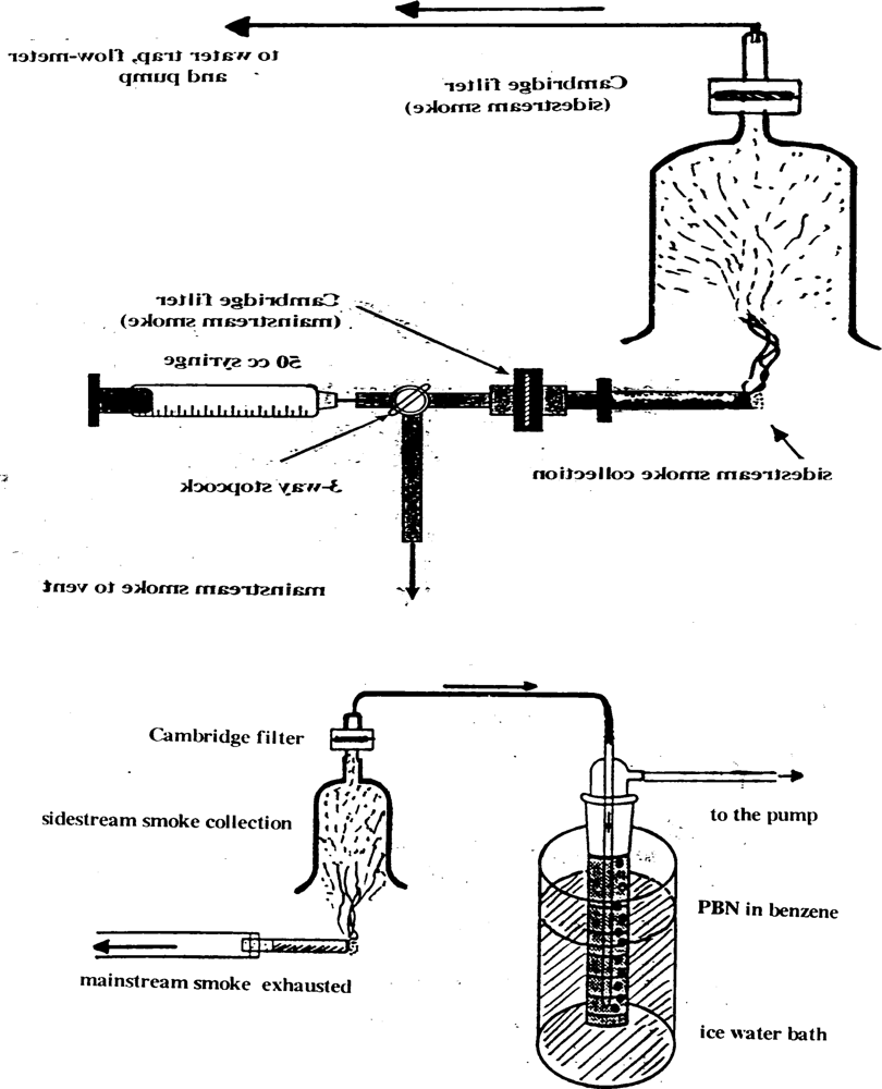
2.2. Cigarette Smoking Methods: The standard puff protocol

Puffs of 30 mL were pulled (3 s duration) at 1 (one) min intervals, using a hand-operated 50 mL syringe, until a fixed butt length was reached (∼10 puffs per cigarette) and passed through a Cambridge filter (glass fibre filter retaining 99% of particles larger than 0.1 micron) which collected the tar. Three different cigarettes were used: (1) low tar, ∼3 mg/cigarette, (2) middle tar, ∼8 mg/c, and (3) high tar, ∼14 mg/c. Tar was extracted with 20 mL of benzene (sonication) and the volume was reduced under reduced pressure in a rotavapor to dryness. The tar of the sidestream was collected in the intervals after passing by a second Cambridge filter, but not used in our experiments, since the smoker breaths only a small amount depending on the enclosed space of smoking. In the second method the gas-phase of the cigarette smoke (after passing though a Cambridge filter to retain tar) was bubbled through 10 mL solution of the spin-trap (0.1 M, in benzene) phenyl-tert-butyl nitrone, (PBN). In all experiments five cigarettes were smoked for every experimental measurement. Both diagrams of apparatus used in our cigarette smoke collection are shown in Figure 1. A more detailed analysis of their function is explained in detail in a previous paper [61].

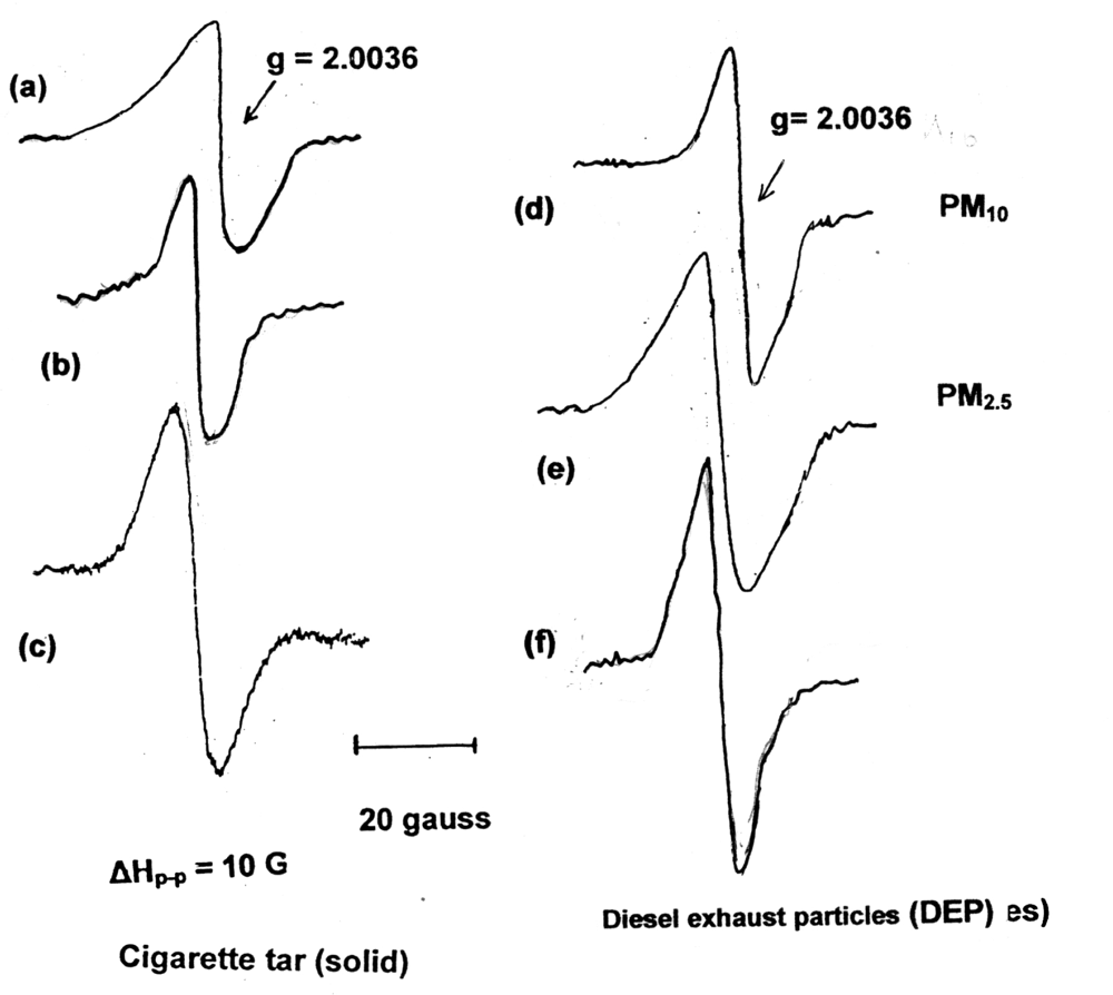
2.3. Measurements of Stable Free Radical in Cigarette Tar by EPR

Tar collected in the Cambridge filter of the mainstream smoke of five cigarettes (the experimental procedure was the same for all three brands of cigarettes) was extracted with distilled benzene (sonicated for 20 min). The solvent was evaporated to dryness and was dried further under vacuum and under a dry nitrogen stream. The dried residue is a thick dark brown colour tarry substance. Tar was weighed (∼5 mg) inside an EPR quartz tube for spectroscopic measurements. Spectral parameters (Varian E-4, X-band spectrometer with 100 kHz field modulation): microwave power, 10–20 mW; modulation amplitude, 1–1.4 G (Gauss); scan range 100 G; time constant, 1.0 s; scan time, 16 min; receiver gain, 1–8 X 10³ (depending on the intensity of the EPR lines). The radical spins/g values of equal amounts of samples of the three brands of cigarette tar were determined by comparison with known amounts of the free radical DPPH. The area under the EPR single-broad peak was cut carefully and the paper was weighed, representing a quantitative measure of the concentration of radical spins per unit of weight (1 g).

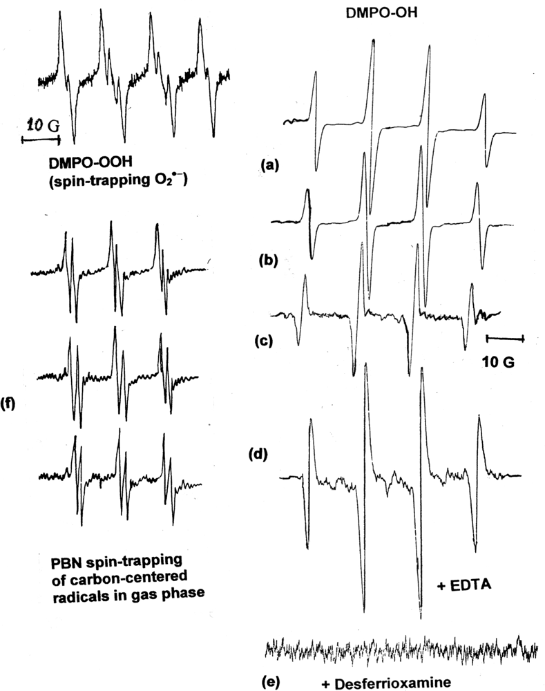
2.4. Spin-Trapping and Measurements of O2•− and HO Generated by Aqueous Cigarette Tar (ACT) Solutions at Physiological pH

Experiments for 50 mg of dried cigarette tar residue was added in 10 mL phosphate buffered solutions (pH =7.4) and 1 mL of 0.08 M aqueous DMPO spin-trap was added. The mixture was shaken gently by a mechanical shaker in the dark (covered with aluminum foil), at room temperature for 2–3 min and placed in an EPR quartz flat cell prior to recording its EPR spectrum. Spectral parameters (Varian E-4, X-band spectrometer with 100 kHz field modulation): microwave power, 20 mW; modulation amplitude, 1.0 G; scan range 100 G; time constant, 1.0 s; scan time, 16 min; receiver gain, 5–8 X 10³. The 6-line EPR spectrum of spin DMPO-OOH of O2•− spin adduct was recorded in less than 8 min after mixing (time is crucial because of the instability of the EPR spin adduct of the O2•−which reverts to the hydroxyl spin adduct). The same experiment can be repeated, but with 15 min shaking and degassing with dry nitrogen under the same conditions of spectral parameters. The 4-line EPR spectrum (1:2:2:1 quartet pattern) EPR spectrum of DMPO-OH for the HO spin adduct was recorded after 20 min in a EPR quartz flat cell. Complimentary experiments were carried out with ACT in the presence of 0.001 M H2O2 and in the presence of 0.02 M of the chelating agent EDTA-Na2 in order to test the increase of HO, through the Fenton reaction by the Fe(II) ions in the tar. EPR spectra of the DMPO-OH adduct were recorded 15 min after mixing and degassed with dry nitrogen. Addition in the ACT mixture of desferrioxamine chelating agent suppress the formation of HO. All measurements were repeated in triplicate. These EPR spectra are presented in Figure 3.

2.5. Spin-Trapping Measurements of the Gaseous Phase of Mainstream Smoke

Five cigarettes were smoked by the continuous flow system and the mainstream smoke, after passing the Cambridge filter, was bubbled through a glass tube containing 0.1 M of the spin-trap PBN in benzene (Figure 1). EPR spectra were recorded by the use of an EPR quartz flat cell and EPR spectral parameters were regulated to give the best resolved spectra of the spin adduct. All experiments were repeated in triplicate.

2.6. HPLC/UV-EC Quantitative Measurements of the 8-OHdG from Mixtures of ACT with 2’-dG in Buffered Solutions

Samples were prepared by mixing cigarette tar (50 mg) in phosphate buffered solution (pH 7.4, 10 mL) with 0.05 M 2’-deoxyguanosine (dG, 1 mL). The mixture was shaken gently in the dark for 1 hr at room temperature. The mixture was shaken gently in the dark for 1 hr at room temperature. The mixture was filtered (Gelman Acrodisc 0.2 μm). A control containing only dG was analysed in parallel with the reaction mixture. HPLC (Hewlett-Packard, Agilent 1100), reverse phase-HPLC, Column Lichro (250 x 4 mm), Lichorospher 100 RP-18, 5 μm column (25 cm X 4.6 mm), under isocratic conditions. The mobile phase was 7% methanol-93% buffer solution KH2PO4, 50 mM (pH 7.4) with a flow rate of 1.2 mL/min. The 8-OHdG and dG were monitored at 254 nm, and by an electrochemical detector (Coulotherm II EC, ESA, Inc, Chelmsford, MA) set at 400 mV and 20 nA full scale. Standards of 2’-dG and 8-OHdG help for the calibration. Detection limit of 8-OHdG estimated to 2–3 μM. Each reaction was repeated three times, values, average of 3 trials ± S.D.

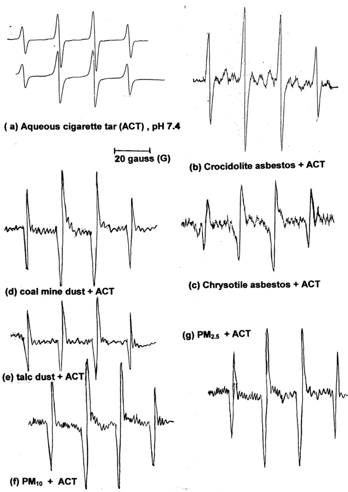
2.7. Synergistic Effect of ACT with Various Fibres and Respirable Particles

50 mg of dried mainstream cigarette tar were dissolved in 20 mL aqueous phosphate buffered solution (physiological pH 7.4). To the solution was added 50 mg of asbestos (separately for crocidolite and chrysotile). Also, experiments were repeated with other respirable particles, such as coal dust, talc dust [Mg3(OH)2Si4O10], airborne particulate matter PM10 and PM2.5 and diesel exhaust particles (DEP).. One (1) mL of 0.08 M of spin-trap DMPO was added in the mixture and the flask was shaken gently for 10 min. In the case of asbestos fibres, the reaction mixture is a suspension of freshly ground asbestos fibres. Aliquots of the suspension were withdrawn at 20 min, filtered and the clear solution was transferred to an EPR quartz flat cell and the EPR spectrum was recorded. EPR instrument parameters: microwave power, 20 mW; scan range 100 G; modulation amplitude 1.0 G, receiver gain 2.5–8 X10³; time constant, 0.1 s; scan time 8 or 16 min. Representative EPR spectra in Figure 4.

2.8. Comparison of Conventional Cellulose Acetate Filters and “Bio-Filters” (these experimental results were presented in our previous paper) [61]

Cigarette with conventional acetate filters were compared with cigarettes with “bio-filters”. The standard puff protocol and the continuous flow system were used to compare mainstream cigarette tar (trapped on Cambridge filters) and the gas-phase sidestream. The “bio-filter” (BF-filter) used was from a SEKAP (Greek tobacco company) cigarette, and is an extra filter containing activated carbon impregnated with dry hemoglobin, and advertised in 1994–1995 as a filter that reduces considerably free radicals, reactive oxidants, volatile carcinogens, etc [61, 62].

3. Results and Discussion

Cigarette tar is a carbonaceous tarry substance with mainly long-lived radicals, but also contains adsorbed heavy metals and carcinogenic organic compounds in the porous polymeric matrix. Whereas the gas-phase is a mixture of oxidative gases, VOCs and a great variety of small reactive radicals, mainly carbon- and oxygen-centered radicals, of short lifetimes [63, 64].
In the first series of experimental observations we focused on the stable free radicals in the cigarette tar of the mainstream. The three types of cigarette all showed a single broad EPR signal with g value of 2.0035 (g, spectroscopic spitting factor), which has been assigned to the semiquinone radical system. The g value is typical of organic semiquinone (QH) radicals previously observed in aqueous solutions at pH 8.0 [65]. Results are presented in Figure 2.
Comparison of cigarette tar for the three different types of cigarettes showed that the lower tar content the smaller the EPR signal. Although the quantitative comparison is of limited accuracy (estimated to 85–90%), the concentrations of spins/g of tar (using the free radical DPPH as a standard) were estimated as: a. low tar cigarette: ∼1015 spins/g; b. middle tar,1015–1016 spins/g; c. high tar, 1016–1017 spins/g. Similar results were reported by Pryor et al. [66]. Additionally, we compared cigarette tar with similar EPR spectra of particulate matter of atmospheric pollution in urban areas (diesel exhaust soot, PM10 and PM2.5 collected in the center of Athens, Greece). Diesel exhaust particles (DEP) showed concentrations in the range 1016–1017 spins/g, which is in the same range with the high tar cigarettes. It is well known that both contain fine and superfine carbonaceous particles in a polymeric matrix [67].
The broad single EPR signal of cigarette tar is known to represent at least six different radicals (some not identifiable) [11], the most important being the semiquinone radical system. Washing the cigarette tar with methanol we removed the semiquinone radical system, the remaining tar showed a much narrower single EPR signal with g 2.0028, which represents a stable carbon-centered radical (-C-) embedded in the polymeric matrix. This radical showed lower potential from the production of HO radicals (results not shown), as was observed by Sagai and co-workers in washed DEP [68]. Thus, it was concluded that the semiquinone radical system is the potential toxic species (with redox potential) in the cigarette tar and in DEP.
The toxicological implications of cigarette tar due to the presence of these stable free radicals are obvious. Fine and superfine tar particles are deposited in the pulmonary alveoli coming into contact with pulmonary fluids that wash over it and extract the water soluble components. Although cigarette tar radicals do not bind to DNA, the semiquinone system reduces oxygen into O2•−, which in turn is dismutated into H2O2 and is decomposed by transition metals, such as iron (ferrous) and copper, to form extremely reactive HO species. These reactions can take place inside the cellular nucleous and HO can attack DNA to produce large number of modified nucleobases, strand breaks and other DNA lesions and oxidative damage in mammalian cells (mutagenic adduct 8-OHdG, single-strand DNA breaks) [21, 69].
EPR measurements of the generation of O2•− and HO radicals with ACT were performed in phosphate buffer solution at physiological pH (7.4). In the first experiment we mixed the aqueous solutions of tar with the spin-trap DMPO for only 2–3 min and the EPR spectrum was recorded very quickly (within 8 min) because of its short lifetime. The DMPO-OOH adduct of O2•− appears, approximately, for 10 min and then is replaced with the DMPO-OH signal of the HO. Some researchers used DMSO as a solvent to stabilize this particular adduct for a longer period of time [68]. But, all researchers consider that the Fenton reaction (especially Fe2+) is the crucial mechanisms for oxidative damage and responsible for a substantially increased production of HO,
Me n + + H 2 O 2 Me n + 1 + HO + HO
The significance of the generation of HO (through the reactions of cigarette tar and other ambient respirable particles) for carcinogenicity and DNA damage due to their highly reactive and oxidizing nature has been emphasized by many research groups [70, 71].
The involvement of metal ions in the generation of HO radicals has been established by using chelating agents. The addition of the known chelating agent EDTA-Na2 in the mixture increases the EPR signal. This is the result of chelation of iron ions, Fe (II), thus lowering their redox potential which is expressed in higher formation of HO. Dalal et al. [48] showed that coal mine dust with H2O2 produces high concentrations of HO (spin-trapped by DMPO). But the addition of EDTA enhances the HO generation due to the coal dust surface ferrous ions (blocks all valences minus one). The opposite happens with the chelator desferrioxamine, well known EPR signal of DMPO-OH adduct is suppressed, almost completely with the addition of the chelating agent desferrioxamine (known for chelating all valences of iron, thus reducing drastically its redox potential). These experimental observations suggest the important role of iron and other metals in the oxidant generating activity. These experiments were repeated with the addition of 0.01 M H2O2. As was expected the EPR signal of DMPO-OH adduct increased substantially. Results of all EPR spectra are presented in Figure 3.
The gaseous phase of mainstream cigarette smoke contains a great variety of gaseous (including CO and NO) and volatile chemicals but also organic free radicals. The use of spin-trap PBN in benzene was used to trap these radicals. The EPR spectra suggest that the principal radical species in cigarette smoke were oxygen- and carbon-centred radicals (-C-, and -O-) with hyperfine splitting factor g= 2.0028–2.0035, and hyperfine splitting constants of aN=14.0 G and aH=2.0 G. These radicals are difficult to separate, but their structure is probably alkoxyl or/and peroxyl [72]. All types of cigarette gave very similar intensity EPR signals, suggesting that low and high tar cigarettes produce similar gaseous phases.
Incubation of 2’-deoxyguanosine (dG) with ACT resulted in the formation of the mutagenic 8-OHdG (or its stable product 8-oxo-2’-deoxyguanosine, 8-oxodG), which is used for quantitative measurements of oxidative DNA damage and as a biomarker of the initial stages of carcinogenesis [73, 74]. This evidence of oxidative damage in various forms of DNA in vivo is very different than “naked” nucleosides, such as dG. But is a representative oxidative damage to cellular DNA which can formed by HO attacking the electron reach 8-position of the nucleobase guanine. Our results which included the three types of cigarette in phosphate buffer, pH 7.4, the ambient particulate matter (center of Athens) PM10, PM2.5 and diesel exhaust particles (collected on filters from the exhaust pipe of a diesel car under special conditions) and a mixture of asbestos fibres (freshly grinded) with ACT in the presence of dG, are presented in Table 1.
Results showed that ACT is a powerful oxidant generating HO. The higher the tar content of the cigarette the higher the potential for HO production, but the differences are relatively small among the three types of cigarettes. The PM in the presence of H2O2 produces relatively smaller amounts of HO than ACT. PM2.5 and DEP have very similar potential with ACT, possibly because of its hyperfine particles and the adsorbed metal ions (including ferrous ions). The production of HO by mixtures of asbestos fibres and ACT is doubled under the same conditions indicating synergistic effects. Similar results were published by other studies for inhalable PM, oil and fly ash, coarse and fine PM, focusing on the role of redox active metals and their bioreactivity [7578].
Asbestos fibres (crocidolite and chrysotile), coal mine dust, talc dust, PM10 and PM2.5 with aqueous cigarette tar (ACT) in phosphate buffer mixtures generated increasing amounts of HO. The spin-trapped DMPO-OH adducts were measured quantitatively (4-line EPR signal). The asbestos fibres showed an increase of the EPR signal, which was 2.5–3 times in the relative intensity than that found in the absence of ATC extracts. This is a strong indication for a synergistic action of asbestos and ACT in the production of highly damaging HO. In 1996 we published a detailed analysis of this synergistic effect of asbestos fibres and ACT [79]. Similar results and the importance of iron in the increased production of free radicals by asbestos fibres were published by other researchers [8082]. All other respirable particles showed similar synergistic effect in mixtures with ACT, the highest effect was by PM2.5 and DEP, and the lowest by coal mine dust and talc. Representative EPR spectra (DMPO-OH adduct) of the synergistic effect of ACT and respirable fibres or particles mixtures are presented in Figure 4. These spectra are compared to ACT alone in order to show the increase in hydroxyl generation.
Finally, we would like to comment on experimental results (published in our previous paper [61]) on the influence of special antioxidant filters (the so called “bio-filters”) to reduce the oxidants and free radicals in the mainstream smoke and some gases in the gas-phase of the cigarette smoke. Greek scientists invented and promoted a “biological filter” (1995) containing activated carbon impregnated with dry hemoglobin [62]. A well known Greek tobacco company (SEKAP) used the bio-filter and started a wide public campaign (full page adverts in newspapers, billboard posters, etc) to promote the “protective” effects of the bio-filter. Despite the controversy surrounding their claims of reductions of dangerous chemical constituents (mainly gas-phase chemicals, such as CO and NO) and free radicals, the promotional campaign and the public’s awareness about the health effects of smoking increased the tobacco company’s share of the market by 50% (1997). A study in our laboratory compared the bio-filter cigarette with cigarettes of conventional cellulose acetate filters. Results showed that there were relative small differences, except in the case of CO and other nitrogen oxides. Especially the mainstream cigarette tar of the “bio-filter” was very similar with conventional cigarettes of the same nominal tar content [61]. In the last decade the EU legislation promoted a drastic reduction of the tar content and the abolition of the cultivation of Anatolian type of tobacco in Greece (very high tar content). Greece has the second highest proportion of smokers in Europe (after Cyprus). In our opinion, the introduction of the “bio-filter” gave the smokers the illusion that there are ways to restrict the adverse health effects of smoking. Finally, after five years the advertising and promotion of the “bio-filter” ceased and the cigarette was withdrawn from the market.

4. Conclusions

Cigarette smoke is a complex mixture of numerous chemicals with carcinogenic and toxic potential, but also of stable free radicals, reactive oxygen species (ROS) and gaseous free radical species. These chemical species and especially stable semiquinone radicals in tar, have ways to interact with one another and with biopolymers in the smoker’s lungs. The evidence for the significant role of ROS and free radicals in cigarette smoke toxicology has been overwhelming in the last decades. Hydroxyl radical generated by aqueous cigarette tar can cause oxidative DNA damage. Cigarette smoke and respirable fibres and dusts act synergistically in the increasing production of damaging hydroxyl radicals. Filters (so called “bio-filters”) with antioxidant compounds impregnated in active carbon can affect only marginally the composition and toxicity of solid and gaseous phases of cigarette smoke.

Acknowledgments

We would like to recognize with this paper the pioneering and important research work of Prof. W.A. Pryor (retired in 2005), Louisiana State University, Biodynamics Institute and past editor of the journal Free Radical Biology and Medicine, on cigarette smoke and his fundamental studies of the dangerous consequences of smoking. Also, we would like to thank the Research Grants Committee of the University of Athens for financial support.

References and Notes

  1. Vineis, P; Alavanja, M; Buffler, P; Fontham, E; Franceschi, S; Gao, YT; Gupta, PC; Hackshaw, A; Matos, E; Samet, J; Sitas, F; Smith, J; Stayner, L; Straif, K; Thun, MJ; Wichmann, HE; Wu, AH; Zaridze, D; Peto, R; Doll, R. Tobacco smoke and cancer: recent epidemiological evidence. J. Natl. Cancer Inst 2004, 96, 99–105. [Google Scholar]
  2. Ezzati, M; Lopez, AD. Estimates of global mortality attributable to smoking in 2000. Lancet 2003, 362, 847–852. [Google Scholar]
  3. Peto, R; Lopez, AD. Future worldwide health effects of current smoking patterns. In Critical Issues in Global Health; Koop, CE, Pearson, CE, Schwartz, MR, Eds.; Jossey-Bass: San Francisco, CA, USA, 2001; pp. 150–167. [Google Scholar]
  4. Hecht, SS. Tobacco smoke carcinogens and lung cancer. J. Natl. Cancer Inst 1999, 91, 1194–1210. [Google Scholar]
  5. Hoffman, D; Hoffmann, I. The changing cigarette, 1950–1995. J. Toxicol. Environ. Health 1997, 50, 307–364. [Google Scholar]
  6. Hoffmann, D; Wynder, EL. Chemical constituents and bioactivity of tobacco smoke. In Tobacco: A Major International Hazard; 74, Zaridze, DG, Peto, R, Eds.; International Agency for Research on Cancer, IARC Scientific Publications: Lyon, France, 1986; pp. 145–166. [Google Scholar]
  7. Pryor, WA; Stone, K. Oxidants in cigarette smoke. Radicals, hydrogen peroxide, peroxynitrate and peroxynitrite. Ann. NY Acad. Sci 1993, 686, 28. [Google Scholar]
  8. Norman, V. An overview of the vapor phase, semivolatile and nonvolatile components of cigarette smoke. Recent Adv. Tob. Sci 1977, 3, 58. [Google Scholar]
  9. Brunnemann, KD; Hoffmann, D. The pH of tobacco smoke. J. Food Cosmet. Toxicol 1974, 12, 5–124. [Google Scholar]
  10. Pryor, WA; Doodley, MM; Church, DF. Mechanisms of cigarette smoke toxicity: the inactivation of human alpha-1-proteinase inhibitor by nitric oxide/isoprene mixtures in air. Chem.-Biol. Interact 1985, 54, 71–183. [Google Scholar]
  11. Church, DF; Pryor, WA. Free radical chemistry of cigarette smoke and its toxicological implications. Environ. Health Perspect 1985, 64, 1–126. [Google Scholar]
  12. Eiserich, JP; van der Vliet, A; Handelman, GJ; Halliwell, B; Cross, CE. Dietary antioxidants and cigarette smoke-induced biomolecular damage: a complex interaction. Am. J. Clin. Nutr 1996, 62, 90S–150S. [Google Scholar]
  13. Pryor, WA; Curch, DF; Evans, MD; Rice, WY; Hayes, JR. A comparison of the free radical chemistry of tobacco-burning cigarettes and cigarettes that only heat tobacco. Free Radic. Biol. Med 1990, 8, 275–279. [Google Scholar]
  14. Environmental Protection Agency, Respiratory Health Effects of Passive Smoking: Lung Cancer and Other Disorders; EPA Publications: Washington DC, USA, 1992.
  15. Hackshaw, AK; Law, MR; Wald, NJ. The accumulated evidence on lung cancer and environmental tobacco smoke. Br Med J 1997, 315, 980–988. [Google Scholar]
  16. Eisner, MD; Balmes, J; Katz, PP; Trupin, L; Yelin, EH; Blanc, PD. Lifetime environmental tobacco smoke exposure and the risk of chronic obstructive pulmonary disease. Environ. Health 2005, 4, 7–9. [Google Scholar]
  17. Heidrich, J; Wellmann, J; Heuschmann, PU; Kraywinkel, K; Keil, U. Mortality and morbidity from coronary heart disease attributable to passive smoking. Eur. Heart J 2007, 28, 2498–2502. [Google Scholar]
  18. Hecht, SS. Biochemistry, biology, and carcinogenicity of tobacco-specific N-nitrosamines. Chem. Res. Toxicol 1998, 11, 559–603. [Google Scholar]
  19. Philips, DH. DNA adducts in human tissues: biomarkers of exposure to carcinogens in tobacco smoke. Environ Health Perspect 1996, 104, 453–458. [Google Scholar]
  20. Pryor, WA; Stone, K; Zang, LY; Bermudez, E. Fractionation of aqueous cigarette tar extracts: fractions that contain the tar radical cause DNA damage. Chem. Res. Toxicol 1998, 11, 441–448. [Google Scholar]
  21. Leaderson, P; Tagesson, C. Cigarette smoke-induced DNA damage in cultured human cells: role of hydroquinone and catechol in the formation of oxidative DNA-adduct, 8-hydroxydeoxyguanosine. Chem.-Biol. Interact 1990, 75, 71–81. [Google Scholar]
  22. Asami, S; Manabe, H; Miyake, J; Tsurudone, Y; Hirano, T. Cigarette smoking induces an increase in oxidative DNA damage, 8-hydroxydeoxyguanosine, in a central site of human lung. Carcinogenesis 1997, 18, 1763–1766. [Google Scholar]
  23. Huang, MF; Lin, WL; Ma, YC. A study of reactive oxygen species in mainstream of cigarette. Indoor Air 2005, 15, 135–140. [Google Scholar]
  24. Pryor, WA. Biological effects of cigarette smoke, wood smoke, and the smoke from plastics: the use of Electron spin resonance. Free Radic. Biol. Med 1992, 13, 659–676. [Google Scholar]
  25. Pryor, WA; Hales, BJ; Premovic, PI; Church, DF. The radicals in cigarette tar: their nature and suggested physiological implications. Science 1983, 220, 425–427. [Google Scholar]
  26. Bermudez, E; Stone, K; Carter, KM; Pryor, WA. Environmental tobacco smoke is just as damaging to DNA as mainstream smoke. Environ. Health Perspect 1994, 102, 870–874. [Google Scholar]
  27. Nakayama, T; Church, DF; Pryor, WA. Quantitative analysis of hydrogen peroxide formed in aqueous cigarette tar extracts. Free Radic. Biol. Med 1989, 7, 9–15. [Google Scholar]
  28. Moreno, JJ; Foroozesh, M; Church, DF; Pryor, WA. Release of iron from ferritin by aqueous extracts of cigarette smoke. Chem. Res. Toxicol 1992, 5, 116–123. [Google Scholar]
  29. Lapenna, D; de Gioia, S; Mezzeti, A; Ciofani, G; Consoli, A; Marzio, L; Cuccurullo, F. Cigarette smoke, ferritin, and lipid peroxidation. Am. J. Respir. Crit. Care Med 1995, 151, 431–435. [Google Scholar]
  30. Frei, B; Forte, TM; Ames, BN; Cross, CE. Gas phase oxidants of cigarette smoke induce lipid peroxidation and changes in lipoprotein properties in human blood plasma. Biochem. J 1991, 277, 133–138. [Google Scholar]
  31. Santanam, N; Sanchez, R; Hendler, S; Parthasarathy, S. Aqueous extracts of cigarette smoke promote the oxidation of low density lipoprotein by peroxidases. FEBS Lett 1997, 414, 549–551. [Google Scholar]
  32. Ambrose, JA; Barua, RS. The pathophysiology of cigarette smoking and cardiovascular disease. An update. J. Am. Coll. Cardiol 2004, 43, 1731–1737. [Google Scholar]
  33. Cross, CE; Traber, M; Eiserich, J; van der Vliet, A. Micronutrient antioxidants and smoking. Br. Med. Bull 1999, 55, 691–704. [Google Scholar]
  34. Panta, K; Chattopadyay, R; Chattopadyay, DJ; Chatterjee, IB. Vitamin C prevents cigarette smoke-induced oxidative damage in vivo. Free Radic. Biol. Med 2000, 29, 115–124. [Google Scholar]
  35. Traber, MG; van der Vliet, A; Reznick, AZ; Cross, CE. Tobacco-related diseases. Is there a role for antioxidant micronutrient supplementation? Clin. Chest Med 2000, 21, 173–187. [Google Scholar]
  36. Henneken, CH; Buring, JE; Manson, JE; Stampfer, M; Rosner, B; Cook, NR; Belanger, C; LaMotte, F; Gaziano, JM; Ridker, PM; Willett, W; Peto, R. Lack of effect of long-term supplementation with beta carotene on the incidence of malignant neoplasms and cardiovascular disease. N. Engl. J. Med 1996, 334, 1145–1149. [Google Scholar]
  37. Prieme, H; Loft, S; Nyyssonen, K; Salonen, JT; Poulsen, HE. No effect of supplementation with vitamin E, ascorbic acid, or coenzyme Q10 on oxidative DNA damage estimated by 8-oxo-7,8-dihydro-2’-deoxyguanosine excretion in smokers. Am. J. Clin. Nutr 1997, 65, 503–507. [Google Scholar]
  38. Saracci, R. The interaction of tobacco smoking and other agents in cancer etiology. Epidemiol. Rev 1987, 9, 175–193. [Google Scholar]
  39. Reif, AE; Heeren, T. Consencus on synergism between cigarette smoke and other environmental carcinogens in the causation of lung cancer. Adv. Cancer Res 1999, 76, 161–186. [Google Scholar]
  40. Kamp, DW; Graceffa, P; Pryor, WA; Weitzman, SA. The role of free radicals in asbestos-induced disease. Free Radic. Biol. Med 1992, 12, 293–315. [Google Scholar]
  41. Jackson, JH; Schraufstatter, IU; Hyslop, PA; Vosbeck, K; Sauerheber, R; Weitzman, SA; Cochrane, CG. Role of oxidants in DNA damage: hydroxyl radical mediates the synergistic DNA damaging effects of asbestos and cigarette smoke. J. Clin. Invest 1987, 80, 1090–1095. [Google Scholar]
  42. Valavanidis, A; Balomenou, H; Macropoulou, I; Zarodimos, I. A study of the synergistic interaction of asbestos fibers with cigarette tar extracts for the generation of hydroxyl radicals in aqueous buffer solution. Free Radic. Biol. Med 1996, 20, 853–858. [Google Scholar]
  43. Leanderson, P; Tagesson, C. Mineral fibers, cigarette smoke, and oxidative DNA damage. In DNA and Free Radicals; Halliwell, B, Aruoma, OI, Eds.; Ellis Harwood: Chichester, UK, 1993; pp. 293–314. [Google Scholar]
  44. Vainio, H; Boffetta, P. Mechanisms of the combined effect of asbestos and smoking in the etiology of cancer. Review. Scand J. Work Environ. Health 1994, 20, 235–242. [Google Scholar]
  45. Jung, M; Davis, WP; Taatjes, DJ; Churg, A; Mossman, BT. Asbestos and cigarette smoke cause increased DNA strand breaks and neutrophils in bronchial epithelial cells in vivo. Free Radic. Biol. Med 2000, 28, 1295–1299. [Google Scholar]
  46. Band, P; Feldstein, M; Saccomanno, G; Watson, L; King, G. Potentiation of cigarette smoking and radiation. Evidence from a sputum cytology survey among uranium miners and controls. Cancer 1980, 45, 1273–1277. [Google Scholar]
  47. Damber, L; Larsson, LG. Combined effects of mining and smoking in the causation of lung carcinoma: a case-control study in northern Sweden. Acta Radiol. Oncol 1982, 21, 305–13. [Google Scholar]
  48. Dalal, NS; Newman, J; Rack, D; Leonard, S; Vallyathan, V. Hydroxyl radical generation by coal mine dust: possible implications to coal worker’s pneumoconiosis. Free Radic. Biol. Med 1995, 18, 11–20. [Google Scholar]
  49. Liu, X; Lu, J; Liu, S. Synergistic induction of hydroxyl radical-induced DNA single-strand breaks by chromium(VI) compounds and cigarette smoke solution. Mutat. Res 1999, 440, 109–117. [Google Scholar]
  50. Pershagen, G; Walls, S; Taube, A; Linnman, L. On the interaction between occupational arsenic exposure and smoking and its relationship to cancer. Scand. J. Work Environ. Health 1981, 7, 302–307. [Google Scholar]
  51. Welch, K; Higgins, I; Oh, M; Burchfield, C. Arsenic exposure, smoking, and respiratory cancer in copper smelter workers. Arch. Environ. Health 1982, 37, 325–329. [Google Scholar]
  52. Molyneux, MJ; Davies, MJ. Direct evidence for hydroxyl-induced damage to nuclei acids by chromium(VI)-derived species: implications for chromium carcinogenesis. Carcinogenesis 1995, 16, 875–882. [Google Scholar]
  53. Costa, D; Guignard, J; Pezerat, H. Production of free radicals arising from the surface activity of minerals and oxygen. Part II. Arsenites, sulfides, and sulfoarsenites of iron, nickel and copper. Toxicol. Ind. Health 1989, 5, 1079–1097. [Google Scholar]
  54. Landolph, JR. Role of free radicals in metal-induced carcinogenesis. Metal Ion Biol. Syst 1999, 36, 445–483. [Google Scholar]
  55. Elwood, JM; Pearson, JCG; Skippen, DH. Alcohol, smoking, social and occupational factors in the aetiology of cancer of the oral cavity, pharynx and larynx. Int. J. Cancer 1984, 34, 603–612. [Google Scholar]
  56. Franceschi, S; Levi, F; La Vecchia, C; Conti, E; Dal Maso, L; Barzan, L; Talamini, R. Comparison of the effect of smoking and alcohol drinking between oral and pharyngeal cancer. Int. J. Cancer 1999, 83, 1–4. [Google Scholar]
  57. Moncada, S; Higgs, EA. Endogenous nitric oxide: physiology, pathology and clinical relevance. Eur. J. Clin. Invest 1991, 21, 361–374. [Google Scholar]
  58. Beckman, JS; Crow, JP. Pathological implications of nitric oxide, superoxide and peroxynitrite formation. Biochem. Soc. Trans 1993, 21, 330–334. [Google Scholar]
  59. Ischiropoulos, H; Zhu, L; Chen, J; Tsai, M; Martin, JC; Smith, CD; Beckman, JS. Peroxynitrite-mediated tyrosine nitration catalyzed by superoxide dismutase. Arch. Biochem. Biophys 1992, 298, 431–437. [Google Scholar]
  60. Lala, PK; Chakraborty, C. Role of nitric oxide in carcinogenesis and tumour progression. Review. Lancet Oncol 2001, 2, 149–156. [Google Scholar]
  61. Valavanidis, A; Haralambous, E. A comparative study by electron paramagnetic resonance of free radical species in the mainstream and sidestream smoke of cigarettes with conventional acetate filters and “bio-filters”. Redox Report 2001, 6, 161–171. [Google Scholar]
  62. Deliconstantinos, G; Villiotou, V; Stavrides, J. Scavenging effects of hemoglobin and related heme containing compounds on nitric oxide, reactive and carcinogenic volatile compounds of cigarette smoke. A new method for protection against the dangerous cigarette constituents. Anticancer Res 1994, 14, 2717–2726. [Google Scholar]
  63. Pryor, WA; Tamura, M; Church, DF. ESR spin trapping study of the radicals produced in NOx/olefin reactions: A mechanism for the production of the apparently long-lived radicals in gas-phase cigarette smoke. J. Am. Chem. Soc 1984, 106, 5073–5079. [Google Scholar]
  64. Cueto, R; Church, DF; Pryor, WA. Quantitative fourier transform infrared analysis of gas phase cigarette smoke and other gas mixtures. Anal. Lett 1989, 22, 751–763. [Google Scholar]
  65. Schreiber, J; Mottley, C; Sinha, BK; Kalyanaraman, B; Mason, RP. One-electron reduction of dianomycin, daunomycinone, and 7-deoxydaumycinone by the xanthine/xanthine oxidase system: detection of semiquinone free radicals by electron spin resonance. J. Am. Chem. Soc 1987, 109, 348–351. [Google Scholar]
  66. Pryor, WA; Prier, DG; Church, DF. Electron-spin resonance study of mainstream and sidestream cigarette smoke: nature of the free radicals in gas-phase smoke and cigarette tar. Environ. Health Perspect 1983, 47, 345–355. [Google Scholar]
  67. Ross, MM; Chedekel, MR; Risby, TH. Electron paramagnetic resonance spectrometry of diesel particulate matter. Environ. Int 1982, 7, 325–329. [Google Scholar]
  68. Sagai, M; Saito, H; Ichinose, T; Kodama, M; Mori, Y. Biological effects of diesel exhaust particles. I. In vivo production of superoxide and in vivo toxicity in mouse. Free Radic. Biol. Med 1993, 14, 37–47. [Google Scholar]
  69. Stone, KK; Bermudez, E; Pryor, WA. Aqueous extracts of cigarette tar containing the tar free radical cause DNA nicks in mammalian cells. Environ. Health Perspect 1994, 102, 173–178. [Google Scholar]
  70. Donaldson, K; Brown, DM; Mitchell, C; Dineva, M; Beswick, PH; Gilmour, P; MacNee, W. Free radical activity of PM10: iron-mediated generation of hydroxyl radicals. Environ. Health Perspect 1997, 105, 1285–1289. [Google Scholar]
  71. Shi, T; Schins, RPF; Knaapen, AM; Kuhlbusch, T; Pitz, M; Heinrich, J; Borm, PJ. Hydroxyl radical generation by electron paramagnetic resonance as a new method to monitor ambient particulate matter composition. J. Environ. Monitor 2003, 5, 550–556. [Google Scholar]
  72. Flicker, TM; Green, SA. Detection and separation of gas-phase carbon-centered radicals from cigarette smoke and diesel exhaust. Anal. Chem 1998, 70, 2208–2212. [Google Scholar]
  73. Ichinose, T; Yajima, Y; Nagashima, M; Takenoshita, S; Nagamachi, Y; Sagai, M. Lung carcinogenesis and formation of 8-hydroxy-deoxyguanosine in mice by diesel exhaust particles. Carcinogenesis 1997, 18, 185–192. [Google Scholar]
  74. Kim, JY; Mukherjee, S; Ngo, LC; Christiani, DC. Urinary 8-hydroxy-2’0-deoxyguanosine as a biomarker of oxidative DNA damage in workers exposed to fine particulates. Environ. Health Perspect 2004, 112, 666–671. [Google Scholar]
  75. Prahalat, AK; Inmon, J; Dailey, LA; Madden, MC; Ghio, AJ; Gallagher, JE. Air pollution particles mnediated oxidative DNA base damage in a cell free system and in human airway epithelial cells in relation to particulate metal content and bioreactivity. Chem. Res. Toxicol 2001, 14, 879–887. [Google Scholar]
  76. Squadrito, GL; Cueto, R; Dellinger, B; Pryor, WA. Quinoid redox cycling as a mechanism for sustained free radical generation by inhaled airborne particulate matter. Free Radic. Biol. Med 2001, 31, 1132–1138. [Google Scholar]
  77. Shi, T; Knaapen, AM; Begerow, J; Birmili, W; Borm, PJ; Schins, RP. Temporal variation of hydroxyl radical generation and formation of 8-hydroxy-2’-deoxyguanosine formation by coarse and fine particulate matter. Occup. Environ. Med 2003, 60, 315–321. [Google Scholar]
  78. Valavanidis, A; Vlachogianni, T; Fiotakis, K. Comparative study of the formation of oxidative damage marker 8-hydroxy-2’-deoxyguanosine (8-OHdG) adduct from the nucleoside 2’-deoxyguanosine by transition metals and suspensions of particulate matter in relation to metal content and redox reactivity. Free Radic. Res 2005, 39, 1071–1081. [Google Scholar]
  79. Valavanidis, A; Balomenou, H; Macropoulou, I; Zarodimos, I. A study of the synergistic interaction of asbestos fibers with cigarette tar ectracts for the generation of hydroxyl radicals in aqueous buffer solutions. Free Radic. Biol. Med 1996, 20, 853–858. [Google Scholar]
  80. Lund, LG; Aust, AE. Iron-catalyzed reactions may be responsible for the biochemical and biological effects of asbestos. Biofactors 1991, 3, 83–89. [Google Scholar]
  81. Shukla, A; Gulumian, M; Hei, TK; Kamp, DW; Rahman, Q; Mossman, BT. Multiple roles of oxidants in the pathogenesis of asbestos-induced diseases. Review. Free Radic. Biol. Med 2003, 34, 1117–1129. [Google Scholar]
  82. Kamp, DW; Greenberger, MJ; Sbalchierro, JS; Preusen, SE; Weitzman, SA. Cigarette smoke augments asbestos-induced alveolar epithelial cell injury: role of free radicals. Free Radic. Biol. Med 1998, 25, 728–739. [Google Scholar]
Figure 1. Diagrams of the methods used for the collection of mainstream and sidestream cigarette smoke. The standard puff protocol: a hand held syringe was used to draw puffs of smoke (30 mL puff x 3 sec duration at 1 min. intervals, approx. 10 puffs per cigarette). Tar was collected on Cambridge filters. The second diagram shows the method used for the collection of the tar and gas-phase of sidestream cigarette smoke. The gas-phase (after retaining tar with a filter) was bubbled through 10 mL of the spin-trap PBN in benzene.
Ijerph 06 00445f1
Figure 2. Electron Paramagnetic Resonance (EPR) spectra for the stable free radicals of cigarette tar: (a) cigarette with low tar content, (b) middle tar content, (c) high tar content. The following EPR spectra are representative of ambient airborne particulate matter (PM) in the urban environment: (d) PM10, (e) PM2.5, (f) Diesel exhaust particles (DEP).
Ijerph 06 00445f2
Figure 3. EPR spectra of measurements of and HO generation from cigarette aqueous tar (CAT) and spin-trapped by DMPO (DMPO-OH 4-line 1:2:2:1): (a) low tar, (b) middle tar, (c) high tar. Representative EPR spectrum of O2•− and spin-trapped by DMPO (short-lived, DMPO-OOH adduct). The addition of chelating agents (d) EDTA increases substantially the DMPO-OH radical adduct; (e) desferrioxamine suppresses the radical formation by complexing iron ions. Representative EPR spectra, (f) of the spin adduct with PBN of the gaseous phase organic radical for the three brands of cigarette.
Ijerph 06 00445f3
Figure 4. Representative EPR spectra of the synergistic effect of ACT with various respirable particles: (a) only ACT in aqueous buffer (pH 7.4), (b) asbestos fibres (crocidolite) and ACT, (c) asbestos (chrysotile) and ACT, (d) coal mine dust and ACT, (e) talc dust and ACT, (f) PM10 and ACT, (g) PM2.5 and ACT.
Ijerph 06 00445f4
Table 1. Quantitative HPLC measurements of 8-OHdG formation from 2’-dG by incubation for 1 hr (pH 7.4, at room temperature) with aqueous cigarette tar (ACT). Also, Quantitative measurements with mixtures of ambient particulate matter (PM) and 0.01 M H2O2 and asbestos fibres with ACT. Units of measurements are μg 8-OHdG per 106 dG.
MaterialMixture8-OHdG (μg/106 dG)
ACT (low tar)50 mg120 ± 10
ACT (middle tar)50 mg145 ± 12
ACT (high tar)50 mg170 ± 20
PM10 + H2O250 mg + 0.001 H2O285 ± 4
PM2.5 + H2O250 mg + 0.001 H2O2100 ± 6
DEP + H2O2(diesel exhaust particles)50 mg + 0.001 H2O2115 ± 10
Asbestos fibres (fresh grinding) +ACT10 mg +50 mg250 ± 35
*All results are expressed as mean value ± S.D. (n=3).

