
Markedly Enhanced Surface Hydroxyl Groups of TiO2 Nanoparticles with Superior Water-Dispersibility for Photocatalysis

Abstract
:1. Introduction
2. Materials and Methods
2.1. Materials
2.2. Characterization
2.3. Light Source and Photocatalytic Efficiency
2.4. Determination of Hydroxyl Radical Species
3. Results and Discussion
3.1. Characterization of AHP-Treated TiO2 NPs
3.2. Photocatalytic Activity
3.3. Formation of TiO2 NPs with High Surface Hydroxyl Groups
4. Conclusions
Supplementary Materials
Acknowledgments
Author Contributions
Conflicts of Interest
References
- Zhang, G.; Kim, G.; Choi, W. Visible light driven photocatalysis mediated via ligand-to-metal charge transfer (LMCT): An alternative approach to solar activation of titania. Energy Environ. Sci. 2014, 7, 954–966. [Google Scholar] [CrossRef]
- Park, H.; Kim, H.I.; Moon, G.H.; Choi, W. Photoinduced charge transfer processes in solar photocatalysis based on modified TiO2. Energy Environ. Sci. 2016, 9, 411–433. [Google Scholar] [CrossRef]
- Zheng, Z.; Teo, J.; Chen, X.; Liu, H.; Yuan, Y.; Waclawik, E.R.; Zhong, Z.; Zhu, H. Correlation of the catalytic activity for oxidation taking place on various TiO2 surfaces with surface OH groups and surface oxygen vacancies. Chem. Eur. J. 2010, 16, 1202–1211. [Google Scholar] [CrossRef] [PubMed]
- Li, W.; Li, D.; Lin, Y.; Wang, P.; Chen, W.; Fu, X.; Shao, Y. Evidence for the active species involved in the photodegradation process of methyl orange on TiO2. J. Phys. Chem. 2012, 116, 3552–3560. [Google Scholar] [CrossRef]
- Wu, C.Y.; Lee, Y.L.; Lo, Y.S.; Lin, C.J.; Wu, C.H. Thickness-dependent phototocatalytic performance of nanocrystalline TiO2 thin films prepared by sol–gel spin coating. Appl. Surf. Sci. 2013, 280, 737–744. [Google Scholar] [CrossRef]
- Thompson, T.L.; Yates, J.T., Jr. Surface science studies of the photoactivation of TiO2-new photochemical processes. Chem. Rev. 2006, 106, 4428–4453. [Google Scholar] [CrossRef] [PubMed]
- Deiana, C.; Fois, E.; Coluccia, S.; Martra, G. Surface structure of TiO2 P25 nanoparticles: Infrared study of hydroxy groups on coordinative defect sites. J. Phys. Chem. 2010, 114, 21531–21538. [Google Scholar]
- Shirai, K.; Sugimoto, T.; Watanabe, K.; Haruta, M.; Kurata, H.; Matsumoto, Y. Effect of water adsorption on carrier trapping dynamics at the surface of anatase TiO2 nanoparticles. Nano Lett. 2016, 16, 1323–1327. [Google Scholar] [CrossRef] [PubMed]
- Wang, J.; Liu, X.; Li, R.; Qiao, P.; Xiao, L.; Fan, J. TiO2 nanoparticles with increased surface hydroxyl groups and their improved photocatalytic activity. Catal. Commun. 2012, 19, 96–99. [Google Scholar] [CrossRef]
- Nie, L.; Yu, J.; Li, X.; Cheng, B.; Liu, G.; Jaroniec, M. Enhanced performance of NaOH-modified Pt/TiO2 toward room temperature selective oxidation of formaldehyde. Environ. Sci. Technol. 2013, 47, 2777–2783. [Google Scholar] [CrossRef] [PubMed]
- Jeong, M.G.; Park, E.J.; Seo, H.O.; Kim, K.D.; Kim, Y.D.; Lim, D.C. Humidity effect on photocatalytic activity of TiO2 and regeneration of deactivated photocatalysts. Appl. Surf. Sci. 2013, 271, 164–170. [Google Scholar] [CrossRef]
- Xu, J.; Li, L.; Yan, Y.; Wang, H.; Wang, X.; Fu, X.; Li, G. Synthesis and photoluminescence of well-dispersible anatase TiO2 nanoparticles. J. Colloid Interface Sci. 2008, 318, 29–34. [Google Scholar] [CrossRef] [PubMed]
- Yan, X.; Pan, D.; Li, Z.; Liu, Y.; Zhang, J.; Xu, G.; Wu, M. Controllable synthesis and photocatalytic activities of water-soluble TiO2 nanoparticles. Mater. Lett. 2010, 64, 1833–1835. [Google Scholar] [CrossRef]
- Jing, J.; Feng, J.; Li, W.; Yu, W.W. Low-temperature synthesis of water-dispersible anatase titanium dioxide nanoparticles for photocatalysis. J. Colloid Interface Sci. 2013, 396, 90–94. [Google Scholar] [CrossRef] [PubMed]
- Guo, J.; Cai, X.; Li, Y.; Zhai, R.; Zhou, S.; Na, P. The preparation and characterization of a three-dimensional titanium dioxide nanostructure with high surface hydroxyl group density and high performance in water treatment. Chem. Eng. J. 2013, 221, 342–352. [Google Scholar] [CrossRef]
- Jang, I.; Park, J.H.; Song, K.; Kim, S.; Lee, Y.; Oh, S.G. Synthesis of micro-sized hierarchical TiO2 particles of nano-scale effectiveness and their photocatalytic activities at various surface hydroxyl concentrations. Mater. Chem. Phys. 2014, 147, 691–700. [Google Scholar] [CrossRef]
- Eskandarloo, H.; Badiei, A.; Behnajady, M.A.; Ziarani, G.M. Photo and chemical reduction of copper onto anatase-type TiO2 nanoparticles with enhanced surface hydroxyl groups as efficient visible light photocatalysts. Photochem. Photobiol. 2015, 91, 797–806. [Google Scholar] [CrossRef] [PubMed]
- Yao, Y.; Zhai, T.; Liu, C.; Guan, Y.; Zhang, J.; Xu, D.; Luo, J. Highly water-dispersible and easily recyclable anatase nanoparticles for photocatalysis. Ceram. Int. 2015, 41, 14740–14747. [Google Scholar] [CrossRef]
- Kasuga, T.; Kondo, H.; Nogami, M. Apatite formation on TiO2 in simulated body fluid. J. Cryst. Growth 2002, 235, 235–240. [Google Scholar] [CrossRef]
- Nosaka, A.Y.; Nishino, J.; Fujiwara, T.; Ikegami, T.; Yagi, H.; Akutsu, H.; Nosaka, Y. Effects of thermal treatments on the recovery of adsorbed water and photocatalytic activities of TiO2 photocatalytic systems. J. Phys. Chem. 2006, 110, 8380–8385. [Google Scholar] [CrossRef] [PubMed]
- Tran, T.H.; Nosaka, A.Y.; Nosaka, Y. Adsorption and photocatalytic decomposition of amino acids in TiO2 photocatalytic systems. J. Phys. Chem. 2006, 110, 25525–25531. [Google Scholar] [CrossRef] [PubMed]
- Pan, L.; Zou, J.J.; Zhang, X.; Wang, L. Water-mediated promotion of dye sensitization of TiO2 under visible light. J. Am. Chem. Soc. 2011, 133, 10000–10002. [Google Scholar] [CrossRef] [PubMed]
- Pan, L.; Zou, J.J.; Liu, X.Y.; Liu, X.J.; Wang, S.; Zhang, X.; Wang, L. Visible-light-induced photodegradation of rhodamine B over hierarchical TiO2: Effects of storage period and water-mediated adsorption switch. Ind. Eng. Chem. Res. 2012, 51, 12782–12786. [Google Scholar] [CrossRef]
- Pan, L.; Zhang, X.; Wang, L.; Zou, J.J. Controlling surface and interface of TiO2 toward highly efficient photocatalysis. Mater. Lett. 2015, 160, 576–580. [Google Scholar] [CrossRef]
- Sun, J.; Guo, L.H.; Zhang, H.; Zhao, L. UV irradiation induced transformation of TiO2 nanoparticles in water: Aggregation and photoreactivity. Environ. Sci. Technol. 2014, 48, 11962–11968. [Google Scholar] [CrossRef] [PubMed]
- Ai, Z.; Wu, N.; Zhang, L. A nonaqueous sol-gel route to highly water dispersible TiO2 nanocrystals with superior photocatalytic performance. Catal. Today 2014, 224, 180–187. [Google Scholar] [CrossRef]
- Qin, Y.; Sun, L.; Li, X.; Cao, Q.; Wang, H.; Tang, X.; Ye, L. Highly water-dispersible TiO2 nanoparticles for doxorubicin delivery: Effect of loading mode on therapeutic efficacy. J. Mater. Chem. 2011, 21, 18003–18010. [Google Scholar] [CrossRef]
- Ren, W.; Zeng, L.; Shen, Z.; Xiang, L.; Gong, A.; Zhang, J.; Mao, C.; Li, A.; Paunesku, T.; Woloschak, G.E.; et al. Enhanced doxorubicin transport to multidrug resistant breast cancer cells via TiO2 nanocarriers. RSC Adv. 2013, 3, 20855–20861. [Google Scholar] [CrossRef]
- Wu, C.Y.; Tu, K.J.; Lo, Y.S.; Pang, Y.L.; Wu, C.H. Alkaline hydrogen peroxide treatment for TiO2 nanoparticles with superior water-dispersibility and visible-light photocatalytic activity. Mater. Chem. Phys. 2016, 181, 82–89. [Google Scholar] [CrossRef]
- Mueller, R.; Kammler, H.K.; Wegner, K.; Pratsinis, S.E. OH surface density of SiO2 and TiO2 by thermogravimetric analysis. Langmuir 2003, 19, 160–165. [Google Scholar] [CrossRef]
- Zhao, D.; Chen, C.; Wang, Y.; Ji, H.; Ma, W.; Zang, L.; Zhao, J. Surface modification of TiO2 by phosphate: Effect on photocatalytic activity and mechanism implication. J. Phys. Chem. 2008, 112, 5993–6001. [Google Scholar]
- Hatchard, C.G.; Parker, C.A. A new sensitive chemical actinometer II. Potassium ferrioxalate as a standard chemical actinometer. Proc. R. Soc. Lond. 1956, 235, 518–536. [Google Scholar] [CrossRef]
- Sugihara, M.N.; Moeller, D.; Paul, T.; Strathmann, T.J. TiO2-photocatalyzed transformation of the recalcitrant X-ray contrast agent diatrizoate. Appl. Catal. B Environ. 2013, 129, 114–122. [Google Scholar] [CrossRef]
- Weller, C.; Horn, S.; Herrmann, H. Effects of Fe(III)-concentration, speciation, excitation-wavelength and light intensity on the quantum yield of iron(III)-oxalato complex photolysis. J. Photochem. Photobiol. A Chem. 2013, 255, 41–49. [Google Scholar] [CrossRef]
- Wang, C.Y.; Rabani, J.; Bahnemann, D.W.; Dohrmann, J.K. Photonic efficiency and quantum yield of formaldehyde formation from methanol in the presence of various TiO2 photocatalysts. J. Photochem. Photobiol. A Chem. 2002, 148, 169–176. [Google Scholar] [CrossRef]
- Grabowska, E.; Reszczynska, J.; Zaleska, A. Mechanism of phenol photodegradation in the presence of pure and modified-TiO2: A review. Water Res. 2012, 46, 5453–5471. [Google Scholar] [CrossRef] [PubMed]
- Lin, Y.L.; Wang, P.Y.; Hsieh, L.L.; Ku, K.H.; Yeh, Y.T.; Wu, C.H. Determination of linear aliphatic aldehydes in heavy metal containing waters by high-performance liquid chromatography using 2,4-dinitrophenylhydrazine derivatization. J. Chromatogr. 2009, 1216, 6377–6381. [Google Scholar] [CrossRef] [PubMed]
- Liu, N.; Chen, X.; Zhang, J.; Schwank, J.W. A review on TiO2-based nanotubes synthesized via hydrothermal method: Formation mechanism, structure modification, and photocatalytic applications. Catal. Today 2014, 225, 34–51. [Google Scholar] [CrossRef]
- Mahl, D.; Diendorf, J.; Meyer-Zaika, W.; Epple, M. Possibilities and limitations of different analytical methods for the size determination of a bimodal dispersion of metallic nanoparticles. Colloids Surf. A Physicochem. Eng. Asp. 2011, 377, 386–392. [Google Scholar] [CrossRef]
- Ryu, J.; Choi, W. Substrate-specific photocatalytic activities of TiO2 and multiactivity test for water treatment application. Environ. Sci. Technol. 2008, 42, 294–300. [Google Scholar] [CrossRef] [PubMed]
- Di Paola, A.; Bellardita, M.; Palmisano, L.; Barbierikova, Z.; Brezova, V. Influence of crystallinity and OH surface density on the photocatalytic activity of TiO2 powders. J. Photochem. Photobiol. A Chem. 2014, 273, 59–67. [Google Scholar] [CrossRef]
- Wang, Y.Q.; Chen, S.G.; Tang, X.H.; Palchik, O.; Zaban, A.; Koltypin, Y.; Gedanken, A. Mesoporous titanium dioxide: Sonochemical synthesis and application in dye-sensitized solar cells. J. Mater. Chem. 2001, 11, 521–526. [Google Scholar] [CrossRef]
- Soria, J.; Sanz, J.; Sobrados, I.; Coronado, J.M.; Maira, A.J.; Hernandez-Alonso, M.D.; Fresno, F. FTIR and NMR study of the adsorbed water on nanocrystalline anatase. J. Phys. Chem. 2007, 111, 10590–10596. [Google Scholar] [CrossRef]
- Lin, H.; Long, J.; Gu, Q.; Zhang, W.; Ruan, R.; Li, Z.; Wang, X. In situ IR study of surface hydroxyl species of dehydrated TiO2: Towards understanding pivotal surface processes of TiO2 photocatalytic oxidation of toluene. Phys. Chem. Chem. Phys. 2012, 14, 9468–9474. [Google Scholar] [CrossRef] [PubMed]
- Erdem, B.; Hunsicker, R.A.; Simmons, G.W.; Sudol, E.D.; Dimonie, V.L.; El-Aasser, M.S. XPS and FTIR surface characterization of TiO2 particles used in polymer encapsulation. Langmuir 2001, 17, 2664–2669. [Google Scholar] [CrossRef]
- Ketteler, G.; Yamamoto, S.; Bluhm, H.; Andersson, K.; Starr, D.E.; Ogletree, D.F.; Ogasawara, H.; Nilsson, A.; Salmeron, M. The nature of water nucleation sites on TiO2(110) surfaces revealed by ambient pressure X-ray photoelectron spectroscopy. J. Phys. Chem. 2007, 111, 8278–8282. [Google Scholar] [CrossRef]
- Carneiro, J.T.; Savenije, T.J.; Moulijn, J.A.; Mul, G. Toward a physically sound structure—Activity relationship of TiO2-based photocatalysts. J. Phys. Chem. 2010, 114, 327–332. [Google Scholar] [CrossRef]
- Tamura, H.; Tanaka, A.; Mita, K.Y.; Furuichi, R. Surface hydroxyl site densities on metal oxides as a measure for the ion-exchange capacity. J. Colloid Interf. Sci. 1999, 209, 225–231. [Google Scholar] [CrossRef] [PubMed]
- Nosaka, A.Y.; Fujiwara, T.; Yagi, H.; Akutsu, H.; Nosaka, Y. Characteristics of water adsorbed on TiO2 photocatalytic systems with increasing temperature as studied by solid-state 1H NMR spectroscopy. J. Phys. Chem. 2004, 108, 9121–9125. [Google Scholar] [CrossRef]
- Zhang, Z.; Bondarchuk, O.; Kay, B.D.; White, J.M.; Dohnalek, Z. Imaging water dissociation on TiO2(110): Evidence for inequivalent geminate OH groups. J. Phys. Chem. 2006, 110, 21840–21845. [Google Scholar] [CrossRef] [PubMed]
- Liu, P.; Duan, W.; Liang, W.; Li, X. Thermokinetic studies of the groups on TiO2 surface. Surf. Interface Anal. 2009, 41, 394–398. [Google Scholar] [CrossRef]
- Du, Y.; Deskins, N.A.; Zhang, Z.; Dohnalek, Z.; Dupuis, M.; Lyubinetsky, I. Water interactions with terminal hydroxyls on TiO2(110). J. Phys. Chem. 2010, 114, 17080–17084. [Google Scholar] [CrossRef]
- Hammer, B.; Wendt, S.; Besenbacher, F. Water adsorption on TiO2. Top. Catal. 2010, 53, 423–430. [Google Scholar] [CrossRef]
- Hanwa, T. A comprehensive review of techniques for biofunctionalization of titanium. J. Periodontal Implant Sci. 2011, 41, 263–272. [Google Scholar] [CrossRef] [PubMed]
- Zhu, L.; Gu, Q.; Sun, P.; Chen, W.; Wang, X.; Xue, G. Characterization of the mobility and reactivity of water molecules on TiO2 nanoparticles by 1H solid-state nuclear magnetic resonance. ACS Appl. Mater. Interfaces 2013, 5, 10352–10356. [Google Scholar] [CrossRef] [PubMed]
- Imanishi, A.; Fukui, K.I. Atomic-scale surface local structure of TiO2 and its influence on the water photooxidation process. J. Phys. Chem. Lett. 2014, 5, 2108–2117. [Google Scholar] [CrossRef] [PubMed]
- Nguyen, T.B.; Doong, R.A. Fabrication of highly visible-light-responsive ZnFe2O4/TiO2 heterostructures for the enhanced photocatalytic degradation of organic dyes. RSC Adv. 2016, 6, 103428–103437. [Google Scholar] [CrossRef]


| Sample | Surface Area a (m2/g) | Crystalline Size b (nm) | Hydrodynamic Size c (nm) | Zeta Potential d (mV) | pHIEP | Surface OH f (mmol/g) | Relative Amount of Surface OH g |
|---|---|---|---|---|---|---|---|
| Degussa P25 | 56 | 20 | 259 ± 5 | 35 ± 5 e | 6.3 e | 0.49 | 1 |
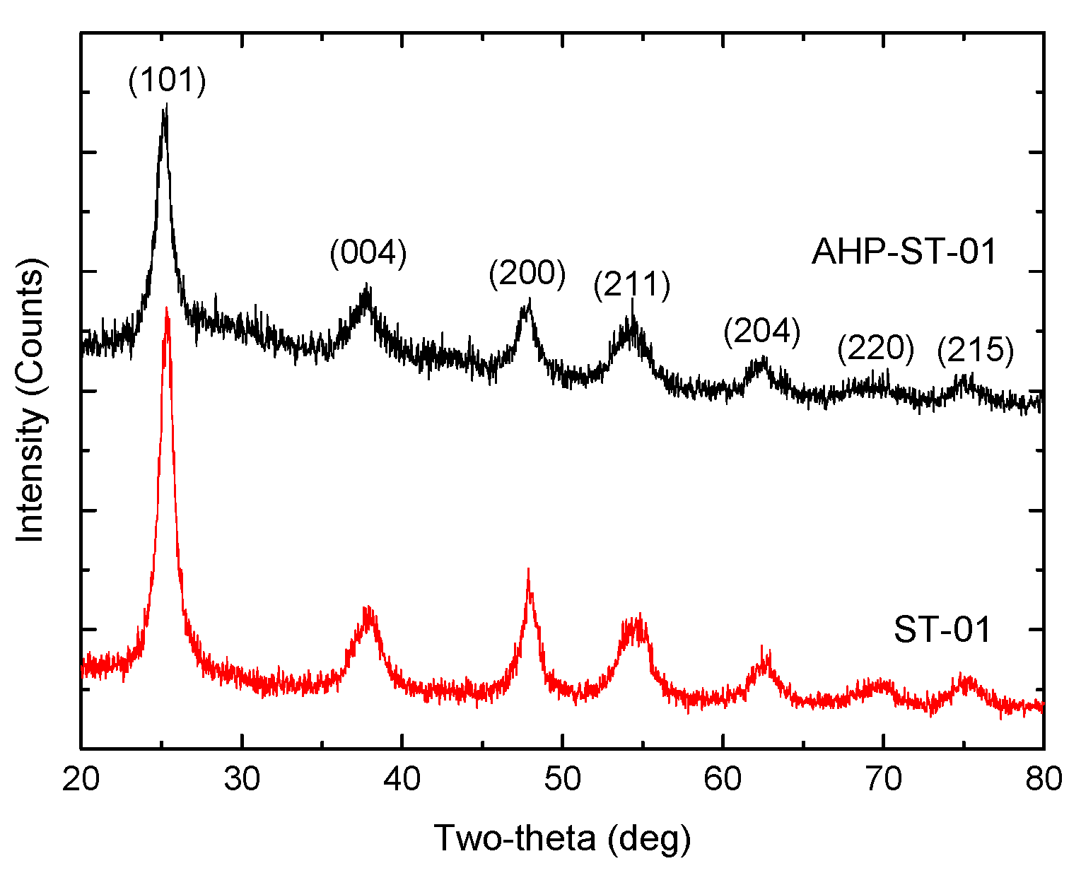
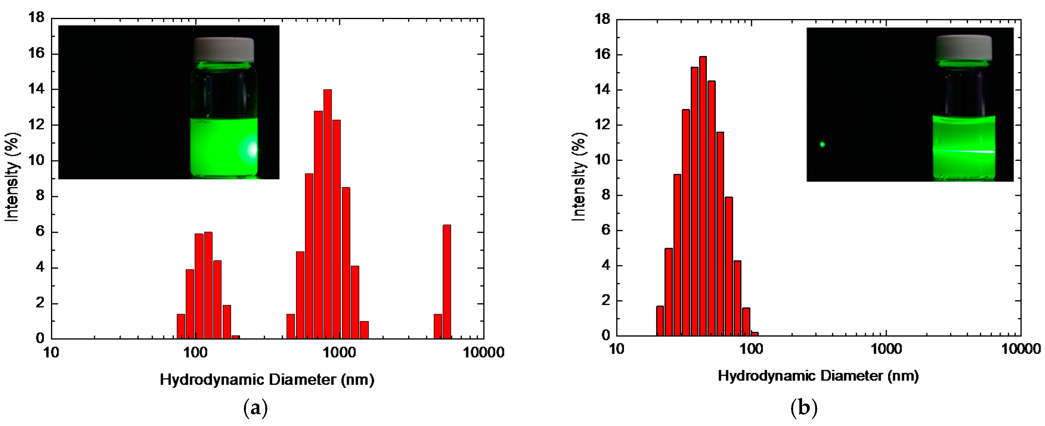
| Ishihara ST-01 | 290 | 7 | 821 ± 97 | 38 ± 1 | 6.3 | 2.32 | 1.9 |
| AHP-ST-01 | 304 | 5 | 41 ± 1 | 48 ± 2 | 6.7 | 6.08 | 3.2 |
| Sample | TGA | XPS | ||||
|---|---|---|---|---|---|---|
| OHweak (mmol/g) | OHstrong (mmol/g) | Relative Amount of OHstrong | TiOH a (mmol/g) | Ti-OH b (mmol/g) | Relative Amount of Ti-OH | |
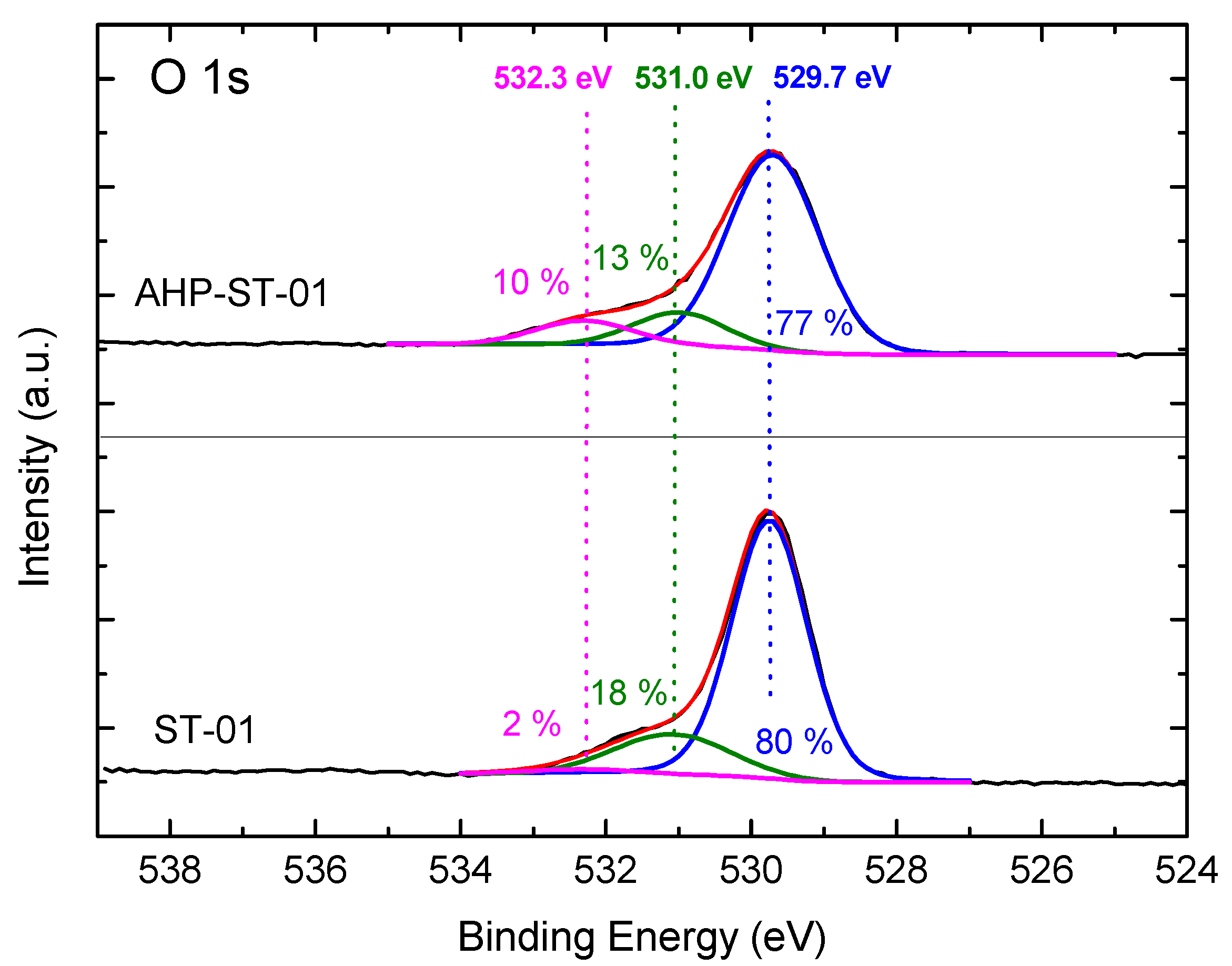
| ST-01 | 1.73 | 0.59 | 1 | 4.67 | 0.51 | 1 |
| AHP-ST-01 | 2.47 | 3.61 | 6.1 | 3.62 | 2.74 | 5.4 |
| Sample | SBET (m2/g) | Surface OH Conc. (mmol/g) | Surface OH Density (/nm2) | Analytical Method | Reference |
|---|---|---|---|---|---|
| Degussa P25 | 56 | 0.44 | 4.8 | TGA | Mueller et al., 2003 [30] |
| Degussa P25 | 56 | 0.49 | 5.3 | TGA | This work |
| Hombikat UV 100 | 337 | 1.15 | 2.5 | NH3-TPD | Carneiro et al., 2010 [47] |
| Kanto | 22 | 0.012 | 0.3 | Ion-Exchange | Tamura et al., 1999 [48] |
| TiO2 by sol-gel | 85 | 1.44 | 10.2 | TGA | Zhao et al., 2008 [31] |
| Three-dimensional TiO2 by sol-gel | 203 | 4.01 | 11.9 | XPS | Guo et al., 2013 [15] |
| NaCl-TiO2 by sol-gel | — a | 2.31 | — | TGA | Wang et al., 2012 [9] |
| NaOH-Degussa P25 | — | 1.60 | — | TGA | Eskandarloo et al., 2015 [17] |
| Ishihara ST-01 | 290 | 2.32 | 4.8 | TGA | This work |
| AHP-ST-01 | 304 | 6.08 | 12.0 | TGA | This work |
© 2017 by the authors. Licensee MDPI, Basel, Switzerland. This article is an open access article distributed under the terms and conditions of the Creative Commons Attribution (CC BY) license (http://creativecommons.org/licenses/by/4.0/).
Share and Cite
Wu, C.-Y.; Tu, K.-J.; Deng, J.-P.; Lo, Y.-S.; Wu, C.-H. Markedly Enhanced Surface Hydroxyl Groups of TiO2 Nanoparticles with Superior Water-Dispersibility for Photocatalysis. Materials 2017, 10, 566. https://0-doi-org.brum.beds.ac.uk/10.3390/ma10050566
Wu C-Y, Tu K-J, Deng J-P, Lo Y-S, Wu C-H. Markedly Enhanced Surface Hydroxyl Groups of TiO2 Nanoparticles with Superior Water-Dispersibility for Photocatalysis. Materials. 2017; 10(5):566. https://0-doi-org.brum.beds.ac.uk/10.3390/ma10050566
Chicago/Turabian StyleWu, Chung-Yi, Kuan-Ju Tu, Jin-Pei Deng, Yu-Shiu Lo, and Chien-Hou Wu. 2017. "Markedly Enhanced Surface Hydroxyl Groups of TiO2 Nanoparticles with Superior Water-Dispersibility for Photocatalysis" Materials 10, no. 5: 566. https://0-doi-org.brum.beds.ac.uk/10.3390/ma10050566