Ag-Coated Heterostructures of ZnO-TiO2/Delaminated Montmorillonite as Solar Photocatalysts

 , , ,
, , ,
Abstract
:1. Introduction
2. Experimental
2.1. Synthesis of Ag/ZnO-TiO2/Clay Materials
2.2. Characterization of the Solids
2.3. Photocatalytic Experiments
3. Results and Discussion
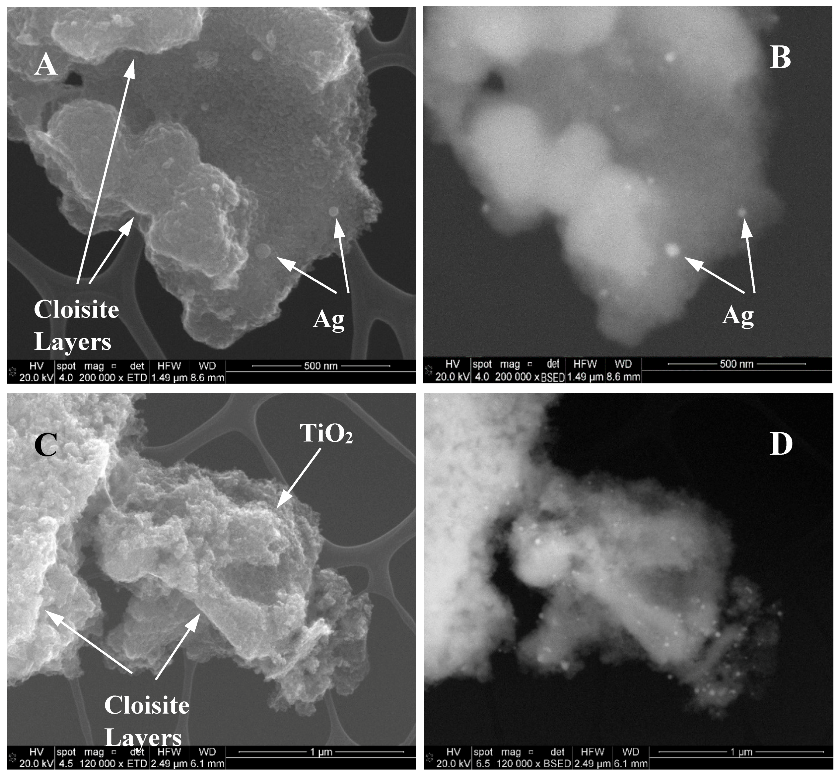
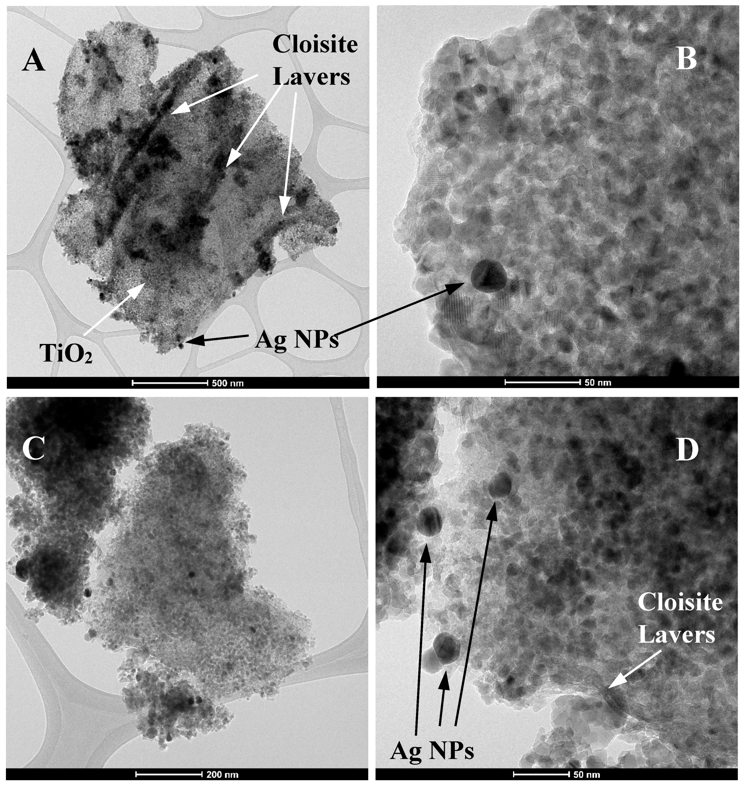
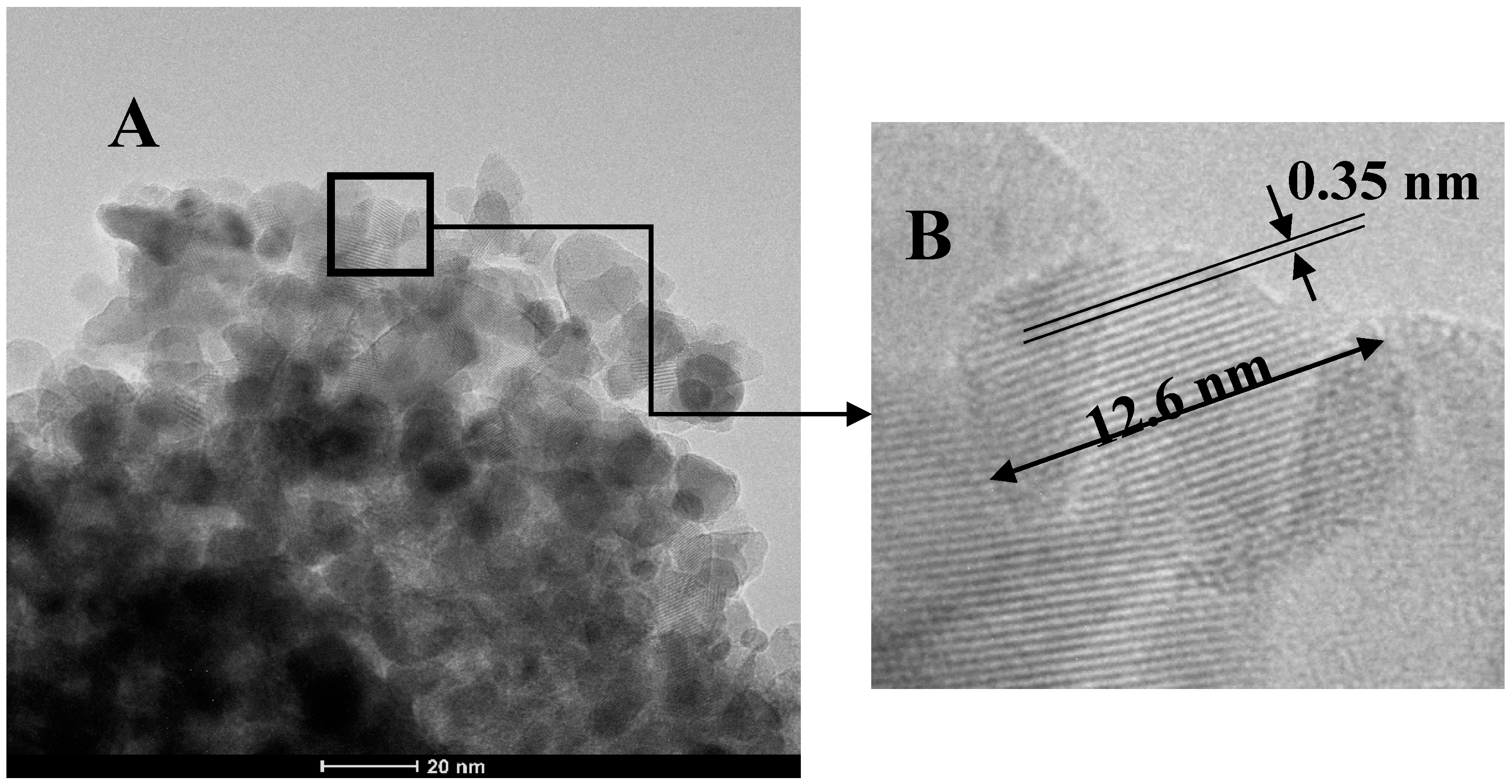
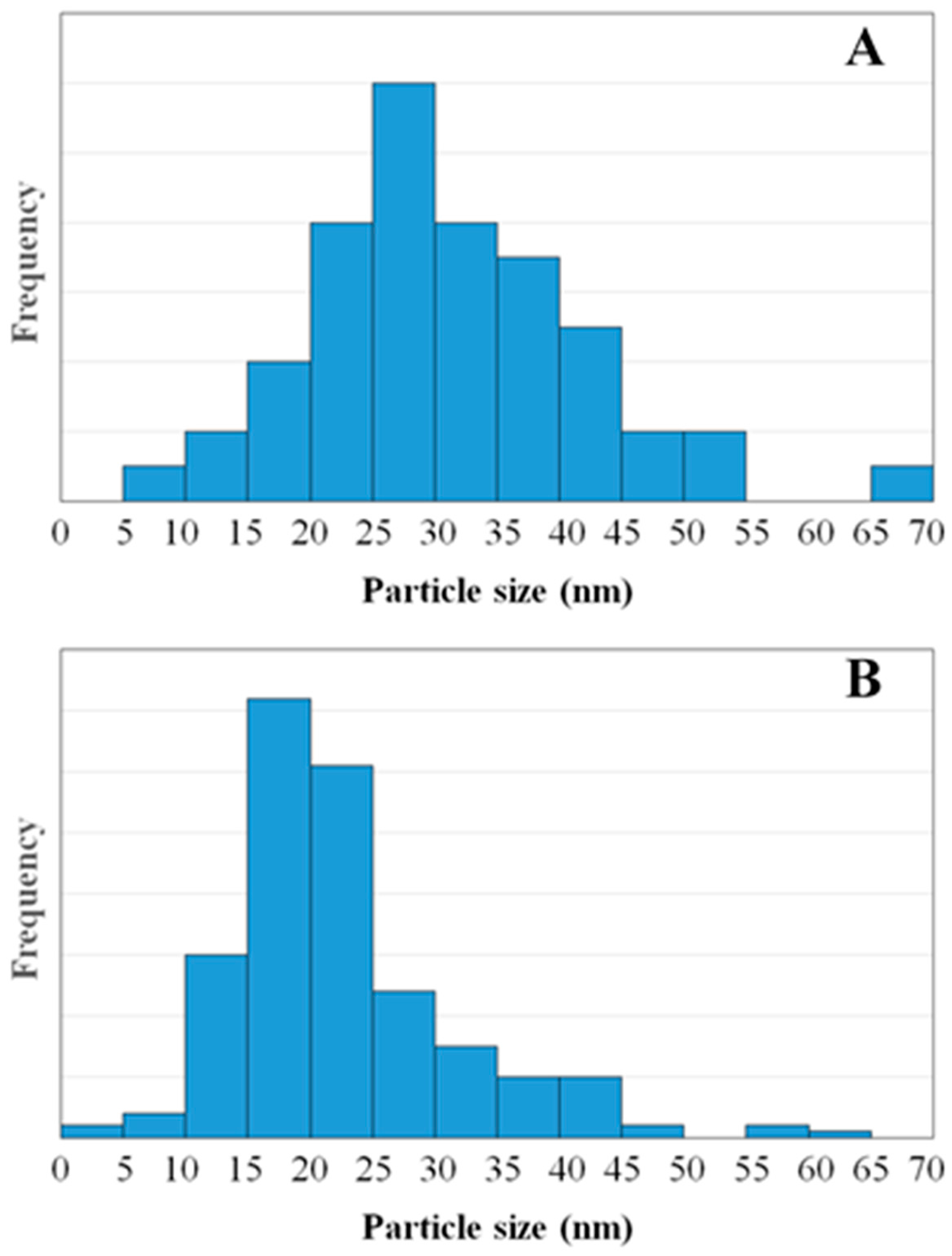
3.1. Characterization of ZnO-TiO2/Delaminated Montmorillonite Coated with Ag
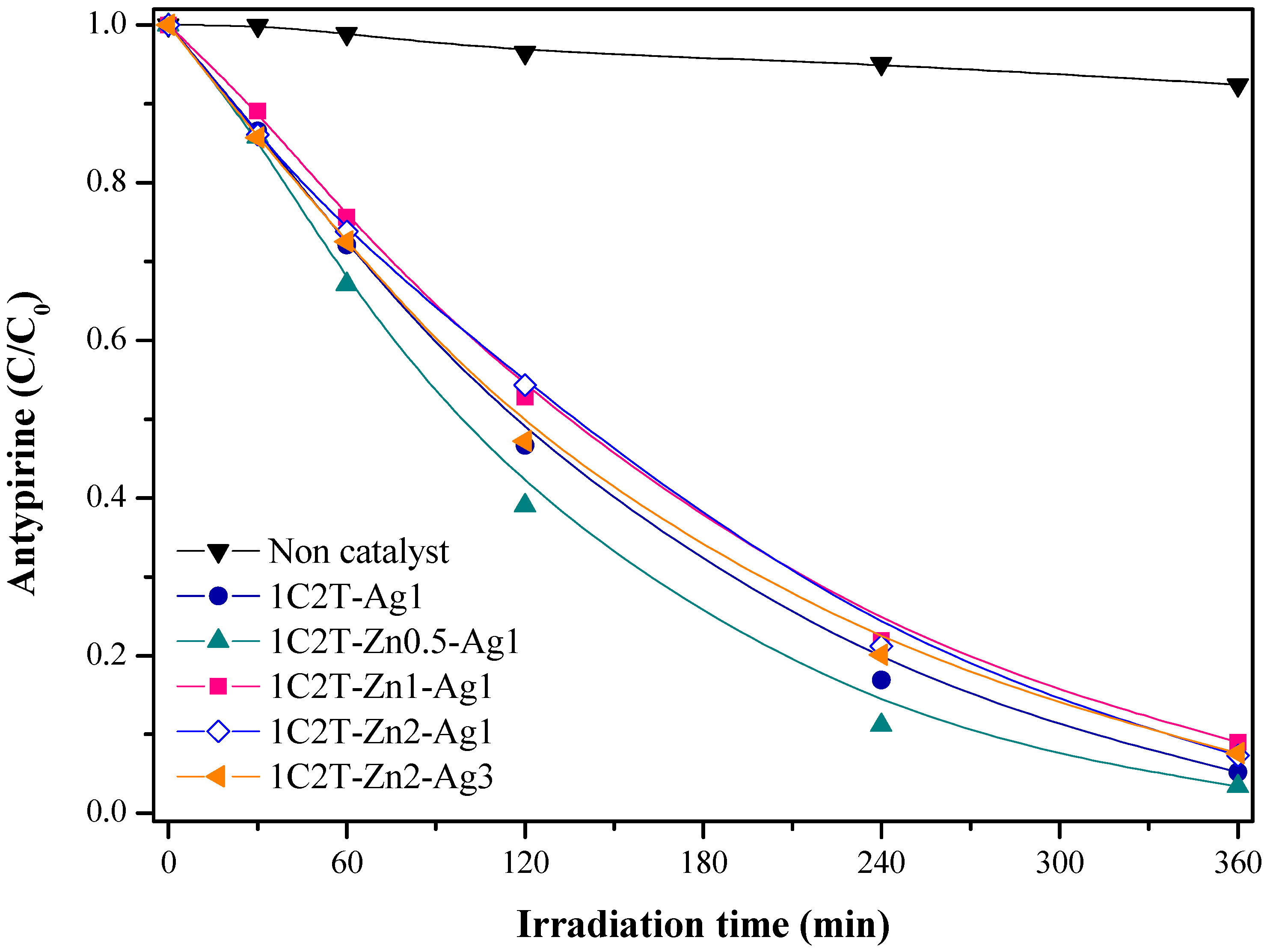
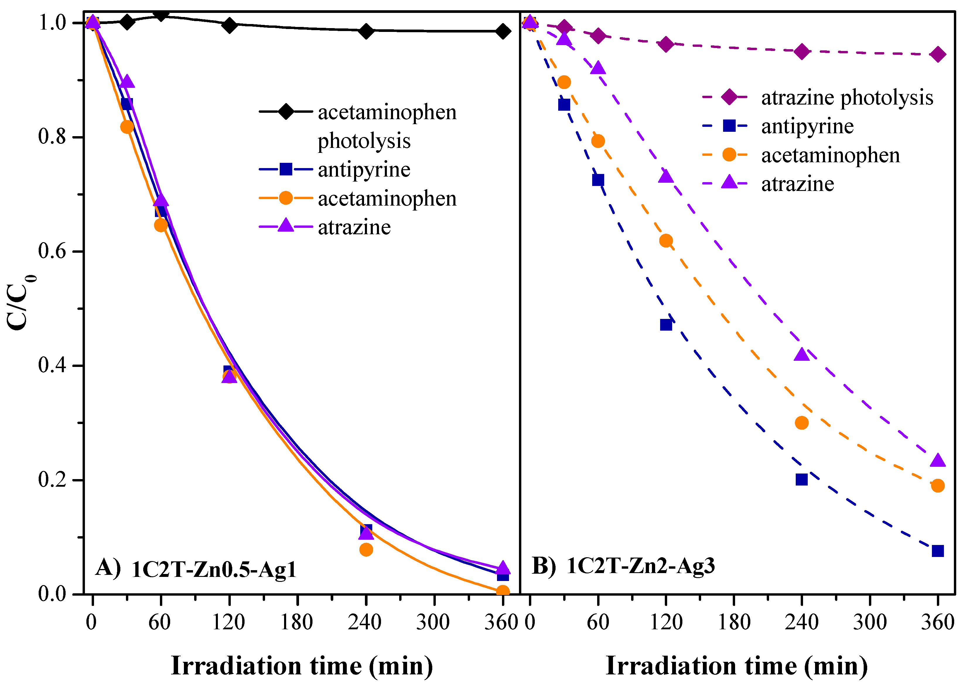
3.2. Photocatalytic Activity
4. Conclusions
Supplementary Materials
Acknowledgments
Author Contributions
Conflicts of Interest
References
- Brindley, G.W.; Sempels, R.E. Preparation and properties of some hydroxy-aluminum beidellites. Clay Miner. 1977, 12, 229–237. [Google Scholar] [CrossRef]
- Lahav, N.; Shani, U.; Shabtai, J. Cross-linked smectites. I. Synthesis and properties of hydroxy-aluminum montmorillonite. Clays Clay Miner. 1978, 26, 107–115. [Google Scholar] [CrossRef]
- Gil, A.; Korili, S.A.; Vicente, M.A. Recent advances in the control and characterization of the porous structure of pillared clay catalysts. Catal. Rev. 2008, 50, 153–221. [Google Scholar] [CrossRef]
- Gil, A.; Korili, S.A.; Trujillano, R.; Vicente, M.A. Pillared Clays and Related Catalysts, 1st ed.; Springer: New York, NY, USA, 2010; ISBN 978-1-44196670-4. [Google Scholar]
- Galarneau, A.; Barodawalla, A.; Pinnavaia, T.J. Porous clay heterostructures formed by gallery-templated synthesis. Nature 1995, 374, 529–531. [Google Scholar] [CrossRef]
- Galarneau, A.; Barodawalla, A.; Pinnavaia, T.J. Porous clay heterostructures (PCH) as acid catalysts. Chem. Commun. 1997, 1661–1662. [Google Scholar] [CrossRef]
- Polverejan, M.; Pauly, T.R.; Pinnavaia, T.J. Acidic porous clay heterostructures (PCN): Intragallery assembly of mesoporous silica in synthetic saponite clays. Chem. Mater. 2000, 12, 2698–2704. [Google Scholar] [CrossRef]
- Chmielarz, L.; Kustrowski, P.; Dziembaj, R.; Cool, P.; Vansant, E.F. Selective catalytic reduction of NO with ammonia over porous clay heterostructures modified with copper and iron species. Catal. Today 2007, 119, 181–186. [Google Scholar] [CrossRef]
- Tchinda, A.J.; Ngameni, E.; Kenfack, I.T.; Walkarius, A. One-Step preparation of thiol-functionalized porous clay heterostructures: Application to Hg(II) binding and characterization of mass transport issues. Chem. Mater. 2009, 21, 4111–4121. [Google Scholar] [CrossRef]
- Qu, F.; Zhu, L.; Yang, K. Adsorption behaviors of volatile organic compounds (VOCs) on porous clay heterostructures (PCH). J. Hazard. Mater. 2009, 170, 7–12. [Google Scholar] [CrossRef] [PubMed]
- Aranda, P.; Belver, C.; Ruiz-Hitzky, E. Inorganic heterostructured materials based on clay mineral. In Materials and Clay Minerals, 1st ed.; Drummy, L.F., Ogawa, M., Aranda, P., Eds.; CMS Workshop Lectures; The Clay Minerals Society: Chantilly, VA, USA, 2013; Volume 18, pp. 21–40. [Google Scholar] [CrossRef]
- Letaïef, S.; Ruiz-Hitzky, E. Silica-clay nanocomposites. Chem. Commun. 2003, 2996–2997. [Google Scholar] [CrossRef]
- Letaïef, S.; Martín-Luengo, M.A.; Aranda, P.; Ruiz-Hitky, E. A colloidal route for delamination of layered solids: Novel porous-clay nanocomposites. Adv. Funct. Mater. 2006, 16, 401–409. [Google Scholar] [CrossRef]
- Belver, C.; Aranda, P.; Martín-Luengo, M.A.; Ruiz-Hitzky, E. New silica/alumina-clay heterostructures: Properties as acid catalysts. Microporous Mesoporous Mater. 2012, 147, 157–166. [Google Scholar] [CrossRef]
- Manova, E.; Aranda, P.; Martín-Luengo, M.A.; Letaief, S.; Ruiz-Hitzky, E. New titania-clay nanostructured porous materials. Microporous Mesoporous Mater. 2010, 131, 252–260. [Google Scholar] [CrossRef]
- Belver, C.; Bedia, J.; Rodriguez, J.J. Titania-clay heterostructures with solar photocatalytic applications. Appl. Catal. B Environ. 2015, 176–177, 278–287. [Google Scholar] [CrossRef]
- Belver, C.; Bedia, J.; Álvarez-Montero, M.A.; Rodriguez, J.J. Solar photocatalytic purification of water with Ce-doped TiO2/clay heterostructures. Catal. Today 2016, 266, 36–45. [Google Scholar] [CrossRef]
- Belver, C.; Bedia, J.; Rodriguez, J.J. Zr-doped TiO2 supported on delaminated clay materials for solar photocatalytic treatment of emerging pollutants. J. Hazard. Mater. 2017, 322, 233–242. [Google Scholar] [CrossRef] [PubMed]
- Tobajas, M.; Belver, C.; Rodriguez, J.J. Degradation of emerging pollutants in water under solar irradiation using novel TiO2-ZnO/clay nanoarchitectures. Chem. Eng. J. 2017, 309, 596–606. [Google Scholar] [CrossRef]
- Liu, J.; Zhang, G. Recent advances in synthesis and applications of clay-based photocatalysts: A review. Phys. Chem. Chem. Phys. 2014, 16, 8178–8192. [Google Scholar] [CrossRef] [PubMed]
- Richardson, S.D.; Ternes, T.A. Water analysis: Emerging contaminants and current issues. Anal. Chem. 2011, 83, 4614–4648. [Google Scholar] [CrossRef] [PubMed]
- Pal, A.; Gin, K.Y.-H.; Lin, A.Y.-C.; Reinhard, M. Impacts of emerging contaminants on freshwater resources: Review of recent occurrences, sources, fate and effects. Sci. Total Environ. 2010, 408, 6062–6069. [Google Scholar] [CrossRef] [PubMed]
- Hoffmann, M.R.; Martin, S.T.; Choi, W.; Bahnemann, D.W. Environmental applications of semiconductor photocatalysis. Chem. Rev. 1995, 95, 69–96. [Google Scholar] [CrossRef]
- Mills, A.; Le Hunte, S. An overview of semiconductor photocatalysis. J. Photochem. Photobiol. A 1997, 108, 1–35. [Google Scholar] [CrossRef]
- Hernández-Gordillo, A.; Rodríguez-González, V. Silver nanoparticles loaded on Cu-doped TiO2 for the effective reduction of nitro-aromatic contaminants. Chem. Eng. J. 2015, 261, 53–59. [Google Scholar] [CrossRef]
- Brunauer, S.; Emmett, P.H.; Teller, E. Adsorption of Gases in Multimolecular Layers. J. Am. Chem. Soc. 1938, 60, 309–319. [Google Scholar] [CrossRef]
- Lippens, B.C.; de Boer, J.H. Studies on pore systems in catalysts: V. The t method. J. Catal. 1965, 4, 319–323. [Google Scholar] [CrossRef]
- Tauc, J. Absorption edge and internal electric fields in amorphous semiconductors. Mater. Res. Bull. 1970, 5, 721–726. [Google Scholar] [CrossRef]
- Turova, N.Y.; Turevskaya, E.P.; Kessler, V.G.; Yanovskaya, M.I. The Chemistry of Metal Alkoxides; Kluwer Academic Publishers: New York, NY, USA, 2002; ISBN 0-7923-7521-1. [Google Scholar]
- Thommes, M.; Kaneko, K.; Neimark, A.V.; Olivier, J.P.; Rodriguez-Reinoso, F.; Rouquerol, J.; Sing, K.S.W. Physisorption of gases, with special reference to the evaluation of surface area and pore size distribution (IUPAC Technical Report). Pure Appl. Chem. 2015, 87, 1051–1069. [Google Scholar] [CrossRef]
- Ruiz-Hitzky, E.; Aranda, P.; Belver, C. Nanoarchitectures based on clay materials. In Manipulation on Nanoscale Materials: An Introduction to Nanoarchitectonics; Ariga, K., Ed.; The Royal Society of Chemistry: Cambridge, UK, 2012; pp. 87–111. ISBN 978-1-84973-415-8. [Google Scholar]
- Li, K.; Lei, J.; Yuan, G.; Weerachanchai, P.; Wang, J.-Y.; Zhao, J.; Yang, Y. Fe-, Ti-, Zr- and Al-pillared clays for efficient catalytic pyrolysis of mixed plastics. Chem. Eng. J. 2017, 317, 800–809. [Google Scholar] [CrossRef]
- Belessi, V.; Lambropoulou, D.; Konstantinou, I.; Katsoulidis, A.; Ponomis, P.; Petridis, D.; Albanis, T. Structure and photocatalytic performance of TiO2/clay nanocomposites for the degradation of dimethachlor. Appl. Catal. B Environ. 2007, 73, 292–299. [Google Scholar] [CrossRef]
- Pelaez, M.; Nolan, N.T.; Pillai, S.C.; Seery, M.K.; Falaras, P.; Kontos, A.G.; Dunlop, P.S.M.; Hamilton, J.W.J.; Byrne, J.A.; O’shea, K.; et al. A review on the visible light active titanium dioxide photocatalysts for environmental applications. Appl. Catal. B 2012, 125, 331–349. [Google Scholar] [CrossRef]
- Chen, J.; Qiu, F.; Xu, W.; Cao, S.; Zhu, H. Recent progress in enhancing photocatalytic efficiency of TiO2-based materials. Appl. Catal. A 2015, 495, 131–140. [Google Scholar] [CrossRef]
- Holtz, R.D.; Souza-Filho, A.G.; Brocchi, M.; Martins, D.; Durán, N.; Alves, O.L. Development of nanostructured silver vanadates decorated with silver nanoparticles as a novel antibacterial agent. Nanotechnology 2010, 21, 185102–185110. [Google Scholar] [CrossRef] [PubMed]
- Kwiatkowski, M.; Bezberkhyy, I.; Skompska, M. ZnO nanorods covered with a TiO2 layer: Simple sol-gel preparation, and optical, photocatalytic and photoelectrochemical properties. J. Mater. Chem. A 2015, 2, 12748–12760. [Google Scholar] [CrossRef]
- Li, D.; Jiang, X.; Zhang, Y.; Zhang, B. A novel route to ZnO/TiO2 heterojunction composite fibers. J. Mater. Res. 2013, 28, 507–512. [Google Scholar] [CrossRef]
- Belver, C.; Adan, C.; García-Rodríguez, S.; Fernández-García, M. Photocatalytic behavior of silver vanadates: Microemulsion synthesis and post-reaction characterization. Chem. Eng. J. 2013, 224, 24–31. [Google Scholar] [CrossRef]
- Mangayayam, M.; Kiwi, J.; Giannakis, S.; Pulgarin, C.; Zivkovic, I.; Magrez, A.; Rtimi, S. FeOx magnetization enhancing E. coli inactivation by orders of magnitude on Ag-TiO2 nanotubes under sunlight. Appl. Catal. B 2017, 202, 438–445. [Google Scholar] [CrossRef]
- Deng, X.; Li, M.; Zhang, J.; Hu, X.; Zheng, J.; Zhang, N.; Chen, B.H. Constructing nano-structure on silver/ceria-zirconia towards highly active and stable catalyst for soot oxidation. Chem. Eng. J. 2017, 313, 544–555. [Google Scholar] [CrossRef]
- Pirhashemi, M.; Habibi-Yangjeh, A. Ultrasonic-assisted preparation of plasmonic ZnO/Ag/Ag2WO4 nanocomposites with high visible-light photocatalytic performance for degradation of organic pollutants. J. Colloid Interface Sci. 2017, 491, 216–229. [Google Scholar] [CrossRef] [PubMed]
- Xu, H.; Liao, J.; Yuan, S.; Zhao, Y.; Zhang, M.; Wang, Z.; Shi, L. Tuning the morphology, stability and photocatalytic activity of TiO2 nanocrystal colloids by tungsten doping. Mater. Res. Bull. 2014, 51, 326–331. [Google Scholar] [CrossRef]
- Hoflund, G.B.; Hazos, Z.F.; Salaita, G.N. Surface characterization study of Ag, AgO, and Ag2O using X-ray photoelectron spectroscopy and electron energy-loss spectroscopy. Phys. Rev. B 2000, 62, 11126–11133. [Google Scholar] [CrossRef]
- Lee, A.Y.; Blakeslee, D.M.; Powell, C.J.; Rumble, J.R. Development of the web-based NIST X-ray Photoelectron Spectroscopy (XPS) Database. Data Sci. J. 2002, 1, 1–12. [Google Scholar] [CrossRef]
- Tahir, M.; Amin, N.S. Photocatalytic reduction of carbon dioxide with water vapors over montmorillonite modified TiO2 nanocomposites. Appl. Catal. B 2013, 142–143, 512–522. [Google Scholar] [CrossRef]


| Sample | D (nm) | TiO2 | ZnO | Ag | ZnO/TiO2 |
|---|---|---|---|---|---|
| 1C2T-Ag1 | 12.4 | 74.30 | 0.00 | 1.13 | n.d. |
| 1C2T-Zn05-Ag1 | 13.4 | 72.70 | 0.26 | 0.91 | 0.36 |
| 1C2T-Zn1-Ag1 | 15.4 | 71.50 | 0.49 | 1.18 | 0.69 |
| 1C2T-Zn2-Ag1 | 14.0 | 70.00 | 1.03 | 1.17 | 1.47 |
| 1C2T-Zn2-Ag3 | 13.7 | 69.80 | 0.95 | 2.38 | 1.36 |
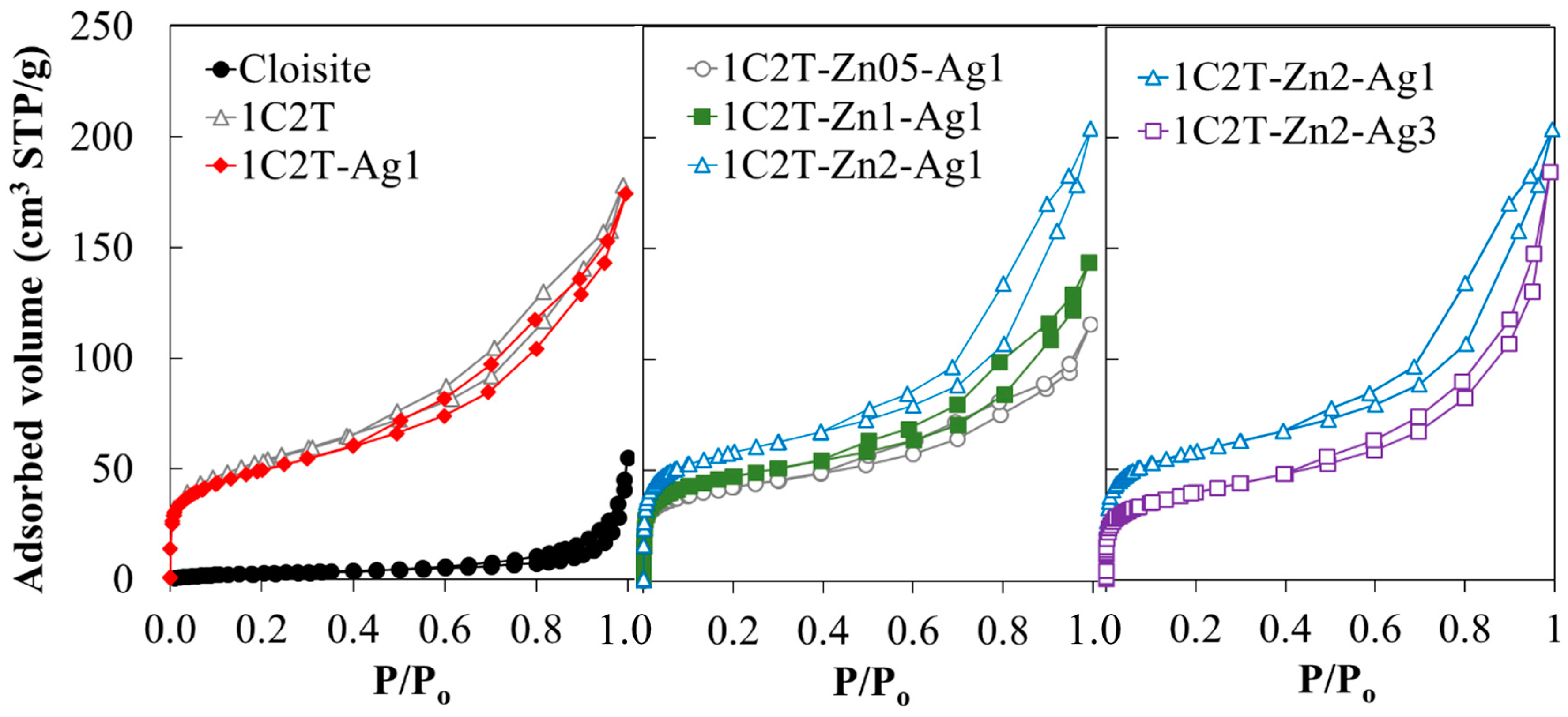
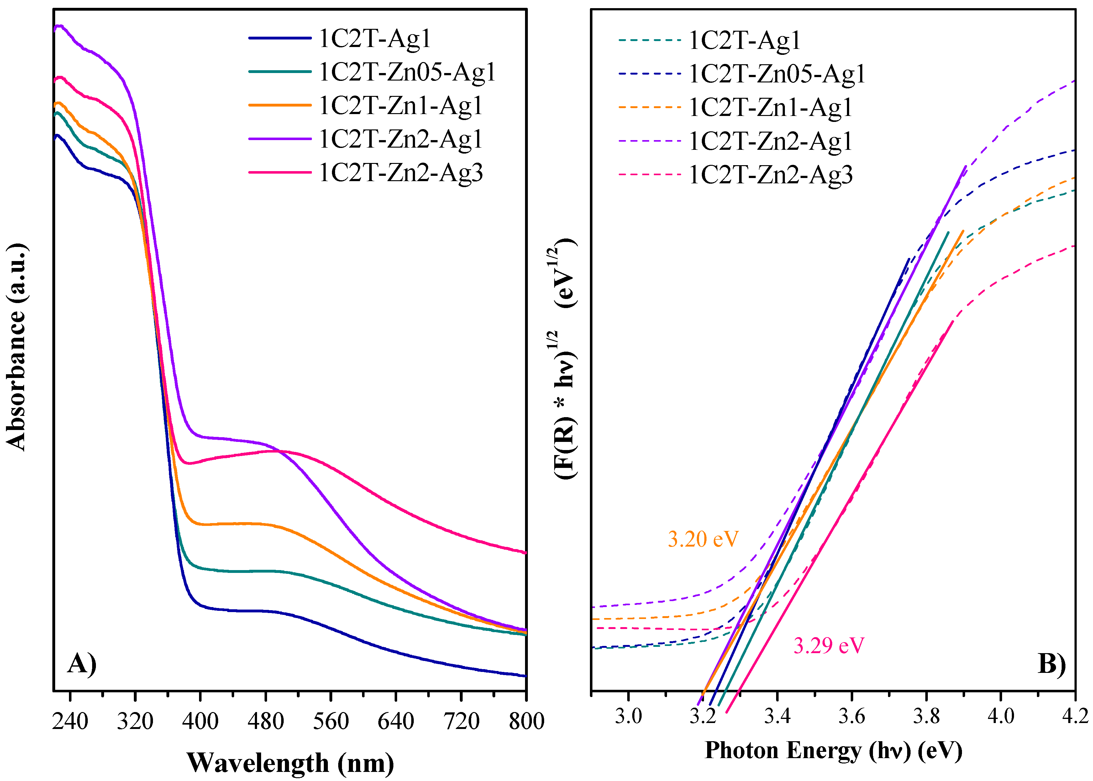
| Sample | SBET (m2 g−1) | SEXT (m2 g−1) | VMP (cm3 g−1) | VT (cm3 g−1) | Band Gap (eV) |
|---|---|---|---|---|---|
| Cloisite | 11 | 11 | n.d. | 0.085 | n.d. |
| 1C2T | 182 | 152 | 0.016 | 0.275 | 3.21 |
| 1C2T-Ag1 | 173 | 127 | 0.021 | 0.270 | 3.25 |
| 1C2T-Zn0.5-Ag1 | 144 | 74 | 0.033 | 0.179 | 3.20 |
| 1C2T-Zn1-Ag1 | 162 | 83 | 0.037 | 0.222 | 3.20 |
| 1C2T-Zn2-Ag1 | 200 | 109 | 0.043 | 0.316 | 3.23 |
| 1C2T-Zn2-Ag3 | 138 | 98 | 0.018 | 0.285 | 3.29 |
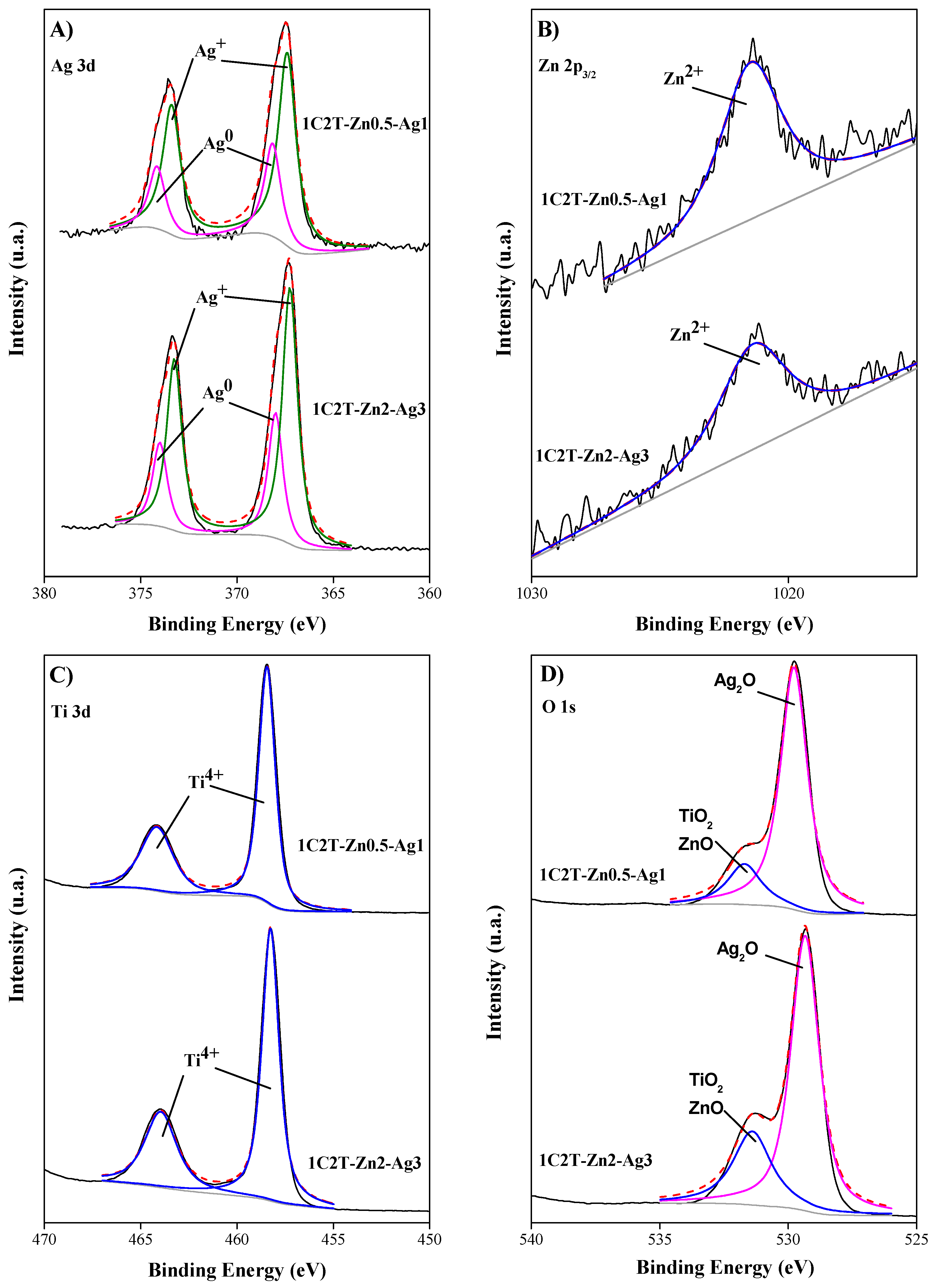
| Sample | Ag+ (%) | Ag0 (%) | Ag+/Ag0 |
|---|---|---|---|
| 1C2T-Zn0.5-Ag1 | 69.5 | 30.5 | 2.3 |
| 1C2T-Zn2-Ag3 | 68.5 | 31.5 | 2.2 |
© 2017 by the authors. Licensee MDPI, Basel, Switzerland. This article is an open access article distributed under the terms and conditions of the Creative Commons Attribution (CC BY) license (http://creativecommons.org/licenses/by/4.0/).
Share and Cite
Belver, C.; Hinojosa, M.; Bedia, J.; Tobajas, M.; Alvarez, M.A.; Rodríguez-González, V.; Rodriguez, J.J. Ag-Coated Heterostructures of ZnO-TiO2/Delaminated Montmorillonite as Solar Photocatalysts. Materials 2017, 10, 960. https://0-doi-org.brum.beds.ac.uk/10.3390/ma10080960
Belver C, Hinojosa M, Bedia J, Tobajas M, Alvarez MA, Rodríguez-González V, Rodriguez JJ. Ag-Coated Heterostructures of ZnO-TiO2/Delaminated Montmorillonite as Solar Photocatalysts. Materials. 2017; 10(8):960. https://0-doi-org.brum.beds.ac.uk/10.3390/ma10080960
Chicago/Turabian StyleBelver, Carolina, Mariana Hinojosa, Jorge Bedia, Montserrat Tobajas, Maria Ariadna Alvarez, Vicente Rodríguez-González, and Juan Jose Rodriguez. 2017. "Ag-Coated Heterostructures of ZnO-TiO2/Delaminated Montmorillonite as Solar Photocatalysts" Materials 10, no. 8: 960. https://0-doi-org.brum.beds.ac.uk/10.3390/ma10080960