
Antioxidants: Classification, Natural Sources, Activity/Capacity Measurements, and Usefulness for the Synthesis of Nanoparticles


Abstract
:1. Introduction
- (i)
- The wide range of industrial and biomedical applications of antioxidants requires effective and rapid in vitro tests to evaluate total antioxidant activity. Various methods were collected in the review, i.e., chromatographic, spectrometric, and electrochemical. Particular attention was paid to the method based on metallic nanoparticles, which are used as optical probes (SNPAC). The method is useful for measuring the antioxidant activity of both simple chemical compounds and mixtures of natural origin. The SNAPC tests are effective in assessing electron transfer but are not used very often. The review includes information on the extracts from plants, lichens, fungi, algae, and actinomycetes (reference antioxidants, extraction process, antioxidant activity tests, and activity parameters).
- (ii)
- Natural extracts as a source of both reducing and stabilizing substances are used for the green synthesis of nanoparticles. The review includes examples of the synthesis of metallic/metal oxides of nanoparticles using extracts from various plant species and microorganisms (yeast, algae, cyanobacteria, fungi, and bacteria). The information collected allows us to trace the links between the type of antioxidant, its origin, activity, and suitability for the efficient synthesis of nanoparticles. Extensive data were collected on the methods of extract preparation, antioxidant activity tests, detection methods, NPs synthesis conditions, and the morphology of the obtained nanoparticles.
2. Free Radicals/Antioxidants
2.1. Free Radicals vs. Oxidative Stress
2.2. Antioxidants
3. Antioxidant Capacity/Activity Measurements
3.1. Techniques Used to Assess Antioxidant Capacity
3.1.1. DPPH Free Radical Scavenging Assay
DPPH Free Radical Scavenging Kinetics
Parameters Used to Express the Antioxidant Potential
DPPH Assay Approaches
3.1.2. Electrochemical Methods
The Cyclic Voltammetry Method
Biamperometry
3.1.3. Nanoparticle-Based Approach for the Antioxidant Activity Measurement
4. Antioxidant Capacity of Extracts from Natural Sources
5. Synthesis of Nanoparticles (NPs) by Natural Extracts
5.1. Microbial Synthesis of NPs
5.2. Plant Extracts-Mediated NPs Synthesis
5.3. Trends of NPs Modification
6. Concluding Remarks and Future Perspectives
Author Contributions
Funding
Institutional Review Board Statement
Informed Consent Statement
Conflicts of Interest
References
- Choi, H.J.; Kim, J.H.; Lee, C.H.; Ahn, Y.J.; Song, J.H.; Baek, S.H.; Kwon, D.H. Antiviral activity of quercetin 7-rhamnoside against porcine epidemic diarrhea virus. Antiviral Res. 2009, 81, 77–81. [Google Scholar] [CrossRef] [PubMed]
- Song, J.H.; Shim, J.K.; Choi, H.J. Quercetin 7-rhamnoside reduces porcine epidemic diarrhea virus replication via independent pathway of viral induced reactive oxygen species. Virol. J. 2011, 8, 460. [Google Scholar] [CrossRef] [PubMed] [Green Version]
- Liang, W.; He, L.; Ning, P.; Lin, J.; Li, H.; Lin, Z.; Kang, K.; Zhang, Y. (+)-Catechin inhibition of transmissible gastroenteritis coronavirus in swine testicular cells is involved its antioxidation. Res. Vet. Sci. 2015, 103, 28–33. [Google Scholar] [CrossRef]
- Diniz, L.R.L.; Bezerra Filho, C.D.S.M.; Fielding, B.C.; de Sousa, D.P. Natural Antioxidants: A Review of Studies on Human and Animal Coronavirus. Oxid. Med. Cell. Longev. 2020, 1–14. [Google Scholar] [CrossRef] [PubMed]
- Delgado-Roche, L.; Mesta, F. Oxidative Stress as Key Player in Severe Acute Respiratory Syndrome Coronavirus (SARS-CoV) Infection. Arch. Med. Res. 2020, 51, 384–387. [Google Scholar] [CrossRef]
- Jo, S.; Kim, S.; Shin, D.H.; Kim, M.S. Inhibition of SARS-CoV 3CL protease by flavonoids. J. Enzyme Inhib. Med. Chem. 2020, 35, 145–151. [Google Scholar] [CrossRef] [Green Version]
- Chen, L.; Li, J.; Luo, C.; Liu, H.; Xu, W.; Chen, G.; Liew, O.W.; Zhu, W.; Puah, C.M.; Shen, X.; et al. Binding interaction of quercetin-3-beta-galactoside and its synthetic derivatives with SARS-CoV 3CL(pro): Structure-activity relationship studies reveal salient pharmacophore features. Bioorg. Med. Chem. 2006, 14, 8295–8306. [Google Scholar] [CrossRef] [PubMed]
- Ryu, Y.B.; Jeong, H.J.; Kim, J.H.; Kim, Y.M.; Park, J.Y.; Kim, D.; Nguyen, T.T.; Park, S.J.; Chang, J.S.; Park, K.H.; et al. Biflavonoids from Torreya nucifera displaying SARS-CoV 3CL(pro) inhibition. Bioorg. Med. Chem. 2010, 18, 7940–7947. [Google Scholar] [CrossRef]
- Kim, D.W.; Seo, K.H.; Curtis-Long, M.J.; Oh, K.Y.; Oh, J.W.; Cho, J.K.; Lee, K.H.; Park, K.H. Phenolic phytochemical displaying SARS-CoV papain-like protease inhibition from the seeds of Psoralea corylifolia. J. Enzyme Inhib. Med. Chem. 2014, 29, 59–63. [Google Scholar] [CrossRef] [Green Version]
- Yu, M.S.; Lee, J.; Lee, J.M.; Kim, Y.; Chin, Y.W.; Jee, J.G.; Keum, Y.S.; Jeong, Y.J. Identification of myricetin and scutellarein as novel chemical inhibitors of the SARS coronavirus helicase, nsP13. Bioorg. Med. Chem. Lett. 2012, 22, 4049–4054. [Google Scholar] [CrossRef]
- Cárdenas-Rodríguez, N.; Bandala, C.; Vanoye-Carlo, A.; Ignacio-Mejía, I.; Gómez-Manzo, S.; Hernández-Cruz, E.Y.; Pedraza-Chaverri, J.; Carmona-Aparicio, L.; Hernández-Ochoa, B. Use of Antioxidants for the Neuro-Therapeutic Management of COVID-19. Antioxidants 2021, 10, 971. [Google Scholar] [CrossRef]
- Forman, H.J.; Zhang, H. Targeting oxidative stress in disease: Promise and limitations of antioxidant therapy. Nat. Rev. Drug Discov. 2021. [Google Scholar] [CrossRef]
- Forman, H.J.; Davies, K.J.; Ursini, F. How do nutritional antioxidants really work: Nucleophilic tone and para-hormesis versus free radical scavenging in vivo. Free Radic. Biol. Med. 2014, 66, 24–35. [Google Scholar] [CrossRef] [Green Version]
- Sies, H. Strategies of antioxidant defense. Eur. J. Biochem. 1993, 215, 213–219. [Google Scholar] [CrossRef]
- Bansal, S.; Choudhary, S.; Sharma, M.; Kumar, S.S.; Lohan, S.; Bhardwaj, V.; Syan, N.; Jyoti, S. Tea: A native source of antimicrobial agents. Food Res. Int. 2013, 53, 568–584. [Google Scholar] [CrossRef]
- Jiang, J.; Xiong, Y.L. Natural antioxidants as food and feed additives to promote health benefits and quality of meat products: A review. Meat Sci. 2016, 120, 107–117. [Google Scholar] [CrossRef] [PubMed] [Green Version]
- Lü, J.-M.; Lin, P.H.; Yao, Q.; Chen, C. Chemical and molecular mechanisms of antioxidants: Experimental approaches and model systems. J. Cell. Mol. Med. 2009, 14, 840–860. [Google Scholar] [CrossRef]
- Cetin-Karaca, H.; Newman, M.C. Antimicrobial efficacy of plant phenolic compounds against Salmonella and Escherichia coli. Food Biosci. 2015, 11, 8–16. [Google Scholar] [CrossRef]
- Caleja, C.; Barros, L.; Antonio, A.L.; Ćirić, A.; Barreira, J.C.; Sokovic, M.; Oliveira, M.B.P.; Santos-Buelga, C.; Ferreira, I.C. Development of a functional dairy food: Exploring bioactive and preservation effects of chamomile (Matricaria recutita L.). J. Funct. Foods. 2015, 16, 114–124. [Google Scholar] [CrossRef] [Green Version]
- De Dicastillo, C.L.; Rodríguez, F.; Guarda, A.; Galotto, M.J. Antioxidant films based on cross-linked methyl cellulose and native Chilean berry for food packaging applications. Carbohydr. Polym. 2016, 136, 1052–1060. [Google Scholar] [CrossRef]
- Galanakis, C.M.; Tsatalas, P.; Charalambous, Z.; Galanakis, I.M.; Charalambous, Z. Polyphenols recovered from olive mill wastewater as natural preservatives in extra virgin olive oils and refined olive kernel oils. Environ. Technol. Innov. 2018, 10, 62–70. [Google Scholar] [CrossRef]
- Bodoira, R.M.; Penci, M.C.; Ribotta, P.D.; Martinez, M.L. Chia (Salvia hispanica L.) oil stability: Study of the effect of natural antioxidants. LWT 2017, 75, 107–113. [Google Scholar] [CrossRef] [Green Version]
- Özcan, M.M.; Arslan, D. Antioxidant effect of essential oils of rosemary, clove and cinnamon on hazelnut and poppy oils. Food Chem. 2011, 129, 171–174. [Google Scholar] [CrossRef]
- Wibowo, S.; Vervoort, L.; Tomic, J.; Santiago, J.S.; Lemmens, L.; Panozzo, A.; Grauwet, T.; Hendrickx, M.; Van Loey, A. Colour and carotenoid changes of pasteurised orange juice during storage. Food Chem. 2015, 171, 330–340. [Google Scholar] [CrossRef]
- Oswell, N.J.; Thippareddi, H.; Pegg, R.B. Practical use of natural antioxidants in meat products in the U.S.: A review. Meat Sci. 2018, 145, 469–479. [Google Scholar] [CrossRef] [PubMed]
- Das, A.K.; Rajkumar, V.; Nanda, P.K.; Chauhan, P.; Pradhan, S.R.; Biswas, S. Antioxidant Efficacy of Litchi (Litchi chinensis Sonn.) Pericarp Extract in Sheep Meat Nuggets. Antioxidants 2016, 5, 16. [Google Scholar] [CrossRef]
- Caleja, C.; Barros, L.; Antonio, A.L.; Oliveira, M.B.P.; Ferreira, I.C. A comparative study between natural and synthetic antioxidants: Evaluation of their performance after incorporation into biscuits. Food Chem. 2017, 216, 342–346. [Google Scholar] [CrossRef] [Green Version]
- Embuscado, M.E. Spices and herbs: Natural sources of antioxidants—A mini review. J. Funct. Foods. 2015, 18, 811–819. [Google Scholar] [CrossRef]
- Lourenço, S.C.; Moldão-Martins, M.; Alves, V.D. Antioxidants of Natural Plant Origins: From Sources to Food Industry Applications. Molecules 2019, 24, 4132. [Google Scholar] [CrossRef] [PubMed] [Green Version]
- Freitas, A.; Moldão-Martins, M.; Costa, H.S.; Albuquerque, T.G.; Valente, A.; Sanches-Silva, A. Effect of UV-C radiation on bioactive compounds of pineapple (Ananas comosus L. Merr.) by-products. J. Sci. Food Agric. 2015, 95, 44–52. [Google Scholar] [CrossRef]
- Panić, M.; Stojković, M.R.; Kraljić, K.; Škevin, D.; Redovniković, I.R.; Srček, V.G.; Radošević, K. Ready-to-use green polyphenolic extracts from food by-products. Food Chem. 2019, 283, 628–636. [Google Scholar] [CrossRef]
- Corrêa-Filho, L.C.; Lourenço, S.C.; Duarte, D.F.; Moldão-Martins, M.; Alves, V.D. Microencapsulation of Tomato (Solanum lycopersicum L.) Pomace Ethanolic Extract by Spray Drying: Optimization of Process Conditions. Appl. Sci. 2019, 9, 612. [Google Scholar] [CrossRef] [Green Version]
- Brezoiu, A.-M.; Matei, C.; Deaconu, M.; Stanciuc, A.-M.; Trifan, A.; Gaspar-Pintiliescu, A.; Berger, D. Polyphenols extract from grape pomace. Characterization and valorisation through encapsulation into mesoporous silica-type matrices. Food Chem. Toxicol. 2019, 133, 110787. [Google Scholar] [CrossRef]
- Da Silva, D.I.; Nogueira, G.D.; Duzzioni, A.G.; Barrozo, M.A. Changes of antioxidant constituents in pineapple (Ananas comosus) residue during drying process. Ind. Crop. Prod. 2013, 50, 557–562. [Google Scholar] [CrossRef]
- Goodsell, D.S. Bionanomedicine in Action. In Bionanotechnology: Lessons from Nature; John Wiley & Sons, Inc.: Hoboken, NJ, USA, 2004. [Google Scholar]
- Chrząszcz, M.; Krzemińska, B.; Celiński, R.; Szewczyk, K. Phenolic Composition and Antioxidant Activity of Plants Belonging to the Cephalaria (Caprifoliaceae) Genus. Plants 2021, 10, 952. [Google Scholar] [CrossRef]
- Rani, A.; Saini, K.C.; Bast, F.; Mehariya, S.; Bhatia, S.K.; Lavecchia, R.; Zuorro, A. Microorganisms: A Potential Source of Bioactive Molecules for Antioxidant Applications. Molecules 2021, 26, 1142. [Google Scholar] [CrossRef] [PubMed]
- Gülçin, İ. Antioxidant activity of food constituents: An overview. Arch. Toxicol. 2011, 86, 345–391. [Google Scholar] [CrossRef]
- Marcelino, G.; Machate, D.J.; Freitas, K.d.C.; Hiane, P.A.; Maldonade, I.R.; Pott, A.; Asato, M.A.; Candido, C.J.; Guimarães, R.d.C.A. β-Carotene: Preventive Role for Type 2 Diabetes Mellitus and Obesity: A Review. Molecules 2020, 25, 5803. [Google Scholar] [CrossRef] [PubMed]
- Scarano, A.; Chieppa, M.; Santino, A. Plant Polyphenols-Biofortified Foods as a Novel Tool for the Prevention of Human Gut Diseases. Antioxidants 2020, 9, 1225. [Google Scholar] [CrossRef]
- Talib, W.H.; AL-Ataby, I.A.; Mahmod, A.I.; Jawarneh, S.; Al Kury, L.T.; AL-Yasari, I.H. The Impact of Herbal Infusion Consumption on Oxidative Stress and Cancer: The Good, the Bad, the Misunderstood. Molecules 2020, 25, 4207. [Google Scholar] [CrossRef] [PubMed]
- Mittal, A.K.; Chisti, Y.; Banerjee, U.C. Synthesis of metallic nanoparticles using plant extracts. Biotechnol. Adv. 2013, 31, 346–356. [Google Scholar] [CrossRef] [PubMed]
- Cheeseman, K.H.; Slater, T.F. An introduction to free radicals chemistry. Br. Med. Bull. 1993, 49, 481–493. [Google Scholar] [CrossRef] [PubMed]
- Liu, T.; Stern, A.; Roberts, L.J.; Morrow, J.D. The isoprostanes: Novel prostanglandin-like products of the free radical catalyzed peroxidation of arachidonic acid. J. Biomed. Sci. 1999, 6, 226–235. [Google Scholar] [CrossRef] [PubMed]
- Beers, R.F.; Sizer, I.W. A spectrophotometric method for measuring the breakdown of hydrogen peroxide by catalase. J. Biol. Chem. 1952, 195, 133–140. [Google Scholar] [CrossRef]
- Apak, R.; Özyürek, M.; Güçlü, K.; Çapanoğlu, E. Antioxidant Activity/Capacity Measurement. 1. Classification, Physicochemical Principles, Mechanisms, and Electron Transfer (ET)-Based Assays. J. Agric. Food Chem. 2016, 64, 997–1027. [Google Scholar] [CrossRef]
- Graves, D.B. The emerging role of reactive oxygen and nitrogen species in redox biology and some implications for plasma applications to medicine and biology. J. Phys. D Appl. Phys. 2012, 45, 263001. [Google Scholar] [CrossRef]
- Poljsak, B.; Šuput, D.; Milisav, I. Achieving the Balance between ROS and Antioxidants: When to Use the Synthetic Antioxidants. Oxidative Med. Cell. Longev. 2013, 1–11. [Google Scholar] [CrossRef]
- Bagchi, K.; Puri, S. Free radicals and antioxidants in health and disease. East Mediterr. Health J. 1998, 4, 350–360. [Google Scholar]
- Hollman, P.C.; Cassidy, A.; Comte, B.; Heinonen, M.; Richelle, M.; Richling, E.; Serafini, M.; Scalbert, A.; Sies, H.; Vidry, S. The biological relevance of direct antioxidant effects of polyphenols for cardiovascular health in humans is not established. J. Nutr. 2011, 141, 989S–1009S. [Google Scholar] [CrossRef] [Green Version]
- Roediger, B.; Armati, P.J. Oxidative stress induces axonal beading in cultured human brain tissue. Neurobiol. Dis. 2003, 13, 222–229. [Google Scholar] [CrossRef]
- Young, I.S.; Woodside, J.V. Antioxidants in health and disease. J. Clin. Pathol. 2001, 54, 176–186. [Google Scholar] [CrossRef] [Green Version]
- Pisoschi, A.M.; Pop, A. The role of antioxidants in the chemistry of oxidative stress: A review. Eur. J. Med. Chem. 2015, 97, 55–74. [Google Scholar] [CrossRef]
- Singh, U.; Jialal, I. Oxidative stress and atherosclerosis. Pathophysiology 2006, 13, 129–142. [Google Scholar] [CrossRef] [PubMed]
- Rao, A.L.; Bharani, M.; Pallavi, V. Role of antioxidants and free radicals in health and disease. Adv. Pharmacol. Toxicol. 2006, 7, 29–38. [Google Scholar]
- Stefanis, L.; Burke, R.E.; Greene, L.A. Apoptosis in neurodegenerative disorders. Curr. Opin. Neurol. 1997, 10, 299–305. [Google Scholar] [CrossRef]
- Lobo, V.; Patil, A.; Phatak, A.; Chandra, N. Free radicals, antioxidants and functional foods: Impact on human health. Pharmacogn. Rev. 2010, 4, 118–126. [Google Scholar] [CrossRef] [PubMed] [Green Version]
- Sas, K.; Robotka, H.; Toldi, J.; Vécsei, L. Mitochondria, metabolic disturbances, oxidative stress and the kynurenine system, with focus on neurodegenerative disorders. J. Neurol. Sci. 2007, 257, 221–239. [Google Scholar] [CrossRef]
- Beal, M.F. Therapeutic approaches to mitochondrial dysfunction in Parkinson’s disease. Parkinsonism Relat. Disord. 2009, 15, S189–S194. [Google Scholar] [CrossRef]
- Chong, Z.Z.; Li, F.; Maiese, K. Oxidative stress in the brain: Novel cellular targets that govern survival during neurodegenerative disease. Prog. Neurobiol. 2005, 75, 207–246. [Google Scholar] [CrossRef]
- Halliwell, B. Oxidative stress and neurodegeneration: Where are we now? J. Neurochem. 2006, 97, 1634–1658. [Google Scholar] [CrossRef]
- Sies, H. Oxidative stress: From basic research to clinical application. Am. J. Med. 1991, 91, 31S–38S. [Google Scholar] [CrossRef]
- Brown, G.C.; Borutaite, V. Nitric oxide, mitochondria, and cell death. IUBMB Life 2001, 52, 189–195. [Google Scholar] [CrossRef] [PubMed]
- Aruoma, O.I. Nutrition and health aspects of free radicals and antioxidants. Food Chem. Toxicol. 1994, 32, 671–683. [Google Scholar] [CrossRef]
- McCord, J.M. The evolution of free radicals and oxidative stress. Am. J. Med. 2000, 108, 652–659. [Google Scholar] [CrossRef]
- Fang, Y.Z.; Yang, S.; Wu, G. Free radicals, antioxidants, and nutrition. Nutrition 2002, 18, 872–879. [Google Scholar] [CrossRef]
- Knight, J.A. Free radicals: Their history and current status in aging and disease. Ann. Clin. Lab. Sci. 1998, 28, 331–346. [Google Scholar]
- Jacob, R.A. Three eras of vitamin C discovery. Subcell. Biochem. 1996, 25, 1–16. [Google Scholar]
- Moreau, D. Comptes Rendus des Séances et Mémoires de la Société de Biologie; Au Bureau de la Gazette médicale: Paris, France, 1922; Volume 86, p. 321. [Google Scholar]
- Mattill, H.A. Antioxidants. Annu. Rev. Biochem. 1947, 16, 177–192. [Google Scholar] [CrossRef]
- German, J.B. Food processing and lipid oxidation. Adv. Exp. Med. Biol. 1999, 459, 23–50. [Google Scholar] [CrossRef]
- Sies, H. Oxidative stress: Oxidants and antioxidants. Exp. Physiol. 1997, 82, 291–295. [Google Scholar] [CrossRef]
- Shi, H.; Noguchi, N.; Niki, E. Comparative study on dynamics of antioxidative action of alpha-tocopheryl hydroquinone, ubiquinol, and alpha-tocopherol against lipid peroxidation. Free Radic. Biol. Med. 1999, 27, 334–346. [Google Scholar] [CrossRef]
- Weiss, J.F.; Landauer, M.R. Protection against ionizing radiation by antioxidant nutrients and phytochemicals. Toxicology 2003, 189, 1–20. [Google Scholar] [CrossRef]
- Blasko, G.; Cordell, G.A. Recent developments in chemistry of plant derived anticancer agents. In Economic and Medicinal Plant Research; Wagner, H., Hiroshi, H., Farnsworth, N.R., Eds.; Academic Press: New York, NY, USA, 1998. [Google Scholar]
- Decramer, M.; Rutten-van Mölken, M.; Dekhuijzen, P.N.; Troosters, T.; van Herwaarden, C.; Pellegrino, R.; van Schayck, C.P.; Olivieri, D.; Del Donno, M.; De Backer, W.; et al. Effects of N-acetylcysteine on outcomes in chronic obstructive pulmonary disease (Bronchitis Randomized on NAC Cost-Utility Study, BRONCUS): A randomised placebo-controlled trial. Lancet 2005, 365, 1552–1560. [Google Scholar] [CrossRef]
- Bast, A. Antioxidant pharmacotherapy. Drug News Perspect. 1994, 7, 465–472. [Google Scholar]
- Rahbar Saadat, Y.; Yari Khosroushahi, A.; Pourghassem Gargari, B. A comprehensive review of anticancer, immunomodulatory and health beneficial effects of the lactic acid bacteria exopolysaccharides. Carbohydr. Polym. 2019, 217, 79–89. [Google Scholar] [CrossRef]
- Chandra, P.; Sharma, R.K.; Arora, D.S. Antioxidant compounds from microbial sources: A review. Food Res. Int. 2020, 129, 108849. [Google Scholar] [CrossRef] [PubMed]
- Yang, M.-H.; Lin, H.-J.; Choong, Y.-M. A rapid gas chromatographic method for direct determination of BHA, BHT and TBHQ in edible oils and fats. Food Res. Int. 2002, 35, 627–633. [Google Scholar] [CrossRef]
- Bendary, E.; Francis, R.R.; Ali, H.M.G.; Sarwat, M.I.; El Hady, S. Antioxidant and structure–activity relationships (SARs) of some phenolic and anilines compounds. Ann. Agric. Sci. 2013, 58, 173–181. [Google Scholar] [CrossRef] [Green Version]
- Madhavi, D.L.; Deshpande, S.S.; Salunkhe, D.K. Introduction. In Food Antioxidants: Technological, Toxicological, and Health Perspectives; Madhavi, D.L., Deshpande, S.S., Salunkhe, D.K., Eds.; Dekker: New York, NY, USA, 1996; pp. 1–4. [Google Scholar]
- Craft, B.D.; Kerrihard, A.L.; Amarowicz, R.; Pegg, R.B. Phenol-based antioxidants and the in vitro methods used for their assessment. Compr. Rev. Food Sci. Food Saf. 2012, 11, 148–173. [Google Scholar] [CrossRef]
- Amarowicz, R.; Pegg, R.B. Natural antioxidants of plant origin. Adv. Food Nutr. Res. 2019, 90, 1–81. [Google Scholar] [CrossRef]
- Weseler, A.R.; Bast, A. Oxidative stress and vascular function: Implications for pharmacologic treatments. Curr. Hypertens. Rep. 2010, 12, 154–161. [Google Scholar] [CrossRef] [PubMed] [Green Version]
- Bast, A.; Haenen, G.R. Ten misconceptions about antioxidants. Trends Pharmacol. Sci. 2013, 34, 430–436. [Google Scholar] [CrossRef]
- Reuland, D.J.; McCord, J.M.; Hamilton, K.L. The role of Nrf2 in the attenuation of cardiovascular disease. Exerc. Sport Sci. Rev. 2013, 41, 162–168. [Google Scholar] [CrossRef]
- Anson, N.M.; Selinheimo, E.; Havenaar, R.; Aura, A.M.; Mattila, I.; Lehtinen, P.; Bast, A.; Poutanen, K.; Haenen, G.R. Bioprocessing of wheat bran improves in vitro bioaccessibility and colonic metabolism of phenolic compounds. J. Agric. Food Chem. 2009, 57, 6148–6155. [Google Scholar] [CrossRef]
- Bjelakovic, G.; Nikolova, D.; Gluud, L.L.; Simonetti, R.G.; Gluud, C. Mortality in randomized trials of antioxidant supplements for primary and secondary prevention: Systematic review and meta-analysis. JAMA 2007, 297, 842–857. [Google Scholar] [CrossRef]
- Biesalski, H.K.; Grune, T.; Tinz, J.; Zöllner, I.; Blumberg, J.B. Reexamination of a meta-analysis of the effect of antioxidant supplementation on mortality and health in randomized trials. Nutrients 2010, 2, 929–949. [Google Scholar] [CrossRef] [PubMed]
- Stampfer, M.J.; Hennekens, C.H.; Manson, J.E.; Colditz, G.A.; Rosner, B.; Willett, W.C. Vitamin E consumption and the risk of coronary disease in women. N. Engl. J. Med. 1993, 328, 1444–1449. [Google Scholar] [CrossRef]
- Vivekananthan, D.P.; Penn, M.S.; Sapp, S.K.; Hsu, A.; Topol, E.J. Use of antioxidant vitamins for the prevention of cardiovascular disease: Meta-analysis of randomised trials. Lancet 2003, 361, 2017–2023. [Google Scholar] [CrossRef]
- Eidelman, R.S.; Hollar, D.; Hebert, P.R.; Lamas, G.A.; Hennekens, C.H. Randomized trials of vitamin E in the treatment and prevention of cardiovascular disease. Arch. Intern. Med. 2004, 164, 1552–1556. [Google Scholar] [CrossRef] [Green Version]
- Halliwell, B. The antioxidant paradox. Lancet 2000, 355, 1179–1180. [Google Scholar] [CrossRef]
- Schürks, M.; Glynn, R.J.; Rist, P.M.; Tzourio, C.; Kurth, T. Effects of vitamin E on stroke subtypes: Meta-analysis of randomised controlled trials. BMJ 2010, 341, c5702. [Google Scholar] [CrossRef] [Green Version]
- Watson, J. Oxidants, antioxidants and the current incurability of metastatic cancers. Open Biol. 2013, 3, 120144. [Google Scholar] [CrossRef] [Green Version]
- Alpha-Tocopherol Beta Carotene Cancer Prevention Study Group. The effect of vitamin E and beta carotene on the incidence of lung cancer and other cancers in male smokers. N. Engl. J. Med. 1994, 330, 1029–1035. [Google Scholar] [CrossRef]
- Pauling, L. Vitamin C and Common Cold. JAMA 1971, 216, 332. [Google Scholar] [CrossRef] [Green Version]
- Bast, A.; Haenen, G.R.; Doelman, C.J. Oxidants and antioxidants: State of the art. Am. J. Med. 1991, 91, 2S–13S. [Google Scholar] [CrossRef]
- Hrelia, S.; Angeloni, C. New Mechanisms of Action of Natural Antioxidants in Health and Disease. Antioxidants 2020, 9, 344. [Google Scholar] [CrossRef] [Green Version]
- Lunec, J.; Griffits, H.R. Measuring In Vivo Oxidative Damage; John Willey and Sons: New York, NY, USA, 2000. [Google Scholar]
- Poljsak, B.; Jamik, P. Methodology for oxidative state detection in biological systems. In Handbook of Free Radicals: Formation, Types and Effects; Cell Biology Research Progress Series; Kozyrev, D., Slutsky, V., Eds.; Nova Science: New York, NY, USA, 2010. [Google Scholar]
- Argüelles, S.; Gómez, A.; Machado, A.; Ayala, A. A preliminary analysis of within-subject variation in human serum oxidative stress parameters as a function of time. Rejuvenation Res. 2007, 10, 621–636. [Google Scholar] [CrossRef]
- Sahnoun, Z.; Jamoussi, K.; Zeghal, K.M. Free radicals and antioxidants: Human physiology, pathology and therapeutic aspects. Therapie 1997, 52, 251–270. [Google Scholar] [PubMed]
- Bunaciu, A.A.; Danet, A.F.; Fleschin, Ş.; Aboul-Enein, H.Y. Recent Applications for in Vitro Antioxidant Activity Assay. Crit. Rev. Anal. Chem. 2016, 46, 389–399. [Google Scholar] [CrossRef] [PubMed]
- Huang, D.; Ou, B.; Prior, R.L. The chemistry behind antioxidant capacity assays. J. Agric. Food Chem. 2005, 53, 1841–1856. [Google Scholar] [CrossRef] [PubMed]
- Pérez-Jiménez, J.; Arranz, S.; Tabernero, M.; Díaz- Rubio, M.E.; Serrano, J.; Goñi, I.; Saura-Calixto, F. Updated methodology to determine antioxidant capacity in plant foods, oils and beverages: Extraction, measurement and expression of results. Food Res. Int. 2008, 41, 274–285. [Google Scholar] [CrossRef]
- Niki, E.; Noguchi, N. Evaluation of antioxidant capacity. What capacity is being measured by which method? IUBMB Life 2000, 50, 323–329. [Google Scholar] [CrossRef]
- Magalhães, L.M.; Segundo, M.A.; Reis, S.; Lima, J.L. Methodological aspects about in vitro evaluation of antioxidant properties. Anal. Chim. Acta 2008, 613, 1–19. [Google Scholar] [CrossRef]
- Bartosz, G. Total antioxidant capacity. In Advances in Clinical Chemistry, 1st ed.; Spiegel, H.E., Ed.; Academic Press: New York, NY, USA, 2003; Volume 37, pp. 219–292. [Google Scholar]
- Leopoldini, M.; Marino, T.; Russo, N.; Toscano, M. Antioxidant properties of phenolic compounds: H-atom versus electron transfer mechanism. J. Phys. Chem. A 2004, 108, 4916–4922. [Google Scholar] [CrossRef]
- Wright, J.S.; Johnson, E.R.; DiLabio, G.A. Predicting the activity of phenolic antioxidants: Theoretical method, analysis of substituent effects, and application to major families of antioxidants. J. Am. Chem. Soc. 2001, 123, 1173–1183. [Google Scholar] [CrossRef]
- Noipa, T.; Srijaranai, S.; Tuntulani, T.; Ngeontae, W. New approach for evaluation of the antioxidant capacity based on scavenging DPPH free radical in micelle systems. Food Res. Int. 2011, 44, 798–806. [Google Scholar] [CrossRef]
- Boudier, A.; Tournebize, J.; Bartosz, G.; El Hani, S.; Bengueddour, R.; Sapin-Minet, A.; Leroy, P. High performance liquid chromatographic method to evaluate the hydrogen atom transfer during reaction between 1,1-diphenyl-2-picryl-hydrazyl radical and antioxidants. Anal. Chim. Acta 2012, 711, 97–106. [Google Scholar] [CrossRef] [PubMed]
- Shahidi, F.; Ambigaipalan, P. Phenolics and polyphenolics in foods, beverages and spices: Antioxidant activity and health effects—A review. J. Funct. Foods 2015, 18, 820–897. [Google Scholar] [CrossRef]
- Shahidi, F.; Zhong, Y. Measurement of antioxidant activity. J. Funct. Foods 2015, 18, 757–781. [Google Scholar] [CrossRef]
- Scampicchio, M.; Wang, J.; Blasco, A.J.; Sanchez Arribas, A.; Mannino, S.; Escarpa, A. Nanoparticle-based assays of antioxidant activity. Anal. Chem. 2006, 78, 2060–2063. [Google Scholar] [CrossRef]
- Özyürek, M.; Güngör, N.; Baki, S.; Güçlü, K.; Apak, R. Development of a Silver Nanoparticle-Based Method for the Antioxidant Capacity Measurement of Polyphenols. Anal. Chem. 2012, 84, 8052–8059. [Google Scholar] [CrossRef]
- Antolovich, M.; Prenzler, P.D.; Patsalides, E.; McDonald, S.; Robards, K. Methods for testing antioxidant activity. Analyst 2002, 127, 183–198. [Google Scholar] [CrossRef]
- Arnao, M.B. Some methodological problems in the determination of antioxidant activity using chromogen radicals: A practical case. Trends Food Sci. Technol. 2000, 11, 419–421. [Google Scholar] [CrossRef]
- Iveković, D.; Milardović, S.; Roboz, M.; Grabarić, B.S. Evaluation of the antioxidant activity by flow injection analysis method with electrochemically generated ABTS radical cation. Analyst 2005, 130, 708. [Google Scholar] [CrossRef] [PubMed]
- Apak, R.; Güçlü, K.; Ozyürek, M.; Karademir, S.E. Novel total antioxidant capacity index for dietary polyphenols and vitamins C and E, using their cupric ion reducing capability in the presence of neocuproine: CUPRAC method. J. Agric. Food Chem. 2004, 52, 7970–7981. [Google Scholar] [CrossRef]
- Mannino, S.; Brenna, O.; Buratti, S.; Cosio, M.S. A New Method for the Evaluation of the “Antioxidant Power” of Wines. Electroanalysis 1998, 10, 908–912. [Google Scholar] [CrossRef]
- Amorati, R.; Valgimigli, L. Methods To Measure the Antioxidant Activity of Phytochemicals and Plant Extracts. J. Agric. Food Chem. 2018, 66, 3324–3329. [Google Scholar] [CrossRef] [PubMed]
- Ordoudi, S.A.; Tsimidou, M.Z. Crocin bleaching assay step by step: Observations and suggestions for an alternative validated protocol. J. Agric. Food Chem. 2006, 54, 1663–1671. [Google Scholar] [CrossRef] [PubMed]
- Prieto, M.A.; Vázquez, J.A.; Murado, M.A. Crocin bleaching antioxidant assay revisited: Application to microplate to analyse antioxidant and pro-oxidant activities. Food Chem. 2015, 167, 299–310. [Google Scholar] [CrossRef] [Green Version]
- Lussignoli, S.; Fraccaroli, M.; Andrioli, G.; Brocco, G.; Bellavite, P. A microplate-based colorimetric assay of the total peroxyl radical trapping capability of human plasma. Anal. Biochem. 1999, 269, 38–44. [Google Scholar] [CrossRef]
- Gupta, D. Methods for Determination of Antioxidant Capacity: A Review. Int. J. Pharm. Sci. Res. 2015, 6, 546–566. [Google Scholar] [CrossRef]
- Pisoschi, A.M.; Negulescu, G.P. Methods for Total Antioxidant Activity Determination: A Review. Biochem. Anal. Biochem. 2011, 1, 106. [Google Scholar] [CrossRef] [Green Version]
- Ndhlala, A.R.; Moyo, M.; Van Staden, J. Natural antioxidants: Fascinating or mythical biomolecules? Molecules 2010, 15, 6905–6930. [Google Scholar] [CrossRef] [Green Version]
- Kaur, I.P.; Geetha, T. Screening methods for antioxidants-a review. Mini Rev. Med. Chem. 2006, 6, 305–312. [Google Scholar] [CrossRef]
- Christodouleas, D.; Fotakis, C.; Papadopoulos, K.; Dimotikali, D.; Calokerinos, A.C. Luminescent Methods in the Analysis of Untreated Edible Oils: A Review. Anal. Lett. 2012, 45, 625–641. [Google Scholar] [CrossRef]
- Gámiz-Gracia, L.; García-Campaña, A.M.; Huertas-Pérez, J.F.; Lara, F.J. Chemiluminescence detection in liquid chromatography: Applications to clinical, pharmaceutical, environmental and food analysis—A review. Anal. Chim. Acta 2009, 640, 7–28. [Google Scholar] [CrossRef] [PubMed]
- Popov, I.; Lewin, G. Antioxidative homeostasis: Characterization by means of chemiluminescent technique. Methods Enzymol. 1999, 300, 437–456. [Google Scholar] [CrossRef] [PubMed]
- Robak, J.; Gryglewski, R.J. Flavonoids are scavengers of superoxide anions. Biochem. Pharmacol. 1988, 37, 837–841. [Google Scholar] [CrossRef]
- Apak, R.; Güçlü, K.; Ozyürek, M.; Karademir, S.E.; Altun, M. Total antioxidant capacity assay of human serum using copper(II)-neocuproine as chromogenic oxidant: The CUPRAC method. Free Radic. Res. 2005, 39, 949–961. [Google Scholar] [CrossRef] [PubMed]
- Ozyurt, D.; Demirata, B.; Apak, R. Determination of total antioxidant capacity by a new spectrophotometric method based on Ce(IV) reducing capacity measurement. Talanta 2007, 71, 1155–1165. [Google Scholar] [CrossRef] [PubMed]
- Ozyurt, D.; Demirata, B.; Apak, R. Modified cerium(IV)-based antioxidant capacity (CERAC) assay with selectivity over citric acid and simple sugars. J. Food Compos. Anal. 2010, 23, 282–288. [Google Scholar] [CrossRef]
- Işık, E.; Şahin, S.; Demir, C. Development of a new chromium reducing antioxidant capacity (CHROMAC) assay for plants and fruits. Talanta 2013, 111, 119–124. [Google Scholar] [CrossRef]
- Priya, D.; Rajaram, K.; Suresh-kumar, P. In vitro antioxidant and preliminary phytochemical studies of Caralluma fimbriata wall. Int. J. Pharm. Res. 2012, 4, 44–48. [Google Scholar]
- Kumar, C.S.; Loh, W.S.; Ooi, C.W.; Quah, C.K.; Fun, H.K. Structural correlation of some heterocyclic chalcone analogues and evaluation of their antioxidant potential. Molecules 2013, 18, 11996–12011. [Google Scholar] [CrossRef] [PubMed] [Green Version]
- Kikuzaki, H.; Nakatani, N. Antioxidant Effects of Some Ginger Constituents. J. Food Sci. 1993, 58, 1407–1410. [Google Scholar] [CrossRef]
- Sharma, B.; Purkayastha, D.D.; Hazra, S.; Thajamanbi, M.; Bhattacharjee, C.R.; Ghosh, N.N.; Rout, J. Biosynthesis of fluorescent gold nanoparticles using an edible freshwater red alga, Lemanea fluviatilis (L.) C.Ag. and antioxidant activity of biomatrix loaded nanoparticles. Bioprocess Biosyst. Eng. 2014, 37, 2559–2565. [Google Scholar] [CrossRef] [PubMed]
- Friel, J.K.; Diehl-Jones, W.L.; Suh, M.; Tsopmo, A.; Shirwadkar, V.P. Impact of iron and vitamin C-containing supplements on preterm human milk: In vitro. Free Radic. Biol. Med. 2007, 42, 1591–1598. [Google Scholar] [CrossRef] [PubMed]
- Özyürek, M.; Güçlü, K.; Tütem, E.; Başkan, K.S.; Erçağ, E.; Esin Çelik, S.; Baki, S.; Yildiz, L.; Karaman, S.; Apak, R. A comprehensive review of CUPRAC methodology. Anal. Methods 2011, 3, 2439–2453. [Google Scholar] [CrossRef]
- Kunchandy, E.; Rao, M.N.A. Oxygen radical scavenging activity of curcumin. Int. J. Pharm. 1990, 58, 237–240. [Google Scholar] [CrossRef]
- Aguilar Diaz De Leon, J.; Borges, C.R. Evaluation of Oxidative Stress in Biological Samples Using the Thiobarbituric Acid Reactive Substances Assay. J. Vis. Exp. 2020, 159. [Google Scholar] [CrossRef]
- Moon, J.K.; Shibamoto, T. Antioxidant Assays for Plant and Food Components. J. Agric. Food Chem. 2009, 57, 1655–1666. [Google Scholar] [CrossRef] [PubMed]
- Re, R.; Pellegrini, N.; Proteggente, A.; Pannala, A.; Yang, M.; Rice-Evans, C. Antioxidant activity applying an improved ABTS radical cation decolorization assay. Free Radic. Biol. Med. 1999, 26, 1231–1237. [Google Scholar] [CrossRef]
- Asghar, M.N.; Khan, I.U.; Arshad, M.N.; Sherin, L. Evaluation of Antioxidant Activity Using an Improved DMPD Radical Cation Decolorization Assay. Acta Chim. Slov. 2007, 54, 295–300. [Google Scholar]
- Damien Dorman, H.J.; Shikov, A.N.; Pozharitskaya, O.N.; Hiltunen, R. Antioxidant and pro-oxidant evaluation of a Potentilla alba L. rhizome extract. Chem. Biodivers. 2011, 8, 1344–1356. [Google Scholar] [CrossRef] [PubMed]
- Fontana, M.; Mosca, L.; Rosei, M.A. Interaction of enkephalins with oxyradicals. Biochem. Pharmacol. 2001, 61, 1253–1257. [Google Scholar] [CrossRef]
- Habu, J.B.; Ibeh, B.O. In vitro antioxidant capacity and free radical scavenging evaluation of active metabolite constituents of Newbouldia laevis ethanolic leaf extract. Biol. Res. 2015, 48, 16. [Google Scholar] [CrossRef] [PubMed] [Green Version]
- Bailly, F.; Zoete, V.; Vamecq, J.; Catteau, J.P.; Bernier, J.L. Antioxidant actions of ovothiol-derived 4-mercaptoimidazoles: Glutathione peroxidase activity and protection against peroxynitrite-induced damage. FEBS Lett. 2000, 486, 19–22. [Google Scholar] [CrossRef] [Green Version]
- Ou, B.; Hampsch-Woodill, M.; Flanagan, J.; Deemer, E.K.; Prior, R.L.; Huang, D. Novel Fluorometric Assay for Hydroxyl Radical Prevention Capacity Using Fluorescein as the Probe. J. Agric. Food Chem. 2002, 50, 2772–2777. [Google Scholar] [CrossRef] [PubMed]
- Chu, Y.H.; Chang, C.L.; Hsu, H.F. Flavonoid content of several vegetables and their antioxidant activity. J. Sci. Food Agric. 2000, 80, 561–566. [Google Scholar] [CrossRef]
- Cheng, Z.; Zhou, H.; Yin, J.; Yu, L. Electron spin resonance estimation of hydroxyl radical scavenging capacity for lipophilic antioxidants. J. Agric. Food Chem. 2007, 55, 3325–3333. [Google Scholar] [CrossRef] [PubMed]
- Carini, M.; Aldini, G.; Piccone, M.; Facino, R.M. Fluorescent probes as markers of oxidative stress in keratinocyte cell lines following UVB exposure. Farmaco 2000, 55, 526–534. [Google Scholar] [CrossRef]
- Nabavi, S.M.; Ebrahimzadeh, M.A.; Nabavi, S.F.; Hamidinia, A.; Bekhradnia, A.R. Determination of antioxidant activity, phenol and flavonoids content of Parrotia persica Mey. Pharmacologyonline 2008, 2, 560–567. [Google Scholar]
- Pedraza-Chaverri, J.; Barrera, D.; Maldonado, P.D.; Chirino, Y.I.; Macias-Ruvalcaba, N.A.; Medina-Campos, O.N.; Castro, L.; Salcedo, M.I.; Hernandez-Pando, R. S-allylmercaptocysteine scavenges hydroxyl radical and singlet oxygen in vitro and attenuates gentamicin-induced oxidative and nitrosative stress and renal damage in vivo. BMC Clin. Pharmacol. 2004, 4, 5. [Google Scholar] [CrossRef] [Green Version]
- Macku, C.; Shibamoto, T. Volatile antioxidants produced from heated corn oil/glycine model system. J. Agric. Food Chem. 1991, 39, 1990–1993. [Google Scholar] [CrossRef]
- Sudan, R.; Bhagat, M.; Gupta, S.; Singh, J.; Koul, A. Iron (FeII) Chelation, Ferric Reducing Antioxidant Power, and Immune Modulating Potential of Arisaema jacquemontii (Himalayan Cobra Lily). BioMed. Res. Int. 2014. [Google Scholar] [CrossRef] [Green Version]
- Sousa, S.J.; Brizola, V.R.A.; Granato, D. High-throughput assay comparison and standardization for metal chelating capacity screening: A proposal and application. Food Chem. 2017, 214, 515–522. [Google Scholar] [CrossRef]
- Rodriguez-Amaya, D.B. Quantitative analysis, in vitro assessment of bioavailability and antioxidant activity of food carotenoids -A review. J. Food Compos. Anal. 2010, 23, 726–740. [Google Scholar] [CrossRef]
- Fukai, T.; Ushio-Fukai, M. Superoxide dismutases: Role in redox signaling, vascular function, and diseases. Antioxid. Redox Signal. 2011, 15, 1583–1606. [Google Scholar] [CrossRef] [Green Version]
- Brigelius-Flohé, R.; Maiorino, M. Glutathione peroxidases. Biochim. Biophys. Acta 2013, 1830, 3289–3303. [Google Scholar] [CrossRef]
- Rhee, S.G.; Kang, S.W.; Chang, T.S.; Jeong, W.; Kim, K. Peroxiredoxin, a novel family of peroxidases. IUBMB Life 2001, 52, 35–41. [Google Scholar] [CrossRef] [PubMed]
- Arnér, E.S.; Holmgren, A. Physiological functions of thioredoxin and thioredoxin reductase. Eur. J. Biochem. 2000, 267, 6102–6109. [Google Scholar] [CrossRef] [PubMed]
- Pellegrini, N.; Serafini, M.; Colombi, B.; Del Rio, D.; Salvatore, S.; Bianchi, M.; Brighenti, F. Total antioxidant capacity of plant foods, beverages and oils consumed in Italy assessed by three different in vitro assays. J. Nutr. 2003, 133, 2812–2819. [Google Scholar] [CrossRef] [Green Version]
- Gómez-Alonso, S.; Mancebo-Campos, V.; Salvador, M.D.; Fregapane, G. Evolution of major and minor components and oxidation indices of virgin olive oil during 21 months storage at room temperature. Food Chem. 2007, 100, 36–42. [Google Scholar] [CrossRef]
- Reis, F.S.; Martins, A.; Barros, L.; Ferreira, I.C. Antioxidant properties and phenolic profile of the most widely appreciated cultivated mushrooms: A comparative study between in vivo and in vitro samples. Food Chem. Toxicol. 2012, 50, 1201–1207. [Google Scholar] [CrossRef] [PubMed]
- Marcocci, L.; Maguire, J.J.; Droy-Lefaix, M.T.; Packer, L. The nitric oxide-scavenging properties of Ginkgo biloba extract EGb 761. Biochem. Biophys. Res. Commun. 1994, 201, 748–755. [Google Scholar] [CrossRef]
- Prior, R.L.; Wu, X.; Schaich, K. Standardized methods for the determination of antioxidant capacity and phenolics in foods and dietary supplements. J. Agric. Food Chem. 2005, 53, 4290–4302. [Google Scholar] [CrossRef]
- Prieto, P.; Pineda, M.; Aguilar, M. Spectrophotometric quantitation of antioxidant capacity through the formation of a phosphomolybdenum complex: Specific application to the determination of vitamin E. Anal. Biochem. 1999, 269, 337–341. [Google Scholar] [CrossRef]
- Goldschmidt, S.; Renn, K. Zweiwertiger Stickstoff: Über das α, α-Diphenyl-β-trinitrophenyl-hydrazyl. (IV. Mitteilung über Amin-Oxydation). Chem. Ber. 1922, 55, 628–643. [Google Scholar] [CrossRef]
- Molyneux, P. The use of the stable free radical diphenylpicrylhydrazyl (DPPH) for estimating antioxidant activity. Songklanakarin. J. Sci. Technol. 2004, 26, 211–219. [Google Scholar]
- Blois, M. Antioxidant Determinations by the Use of a Stable Free Radical. Nature 1958, 181, 1199–1200. [Google Scholar] [CrossRef]
- Mishra, K.; Ojha, H.; Chaudhury, N.K. Estimation of antiradical properties of antioxidants using DPPH• assay: A critical review and results. Food Chem. 2012, 130, 1036–1043. [Google Scholar] [CrossRef]
- Osman, A.M. Multiple pathways of the reaction of 2,2-diphenyl-1-picrylhydrazyl radical (DPPH•) with (+)-catechin: Evidence for the formation of a covalent adduct between (DPPH•) and the oxidized form of the polyphenol. Biochem. Biophys. Res. Commun. 2011, 412, 473–478. [Google Scholar] [CrossRef]
- Passari, A.K.; Leo, V.V.; Singh, G.; Samanta, L.; Ram, H.; Siddaiah, C.N.; Hashem, A.; Al-Arjani, A.-B.F.; Alqarawi, A.A.; Fathi Abd_Allah, E.; et al. In Vivo Studies of Inoculated Plants and In Vitro Studies Utilizing Methanolic Extracts of Endophytic Streptomyces sp. Strain DBT34 Obtained from Mirabilis jalapa L. Exhibit ROS-Scavenging and Other Bioactive Properties. Int. J. Mol. Sci. 2020, 21, 7364. [Google Scholar] [CrossRef]
- Ražná, K.; Sawinska, Z.; Ivanišová, E.; Vukovic, N.; Terentjeva, M.; Stričík, M.; Kowalczewski, P.Ł.; Hlavačková, L.; Rovná, K.; Žiarovská, J.; et al. Properties of Ginkgo biloba L.: Antioxidant Characterization, Antimicrobial Activities, and Genomic MicroRNA Based Marker Fingerprints. Int. J. Mol. Sci. 2020, 21, 3087. [Google Scholar] [CrossRef]
- Gois Ruivo da Silva, M.; Skrt, M.; Komes, D.; Poklar Ulrih, N.; Pogačnik, L. Enhanced Yield of Bioactivities from Onion (Allium cepa L.) Skin and Their Antioxidant and Anti-α-Amylase Activities. Int. J. Mol. Sci. 2020, 21, 2909. [Google Scholar] [CrossRef]
- Pisoschi, A.M.; Cheregi, M.C.; Danet, A.F. Total Antioxidant Capacity of Some Commercial Fruit Juices: Electrochemical and Spectrophotometrical Approaches. Molecules 2009, 14, 480–493. [Google Scholar] [CrossRef]
- McGowan, J.C.; Powell, T.; Raw, R. The rates of reaction of α,α-diphenyl-β-picrylhydrazyl with certain amines and phenols. J. Chem. Soc. 1959, 1959, 3103–3110. [Google Scholar] [CrossRef]
- Hogg, J.S.; Lohmann, D.H.; Russell, K.S. The kinetics of reaction of 2,2-diphenyl-1-picrylhydrazyl with phenols. Can. J. Chem. 1961, 39, 1588–1594. [Google Scholar] [CrossRef] [Green Version]
- Apak, R.; Gorinstein, S.; Böhm, V.; Schaich, K.M.; Özyürek, M.; Güçlü, K. Methods of measurement and evaluation of natural antioxidant capacity/activity. Pure Appl. Chem. 2013, 85, 957–998. [Google Scholar] [CrossRef] [Green Version]
- Brand-Williams, W.; Cuvelier, M.E.; Berset, C. Use of a free radical method to evaluate antioxidant activity. LWT-Food Sci. Technol. 1995, 28, 25–30. [Google Scholar] [CrossRef]
- Cheng, Z.; Moore, J.; Yu, L. High-throughput relative DPPH radical scavenging capacity assay. J. Agric Food Chem. 2006, 54, 7429–7436. [Google Scholar] [CrossRef]
- Bondet, V.; Brand-Williams, W.; Berset, C. Kinetics and mechanisms of antioxidant activity using the DPPH radical method. LWT-Food Sci. Technol. 1997, 30, 609–615. [Google Scholar] [CrossRef]
- Xie, J.; Schaich, K.M. Re-evaluation of the 2,2-Diphenyl-1-picrylhydrazyl Free Radical (DPPH) Assay for Antioxidant Activity. J. Agric. Food Chem. 2014, 62, 4251–4260. [Google Scholar] [CrossRef]
- Alexander, B.; Browse, D.J.; Reading, S.J.; Benjamin, I.S. A simple and accurate mathematical method for calculation of the EC50. J. Pharmacol. Toxicol. Methods 1999, 41, 55–58. [Google Scholar] [CrossRef]
- Chen, Z.; Bertin, R.; Froldi, G. EC50 estimation of antioxidant activity in DPPH·assay using several statistical programs. Food Chem. 2013, 138, 414–420. [Google Scholar] [CrossRef] [PubMed]
- Van der Graaf, P.H.; Schoemaker, R.C. Analysis of asymmetry of agonist concentration-effect curves. J. Pharmacol. Toxicol. Methods 1999, 41, 107–115. [Google Scholar] [CrossRef]
- Ukeda, H.; Adachi, Y.; Sawamura, M. Flow injection analysis of DPPH radical based on electron spin resonance. Talanta 2002, 58, 1279–1283. [Google Scholar] [CrossRef]
- Ukeda, H. Flow-injection analytical system for the evaluation of antioxidative activity. Bunseki Kagaku 2004, 53, 221. [Google Scholar] [CrossRef] [Green Version]
- Polásek, M.; Skála, P.; Opletal, L.; Jahodár, L. Rapid automated assay of anti-oxidation/radical-scavenging activity of natural substances by sequential injection technique (SIA) using spectrophotometric detection. Anal. Bioanal. Chem. 2004, 379, 754–758. [Google Scholar] [CrossRef]
- Yamaguchi, T.; Takamura, H.; Matoba, T.; Terao, J. HPLC method for evaluation of the free radical-scavenging activity of foods by using 1,1-diphenyl-2-picrylhydrazyl. Biosci. Biotechnol. Biochem. 1998, 62, 1201–1204. [Google Scholar] [CrossRef]
- Koleva, I.I.; Niederländer, H.A.; van Been, T.A. An on-line HPLC method for detection of radical scavenging compounds in complex mixtures. Anal. Chem. 2000, 72, 2323–2328. [Google Scholar] [CrossRef]
- Cerdà, V.; Estela, J.M.; Forteza, R.; Cladera, A.; Becerra, E.; Altimira, P.; Sitjar, P. Flow techniques in water analysis. Talanta 1999, 50, 695–705. [Google Scholar] [CrossRef]
- Wu, C.; Chen, F.; Wang, X.; Wu, Y.; Dong, M.; He, G.; Galyean, R.D.; He, L.; Huang, G. Tanacetum parthenium by HPLC-ESI-MS/MS and NMR. Phytochem. Anal. 2007, 18, 401–410. [Google Scholar] [CrossRef]
- Nuengchamnong, N.; de Jong, C.F.; Bruyneel, B.; Niessen, W.M.; Irth, H.; Ingkaninan, K. HPLC coupled on-line to ESI-MS and a DPPH-based assay for the rapid identification of anti-oxidants in Butea superba. Phytochem. Anal. 2005, 16, 422–428. [Google Scholar] [CrossRef]
- Tang, D.; Li, H.J.; Chen, J.; Guo, C.W.; Li, P. Rapid and simple method for screening of natural antioxidants from Chinese herb Flos Lonicerae Japonicae by DPPH-HPLC-DAD-TOF/MS. J. Sep. Sci. 2008, 31, 3519–3526. [Google Scholar] [CrossRef]
- Glavind, J.; Holmer, G. Thin-Layer Chromatographic Determination of Antioxidants by the Stable Free Radical Diphenyl-picrylhydrazyl. J. Am. Oil Chem. Soc. 1967, 44, 539–542. [Google Scholar] [CrossRef]
- Li, P.; Anu, H.; Jari, S.; Teijo, Y.; Heikki, V. TLC Method for Evaluation of Free Radical Scavenging Activity of Rapeseed Meal by Video Scanning Technology. In Proceedings of the 10th International Rapseed Congress, Canberra, Australia, 26–29 September 1999; Available online: http://www.regional.org.au/au/gcirc/1/551.htm (accessed on 30 September 2008).
- Blasco, A.J.; Rogerio, M.C.; Gonzalez, M.C.; Escarpa, A. “Electrochemical Index” as a screening method to determine “total polyphenolics” in foods: A proposal. Anal. Chim. Acta 2005, 539, 237–244. [Google Scholar] [CrossRef]
- Chevion, S.; Roberts, M.A.; Chevion, M. The use of cyclic voltammetry for the evaluation of antioxidant capacity. Free Radic. Biol. Med. 2000, 28, 860–870. [Google Scholar] [CrossRef]
- Martinez, S.; Valek, L.; Resetic, J.; Rusic, D.F. Cyclic voltammetry study of plasma antioxidant capacity–comparison with the DPPH and TAS spectrophotometric methods. J. Electroanal. Chem. 2006, 588, 68–73. [Google Scholar] [CrossRef]
- Zielińska, D.; Zieliński, H. Antioxidant activity of flavone C-glucosides determined by updated analytical strategies. Food Chem. 2011, 124, 672–678. [Google Scholar] [CrossRef]
- Zielińska, D.; Turemko, M. Electroactive Phenolic Contributors and Antioxidant Capacity of Flesh and Peel of 11 Apple Cultivars Measured by Cyclic Voltammetry and HPLC–DAD–MS/MS. Antioxidants 2020, 9, 1054. [Google Scholar] [CrossRef] [PubMed]
- Milardovic, S.; Ivekovic, D.; Rumenjak, V.; Bozidar, S.G. Use of DPPH./DPPH redox couple for biamperometric determination of antioxidant activity. Electroanalysis 2005, 17, 1847–1853. [Google Scholar] [CrossRef]
- Baig, N.; Kammakakam, I.; Falath, W. Nanomaterials: A review of synthesis methods, properties, recent progress, and challenges. Mater. Adv. 2021, 2, 1821–1871. [Google Scholar] [CrossRef]
- Katz, E.; Willner, I. Integrated nanoparticle-biomolecule hybrid systems: Synthesis, properties, and applications. Angew. Chem. Int. Ed. Engl. 2004, 43, 6042–6108. [Google Scholar] [CrossRef] [PubMed]
- Katz, E.; Willner, I.; Wang, J. Electroanalytical and Bioelectroanalytical Systems Based on Metal and Semiconductor Nanoparticles. Electroanalysis 2004, 16, 19–44. [Google Scholar] [CrossRef]
- Rosi, N.L.; Mirkin, C.A. Nanostructures in biodiagnostics. Chem. Rev. 2005, 105, 1547–1562. [Google Scholar] [CrossRef]
- Vasilescu, A.; Sharpe, E.; Andreescu, S. Nanoparticle-Based Technologies for the Detection of Food Antioxidants. Curr. Anal. Chem. 2012, 8, 495–505. [Google Scholar] [CrossRef]
- Mourdikoudis, S.; Pallares, R.M.; Thanh, N.T.K. Characterization techniques for nanoparticles: Comparison and complementarity upon studying nanoparticle properties. Nanoscale 2018, 10, 12871–12934. [Google Scholar] [CrossRef] [Green Version]
- Hendrickson, O.D.; Safenkova, I.V.; Zherdev, A.V.; Dzantiev, B.B.; Popov, V.O. Methods of detection and identification of manufactured nanoparticles. Biophysics 2011, 56, 961–986. [Google Scholar] [CrossRef]
- Szydlowska-Czerniak, A.; Tulodziecka, A. Comparison of a silver nanoparticle-based method and the modified spectrophotometric methods for assessing antioxidant capacity of rapeseed varieties. Food Chem. 2013, 141, 1865–1871. [Google Scholar] [CrossRef] [PubMed]
- Rao, Y.; Chen, Q.; Dong, J.; Qian, W. Growth-sensitive 3D ordered gold nanoshells precursor composite arrays as SERS nanoprobes for assessing hydrogen peroxide scavenging activity. Analyst 2011, 136, 769–774. [Google Scholar] [CrossRef] [PubMed]
- Zhao, W.; Brook, M.A.; Li, Y.F. Design of Gold NanoparticleBased Colorimetric Biosensing Assays. ChemBioChem 2008, 9, 2363–2371. [Google Scholar] [CrossRef]
- Ma, X.; Qian, W. Phenolic acid induced growth of gold nanoshells precursor composites and their application in antioxidant capacity assay. Biosens. Bioelectron. 2010, 26, 1049–1055. [Google Scholar] [CrossRef]
- Alzahrani, E. Colorimetric Detection Based on Localised Surface Plasmon Resonance Optical Characteristics for the Detection of Hydrogen Peroxide Using Acacia Gum–Stabilised Silver Nanoparticles. Anal. Chem. Insights 2017. [Google Scholar] [CrossRef] [PubMed] [Green Version]
- Gatselou, V.; Christodouleas, D.C.; Kouloumpis, A.; Gournis, D.; Giokas, D.L. Determination of phenolic compounds using spectral and color transitions of rhodium nanoparticles. Anal. Chim. Acta 2016, 932, 80–87. [Google Scholar] [CrossRef]
- Dahle, T.; Arai, Y. Environmental geochemistry of cerium: Applications and toxicology of cerium oxide nanoparticles. Int. J. Environ. Res. Public Health 2015, 12, 1253–1278. [Google Scholar] [CrossRef]
- Ozdemir Olgun, F.A.; Üzer, A.; Ozturk, B.D.; Apak, R. A novel cerium oxide nanoparticles-based colorimetric sensor using tetramethyl benzidine reagent for antioxidant activity assay. Talanta 2018, 182, 55–61. [Google Scholar] [CrossRef]
- Sharpe, E.; Frasco, T.; Andreescu, D.; Andreescu, S. Portable ceria nanoparticle-based assay for rapid detection of food antioxidants (NanoCerac). Analyst 2013, 138, 249–262. [Google Scholar] [CrossRef] [Green Version]
- Sharpe, E.; Bradley, R.; Frasco, T.; Jayathilaka, D.; Marsh, A.; Andreescu, S. Metal oxide-based multisensor array and portable database for field analysis of antioxidants. Sens. Actuators B-Chem. 2014, 193, 552–562. [Google Scholar] [CrossRef] [Green Version]
- Lopez-Alarcon, C.; Denicola, A. Evaluating the antioxidant capacity of natural products: A review on chemical and cellular-based assays. Anal. Chim. Acta 2013, 763, 1–10. [Google Scholar] [CrossRef]
- Alarcon-Angeles, G.; Alvarez-Romero, G.A.; Merkoci, A. Emerging Nanomaterials for Analytical Detection. Biosensors for Sustainable Food—New Opportunities and Technical Challenges. Compr. Anal. Chem. 2016, 74, 195–246. [Google Scholar] [CrossRef]
- Della Pelle, F.; Compagnone, D. Nanomaterial-Based Sensing and Biosensing of Phenolic Compounds and Related Antioxidant Capacity in Food. Sensors 2018, 18, 462. [Google Scholar] [CrossRef] [Green Version]
- Bedlovičová, Z.; Strapáč, I.; Baláž, M.; Salayová, A. A Brief Overview on Antioxidant Activity Determination of Silver Nanoparticles. Molecules 2020, 25, 3191. [Google Scholar] [CrossRef]
- Vilela, D.; Gonzalez, M.C.; Escarpa, A. Nanoparticles as analytical tools for in-vitro antioxidant-capacity assessment and beyond. TrAC 2015, 64, 1–16. [Google Scholar] [CrossRef]
- Ganesan, K.; Kumar, K.S.; Rao, P.V.S. Comparative assessment of antioxidant activity in three edible species of green seaweed, Enteromorpha from Okha, northwest coast of India. Innov. Food Sci. Emerg. Technol. 2011, 12, 73–78. [Google Scholar] [CrossRef]
- Pisoschi, A.M.; Pop, A.; Cimpeanu, C.; Predoi, G. Antioxidant Capacity Determination in Plants and Plant-Derived Products: A Review. Oxid. Med. Cell Longev. 2016, 2016, 9130976. [Google Scholar] [CrossRef] [Green Version]
- Smith, R.M. Before the injection--modern methods of sample preparation for separation techniques. J. Chromatogr. A 2003, 1000, 3–27. [Google Scholar] [CrossRef]
- Gupta, A.; Naraniwal, M.; Kothari, V. Modern extraction methods for preparation of bioactive plant extracts. Int. J. Appl. Nat. Sci. 2012, 1, 8–26. [Google Scholar]
- Lisitsyn, A.B.; Semenova, A.A.; Gundyreva, M.I. A study of the antioxidant properties of supercritical CO2 extracts. Meat Ind. 2006, 3, 30–35. [Google Scholar]
- Luque de Castro, M.D.; García-Ayuso, L.E. Soxhlet extraction of solid materials: An outdated technique with a promising innovative future. Anal. Chim. Acta 1998, 369, 1–10. [Google Scholar] [CrossRef]
- Buitrago, D.; Buitrago-Villanueva, I.; Barbosa-Cornelio, R.; Coy-Barrera, E. Comparative Examination of Antioxidant Capacity and Fingerprinting of Unfractionated Extracts from Different Plant Parts of Quinoa (Chenopodium quinoa) Grown under Greenhouse Conditions. Antioxidants 2019, 8, 238. [Google Scholar] [CrossRef] [Green Version]
- Singh, B.P.; Rateb, M.E.; Rodriguez-Couto, S.; Polizeli, M.L.T.M.; Li, W.J. Editorial: Microbial secondary metabolites: Recent developments and technological challenges. Front. Microbiol. 2019, 10, 914. [Google Scholar] [CrossRef] [PubMed]
- Demain, A.L. Importance of microbial natural products and the need to revitalize their discovery. J. Ind. Microbiol. Biotechnol. 2014, 41, 185–201. [Google Scholar] [CrossRef] [PubMed]
- Min, W.H.; Fang, X.B.; Wu, T.; Fang, L.; Liu, C.L.; Wang, J. Characterization and antioxidant activity of an acidic exopolysaccharide from Lactobacillus plantarum JLAU103. J. Biosci. Bioeng. 2019, 127, 758–766. [Google Scholar] [CrossRef]
- Yang, Z.; Li, S.; Zhang, X.; Zeng, X.; Li, D.; Zhao, Y.; Zhang, J. Capsular and slime-polysaccharide production by Lactobacillus rhamnosus JAAS8 isolated from Chinese sauerkraut: Potential application in fermented milk products. J. Biosci. Bioeng. 2010, 110, 53–57. [Google Scholar] [CrossRef] [PubMed]
- Dose, J.; Matsugo, S.; Yokokawa, H.; Koshida, Y.; Okazaki, S.; Seidel, U.; Eggersdorfer, M.; Rimbach, G.; Esatbeyoglu, T. Free radical scavenging and cellular antioxidant properties of astaxanthin. Int. J. Mol. Sci. 2016, 17, 103. [Google Scholar] [CrossRef]
- Naguib, Y.M.A. Antioxidant activities of astaxanthin and related carotenoids. J. Agric. Food Chem. 2000, 48, 1150–1154. [Google Scholar] [CrossRef]
- Blagojević, D.; Babić, O.; Rašeta, M.; Šibul, F.; Janjušević, L.; Simeunović, J. Antioxidant activity and phenolic profile in filamentous cyanobacteria: The impact of nitrogen. J. Appl. Phycol. 2018, 30, 2337–2346. [Google Scholar] [CrossRef]
- Kumar, H.; Bhardwaj, K.; Nepovimova, E.; Kuča, K.; Singh Dhanjal, D.; Bhardwaj, S.; Bhatia, S.K.; Verma, R.; Kumar, D. Antioxidant Functionalized Nanoparticles: A Combat against Oxidative Stress. Nanomaterials 2020, 10, 1334. [Google Scholar] [CrossRef]
- Sansone, C.; Brunet, C. Promises and Challenges of Microalgal Antioxidant Production. Antioxidants 2019, 8, 199. [Google Scholar] [CrossRef] [Green Version]
- Pagels, F.; Guedes, A.C.; Amaro, H.M.; Kijjoa, A.; Vasconcelos, V. Phycobiliproteins from cyanobacteria: Chemistry and biotechnological applications. Biotechnol. Adv. 2019, 37, 422–443. [Google Scholar] [CrossRef]
- Wekre, M.E.; Kåsin, K.; Underhaug, J.; Holmelid, B.; Jordheim, M. Quantification of Polyphenols in Seaweeds: A Case Study of Ulva intestinalis. Antioxidants 2019, 8, 612. [Google Scholar] [CrossRef] [PubMed] [Green Version]
- Maroneze, M.M.; Zepka, L.Q.; Lopes, E.J.; Pérez-Gálvez, A.; Roca, M. Chlorophyll Oxidative Metabolism During the Phototrophic and Heterotrophic Growth of Scenedesmus obliquus. Antioxidants 2019, 8, 600. [Google Scholar] [CrossRef] [Green Version]
- Rajauria, G. In-Vitro Antioxidant Properties of Lipophilic Antioxidant Compounds from 3 Brown Seaweed. Antioxidants 2019, 8, 596. [Google Scholar] [CrossRef] [PubMed] [Green Version]
- Nègre, D.; Aite, M.; Belcour, A.; Frioux, C.; Brillet-Guéguen, L.; Liu, X.; Bordron, P.; Godfroy, O.; Lipinska, A.P.; Leblanc, C.; et al. Genome–Scale Metabolic Networks Shed Light on the Carotenoid Biosynthesis Pathway in the Brown Algae Saccharina japonica and Cladosiphon okamuranus. Antioxidants 2019, 8, 564. [Google Scholar] [CrossRef] [PubMed] [Green Version]
- Neumann, U.; Derwenskus, F.; Flaiz Flister, V.; Schmid-Staiger, U.; Hirth, T.; Bischoff, S.C.; Fucoxanthin, A. Carotenoid Derived from Phaeodactylum tricornutum Exerts Antiproliferative and Antioxidant Activities In Vitro. Antioxidants 2019, 8, 183. [Google Scholar] [CrossRef] [Green Version]
- Smerilli, A.; Balzano, S.; Maselli, M.; Blasio, M.; Orefice, I.; Galasso, C.; Sansone, C.; Brunet, C. Antioxidant and Photoprotection Networking in the Coastal Diatom Skeletonema marinoi. Antioxidants 2019, 8, 154. [Google Scholar] [CrossRef] [Green Version]
- Xu, Y.; Harvey, P.J. Red Light Control of β-Carotene Isomerisation to 9-cis β-Carotene and Carotenoid Accumulation in Dunaliella salina. Antioxidants 2019, 8, 148. [Google Scholar] [CrossRef] [Green Version]
- Le, B.; Golokhvast, K.S.; Yang, S.H.; Sun, S. Optimization of Microwave-Assisted Extraction of Polysaccharides from Ulva pertusa and Evaluation of Their Antioxidant Activity. Antioxidants 2019, 8, 129. [Google Scholar] [CrossRef] [PubMed] [Green Version]
- Xu, Y.; Harvey, P.J. Carotenoid Production by Dunaliella salina under Red Light. Antioxidants 2019, 8, 123. [Google Scholar] [CrossRef] [Green Version]
- Merino, J.J.; Parmigiani-Izquierdo, J.M.; Toledano Gasca, A.; Cabaña-Muñoz, M.E. The Long-Term Algae Extract (Chlorella and Fucus sp.) and Aminosulphurate Supplementation Modulate SOD-1 Activity and Decrease Heavy Metals (Hg++, Sn) Levels in Patients with Long-Term Dental Titanium Implants and Amalgam Fillings Restorations. Antioxidants 2019, 8, 101. [Google Scholar] [CrossRef] [PubMed] [Green Version]
- Ranković, B.; Kosanić, M. Lichens as a potential source of bioactive secondary metabolites. In Lichen Secondary Metabolites: Bioactive Properties and Pharmaceutical Potential; Ranković, B., Ed.; Springer International Publishing: Berlin/Heidelberg, Germany, 2015. [Google Scholar] [CrossRef]
- Gülçin, İ.; Oktay, M.; Küfrevioğlu, Ö.İ.; Aslan, A. Determination of antioxidant activity of lichen Cetraria islandica (L.) Ach. J. Ethnopharmacol. 2002, 79, 325–329. [Google Scholar] [CrossRef]
- Behera, B.C.; Verma, N.; Sonone, A.; Makhija, U. Antioxidant and antibacterial activities of lichen Usnea ghattensis in vitro. Biotechnol. Lett. 2005, 27, 991–995. [Google Scholar] [CrossRef]
- Gulluce, M.; Aslan, A.; Sokmen, M.; Sahin, F.; Adiguzel, A.; Agar, G.; Sokmen, A. Screening the antioxidant and antimicrobial properties of the lichens Parmelia saxatilis, Platismatia glauca, Ramalina pollinaria, Ramalina polymorpha and Umbilicaria nylanderiana. Phytomedicine 2006, 13, 515–521. [Google Scholar] [CrossRef] [PubMed]
- Fernández-Moriano, C.; Gómez-Serranillos, M.P.; Crespo, A. Antioxidant potential of lichen species and their secondary metabolites. A systematic review. Pharm. Biol. 2016, 54, 1–17. [Google Scholar] [CrossRef] [PubMed]
- Kosanić, M.; Ranković, B. Lichens as possible sources of antioxidants. Pak. J. Pharm. Sci. 2011, 24, 165–170. [Google Scholar]
- Ranković, B.; Ranković, D.; Kosanić, M.; Marić, D. Antioxidant and antimicrobial properties of the lichens Anaptychya ciliaris, Nephroma parile, Ochrolechia tartarea and Parmelia centrifuga. Cent. Eur. J. Biol. 2010, 5, 649–655. [Google Scholar] [CrossRef]
- Bhattarai, H.D.; Paudel, B.; Hong, S.G.; Lee, H.K.; Yim, J.H. Thin layer chromatography analysis of antioxidant constituents of lichens from Antarctica. J. Nat. Med. 2008, 62, 481–484. [Google Scholar] [CrossRef] [PubMed]
- Rajan, V.P.; Gunasekaran, S.; Ramanathan, S.; Murugaiyah, V.; Samsudin, M.W.; Din, L.B. Biological activities of four Parmotrema species of Malaysian origin and their chemical constituents. J. Appl. Pharm. Sci. 2016, 6, 36–43. [Google Scholar] [CrossRef]
- Tomović, J.; Kosanić, M.; Ristić, S.; Ranković, B.; Stanojković, T.; Manojlović, N. Chemical composition and bioactive properties of the lichen, Pleurosticta acetabulum. Trop. J. Pharm. Res. 2017, 16, 2977–2984. [Google Scholar] [CrossRef] [Green Version]
- Sisodia, R.; Geol, M.; Verma, S.; Rani, A.; Dureja, P. Antibacterial and antioxidant activity of lichen species Ramalina roesleri. Nat. Prod. Res. 2013, 27, 2235–2239. [Google Scholar] [CrossRef]
- Kawahara, T.; Izumikawa, M.; Otoguro, M.; Yamamura, H.; Hayakawa, M.; Takagi, M.; Shin Ya, K. JBIR-94 and JBIR-125, antioxidative phenolic compounds from Streptomyces sp. R56-07. J. Nat. Prod. 2012, 75, 107–110. [Google Scholar] [CrossRef] [PubMed]
- Lertcanawanichakul, M.; Pondet, K.; Kwantep, J. In vitro antimicrobial and antioxidant activities of bioactive compounds (secondary metabolites) extracted from Streptomyces lydicus A2. J. Appl. Pharm. Sci. 2015, 5, 17–21. [Google Scholar] [CrossRef] [Green Version]
- Kiruthika, P.; Durairaju Nisshanthini, S.; Angayarkanni, J. In vitro antimicrobial and antioxidant profile of Streptomyces spp. isolated from coromandel coast region, India. Int. J. Pharma Bio Sci. 2013, 4, 127–136. [Google Scholar]
- Lee, D.R.; Lee, S.K.; Choi, B.K.; Cheng, J.; Lee, Y.S.; Yang, S.H.; Suh, J.W. Antioxidant activity and free radical scavenging activities of Streptomyces sp. strain MJM 10778. Asian Pac. J. Trop. Med. 2014, 7, 962–967. [Google Scholar] [CrossRef]
- Janardhan, A.; Kumar, A.P.; Viswanath, B.; Saigopal, D.V.R.; Narasimha, G. Production of Bioactive Compounds by Actinomycetes and Their Antioxidant Properties. Biotechnol. Res. Int. 2014, 1–8. [Google Scholar] [CrossRef] [PubMed] [Green Version]
- Thenmozhi, M.; Sindhura, S.; Kannabiran, K. Characterization of antioxidant activity of Streptomyces species VITTK3 isolated from Puducherry coast, India. J. Adv. Sci. Res. 2010, 1, 46–52. [Google Scholar]
- Saurav, K.; Kannabiran, K. Cytotoxicity and antioxidant activity of 5-(2,4-dimethylbenzyl)pyrrolidin-2-one extracted from marine Streptomyces VITSVK5 spp. Saudi J. Biol. Sci. 2012, 19, 81–86. [Google Scholar] [CrossRef] [PubMed] [Green Version]
- Komiyama, K.; Funayama, S.; Anraku, Y.; Mita, A.; Takahashi, Y.; Omura, S.; Shimasaki, H. Isolation of isoflavonoids possessing antioxidant activity from the fermentation broth of Streptomyces spp. J. Antibiot. 1989, 42, 1344–1349. [Google Scholar] [CrossRef] [Green Version]
- Ielciu, I.; Frédérich, M.; Hanganu, D.; Angenot, L.; Olah, N.-K.; Ledoux, A.; Crișan, G.; Păltinean, R. Flavonoid Analysis and Antioxidant Activities of the Bryonia alba L. Aerial Parts. Antioxidants 2019, 8, 108. [Google Scholar] [CrossRef] [PubMed] [Green Version]
- Chaves, N.; Santiago, A.; Alías, J.C. Quantification of the Antioxidant Activity of Plant Extracts: Analysis of Sensitivity and Hierarchization Based on the Method Used. Antioxidants 2020, 9, 76. [Google Scholar] [CrossRef] [Green Version]
- Gougoulias, N. Evaluation of antioxidant activity and polyphenol content of leaves from some fruit species. Oxid. Commun. 2015, 38, 35–45. [Google Scholar]
- Katalinic, V.; Mozina, S.S.; Generalic, I.; Skroza, D.; Ljubenkov, I.; Klancnik, A. Phenolic Profile, Antioxidant Capacity, and Antimicrobial Activity of Leaf Extracts from Six Vitis vinifera, L. Varieties. Int. J. Food Prop. 2013, 16, 45–60. [Google Scholar] [CrossRef]
- Medlej, M.K.; Batoul, C.; Olleik, H.; Li, S.; Hijazi, A.; Nasser, G.; Maresca, M.; Pochat-Bohatier, C. Antioxidant Activity and Biocompatibility of Fructo-Polysaccharides Extracted from a Wild Species of Ornithogalum from Lebanon. Antioxidants 2021, 10, 68. [Google Scholar] [CrossRef]
- Attanzio, A.; D’Anneo, A.; Pappalardo, F.; Bonina, F.P.; Livrea, M.A.; Allegra, M.; Tesoriere, L. Phenolic Composition of Hydrophilic Extract of Manna from Sicilian Fraxinus angustifolia Vahl and its Reducing, Antioxidant and Anti-Inflammatory Activity in Vitro. Antioxidants 2019, 8, 494. [Google Scholar] [CrossRef] [Green Version]
- Skenderidis, P.; Lampakis, D.; Giavasis, I.; Leontopoulos, S.; Petrotos, K.; Hadjichristodoulou, C.; Tsakalof, A. Chemical Properties, Fatty-Acid Composition, and Antioxidant Activity of Goji Berry (Lycium barbarum L. and Lycium chinense Mill.) Fruits. Antioxidants 2019, 8, 60. [Google Scholar] [CrossRef] [Green Version]
- Flieger, J.; Flieger, M. The [DPPH●/DPPH-H]-HPLC-DAD Method on Tracking the Antioxidant Activity of Pure Antioxidants and Goutweed (Aegopodium podagraria L.) Hydroalcoholic Extracts. Molecules 2020, 25, 6005. [Google Scholar] [CrossRef] [PubMed]
- Uma Anitha, K.P.G.; Mythili, S. Antioxidant and hepatoprotective potentials of novel endophytic fungus Achaetomium sp., from Euphorbia hirta. Asian Pac. J. Trop. Med. 2017, 10, 588–593. [Google Scholar] [CrossRef] [PubMed]
- Sugiharto, S.; Yudiarti, T.; Isroli, I. Assay of Antioxidant Potential of Two Filamentous Fungi Isolated from the Indonesian Fermented Dried Cassava. Antioxidants 2016, 5, 6. [Google Scholar] [CrossRef] [PubMed] [Green Version]
- Arora, D.S.; Chandra, P. In vitro antioxidant potential of some soil fungi: Screening of functional compounds and their purification from Penicillium citrinum. Appl. Biochem. Biotechnol. 2011, 165, 639–651. [Google Scholar] [CrossRef] [PubMed]
- Yadav, M.; Yadav, A.; Yadav, J.P. In vitro antioxidant activity and total phenolic content of endophytic fungi isolated from Eugenia jambolana Lam. Asian Pac. J. Trop. Med. 2014, 7S1, S256–S261. [Google Scholar] [CrossRef] [Green Version]
- Li, T.X.; Meng, D.D.; Wang, Y.; An, J.L.; Bai, J.F.; Jia, X.W.; Xu, C.P. Antioxidant coumarin and pyrone derivatives from the insect-associated fungus Aspergillus versicolor. Nat. Prod. Res. 2020, 34, 1360–1365. [Google Scholar] [CrossRef]
- Zhang, H.; Wang, Z.Y.; Yang, L.; Yang, X.; Wang, X.; Zhang, Z. In vitro antioxidant activities of sulfated derivatives of polysaccharides extracted from Auricularia auricular. Int. J. Mol. Sci. 2011, 12, 3288–3302. [Google Scholar] [CrossRef]
- Huang, X.Z.; Zhu, Y.; Guan, X.L.; Tian, K.; Guo, J.M.; Wang, H.B.; Fu, G.M. A novel antioxidant isobenzofuranone derivative from fungus Cephalosporium sp.AL031. Molecules 2012, 17, 4219–4224. [Google Scholar] [CrossRef] [Green Version]
- Jaszek, M.; Osińska-Jaroszuk, M.; Janusz, G.; Matuszewska, A.; Stefaniuk, D.; Sulej, J.; Polak, J.; Ruminowicz, M.; Grzywnowicz, K.; Jarosz-Wilkołazka, A. New bioactive fungal molecules with high antioxidant and antimicrobial capacity isolated from Cerrena unicolor idiophasic cultures. Biomed. Res. Int. 2013, 2013, 1–11. [Google Scholar] [CrossRef] [Green Version]
- Shah, S.R.; Ukaegbu, C.I.; Hamid, H.A.; Alara, O.R. Evaluation of antioxidant and antibacterial activities of the stems of Flammulina velutipes and Hypsizygus tessellates (white and brown var.) extracted with different solvents. J. Food Meas. Charact. 2018, 12, 1947–1961. [Google Scholar] [CrossRef]
- Abdullah, N.; Ismail, S.M.; Aminudin, N.; Shuib, A.S.; Lau, B.F. Evaluation of Selected Culinary-Medicinal Mushrooms for Antioxidant and ACE Inhibitory Activities. Evid. Based Complement. Alternat. Med. 2012, 2012, 464238. [Google Scholar] [CrossRef] [Green Version]
- Chandra, P.; Arora, D.S. Antioxidant Potential of Penicillium expansum and Purification of its Functional Compound. Asian J. Biotechnol. 2017, 9, 24–34. [Google Scholar] [CrossRef] [Green Version]
- Arora, D.S.; Sharma, R.K.; Chandra, P. Biodelignification of wheat straw and its effect on in vitro digestibility and antioxidant properties. Int. Biodeterior. Biodegrad. 2011, 65, 352–358. [Google Scholar] [CrossRef]
- Babu, D.R.; Pandey, M.; Rao, G.N. Antioxidant and electrochemical properties of cultivated Pleurotus spp. and their sporeless/low sporing mutants. J. Food Sci. Technol. 2012, 51, 3317–3324. [Google Scholar] [CrossRef] [PubMed] [Green Version]
- Knežević, A.; Stajić, M.; Sofrenić, I.; Stanojković, T.; Milovanović, I.; Tešević, V.; Vukojević, J. Antioxidative, antifungal, cytotoxic and antineurodegenerative activity of selected Trametes species from Serbia. PLoS ONE 2018, 13, e0203064. [Google Scholar] [CrossRef] [PubMed] [Green Version]
- Kodali, V.P.; Sen, R. Antioxidant and free radical scavenging activities of an exopolysaccharide from a probiotic bacterium. Biotechnol. J. 2008, 3, 245–251. [Google Scholar] [CrossRef]
- Adesulu-Dahunsi, A.; Sanni, A.; Jeyaram, K. Production, characterization and in vitro antioxidant activities of exopolysaccharide from Weissella cibaria GA44. LWT Food Sci. Technol. 2018, 87, 432–442. [Google Scholar] [CrossRef] [Green Version]
- Zhang, L.; Liu, C.; Li, D.; Zhao, Y.; Zhang, X.; Zeng, X.; Yang, Z.; Li, S. Antioxidant activity of an exopolysaccharide isolated from Lactobacillus plantarum C88. Int. J. Biol. Macromol. 2013, 54, 270–275. [Google Scholar] [CrossRef] [PubMed]
- Akinsanya, M.A.; Goh, J.K.; Lim, S.P.; Ting, A.S. Diversity, antimicrobial and antioxidant activities of culturable bacterial endophyte communities in Aloe vera. FEMS Microbiol. Lett. 2015, 362, fnv184. [Google Scholar] [CrossRef]
- Horta, A.; Pinteus, S.; Alves, C.; Fino, N.; Silva, J.; Fernandez, S.; Rodrigues, A.; Pedrosa, R. Antioxidant and antimicrobial potential of the Bifurcaria bifurcata epiphytic bacteria. Mar. Drugs 2014, 12, 1676–1689. [Google Scholar] [CrossRef] [Green Version]
- Kato, S.; Shindo, K.; Yamagishi, Y.; Matsuoka, M.; Kawai, H.; Mochizuki, J. Phenazoviridin, a novel free radical scavenger from Streptomyces sp. Taxonomy, fermentation, isolation, structure elucidation and biological properties. J. Antibiot. 1993, 46, 1485–1493. [Google Scholar] [CrossRef]
- Aguilar-Toalá, J.E.; Astiazarán-García, H.; Estrada-Montoya, M.C.; Garcia, H.S.; Vallejo-Cordoba, B.; González-Córdova, A.F.; Hernández-Mendoza, A. Modulatory Effect of the Intracellular Content of Lactobacillus casei CRL 431 Against the Aflatoxin B1-Induced Oxidative Stress in Rats. Probiotics Antimicrob. Proteins. 2019, 11, 470–477. [Google Scholar] [CrossRef]
- Wang, K.; Niu, M.; Yao, D.; Zhao, J.; Wu, Y.; Lu, B.; Zheng, X. Physicochemical characteristics and in vitro and in vivo antioxidant activity of a cell-bound exopolysaccharide produced by Lactobacillus fermentum S1. Int. J. Biol. Macromol. 2019, 139, 252–261. [Google Scholar] [CrossRef]
- Liu, C.F.; Tseng, K.C.; Chiang, S.S.; Lee, B.H.; Hsu, W.H.; Pan, T.M. Immunomodulatory and antioxidant potential of Lactobacillus exopolysaccharides. J. Sci. Food Agric. 2011, 91, 2284–2291. [Google Scholar] [CrossRef] [PubMed]
- González-Ballesteros, N.; Rodríguez-Argüelles, M.C.; Lastra-Valdor, M. Evaluation of the Antioxidant Capacities of Antarctic Macroalgae and Their Use for Nanoparticles Production. Molecules 2021, 26, 1182. [Google Scholar] [CrossRef]
- Régnier, P.; Bastias, J.; Rodriguez-Ruiz, V.; Caballero-Casero, N.; Caballo, C.; Sicilia, D.; Fuentes, A.; Maire, M.; Crepin, M.; Letourneur, D.; et al. Astaxanthin from Haematococcus pluvialis Prevents Oxidative Stress on Human Endothelial Cells without Toxicity. Mar. Drugs 2015, 13, 2857–2874. [Google Scholar] [CrossRef] [PubMed] [Green Version]
- Sueishi, Y.; Ishikawa, M.; Yoshioka, D.; Endoh, N.; Oowada, S.; Shimmei, M.; Fujii, H.; Kotake, Y. Oxygen radical absorbance capacity (ORAC) of cyclodextrin-solubilized flavonoids, resveratrol and astaxanthin as measured with the ORAC-EPR method. J. Clin. Biochem. Nutr. 2012, 50, 127–132. [Google Scholar] [CrossRef]
- Widowati, I.; Zainuri, M.; Kusumaningrum, H.P.; Susilowati, R.; Hardivillier, Y.; Leignel, V.; Bourgougnon, N.; Mouget, J.L. Antioxidant activity of three microalgae Dunaliella salina, Tetraselmis chuii and Isochrysis galbana clone Tahiti. IOP Conf. Ser. Earth Environ. Sci. 2017, 55, 012067. [Google Scholar] [CrossRef] [Green Version]
- Gürlek, C.; Yarkent, C.; Köse, A.; Oral, I.; Öncel, S.S.; Elibol, M. Evaluation of several microalgal extracts as bioactive metabolites as potential pharmaceutical compounds. In Proceedings of the CMBEBIH 2019: Proceedings of the International Conference on Medical and Biological Engineering, Banja Luka, Bosnia and Herzegovina, 16–18 May 2019. [Google Scholar]
- Agregán, R.; Munekata, P.E.S.; Franco, D.; Carballo, J.; Barba, F.J.; Lorenzo, J.M. Antioxidant Potential of Extracts Obtained from Macro- (Ascophyllum nodosum, Fucus vesiculosus and Bifurcaria bifurcata) and Micro-Algae (Chlorella vulgaris and Spirulina platensis) Assisted by Ultrasound. Medicines 2018, 5, 33. [Google Scholar] [CrossRef] [Green Version]
- Dantas, D.M.M.; Oliveira, C.Y.B.; Costa, R.M.P.B.; Carneiro-da-Cunha, M.D.G.; Gálvez, A.O.; Bezerra, R.S. Evaluation of antioxidant and antibacterial capacity of green microalgae Scenedesmus subspicatus. Food Sci. Technol. Int. 2019, 25, 318–326. [Google Scholar] [CrossRef]
- Sansone, C.; Galasso, C.; Orefice, I.; Nuzzo, G.; Luongo, E.; Cutignano, A.; Romano, G.; Brunet, C.; Fontana, A.; Esposito, F.; et al. The green microalga Tetraselmis suecica reduces oxidative stress and induces repairing mechanisms in human cells. Sci. Rep. 2017, 7, 41215. [Google Scholar] [CrossRef]
- Kato, S.; Kawasaki, T.; Urata, T.; Mochizuki, J. In vitro and ex vivo free radical scavenging activities of carazostatin, carbazomycin b and their derivatives. J. Antibiot. 1993, 46, 1859–1865. [Google Scholar] [CrossRef] [Green Version]
- Shin-Ya, K.; Imai, S.; Furihata, K.; Hayakawa, K.; Hayakawa, Y.; Kato, Y.; Vanduyne, G.D.; Clardy, J.; Seto, H. Isolation and structural elucidation of an antioxidative agent, naphterpin. J. Antibiot. 1990, 43, 444–447. [Google Scholar] [CrossRef] [Green Version]
- Karthik, L.; Kumar, G.; Rao, K.V.B. Antioxidant activity of newly discovered lineage of marine actinobacteria. Asian Pac. J. Trop. Med. 2013, 6, 325–332. [Google Scholar] [CrossRef] [Green Version]
- Boisselier, E.; Astruc, D. Gold nanoparticles in nanomedicine: Preparations, imaging, diagnostics, therapies and toxicity. Chem. Soc. Rev. 2009, 38, 1759–1782. [Google Scholar] [CrossRef]
- Eftekhari, A.; Dizaj, S.M.; Chodari, L.; Sunar, S.; Hasanzadeh, A.; Ahmadian, E.; Hasanzadeh, M. The promising future of nano-antioxidant therapy against environmental pollutants induced-toxicities. Biomed. Pharmacother. 2018, 103, 1018–1027. [Google Scholar] [CrossRef]
- Nelson, B.C.; Johnson, M.E.; Walker, M.L.; Riley, K.R.; Sims, C.M. Antioxidant Cerium Oxide Nanoparticles in Biology and Medicine. Antioxidants 2016, 5, 15. [Google Scholar] [CrossRef] [PubMed] [Green Version]
- Herlekar, M.; Barve, S.; Kumar, R. Plant-mediated green synthesis of iron nanoparticles. J. Nanopart. Res. 2014, 2014, 140614. [Google Scholar] [CrossRef] [Green Version]
- Huber, D.L. Synthesis, properties, and applications of iron nanoparticles. Small 2005, 1, 482–501. [Google Scholar] [CrossRef]
- Guo, J.; Wang, R.; Tjiu, W.W.; Pan, J.; Liu, T. Synthesis of Fe nanoparticles@ graphene composites for environmental applications. J. Hazard. Mater. 2012, 225, 63–73. [Google Scholar] [CrossRef] [PubMed]
- Ebrahiminezhad, A.; Zare-Hoseinabadi, A.; Sarmah, A.K.; Taghizadeh, S.; Ghasemi, Y.; Berenjian, A. Plant-mediated synthesis and applications of iron nanoparticles. Mol. Biotechnol. 2018, 60, 154–168. [Google Scholar] [CrossRef] [PubMed]
- Kumar, H.; Bhardwaj, K.; Kuča, K.; Kalia, A.; Nepovimova, E.; Verma, R.; Kumar, D. Flower-Based Green Synthesis of Metallic Nanoparticles: Applications beyond Fragrance. Nanomaterials 2020, 10, 766. [Google Scholar] [CrossRef] [Green Version]
- Henriksen-Lacey, M.; Carregal-Romero, S.; Liz-Marzán, L.M. Current Challenges toward In Vitro Cellular Validation of Inorganic Nanoparticles. Bioconjug. Chem. 2017, 28, 212–221. [Google Scholar] [CrossRef] [Green Version]
- Ariga, K.; Minami, K.; Ebara, M.; Nakanishi, J. What are the emerging concepts and challenges in NANO Nanoarchitectonics, hand-operating nanotechnology and mechanobiology. Polym. J. 2016, 48, 371–389. [Google Scholar] [CrossRef]
- Jessl, S.; Tebbe, M.; Guerrini, L.; Fery, A.; Alvarez-Puebla, R.A.; Pazos-Perez, N. Silver-Assisted Synthesis of Gold Nanorods: The Relation between Silver Additive and Iodide Impurities. Small 2018, 14, e1703879. [Google Scholar] [CrossRef] [PubMed]
- Boström, M.; Williams, D.R.; Ninham, B.W. Specific ion effects: Why DLVO theory fails for biology and colloid systems. Phys. Rev. Lett. 2001, 87, 168103. [Google Scholar] [CrossRef] [PubMed] [Green Version]
- Nel, A.; Xia, T.; Mädler, L.; Li, N. Toxic potential of materials at the nanolevel. Science 2006, 311, 622–627. [Google Scholar] [CrossRef] [Green Version]
- Oberdörster, G.; Oberdörster, E.; Oberdörster, J. Nanotoxicology: An emerging discipline evolving from studies of ultrafine particles. Environ. Health Perspect. 2005, 113, 823–839. [Google Scholar] [CrossRef]
- Gurunathan, S.; Jeyaraj, M.; La, H.; Yoo, H.; Choi, Y.; Do, J.T.; Park, C.; Kim, J.H.; Hong, K. Anisotropic Platinum Nanoparticle-Induced Cytotoxicity, Apoptosis, Inflammatory Response, and Transcriptomic and Molecular Pathways in Human Acute Monocytic Leukemia Cells. Int. J. Mol. Sci. 2020, 21, 440. [Google Scholar] [CrossRef] [PubMed] [Green Version]
- Giordo, R.; Nasrallah, G.K.; Al-Jamal, O.; Paliogiannis, P.; Pintus, G. Resveratrol Inhibits Oxidative Stress and Prevents Mitochondrial Damage Induced by Zinc Oxide Nanoparticles in Zebrafish (Danio rerio). Int. J. Mol. Sci. 2020, 21, 3838. [Google Scholar] [CrossRef] [PubMed]
- Zhou, X.; Zhao, L.; Luo, J.; Tang, H.; Xu, M.; Wang, Y.; Yang, X.; Chen, H.; Li, Y.; Ye, G.; et al. The Toxic Effects and Mechanisms of Nano-Cu on the Spleen of Rats. Int. J. Mol. Sci. 2019, 20, 1469. [Google Scholar] [CrossRef] [Green Version]
- Khanna, P.; Ong, C.; Bay, B.H.; Baeg, G.H. Nanotoxicity: An Interplay of Oxidative Stress, Inflammation and Cell Death. Nanomaterials 2015, 5, 1163–1180. [Google Scholar] [CrossRef] [PubMed] [Green Version]
- Fubini, B.; Hubbard, A. Reactive oxygen species (ROS) and reactive nitrogen species (RNS) generation by silica in inflammation and fibrosis. Free Radic. Biol. Med. 2003, 34, 1507–1516. [Google Scholar] [CrossRef]
- Manke, A.; Wang, L.; Rojanasakul, Y. Mechanisms of nanoparticle-induced oxidative stress and toxicity. Biomed. Res. Int. 2013, 2013, 942916. [Google Scholar] [CrossRef] [Green Version]
- Sims, C.M.; Hanna, S.K.; Heller, D.A.; Horoszko, C.P.; Johnson, M.E.; Montoro Bustos, A.R.; Reipa, V.; Riley, K.R.; Nelson, B.C. Redox-active nanomaterials for nanomedicine applications. Nanoscale 2017, 9, 15226–15251. [Google Scholar] [CrossRef]
- Schubert, D.; Dargusch, R.; Raitano, J.; Chan, S.W. Cerium and yttrium oxide nanoparticles are neuroprotective. Biochem. Biophys. Res. Commun. 2006, 342, 86–91. [Google Scholar] [CrossRef]
- Chen, J.; Patil, S.; Seal, S.; McGinnis, J.F. Rare earth nanoparticles prevent retinal degeneration induced by intracellular peroxides. Nat. Nanotechnol. 2006, 1, 142–150. [Google Scholar] [CrossRef] [PubMed]
- Das, M.; Patil, S.; Bhargava, N.; Kang, J.F.; Riedel, L.M.; Seal, S.; Hickman, J.J. Auto-catalytic ceria nanoparticles offer neuroprotection to adult rat spinal cord neurons. Biomaterials 2007, 28, 1918–1925. [Google Scholar] [CrossRef] [Green Version]
- Korsvik, C.; Patil, S.; Seal, S.; Self, W.T. Superoxide dismutase mimetic properties exhibited by vacancy engineered ceria nanoparticles. Chem. Commun. 2007, 10, 1056–1058. [Google Scholar] [CrossRef] [PubMed]
- Reidy, B.; Haase, A.; Luch, A.; Dawson, K.A.; Lynch, I. Mechanisms of Silver Nanoparticle Release, Transformation and Toxicity: A Critical Review of Current Knowledge and Recommendations for Future Studies and Applications. Materials 2013, 6, 2295–2350. [Google Scholar] [CrossRef] [PubMed] [Green Version]
- AshaRani, P.V.; Low Kah Mun, G.; Hande, M.P.; Valiyaveettil, S. Cytotoxicity and genotoxicity of silver nanoparticles in human cells. ACS Nano 2009, 3, 279–290. [Google Scholar] [CrossRef] [PubMed]
- Hirst, S.M.; Karakoti, A.; Singh, S.; Self, W.; Tyler, R.; Seal, S.; Reilly, C.M. Bio-distribution and in vivo antioxidant effects of cerium oxide nanoparticles in mice. Environ. Toxicol. 2013, 28, 107–118. [Google Scholar] [CrossRef]
- Caputo, F.; De Nicola, M.; Sienkiewicz, A.; Giovanetti, A.; Bejarano, I.; Licoccia, S.; Traversa, E.; Ghibelli, L. Cerium oxide nanoparticles, combining antioxidant and UV shielding properties, prevent UV-induced cell damage and mutagenesis. Nanoscale 2015, 7, 15643–15656. [Google Scholar] [CrossRef]
- Koul, B.; Poonia, A.K.; Yadav, D.; Jin, J.-O. Microbe-Mediated Biosynthesis of Nanoparticles: Applications and Future Prospects. Biomolecules 2021, 11, 886. [Google Scholar] [CrossRef]
- Garmasheva, I.; Kovalenko, N.; Voychuk, S.; Ostapchuk, A.; Livins’ka, O.; Oleschenko, L. Lactobacillus species mediated synthesis of silver nanoparticles and their antibacterial activity against opportunistic pathogens in vitro. BioImpacts 2016, 6, 219–223. [Google Scholar] [CrossRef] [Green Version]
- Vu, X.H.; Duong, T.T.T.; Pham, T.T.H.; Trinh, D.K.; Nguyen, X.H.; Dang, V.-S. Synthesis and study of silver nanoparticles for antibacterial activity against Escherichia coli and Staphylococcus aureus. Adv. Nat. Sci. Nanosci. Nanotechnol. 2018, 9, 025019. [Google Scholar] [CrossRef]
- Rai, M.; Yadav, A.; Gade, A. Silver nanoparticles as a new generation of antimicrobials. Biotechnol. Adv. 2009, 27, 76–83. [Google Scholar] [CrossRef]
- Lemire, J.A.; Harrison, J.J.; Turner, R.J. Antimicrobial activity of metals: Mechanisms, molecular targets and applications. Nat. Rev. Microbiol. 2013, 11, 371–384. [Google Scholar] [CrossRef]
- Lok, C.N.; Ho, C.M.; Chen, R.; He, Q.Y.; Yu, W.Y.; Sun, H.; Tam, P.K.; Chiu, J.F.; Che, C.M. Silver nanoparticles: Partial oxidation and antibacterial activities. J. Biol. Inorg. Chem. 2007, 12, 527–534. [Google Scholar] [CrossRef]
- Percival, S.L.; Hill, K.E.; Williams, D.W.; Hooper, S.J.; Thomas, D.W.; Costerton, J.W. A review of the scientific evidence for biofilms in wounds. Wound Repair. Regen. 2012, 20, 647–657. [Google Scholar] [CrossRef]
- Rendueles, O.; Ghigo, J.M. Multi-species biofilms: How to avoid unfriendly neighbors. FEMS Microbiol. Rev. 2012, 36, 972–989. [Google Scholar] [CrossRef]
- Donlan, R.M.; Costerton, J.W. Biofilms: Survival mechanisms of clinically relevant microorganisms. Clin. Microbiol. Rev. 2002, 15, 167–193. [Google Scholar] [CrossRef] [Green Version]
- Mah, T.F.; O’Toole, G.A. Mechanisms of biofilm resistance to antimicrobial agents. Trends Microbiol. 2001, 9, 34–39. [Google Scholar] [CrossRef]
- Kakoullis, L.; Papachristodoulou, E.; Chra, P.; Panos, G. Mechanisms of Antibiotic Resistance in Important Gram-Positive and Gram-Negative Pathogens and Novel Antibiotic Solutions. Antibiotics 2021, 10, 415. [Google Scholar] [CrossRef]
- Amanatidou, E.; Matthews, A.C.; Kuhlicke, U.; Neu, T.R.; McEvoy, J.P.; Raymond, B. Biofilms facilitate cheating and social exploitation of β-lactam resistance in Escherichia coli. NPJ Biofilms Microbiomes 2019, 5. [Google Scholar] [CrossRef] [PubMed]
- Kalishwaralal, K.; Barath Mani Kanth, S.; Pandian, S.R.K.; Deepak, V.; Gurunathan, S. Silver nanoparticles impede the biofilm formation by Pseudomonas aeruginosa and Staphylococcus epidermidis. Colloids Sur. B Biointerfaces 2010, 79, 340–344. [Google Scholar] [CrossRef]
- Siddique, M.H.; Aslam, B.; Imran, M.; Ashraf, A.; Nadeem, H.; Hayat, S.; Khurshid, M.; Afzal, M.; Malik, I.R.; Shahzad, M.; et al. Effect of Silver Nanoparticles on Biofilm Formation and EPS Production of Multidrug-Resistant Klebsiella pneumoniae. Biomed. Res. Int. 2020, 2020, 6398165. [Google Scholar] [CrossRef] [PubMed] [Green Version]
- Hamida, R.S.; Ali, M.A.; Goda, D.A.; Khalil, M.I.; Al-Zaban, M.I. Novel Biogenic Silver Nanoparticle-Induced Reactive Oxygen Species Inhibit the Biofilm Formation and Virulence Activities of Methicillin-Resistant Staphylococcus aureus (MRSA) Strain. Front. Bioeng. Biotechnol. 2020, 8. [Google Scholar] [CrossRef]
- Jafari, A.; Nagheli, A.; Alavi Foumani, A.; Soltani, B.; Goswami, R. The Role of Metallic Nanoparticles in Inhibition of Mycobacterium Tuberculosis and Enhances Phagosome Maturation into the Infected Macrophage. Oman Med. J. 2020, 35, e194. [Google Scholar] [CrossRef]
- Li, X.; Sun, L.; Zhang, P.; Wang, Y. Novel Approaches to Combat Medical Device-Associated BioFilms. Coatings 2021, 11, 294. [Google Scholar] [CrossRef]
- Liu, Z.; Liu, X.; Ramakrishna, S. Surface engineering of biomaterials in orthopedic and dental implants: Strategies to improve osteointegration, bacteriostatic and bactericidal activities. Biotechnol. J. 2021, e2000116. [Google Scholar] [CrossRef] [PubMed]
- Filipović, U.; Dahmane, R.G.; Ghannouchi, S.; Zore, A.; Bohinc, K. Bacterial adhesion on orthopedic implants. Adv. Colloid Interface Sci. 2020, 283, 102228. [Google Scholar] [CrossRef]
- Chouirfa, H.; Bouloussa, H.; Migonney, V.; Falentin-Daudré, C. Review of titanium surface modification techniques and coatings for antibacterial applications. Acta Biomater. 2019, 83, 37–54. [Google Scholar] [CrossRef]
- Wang, N.; Fuh, J.Y.H.; Dheen, S.T.; Senthil Kumar, A. Functions and applications of metallic and metallic oxide nanoparticles in orthopedic implants and scaffolds. J. Biomed. Mater. Res. B Appl. Biomater. 2021, 109, 160–179. [Google Scholar] [CrossRef]
- Sukhanova, A.; Bozrova, S.; Sokolov, P.; Berestovoy, M.; Karaulov, A.; Nabiev, I. Dependence of Nanoparticle Toxicity on Their Physical and Chemical Properties. Nanoscale Res. Lett. 2018, 13, 44. [Google Scholar] [CrossRef] [Green Version]
- Alphandéry, E. Natural Metallic Nanoparticles for Application in Nano-Oncology. Int. J. Mol. Sci. 2020, 21, 4412. [Google Scholar] [CrossRef]
- Alphandéry, E. Bio-synthesized iron oxide nanoparticles for cancer treatment. Int. J. Pharm. 2020, 586, 119472. [Google Scholar] [CrossRef] [PubMed]
- Fdez-Gubieda, M.L.; Alonso, J.; García-Prieto, A.; García-Arribas, A.; Barquín, L.F.; Muela, A. Magnetotactic bacteria for cancer therapy. J. Appl. Phys. 2020, 128, 070902. [Google Scholar] [CrossRef]
- Bai, Y.; Qiu, X.; Zhang, Q.; Qiu, S.; Qin, Y.; Wang, T. Green Synthesis of Highly Dispersed Ni/SiO2 Catalysts Using Natural Biomass of Sesbania Powder. Ind. Eng. Chem. Res. 2020, 59, 17399–17407. [Google Scholar] [CrossRef]
- Nasrullah, M.; Gul, F.Z.; Hanif, S.; Mannan, A.; Naz, S.; Ali, J.S.; Zia, M. Green and Chemical Syntheses of CdO NPs: A Comparative Study for Yield Attributes, Biological Characteristics, and Toxicity Concerns. ACS Omega 2020, 5, 5739–5747. [Google Scholar] [CrossRef] [Green Version]
- Sriranjani, R.; Srinithya, B.; Vellingiri, V.; Brindha, P.; Anthony, S.P.; Sivasubramanian, A.; Muthuraman, M.S. Silver nanoparticle synthesis using Clerodendrum phlomidis leaf extract and preliminary investigation of its antioxidant and anticancer activities. J. Mol. Liq. 2016, 220, 926–930. [Google Scholar] [CrossRef]
- Misawa, M.; Takahashi, J. Generation of reactive oxygen species induced by gold nanoparticles under X-ray and UV Irradiations. Nanomedicine 2011, 7, 604–614. [Google Scholar] [CrossRef]
- Avalos, A.; Haza, A.I.; Mateo, D.; Morales, P. Cytotoxicity and ROS production of manufactured silver nanoparticles of different sizes in hepatoma and leukemia cells. J. Appl. Toxicol. 2014, 34, 413–423. [Google Scholar] [CrossRef]
- Flores-López, L.Z.; Espinoza-Gómez, H.; Somanathan, R. Silver nanoparticles: Electron transfer, reactive oxygen species, oxidative stress, beneficial and toxicological effects. Mini review. J. Appl. Toxicol. 2019, 39, 16–26. [Google Scholar] [CrossRef] [Green Version]
- Chithrani, B.D.; Ghazani, A.A.; Chan, W.C. Determining the size and shape dependence of gold nanoparticle uptake into mammalian cells. Nano. Lett. 2006, 6, 662–668. [Google Scholar] [CrossRef]
- Rajeshkumar, S.; Menon, S.; Kumar, S.V.; Tambuwala, M.M.; Bakshi, H.A.; Mehta, M.; Satija, S.; Gupta, G.; Chellappan, D.K.; Thangavelu, L.; et al. Antibacterial and antioxidant potential of biosynthesized copper nanoparticles mediated through Cissus arnotiana plant extract. J. Photochem. Photobiol. 2019, 197, 111531. [Google Scholar] [CrossRef] [PubMed]
- Muthukumar, H.; Manickam, M. Amaranthus spinosus leaf extract mediated FeO nanoparticles: Physicochemical traits, photocatalytic and antioxidant activity. ACS Sustain. Chem. Eng. 2015, 3, 3149–3156. [Google Scholar] [CrossRef]
- Babay, S.; Mhiri, T.; Toumi, M. Synthesis, structural and spectroscopic characterizations of maghemite-Fe2O3 prepared by one-step coprecipitation route. J. Mol. Struct. 2015, 1085, 286–293. [Google Scholar] [CrossRef]
- Saleh, N.; Kim, H.J.; Phenrat, T.; Matyjaszewski, K.; Tilton, R.D.; Lowry, G.V. Ionic strength and composition a_ect the mobility of surface-modified Fe0 nanoparticles in water-saturated sand columns. Environ. Sci. Technol. 2008, 42, 3349–3355. [Google Scholar] [CrossRef]
- Kim, H.J.; Kim, D.G.; Yoon, H.; Choi, Y.S.; Yoon, J.; Lee, J.C. Polyphenol/FeIII complex coated membranes having multifunctional properties prepared by a one-step fast assembly. Adv. Mater. Interfaces 2015, 2, 1500298. [Google Scholar] [CrossRef]
- Yang, L.; Cao, Z.; Sajja, H.K.; Mao, H.; Wang, L.; Geng, H.; Xu, H.; Jiang, T.; Wood, W.C.; Nie, S.; et al. Development of receptor targeted magnetic iron oxide nanoparticles for effcient drug delivery and tumor imaging. J. Biomed. Nanotechnol. 2008, 4, 439–449. [Google Scholar] [CrossRef] [PubMed] [Green Version]
- Akhtar, M.S.; Panwar, J.; Yun, Y.S. Biogenic Synthesis of Metallic Nanoparticles by Plant Extracts. ACS Sustain. Chem. Eng. 2013, 1, 591–602. [Google Scholar] [CrossRef]
- Gericke, M.; Pinches, A. Biological synthesis of metal nanoparticles. Hydrometallurgy 2006, 83, 132–140. [Google Scholar] [CrossRef]
- Suresh, A.K.; Pelletier, D.A.; Wang, W.; Broich, M.L.; Moon, J.W.; Gu, B.; Allison, D.P.; Joy, D.C.; Phelps, T.J.; Doktycz, M.J. Biofabrication of discrete spherical gold nanoparticles using the metal-reducing bacterium Shewanella oneidensis. Acta Biomater. 2011, 7, 2148–2152. [Google Scholar] [CrossRef]
- Grzelczak, M.; Pérez-Juste, J.; Mulvaney, P.; Liz-Marzán, L.M. Shape control in gold nanoparticle synthesis. Chem. Soc. Rev. 2008, 37, 1783–1791. [Google Scholar] [CrossRef] [PubMed]
- Patil, S.P.; Kumbhar, S.T. Antioxidant, antibacterial and cytotoxic potential of silver nanoparticles synthesized using terpenes rich extract of Lantana camara L. leaves. Biochem. Biophys. Rep. 2017, 10, 76–81. [Google Scholar] [CrossRef]
- Elemike, E.E.; Fayemi, O.E.; Ekennia, A.C.; Onwudiwe, D.C.; Ebenso, E.E. Silver Nanoparticles Mediated by Costus afer Leaf Extract: Synthesis, Antibacterial, Antioxidant and Electrochemical Properties. Molecules 2017, 22, 701. [Google Scholar] [CrossRef] [Green Version]
- Saratale, R.G.; Benelli, G.; Kumar, G.; Kim, D.S.; Saratale, G.D. Bio-fabrication of silver nanoparticles using the leaf extract of an ancient herbal medicine, dandelion (Taraxacum o_cinale), evaluation of their antioxidant, anticancer potential, and antimicrobial activity against phytopathogens. Environ. Sci. Pollut. Res. 2018, 25, 10392–10406. [Google Scholar] [CrossRef]
- Das, D.; Ghosh, R.; Mandal, P. Biogenic synthesis of silver nanoparticles using S1 genotype of Morus alba leaf extract: Characterization, antimicrobial and antioxidant potential assessment. SN Appl. Sci. 2019, 1, 498. [Google Scholar] [CrossRef] [Green Version]
- Hamelian, M.; Zangeneh, M.M.; Amisama, A.; Varmira, K.; Veisi, H. Green synthesis of silver nanoparticlesusing Thymus kotschyanus extract and evaluation of their antioxidant, antibacterial and cytotoxic effects. Appl. Organomet. Chem. 2018, 32, e4458. [Google Scholar] [CrossRef]
- Keshari, A.K.; Srivastava, R.; Singh, P.; Yadav, V.B.; Nath, G. Antioxidant and antibacterial activity of silver nanoparticles synthesized by Cestrum nocturnum. J. Ayurveda Integr. Med. 2020, 11, 37–44. [Google Scholar] [CrossRef]
- Shanmugasundaram, T.; Radhakrishnan, M.; Gopikrishnan, V.; Pazhanimurugan, R.; Balagurunathan, R. A study of the bactericidal, anti-biofouling, cytotoxic and antioxidant properties of actinobacterially synthesised silver nanoparticles. Colloids Surf. B Biointerfaces 2013, 111, 680–687. [Google Scholar] [CrossRef]
- Dasari, S.; Suresh, K.A.; Rajesh, M.; Reddy, C.S.S.; Hemalatha, C.S.; Wudayagiri, R.; Valluru, L. Biosynthesis, characterization, antibacterial and antioxidant activity of silver nanoparticles produced by lichens. J. Bionanosci. 2013, 7, 237–244. [Google Scholar] [CrossRef]
- Venkatesan, J.; Kim, S.K.; Shim, S.K. Antimicrobial, antioxidant, and anticancer activities of biosynthesized silver nanoparticles using marine algae Ecklonia cava. Nanomaterials 2016, 6, 235. [Google Scholar] [CrossRef] [PubMed]
- Netala, V.R.; Bethu, M.S.; Pushpalatha, B.; Baki, V.B.; Aishwarya, S.; Rao, J.V.; Tartte, V. Biogenesis of silver nanoparticles using endophytic fungus Pestalotiopsis microspora and evaluation of their antioxidant and anticancer activities. Int. J. Nanomed. 2016, 11, 5683–5696. [Google Scholar] [CrossRef] [Green Version]
- Manjunath, H.M.; Joshi, C.G.; Danagoudar, A.; Poyya, J.; Kudva, A.K.; Dhananjaya, B.L. Biogenic synthesis of gold nanoparticles by marine endophytic fungus-Cladosporium cladosporioides isolated from seaweed and evaluation of their antioxidant and antimicrobial properties. Process Biochem. 2017, 63, 137–144. [Google Scholar] [CrossRef]
- Netala, V.R.; Kotakadi, V.S.; Bobbu, P.; Gaddam, S.A.; Tartte, V. Endophytic fungal isolate mediated biosynthesis of silver nanoparticles and their free radical scavenging activity and anti microbial studies. 3 Biotech 2016, 6, 132. [Google Scholar] [CrossRef] [Green Version]
- Saravanakumar, K.; Wang, M.H. Trichoderma based synthesis of anti-pathogenic silver nanoparticles and their characterization, antioxidant and cytotoxicity properties. Microb. Pathog. 2018, 114, 269–273. [Google Scholar] [CrossRef]
- Govindappa, M.; Farheen, H.; Chandrappa, C.P.; Rai, R.V.; Raghavendra, V.B. Mycosynthesis of silver nanoparticles using extract of endophytic fungi, Penicillium species of Glycosmis mauritiana, and its antioxidant, antimicrobial, anti-inflammatory and tyrokinase inhibitory activity. Adv. Nat. Sci. Nanosci. Nanotechnol. 2016, 7, 035014. [Google Scholar] [CrossRef]
- Nagajyothi, P.C.; Sreekanth, T.V.M.; Lee, J.I.; Lee, K.D. Mycosynthesis: Antibacterial, antioxidant and antiproliferative activities of silver nanoparticles synthesized from Inonotus obliquus (Chaga mushroom) extract. J. Photochem. Photobiol. B 2014, 130, 299–304. [Google Scholar] [CrossRef]
- Popli, D.; Anil, V.; Subramanyam, A.B.; Namratha, M.N.; Ranjitha, V.R.; Rao, S.N.; Rai, R.V.; Govindappa, M. Endophyte fungi, Cladosporium species-mediated synthesis of silver nanoparticles possessing in vitro antioxidant, anti-diabetic and anti-Alzheimer activity. Artif. Cells Nanomed. Biotechnol. 2018, 46, 676–683. [Google Scholar] [CrossRef] [Green Version]
- Sriramulu, M.; Sumathi, S. Photocatalytic, antioxidant, antibacterial and anti-inflammatory activity of silver nanoparticles synthesised using forest and edible mushroom. Adv. Nat. Sci. Nanosci. Nanotechnol. 2017, 8, 045012. [Google Scholar] [CrossRef] [Green Version]
- Aygün, A.; Özdemir, S.; Gülcan, M.; Cellat, K.; Sen, F. Synthesis and characterization of Reishi mushroom-mediated green synthesis of silver nanoparticles for the biochemical applications. J. Pharm. Bimed. Anal. 2020, 178, 112970. [Google Scholar] [CrossRef] [PubMed]
- Poudel, M.; Pokharel, R.; Sudip, K.C.; Awal, S.C.; Pradhananga, R. Biosynthesis of silver nanoparticles using Ganoderma Lucidum and assessment of antioxidant and antibacterial activity. Int. J. Appl. Sci. Biotechnol. 2017, 5, 523–531. [Google Scholar] [CrossRef]
- Baygar, T.; Ugur, A. Biosynthesis of silver nanoparticles by Streptomyces griseorubens isolated from soil and their antioxidant activity. IET Nanobiotechnol. 2017, 11, 286–291. [Google Scholar] [CrossRef]
- Ungureanu, C.; Fierascu, I.; Fierascu, R.C.; Costea, T.; Avramescu, S.M.; Călinescu, M.F.; Somoghi, R.; Pirvu, C. In Vitro and In Vivo Evaluation of Silver Nanoparticles Phytosynthesized Using Raphanus sativus L. Waste Extracts. Materials 2021, 14, 1845. [Google Scholar] [CrossRef] [PubMed]
- Debnath, R.; Purkayastha, D.D.; Hazra, S.; Ghosh, N.N.; Bhattacharjee, C.R.; Rout, J. Biogenic synthesis of antioxidant, shape selective gold nanomaterials mediated by high altitude lichens. Mater. Lett. 2016, 169, 58–61. [Google Scholar] [CrossRef]
- Shakibaie, M.; Forootanfar, H.; Mollazadeh-Moghaddam, K.; Bagherzadeh, Z.; Nafissi-Varcheh, N.; Shahverdi, A.R.; Faramarzi, M.A.J.B.; Biochemistry, A. Green synthesis of gold nanoparticles by the marine microalga Tetraselmis suecica. Biotechnol. Appl. Biochem. 2010, 57, 71–75. [Google Scholar] [CrossRef] [PubMed] [Green Version]
- Veeraapandian, S.; Sawant, S.N.; Doble, M. Antibacterial and antioxidant activity of protein capped silver and gold nanoparticles synthesized with Escherichia coli. J. Biomed. Nanotechnol. 2012, 8, 140–148. [Google Scholar] [CrossRef]
- Ramamurthy, C.H.; Padma, M.; Samadanam, I.D.; Mareeswaran, R.; Suyavaran, A.; Kumar, M.S.; Premkumar, K.; Thirunavukkarasu, C. The extra cellular synthesis of gold and silver nanoparticles and their free radical scavenging and antibacterial properties. Colloids Surf. B Biointerfaces 2013, 102, 808–815. [Google Scholar] [CrossRef] [PubMed]
- Parial, D.; Patra, H.K.; Dasgupta, A.K.; Pal, R. Screening of different algae for green synthesis of gold nanoparticles. Eur. J. Phycol. 2012, 47, 22–29. [Google Scholar] [CrossRef]
- Lee, K.D.; Nagajyothi, P.C.; Sreekanth, T.V.M.; Park, S. Eco-friendly synthesis of gold nanoparticles (AuNPs) using Inonotus obliquus and their antibacterial, antioxidant and cytotoxic activities. J. Ind. Eng. Chem. 2015, 26, 67–72. [Google Scholar] [CrossRef]
- Markus, J.; Mathiyalagan, R.; Kim, Y.J.; Abbai, R.; Singh, P.; Ahn, S.; Perez, Z.E.J.; Hurh, J.; Yang, D.C. Intracellularsynthesis of goldnanoparticles with antioxidantactivity by probiotic Lactobacillus kimchicus DCY51T isolated from Koreankimchi. Enzym. Microb. Technol. 2016, 95, 85–93. [Google Scholar] [CrossRef]
- Oladipo, I.C.; Lateef, A.; Elegbede, J.A.; Azeez, M.A.; Asafa, T.M.; Yekeen, T.A.; Akinboro, A.; Gueguim-Kana, E.B.; Beukes, L.S.; Oluyide, T.O.; et al. Enterococcus species for the one-pot biofabrication of gold nanoparticles: Characterization and nanobiotechnological applications. J. Photochem. Photobiol. B 2017, 173, 250–257. [Google Scholar] [CrossRef] [PubMed]
- Singaravelu, G.; Arockiamary, J.; Kumar, V.G.; Govindaraju, K. A novel extracellular synthesis of monodisperse gold nanoparticles using marine alga, Sargassum wightii Greville. Colloids Surf. B Biointerfaces 2007, 57, 97–101. [Google Scholar] [CrossRef]
- Kalabegishvili, T.; Kirkesali, E.I.; Rcheulishvili, A.N.; Ginturi, E.; Murusidze, I.; Kuchava, N.; Bagdavadze, N.; Tsertsvadze, G.; Gabunia, V.; Frontasyeva, M.; et al. Synthesis of gold nanoparticles by blue-green algae Spirulina platensis. Adv. Sci. Eng. Med. 2012, 4, 1–7. [Google Scholar]
- Veena, S.; Devasena, T.; Sathak, S.S.M.; Yasasve, M.; Vishal, L.A. Green synthesis of gold nanoparticles from Vitex negundo leaf extract: Characterization and in vitro evaluation of antioxidant-antibacterial activity. J. Clust. Sci. 2019, 30, 1591–1597. [Google Scholar] [CrossRef]
- Moghaddam, A.B.; Moniri, M.; Azizi, S.; Rahim, R.A.; Ari_, A.B.; Saad, W.Z.; Namvar, F.; Navaderi, M.; Mohamad, R. Biosynthesis of ZnO nanoparticles by a new Pichia kudriavzevii yeast strain and evaluation of their antimicrobial and antioxidant activities. Molecules 2017, 22, 872. [Google Scholar] [CrossRef] [Green Version]
- Chandra, H.; Patel, D.; Kumari, P.; Jangwan, J.S.; Yadav, S. Phyto-mediated synthesis of zinc oxide nanoparticles of Berberis aristata: Characterization, antioxidant activity and antibacterial activity with special reference to urinary tract pathogens. Mater. Sci. Eng. C Mater. Biol. Appl. 2019, 102, 212–220. [Google Scholar] [CrossRef] [PubMed]
- Ramya, S.; Shanmugasundaram, T.; Balagurunathan, R. Biomedical potential of actinobacterially synthesized selenium nanoparticles with special reference to anti-biofilm, anti-oxidant, wound healing, cytotoxic and anti-viral activities. J. Trace Elem. Med. Biol. 2015, 32, 30–39. [Google Scholar] [CrossRef] [PubMed]
- Torres, S.K.; Campos, V.L.; León, C.G.; Rodríguez-Llamazares, S.M.; Rajos, S.M.; González, M.; Smith, C.; Mondaca, M.A. Biosynthesis of selenium nanoparticles by Pantoea agglomerans and their antioxidant activity. J. Nanopart. Res. 2012, 14, 1236. [Google Scholar] [CrossRef]
- El-Zayat, M.M.; Eraqi, M.M.; Alrefai, H.; El-Khateeb, A.Y.; Ibrahim, M.A.; Aljohani, H.M.; Aljohani, M.M.; Elshaer, M.M. The Antimicrobial, Antioxidant, and Anticancer Activity of Greenly Synthesized Selenium and Zinc Composite Nanoparticles Using Ephedra aphylla Extract. Biomolecules 2021, 11, 470. [Google Scholar] [CrossRef]
- Ghosh, S.; More, P.; Nitnavare, R.; Jagtap, S.; Chippalkatti, R.; Derle, A.; Kitture, R.; Asok, A.; Kale, S.; Singh, S.; et al. Antidiabetic and antioxidant properties of copper nanoparticles synthesized by medicinal plant Dioscorea bulbifera. J. Nanomed. Nanotechnol. 2015, S6, 007. [Google Scholar] [CrossRef] [Green Version]
- Sarkar, J.; Chakraborty, N.; Chatterjee, A.; Bhattacharjee, A.; Dasgupta, D.; Acharya, K. Green synthesized copper oxide nanoparticles ameliorate defence and antioxidant enzymes in Lens culinaris. Nanomaterials 2020, 10, 312. [Google Scholar] [CrossRef] [PubMed] [Green Version]
- Dobrucka, R. Antioxidant and catalytic activity of biosynthesized CuO nanoparticles using extract of Galeopsidis herba. J. Inorg. Organomet. Polym. Mater. 2018, 28, 812–819. [Google Scholar] [CrossRef] [Green Version]
- Keabadile, O.P.; Aremu, A.O.; Elugoke, S.E.; Fayemi, O.E. Green and Traditional Synthesis of Copper Oxide Nanoparticles—Comparative Study. Nanomaterials 2020, 10, 2502. [Google Scholar] [CrossRef]
- Harshiny, M.; Iswarya, C.N.; Matheswaran, M. Biogenic synthesis of iron nanoparticles using Amaranthus dubius leaves extract as reducing agents. Powder Technol. 2015, 286, 744–749. [Google Scholar] [CrossRef]
- Tuzun, B.S.; Fafal, T.; Tastan, P.; Kivcak, B.; Yelken, B.O.; Kayabasi, C.; Susluer, S.Y.; Gunduz, C. Structural characterization, antioxidant and cytotoxic e_ects of iron nanoparticles synthesized using Asphodelus aestivus Brot. aqueous extract. Green Process. Synth. 2020, 9, 153–163. [Google Scholar] [CrossRef]
- Srihasam, S.; Thyagarajan, K.; Korivi, M.; Lebaka, V.R.; Mallem, S.P.R. Phytogenic generation of NiO nanoparticles using Stevia leaf extract and evaluation of their in-vitro antioxidant and antimicrobial properties. Biomolecules 2020, 10, 89. [Google Scholar] [CrossRef] [Green Version]
- Uddin, S.; Safdar, L.B.; Anwar, S.; Iqbal, J.; Laila, S.; Abbasi, B.A.; Saif, M.S.; Ali, M.; Rehman, A.; Basit, A.; et al. Green Synthesis of Nickel Oxide Nanoparticles from Berberis balochistanica Stem for Investigating Bioactivities. Molecules 2021, 26, 1548. [Google Scholar] [CrossRef] [PubMed]
- Paul, J.J.P.; Sakunthala, M.; Udhaya, I. Green synthesis of Manganese nanoparticles using the aqueous extract of Ctenolepis garcini (Burm. f.) C.B Clarke. Int. J. Bot. Stud. 2017, 2, 71–75. [Google Scholar]
- Khan, S.A.; Shahid, S.; Shahid, B.; Fatima, U.; Abbasi, S.A. Green Synthesis of MnO Nanoparticles Using Abutilon indicum Leaf Extract for Biological, Photocatalytic, and Adsorption Activities. Biomolecules 2020, 10, 785. [Google Scholar] [CrossRef]
- Fouda, A.; Awad, M.A.; Eid, A.M.; Saied, E.; Barghoth, M.G.; Hamza, M.F.; Awad, M.F.; Abdelbary, S.; Hassan, S.E.-D. An Eco-Friendly Approach to the Control of Pathogenic Microbes and Anopheles stephensi Malarial Vector Using Magnesium Oxide Nanoparticles (Mg-NPs) Fabricated by Penicillium chrysogenum. Int. J. Mol. Sci. 2021, 22, 5096. [Google Scholar] [CrossRef] [PubMed]
- Konopko, A.; Kusio, J.; Litwinienko, G. Antioxidant Activity of Metal Nanoparticles Coated with Tocopherol-Like Residues-The Importance of Studies in Homo- and Heterogeneous Systems. Antioxidants 2019, 9, 5. [Google Scholar] [CrossRef] [PubMed] [Green Version]
- Nie, Z.; Liu, K.J.; Zhong, C.J.; Wang, L.F.; Yang, Y.; Tian, Q.; Liu, Y. Enhanced radical scavenging activity by antioxidant-functionalized gold nanoparticles: A novel inspiration for development of new artificial antioxidants. Free Radic. Biol. Med. 2007, 43, 1243–1254. [Google Scholar] [CrossRef]
- Esumi, K.; Takei, N.; Yoshimura, T. Antioxidant-potentiality of gold-chitosan nanocomposites. Colloids Surf. B Biointerfaces 2003, 32, 117–123. [Google Scholar] [CrossRef]
- Baruwati, B.; Polshettiwar, V.; Varma, R.S. Glutathione promoted expeditious green synthesis of silver nanoparticles in water using microwaves. Green Chem. 2009, 11, 926–930. [Google Scholar] [CrossRef]
- Viglianisi, C.; Di Pilla, V.; Menichetti, S.; Rotello, V.M.; Candiani, G.; Malloggi, C.; Amorati, R. Linking an α-tocopherol derivative to cobalt(0) nanomagnets: Magnetically responsive antioxidants with superior radical trapping activity and reduced cytotoxicity. Chemistry 2014, 20, 6857–6860. [Google Scholar] [CrossRef] [PubMed] [Green Version]
- Mohd Taib, S.H.; Shameli, K.; Moozarm Nia, P.; Etesami, M.; Miyake, M.; Rasit Ali, R.; Abouzari-Lotf, E.; Izadiyan, Z. Electrooxidation of nitrite based on green synthesis of gold nanoparticles using Hibiscus sabdariffa leaves. J. Taiwan Inst. Chem. Eng. 2018, 95, 616–626. [Google Scholar] [CrossRef]
- Ouellette, M.; Masse, F.; Lefebvre-Demers, M.; Maestracci, Q.; Grenier, P.; Millar, R.; Bertrand, N.; Prieto, M.; Boisselier, E. Insights into gold nanoparticles as a mucoadhesive system. Sci. Rep. 2018, 8, 14357. [Google Scholar] [CrossRef] [PubMed] [Green Version]
- Choi, K.H.; Nam, K.C.; Lee, S.Y.; Cho, G.; Jung, J.S.; Kim, H.J.; Park, B.J. Antioxidant Potential and Antibacterial Efficiency of Caffeic Acid-Functionalized ZnO Nanoparticles. Nanomaterials 2017, 7, 148. [Google Scholar] [CrossRef] [Green Version]
- Valgimigli, L.; Baschieri, A.; Amorati, R. Antioxidant activity of nanomaterials. J. Mater. Chem. B 2018, 6, 2036–2051. [Google Scholar] [CrossRef] [PubMed]
- Baschieri, A.; Amorati, R.; Benelli, T.; Mazzocchetti, L.; D’Angelo, E.; Valgimigli, L. Enhanced Antioxidant Activity under Biomimetic Settings of Ascorbic Acid Included in Halloysite Nanotubes. Antioxidants 2019, 8, 30. [Google Scholar] [CrossRef] [PubMed] [Green Version]
- Massaro, M.; Riela, S.; Guernelli, S.; Parisi, F.; Lazzara, G.; Baschieri, A.; Valgimigli, L.; Amorati, R. A synergic nanoantioxidant based on covalently modified halloysite-trolox nanotubes with intra-lumen loaded quercetin. J. Mater. Chem. B 2016, 4, 2229–2241. [Google Scholar] [CrossRef] [PubMed] [Green Version]
- Manjunath, H.M.; Joshi, C.G. Characterization, antioxidant and antimicrobial activity of silver nanoparticles synthesized using marine endophytic fungus- Cladosporium cladosporioides. Process Biochem. 2019, 82, 199–204. [Google Scholar] [CrossRef]
- Catauro, M.; Barrino, F.; Dal Poggetto, G.; Crescente, G.; Piccolella, S.; Pacifico, S. Chlorogenic Acid Entrapped in Hybrid Materials with High PEG Content: A Strategy to Obtain Antioxidant Functionalized Biomaterials? Materials 2019, 12, 148. [Google Scholar] [CrossRef] [Green Version]
- Shah, B.R.; Zhang, C.; Li, Y.; Li, B. Bioaccessibility and antioxidant activity of curcumin after encapsulated by nano and Pickering emulsion based on chitosan-tripolyphosphate nanoparticles. Food Res. Int. 2016, 89 (Pt 1), 399–407. [Google Scholar] [CrossRef]
- Pu, H.L.; Chiang, W.L.; Maiti, B.; Liao, Z.X.; Ho, Y.C.; Shim, M.S.; Chuang, E.Y.; Xia, Y.; Sung, H.W. Nanoparticles with dual responses to oxidative stress and reduced ph for drug release and anti-inflammatory applications. ACS Nano 2014, 8, 1213–1221. [Google Scholar] [CrossRef]
- Talelli, M.; Barz, M.; Rijcken, C.J.; Kiessling, F.; Hennink, W.E.; Lammers, T. Core-Crosslinked Polymeric Micelles: Principles, Preparation, Biomedical Applications and Clinical Translation. Nano Today 2015, 10, 93–117. [Google Scholar] [CrossRef] [Green Version]
- Kwon, G.S.; Kataoka, K. Block copolymer micelles as long-circulating drug vehicles. Adv. Drug Deliv. Rev. 2012, 64, 237–245. [Google Scholar] [CrossRef]
- Biswas, S.; Kumari, P.; Lakhani, P.M.; Ghosh, B. Recent advances in polymeric micelles for anti-cancer drug delivery. Eur. J. Pharm. Sci. 2016, 83, 184–202. [Google Scholar] [CrossRef]
- Mitchell, M.J.; Billingsley, M.M.; Haley, R.M.; Wechsler, M.E.; Peppas, N.A.; Langer, R. Engineering precision nanoparticles for drug delivery. Nat. Rev. Drug Discov. 2021, 20, 101–124. [Google Scholar] [CrossRef]
- El Jundi, A.; Buwalda, S.J.; Bakkour, Y.; Garric, X.; Nottelet, B. Double hydrophilic block copolymers self-assemblies in biomedical applications. Adv. Colloid Interface Sci. 2020, 283, 102213. [Google Scholar] [CrossRef]


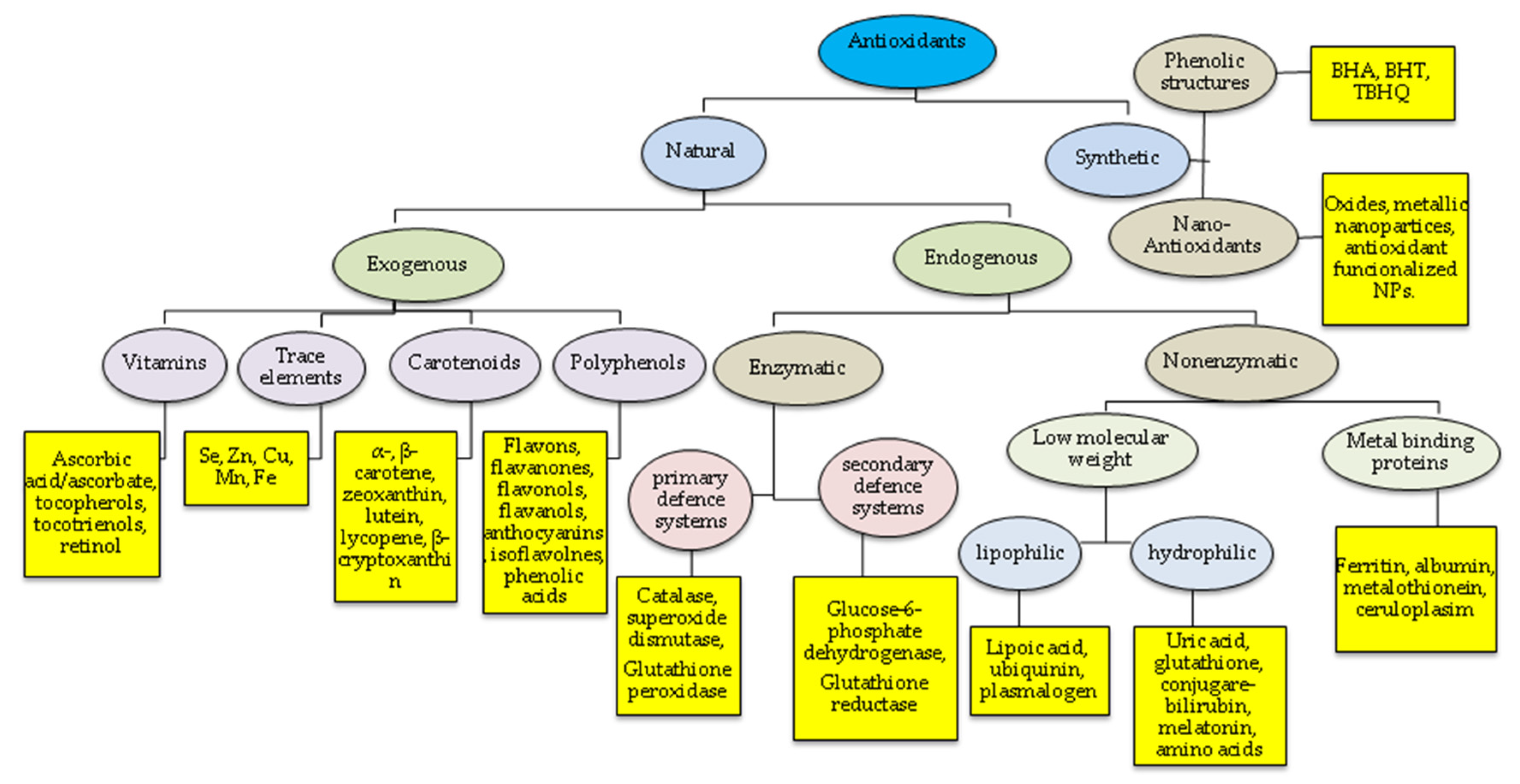
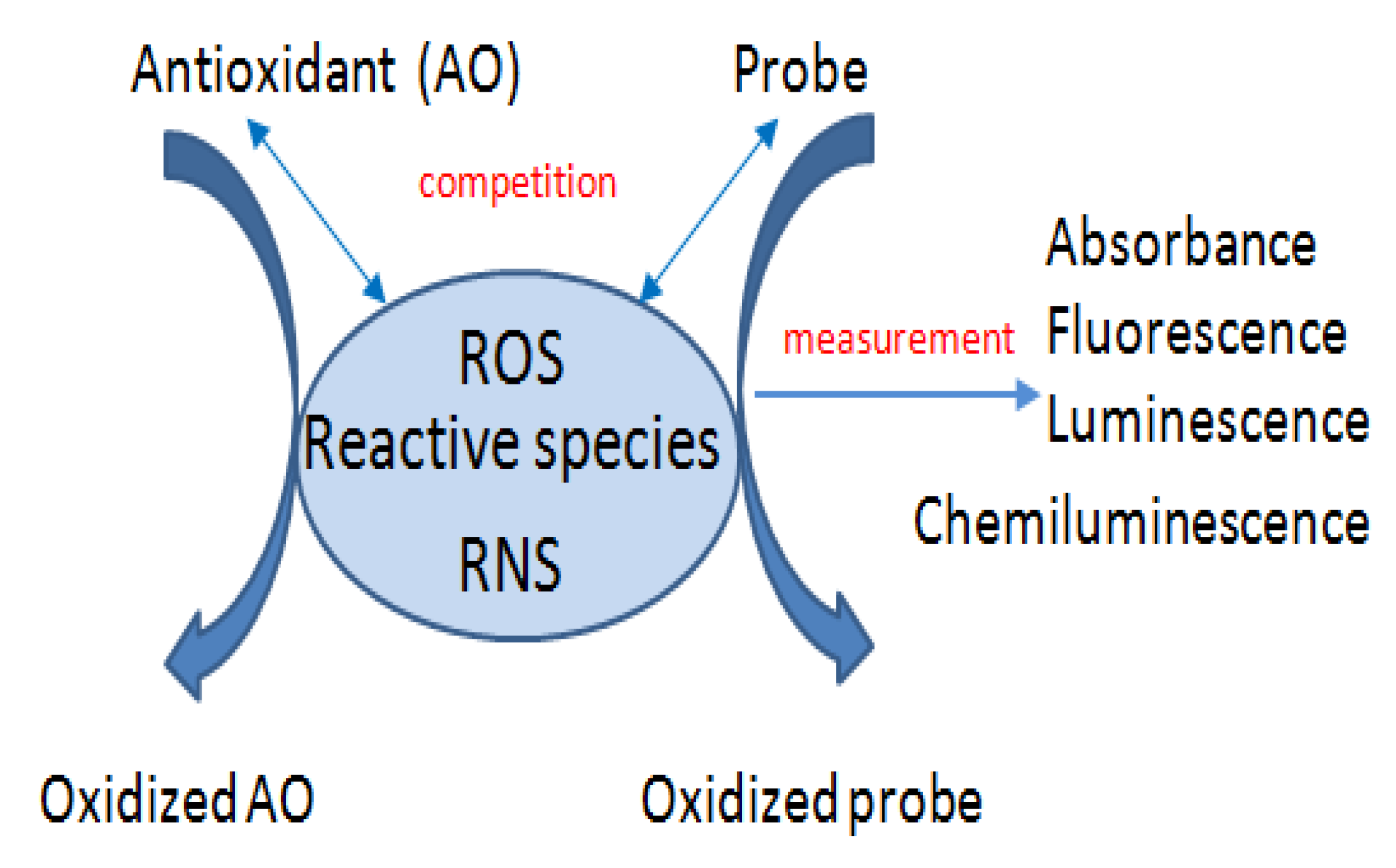
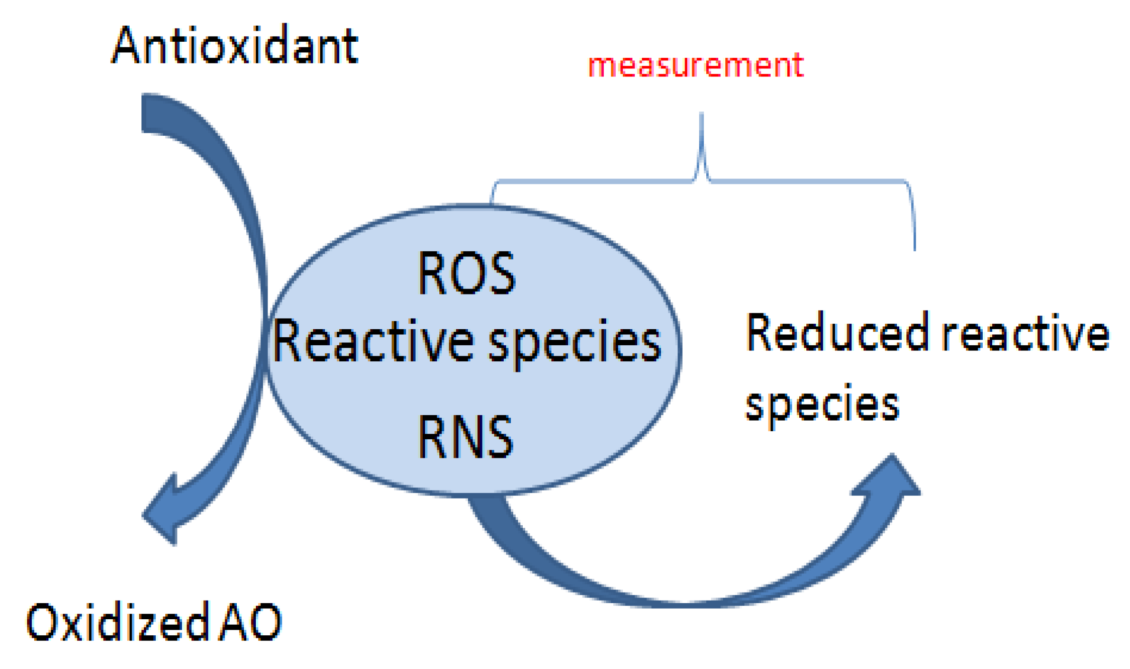
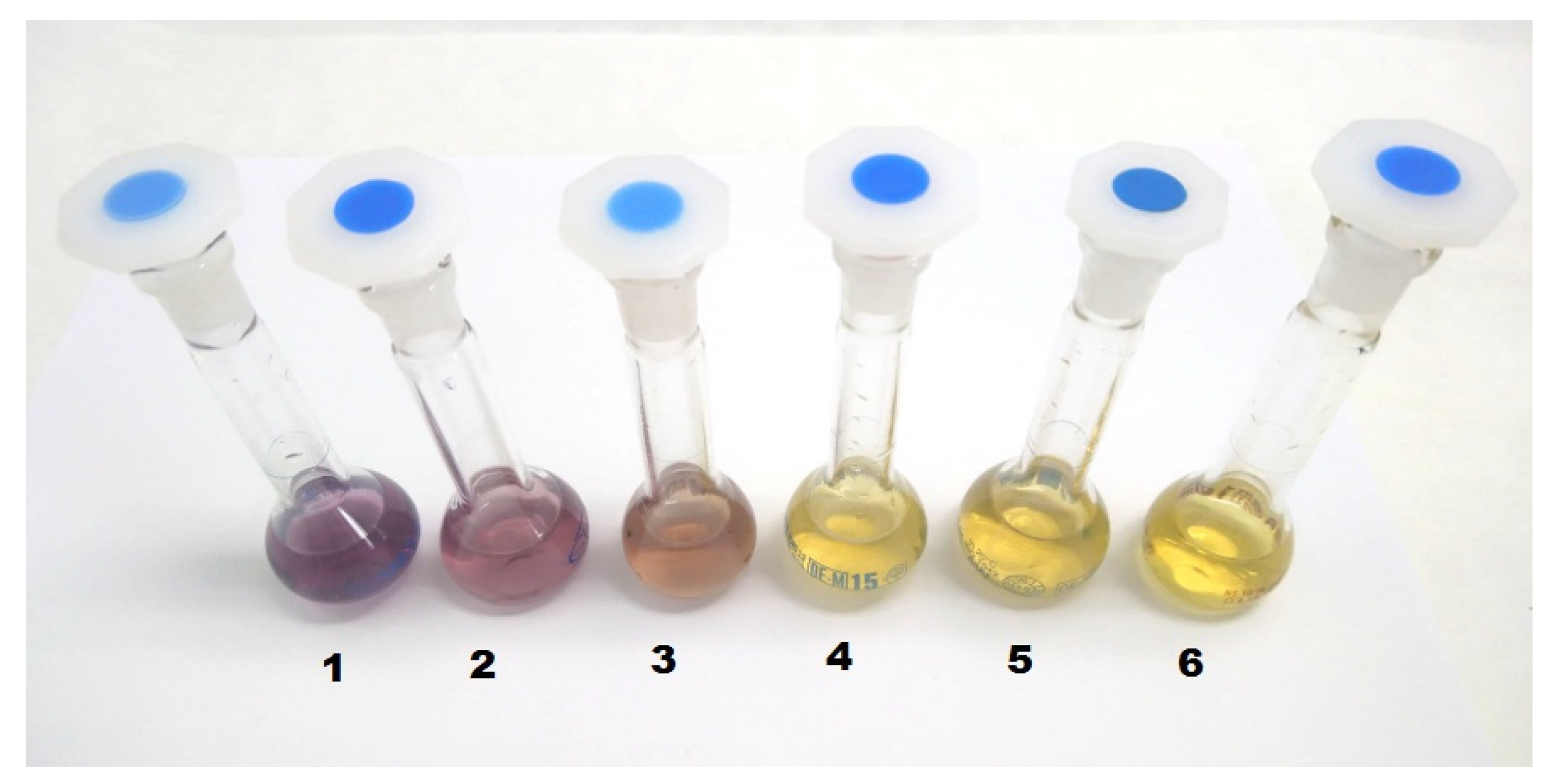
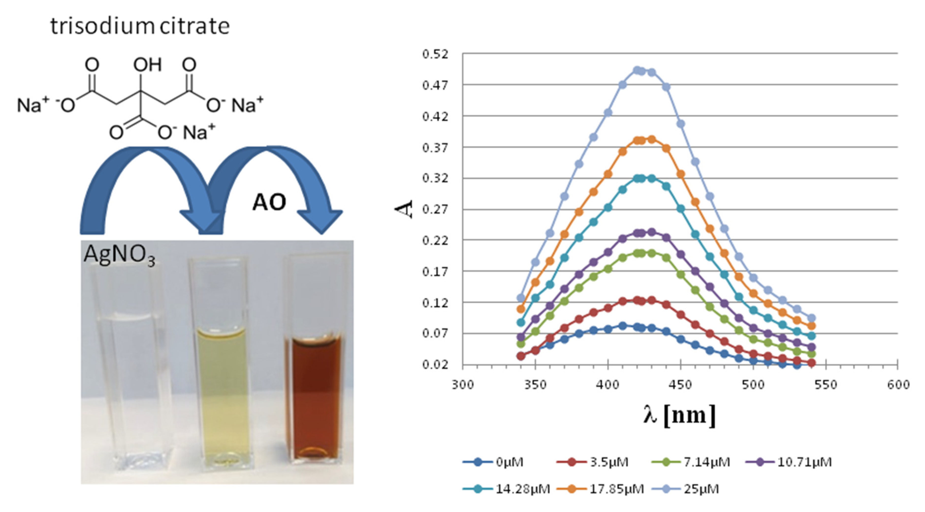
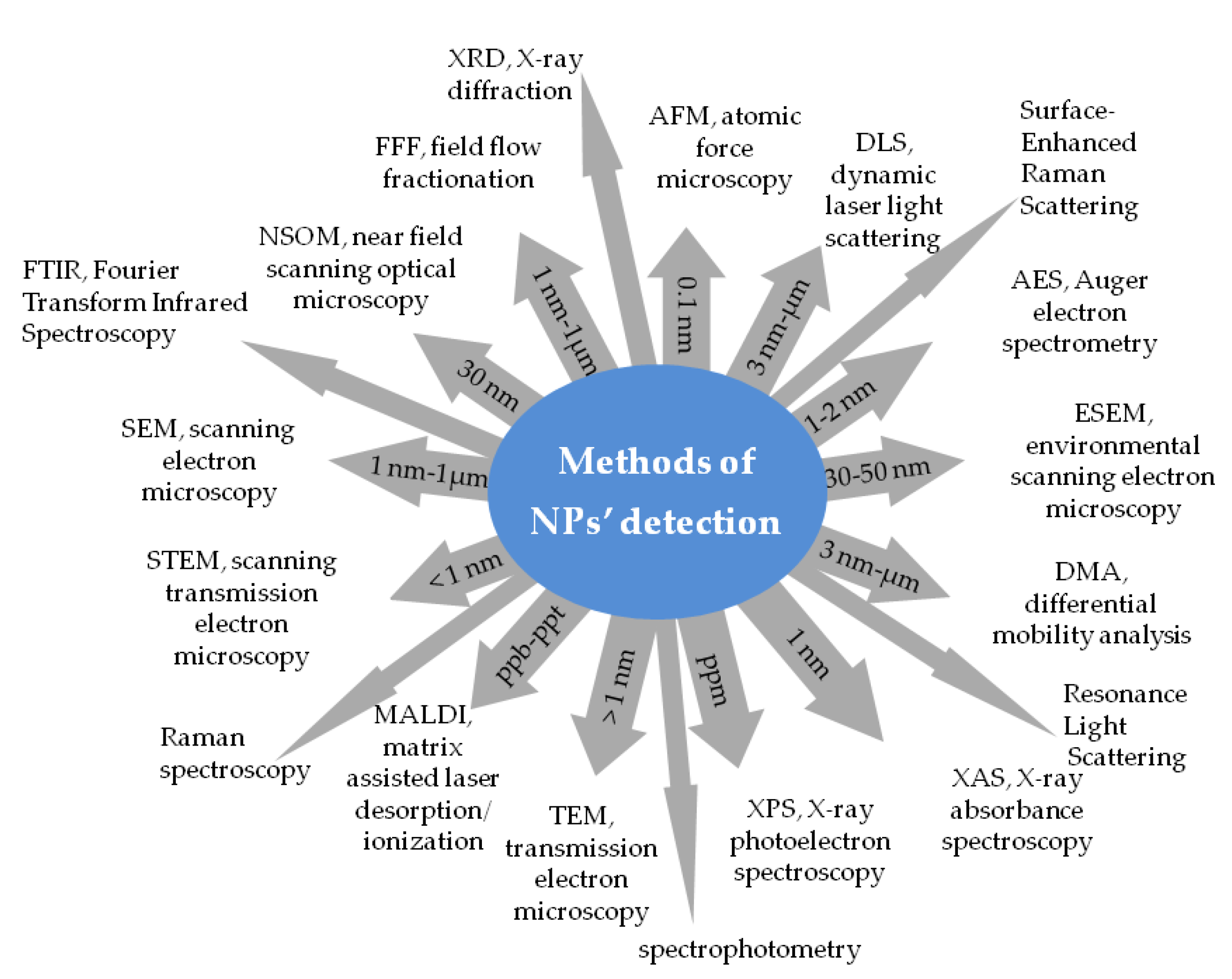
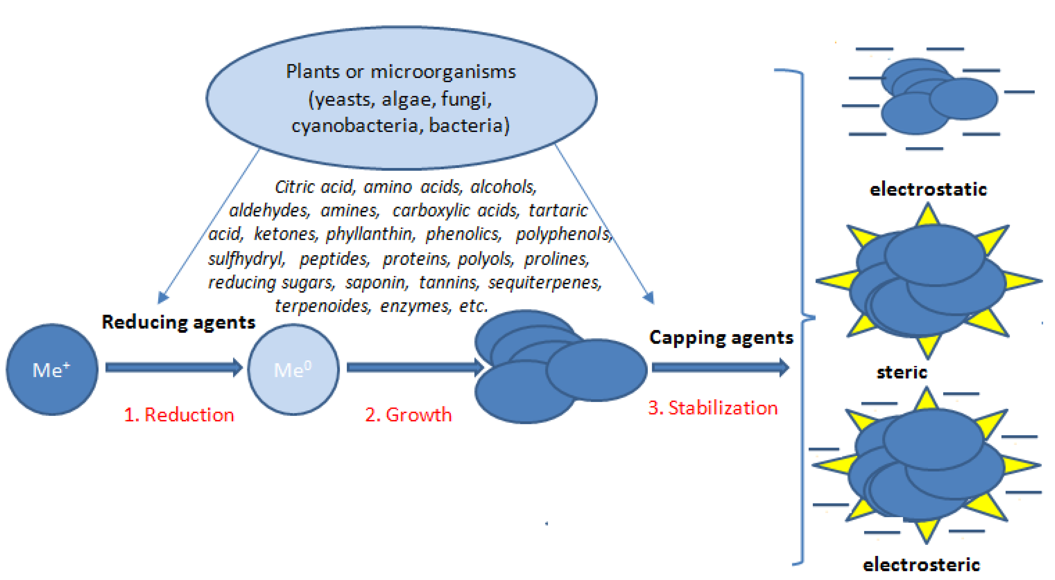
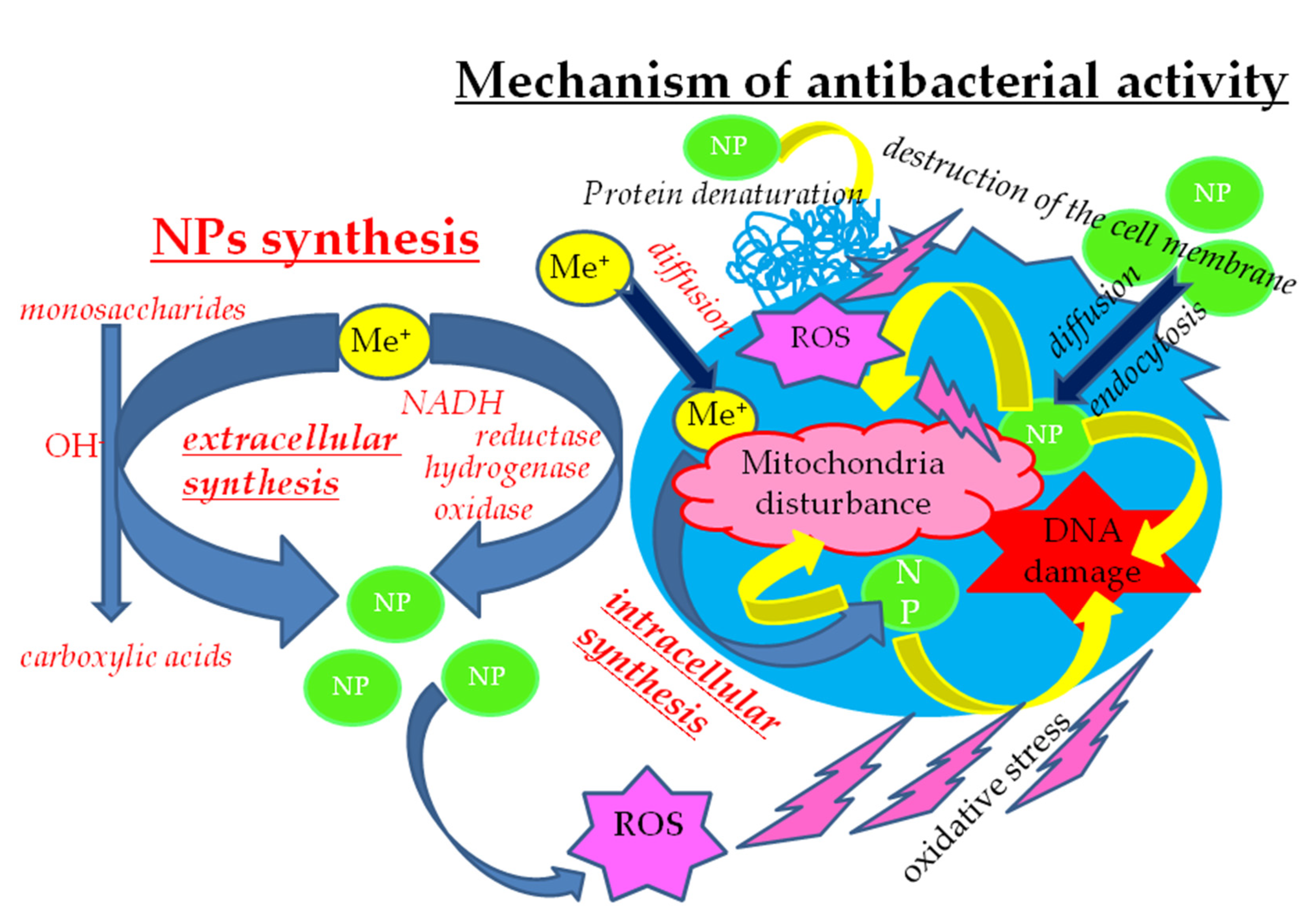
| Reactive Species | Form | Example |
|---|---|---|
| Reactive oxygen species (ROS) | Radical | HO•, 1[O]2, O2•− HOO•, ROO•, RO•, CO2•−, CO3•− |
| Non-radical | O3, H2O2, HOCl, HOI, HOBr, ROOH, CO, ONOOH, ONOO−, O2NOO−, HOOCO2−, (O2 1Dg) | |
| Reactive nitrogen species (RNS) | Radical | NO•, NO2•, NO3• |
| Non-radical | ROONO, RO2ONO, CH3C(O)OONO2, N2O4, N2O3, N2O5, HNO2 NO2Cl NO−, NO+ | |
| Reactive chlorine species | Radical | Cl• |
| Non-radical | ClBr, Cl2, ClO2 | |
| Reactive sulfur species | Radical | S• |
| Non-radical | H2S, RSSR, RS(O)SR, RSOH, RS(O)2SR, RSR’ |
| Assay | The Chromogenic Agents | Observed Changes | Principle of Assay | Mode | Mech | Ref |
|---|---|---|---|---|---|---|
| Total antioxidant capacities | ||||||
| Crocin bleaching | crocin | bleaching of crocin | The ability of AOs to inhibit oxidation of crocin. | Abs. 443 nm pH = 7.0–7.5 | HAT | [125,126] |
| ORAC (Oxygen radical absorbance capacity) | fluorescein, dichloro- fluorescein | fluorescence decay | The fluorescence caused by oxidation of the probe by peroxyl-radical initiated by thermal decomposition of AAPH, is delayed/inhibited by AOs. | Fl. λex = 485 nm λem = 538 nm pH = 7.4 | HAT | [127] |
| TRAP (Total peroxyl radical trapping antioxidant parameter) | β-phycoerythrin | fluorescence decay | Fluorescence decay along time due to oxidation of the probe is delayed by AOs. | Fl. λex = 495 nm λem = 575 nm pH = 7.5 | HAT | [128,129] |
| β-carotene bleaching assay | β-carotene | bleaching yellow color of β-carotene | The ability of AOs to slow down the rate of β-carotene bleaching due to its reaction with peroxyl radicals, which are formed by linolenic acid oxidation. | Abs. 470 nm pH = 5.5–7.5 | HAT | [130,131] |
| PCL (Photochemiluminescence) | luminol | blue light emision | An AO-sensitive inhibition of a photo-induced, chemiluminescence accompanying autooxidation of luminol. | Cl. 360 nm pH = 10.5 | HAT | [132,133,134] |
| Reducing antioxidant power (RP) | ||||||
| FRAP (Ferric reducing antioxidant potential) | ferric tripyridyl triazine | yellow color to blue | AOs as reductant at low pH can reduce ferric tripyridyl triazine to ferrous form, causing absorbance increase. | Abs. 593 nm pH = 3.6 | ET | [135] |
| CUPRAC (cupric ion reducing antioxidant capacity) | Cu(II) complex | light blue to orange-yellow | Ability of AO for the reduction of Cu(II) in bathocuproine(2,9-dimethyl-4,7-diphenyl-1,10-phenanthroline) or neocuproine (2,9-dimethyl-1,10-phenanthroline) complexes to Cu(I) forms. | Abs. 490 nm 450 nm pH = 7 | ET | [136] |
| CERAC (Ce(IV)-based reducing capacity) | Ce (IV) | fluorescence | The ability of AO to reduce Ce(IV) to Ce(III) accopanied with fluorescence elevation. | Fl. λex = 256 nm λem = 360 nm pH acidic | ET | [137,138] |
| CHROMAC (Chromium reducing antioxidant capacity) | Cr (VI) with DPC | red–violet product | The reduction of chromate(VI) to Cr(III) in acidic solution. The remaining Cr(VI) reacts with DPC to produce a chelate complex. The Cr(VI) consumption was correlated with AO’ concentration. | Abs. 540 nm. pH = 2.8 | ET | [139] |
| Phosphomolybdenum assay | Phosphormolybdenum complex | green product | The reduction of Mo(Vl) to Mo(V) by AO. | Abs. 695 nm pH acidic | ET | [140] |
| The Folin–Ciocalteu (FC) assay | Tungstate–molybdate complexes | from yellow to dark blue | FC reagent in a basic medium is able to oxidize reducing substances, mainly phenolic and polyphenolic AOs. The change in color is connected with transformation of Mo(VI) to Mo(V), causing absorbance increase. | Abs. 750–765 nm pH = 10 | ET | [109] |
| PFRAP (Potassium ferricyanide reducing power assay) | Ferricyanide reagent: Fe(III), Fe(CN)63− | prussian blue | The AOs react with potassium ferricyanide Fe(CN)63−) forming potassium ferrocyanide Fe(CN)64− which further reacts with FeCl3 to form prussian blue KFe[Fe(CN)6]. | Abs. 700 nm pH = 6.6 | ET | [141] |
| FTC (Ferric thiocyanate) | Fe(S-CN)2 | red color | A hydroperoxide formed from a lipid (linoleic acid) oxidizes a ferrous ion to a ferric ion. The AO causes an inhibitory effect on hydroperoxide formation or by its ability to donate an electron to ferric ion. | Abs. 500 nm | ET | [142,143] |
| FOX (Ferrous Oxidation-Xylenol Orange Assay) | ferric-XO complex | blue-purple color | The presence of hydroperoxides that oxidize ferrous ion to ferric ion, which subsequently react with xylenol orange (XO). | Abs. 550 nm. | ET | [144] |
| Assays associated with lipid peroxidations | ||||||
| LPO (Lipid peroxidation inhibition assay) | N-methyl-2-phenylindole | dye product | AOs delay radical-induced malonyl dialdehyde generation. MDA and HAE are measured as an indicator of lipid peroxidation. The product -MDA with chromogenic reagent gives carbocyanine adduct. | Abs. 586 nm | ET | [138,145] |
| TBARS (Thiobarbituric acid reactive substances assay) | TBARS | red-pink color | The reaction of lipid peroxidation products (MDA), with TBA, leads to the formation of MDA-TBA adducts (TBARS). | Abs. 532 nm pH = 4 | ET | [130,131,146,147] |
| Conjugated diene assay | linoleic acid | UV absorbance | Antioxidants delay conjugated dienes formation. The AO effect can be evaluated by monitoring the conjugated diene formation. | Abs. 234 nm | [148] | |
| Radical scavenging assays | ||||||
| DPPH | 2,2-diphenyl-1-picrylhydrazyl radical | deep violet to pale yellow or colorless | The decrease in DPPH absorbance depends linearly on AO’ concentration. | Abs. 515–517 nm pH = 7 | HAT/ET | [130] |
| ABTS | 2,2’-azino-bis(3-ethylbenzothiazoline-6-sulfonic acid (ABTS+.) | bluish-green to colorless | ABTS treated with Na/K persulphate or MnO2 gives a radical cation (ABTS+). ABTS+. is reduced by antioxidants. The decrease in absorbance depends linearly on AO’concentration. | Abs. 734 nm pH = 7.4 | HAT/ET | [149] |
| DMPD (N,N-dimethyl-p-phenylene-diamine) | DMPD·+ radical cation | reduction of purple color | DMPD·+ is generated through a reaction between DMPD and potassium persulphate the assay measures scavenging of free radicals by AOs. | Abs. 517 nm pH = 5.25 | HAT | [150,151] |
| SOSA (Superoxide Anion Radical Scavenging Capacity) | NBT | yellow to blue | The ability of the AO to compete with NBT to scavenge O2•− generated by an enzymatic HPX-XOD, X-XOD or PMS/NADH systems. | Abs. 560 nm pH = 7.4 | ET | [135,152] |
| Nitric oxide free radical scavenging activity | Griess reagent | colorless to light pink to deep purple | NO was generated from sodium nitroprusside and measured by the Greiss reaction. AO reduces the amount of nitrite. | Abs. 546 nm pH = 7.4 | ET | [153] |
| Peroxynitrite Scavenging Capacity Assay | Evans Blue | dye bleaching | The percentage of scavenging of ONOO− by the Evans Blue was measured in presence of AO. | Abs. 611 nm pH < 7 | ET | [154] |
| HORAC (Hydroxyl Radical Averting Capacity Assay) | fluorescein | fluorescence decay | OH radicals are generated by a Co(II)-mediated Fenton-like reaction. The reaction is confirmed by the hydroxylation of p-hydroxybenzoic acid. Metal ion-induced OH radical generation reaction can be monitored by the fluorescence decay of fluorescein. In the presence of AO, the formation of OH radicals can be inhibited because the metal is deactivated due to coordination with AO. | Fl. λex = 493 λem = 515 nm | HAT | [130,135,146,147,148,149,150,151,152,153,154,155] |
| HRS (Deoxyribose Degradation Assay) | MDA-TBA adducts | pink | A mixture of Fe(III)-EDTA, H2O2, vit.C generates OH radical, is able to degrade deoxyribose. The products heated under acidic conditions form MDA detected by adduct with TBA. AO can inhibit deoxyribose damage. | Abs. 532 nm pH = 7.4 | ET | [156] |
| Hydroxyl Radical Scavenging Capacity Assay | Fenton-like system Fe(II)/H2O2 | - | The Fenton system generates a constant flux of pure OH radicals. ESR measurements evaluate the OH radicals scavenging capacity of AOs. | Electron spin resonance (ESR) | ET | [157] |
| CAA(Cellular Antioxidant Activity Assays) | DCFH-DA | fluorescence decay | The ability of AOs to prevent oxidation of DCFH by azide generated peroxyl radicals in human hepatocarcinoma HepG2 cells. | Fl. λexc.502 nm, λem 520 nm | ET | [158] |
| Nonradical reactive oxygen species scavenging assay | ||||||
| Hydrogen peroxide scavenging activity | hydrogen peroxide | UV absorbance | Hydroxyl radicals are the byproducts of H2O2 decomposition. They initiate lipid peroxidation. After the addition of AO, the absorbance is measured against blank (phosphate buffer). | Abs. 230 m pH = 7.4 | ET | [159] |
| Singlet oxygen scavenger | RNO | bleaching of RNO | Production of singlet oxygen (1O2) was achieved by monitoring RNO bleaching. Singlet oxygen was generated by a reaction between NaOCl and H2O2. | Abs. 440 pH = 7.1 | ET | [160] |
| ACA (Aldehyde/carboxylic acid assay) | Alkylaldehyde/ alkylcarboxylic acid | - | The stoichiometric conversion from alkylaldehyde (hexanal) to alkylcarboxylic acid in the presence of radicals induced by heat, O2, or H2O2. | GC | ET | [161] |
| Metal chelating capacity assays (MCA) | ||||||
| Ferrous ions chelating assay | Fe(II) with 2,2-bipyridine or ferrozine | blue | The capacity to chelate ferrous ion can be disturbed by the presence of other complexing agents (AOs), which cause a decrease intensity of the complex (Fe(II) and ferrozine). | Abs. 562 nm 522 nm pH = 4–10 | ET | [162] |
| Copper(II) chelating capacity assay | Cu(II)- PV | dark to yellow | The chelating activity can be estimated by the measurement of the rate of color reduction. | Abs. 632 nm pH = 6 | ET | [163] |
| Nanoparticles (NPs)-based assays | ||||||
| Gold nanoparticles (Au-NPs) | NPs | No color into dark red | The highest capacity of reducing gold(III) to gold NPs corresponds to the highest antioxidant activity. Alternatively, cyclic voltammetry measures anodic peak potentials | Abs. 555 nm pH = 8 | ET | [117,118] |
| Silver nanoparticles (Ag-NPs) | NPs | no color into pale yellow | Nanoparticles generated from metal salts upon reduction with antioxidants in the presence of citrate-stabilized silver seeds. | Abs. 423 nm pH = 7 | ET | [118] |
| Analysed Product | Antioxidant Assays | Positive Control | Extraction Procedure | Ref. |
|---|---|---|---|---|
| Plants | ||||
| Bryonia alba L. | DPPH, CUPRAC, FRAP, TEAC, SNPAC | quercetine, BHT, Trolox | Fifty grams of powder was macerated with 500 mL methanol for 24 h. After percolation, extract was evaporated under vacuum at 40 °C. | [279] |
| Cistus ladanifer L., Cistus salvifolius L., Cistus albidus L., Erica australis L., Arbutus unedo L., Pistacia lentiscus L. | DPPH, FRAP, ABTS, RP | quercetin | Twenty grams of grounded leaves was mixed with 200 mL of methanol. The mixture was kept for 24 h at RT. Then, it was filtered. | [280] |
| Prunus avium, Prunus persica, Prunus domestica, Olea europeae, Pirus communis, Pirus maus, Pistacia verra, Castanea sativa | DPPH, FRAP, TAC | Trolox, ascorbic acid | Two grams of sample was mixed with 60% methanol and kept 1 h at dark at RT. The procedure was repeated two times. The combined extracts were centrifuged and filled to 50 mL by aqueous methanol. | [281] |
| Vitis vinifera L. (Maraština, Pošip; Lasin, Merlot, Syrah, Vranac) | DPPH, FRAP | Trolox | The dry plant material (20 g) was extracted using 100 mL of ethanol/water 80/20, (v/v) at 60 °C, for 60 min. The extract was filtered and dried under a vacuum at 50 °C. The dry residues were redissolved with 50% methanol-water and centrifuged at 5000 rpm for 10 min. | [282] |
| Ornithogalum billardieri | β-carotene-linoleic acid assay, ABTS, MCA/ferrous ion, TAC/H2SO4, Na3PO4, (NH4)2MoO4) | ascorbic acid | The combined maceration with sonication either at 25 °C for 50% ultra-sound (US) treatment or the extract obtained under the optimal conditions: extraction time: 37.1 min, temperature: 44.2 °C, water volume-to-mass ratio: 33.8 mL/g, and US%:51.7% | [283] |
| Apple cultivars: antonówka, delikates, Early Geneva, papierówka, Paulared, Sunrise, Quinte Gloster, Jonagored, Ligol, Rubinola | DPPH, FRAP, MCA/ferrous ion | Trolox, catechin | The material was lyophilized. A total of 250 mg of sample was extracted by sonication using 1 mL of 80% methanol for 30 s. Then, the mixture was vortexed, centrifuged for 5 min (13,200 rpm), and sonicated. The extraction procedure was repeated five times. The supernatants were collected together. | [209] |
| Fraxinus angustifolia Vahl | TAC/Folin-Ciocalteu, FRAP, ABTS, DPPH | α-tocopherol | Manna samples (10 g) were dissolved in methanol-water (2:1) and extraction carried out at 25 ± 2 °C and in darkness for 1 h., and centrifuged (3000 g, 10 min). The supernatants were filtered and evaporated at 35 °C. Dried samples were resuspended in 5 mM phosphate buffer saline pH 7.4. | [284] |
| Chenopodium quinoa | DPPH, FRAP | Trolox, gallic acid | Dry plant materials (roots, leaves, stems, flowers, and seeds) were extracted by the use of ultrasound-assisted extraction with 96% ethanol. The extracts were concentrated under reduced pressure in a rotary evaporator. | [239] |
| Lycium barbarum, Lyciumchinense | DPPH, ABTS | - | Samples were dried, powdered, and dissolved in distilled water. | [285] |
| Aegopodium podagraria L. | DPPH | GSH, ascorbic acid | A total of 2.5 g of air-dried or fresh aerial parts was extracted by 100 mL of 80% (v/v) ethanol. The samples were kept at RT for 3 days, three months in dark, or in an ultrasonic bath for 60 min. Then, the extracts were filtered. | [286] |
| Fungi | ||||
| Achaetomium sp. | DPPH | ascorbic acid, BHT, gallic acid, pyrogallol | The organic ethyl acetate extract was evaporated. The crude extract was dissolved in DMSO. | [287] |
| Acremonium charticola, Rhizopus oryzae | ABTS | ascorbic acid | Fungi were cultured in potato dextrose broth at 37 °C. After 3 days, the cultures were centrifugated at 5000 rpm for 10 min. The filtrate (1 g) was mixed with 100 mL methanol and ultrasonicated for 30 min. The homogenate was created for three days at RT. Then, it was evaporated with a rotary vacuum evaporator (50 °C, 100 rpm) to the volume of 25 mL. | [288] |
| Agaricus bisporus, Pleurotus ostreatus, Pleurotus eryngii, Lentinula edodes | TPC/Folin–Ciocalteu, Ferricyanide/prussian blue, DPPH, TBARS β-carotene/linoleate | Trolox, gallic acid | The product was lyophilized. The obtained powder was mixed with methanol and kept at 25 °C at 150 rpm for 1 h. Then, the mixture was and filtered. The extracts were evaporated under reduced pressure and redissolved in methanol at a concentration of 20 mg/mL. | [171] |
| Aspergillus wentii, A. wentii, Penicillium citrinum, Penicillium granulatum | DPPH, FRAP, MCA/ferrous ion, NO• scavenging activity, RP/potassium ferricyanide | ascorbic acid, BHT, rutin, catechin | The fungal mycelia were grown on agar plates with extracts of yeast and glucose. After 6 to 7 days, the Czapek–Dox’s broth was inoculated of fungal mycelia. After 10 days of incubation at 25 °C, the culture broth was filtered. | [289] |
| Aspergillus niger, Aspergillus peyronelii | DPPH, RP H2O2 scavenging activity | ascorbic acid | The dried samples were extracted by the use of ethyl acetate (1:10) applying cold percolation for 48–72 h. Then, obtained extracts were filtered, and concentrated under vacuum at 40 °C. | [290] |
| Aspergillus versicolor | ABTS, DPPH | Trolox | Aspergillus versicolor was cultivated on rice for 30 days. The EtOAc extracts of solid fermentation were fractioned through slica gel and Sephadex LH-20 column chromatography (CC), and were further purified by semi-preparative HPLC. | [291] |
| Auricularia auricular | ABTS, O2●−, OH• scavenging activity, lipid peroxidation | - | A.auricula was extracted by hot water and ultrasonic-assisted extraction. The supernatants were precipitated with absolute ethanol (95%) and maintained at 4 °C overnight. The precipitate was centrifugated, dissolved in DI, and dialyzed. The non-dialyzed portion was lyophilized to give a crude polysaccharide extract. The separation of polysaccharides was performed using CTAB or CPC. | [292] |
| Cephalosporium sp. | DPPH | gallic acid | The fermented material (1.75 kg) was extracted twice with EtOAc (9.0 L) for 3 days at RT, and the extract was evaporated under vacuum. | [293] |
| Cerrena unicolor | ABTS, DPPH | Trolox, ascorbic acid | Ten-day-old cultures were filtered and washed with DW. The fungal biomass was used for the polysaccharides extraction by hot water (90 °C, 4 h). The proteins were separated by anion exchange chromatography on a DEAE Sepharose column with a linear gradient of NaCl (0.1–0.5 M). The culture liquid was subdivided into two fractions by ultrafiltration: substances above 10 kDa (rude lactase) and substances below 10 kDa with low molecular weight metabolites. | [294] |
| Flammulina velutipes, Hypsizygus tessellatus | DPPH, H2O2 scavenging activity, FRAP | gallic acid, quercetin | The stems of the mushrooms were dried at 60 °C and pulverized. Samples (180 g) were extracted overnight with 500 mL of water, absolute methanol, 95% acetone, or 95% ethyl acetate at RT. The extracts were filtered, evaporated to dryness. Aqueous fractions were concentrated to 50 mL, freeze-dried, and stored at 4 °C. | [295] |
| Grifola frondosa | DPPH, β-carotene bleaching assay, inhibition of lipid peroxidation, RP, CUPRAC | quercetin | Mushrooms were boiled in DW at the ratio of 1:10 (w/v) for 30 min. Then, the extract was filtered, and freeze-dried. | [296] |
| Penicillium expansum | DPPH, RP, FRAP MCA/ferrous ion, NO• scavenging activity | - | The fungi were grown on CDM, MEM, PDM, and YEM. After incubation at 25 °C for 10 days, the culture broth was filtered through filter paper. The culture broth was extracted with petroleum ether, chloroform, ethyl acetate, and butanol. Then, the extracts were then evaporated to dryness in a vacuum, and the precipitates were dissolved in DMSO. | [297] |
| Phlebia brevispora, Phlebia floridensis, Phlebia radiate, Phlebia fascicularia | DPPH, FRAP, RP, MCA, NO• scavenging activity | - | Five grams of dried wheat straw was ground, washed, and dried at 90 °C. Next, it was moistened by the use of 25 mL of malt extract (0.5%, w/v) and inoculated with three mycelial discs (8 mm), which were grown on YGA plates for 6 days. The inoculated flasks were kept at 25 °C for 30 days. Then, they were homogenized, filtered, and dried at 90 °C. | [298] |
| Pleurotus florida, Pleurotus sajor-caju, Pleurotus cystidiosus, Pleurotus djamor | FRAP, DPPH, RP, MCA, H2O2, O2●− scavenging activity | - | Sporophores were cleaned and dried at 40 ± 2 °C for 10–12 h. The obtained powders (100 g) were refluxing with light petrol (60–80 °C) for 6 h with the aim of defatting. Then, the material was extracted with 95% ethanol (500 mL × 3) by refluxing for 6 h. The extracts were combined, filtered, and evaporated to dryness at 40 °C. The dried extracts were redissolved in methanol at a concentration of 20 mg/mL for analysis. | [299] |
| Trametes versicolor, Trametes hirsuta, Trametes gibbosa | ABTS, FRAP | ascorbic acid | Dried material (3.0 g) was grounded. Extraction was carried out in 96% ethanol during 72 h. The extracts were centrifuged and supernatants filtrated. The filtrates were concentrated under reduced pressure at 40 °C to dryness and redissolved in 96% ethanol | [300] |
| Bacteria | ||||
| Bacillus coagulans RK-02 | β-carotene-linoleate, O2●−, OH• scavenging activity, DPPH | ascorbic acid, α-tocopherol | The 36 h culture was centrifuged at 10,000× g for 20 min at RT. The supernatant was filtered. The proteins were isolated by the addition of 10% TCA. After 12 h at 4 °C, the mixture was centrifugated. Four volumes of 95% ethanol were added to the supernatant and centrifuged. The pellet was lyophilized, dissolved in 5 mL DW and dialyzed (MWCO 12,000 Da). | [301] |
| Weissella cibaria GA44 | DPPH, RP, O2●−, OH• scavenging activity | ascorbic acid | The cultures were heated at 100 °C for 10 min. The cells were removed by centrifugation at 12,000× g for 15 min at 4 °C. The supernatant was precipitated with double volume of chilled ethanol, shaken, and centrifuged at 98 5000× g for 30 min at 4 °C. The precipitate was dried at 50 °C, and dissolved in water. This step was repeated three times and dialyzed against distilled water for two days at 4 °C using 10 kDa dialysis membrane and then lyophilized. | [302] |
| Lactobacillus plantarum C88 | OH• scavenging activity, DPPH, the LPC-1 on H2O2− induced oxidative stress in Caco-2 cells | ascorbic acid | After 20 h of the incubation period, TCA was added to achieve 4% (w/v). The mixture was stirred for 30 min at RT. Cells were removed by centrifugation (10,000× g, 4 °C, 15 min). Crude EPS was precipitated by the addition of 2 volumes of cold ethanol. Crude EPS was collected by centrifugation. The pellet was dissolved in deionized water and dialyzed (MW cut-off 3500 Da) for 24 h against distilled water at 4 °C and then lyophilized. | [303] |
| Pseudomonas hibiscicola, Macrococcus caseolyticus, Enterobacter ludwigii, Bacillus anthracis | DPPH | ascorbic acid | The bacteria were cultured in 500 mL broth at 35 °C. Then, the culture was centrifuged at 8000× g for 5 min. The supernatant was extracted with ethyl acetate (ratio of 1:1). Then, the extract was concentrated to dryness in a rotatory evaporator at 37 °C. The solids were re-dissolved in 20% DMSO and filtered. | [304] |
| Alteromonas sp. Shewanella sp. Serratia sp., Citricoccus sp., Cellulophaga sp., Ruegeria sp. Staphylococcus sp. | DPPH, ORAC | BHT, Trolox | Isolated bacteria were cultured in 500 mL of Marine Broth for 3 days at 25 °C. Bacteria cells were centrifuged, and the pellet was lyophilized. Then, the lyophilizes were extracted with methanol and dichloromethane (1:1) for 12 h. The solvents were evaporated at 40 °C, and the extracts were re-dissolved in DMSO. | [305] |
| Pseudomonas sp. (HR04) | anti-lipoperoxidative activity | BHT, α-tocopherol | The mycelial cake was extracted with acetone followed by purification by column chromatography on silica gel and Sephadex LH-20. | [306] |
| Lactobacillus casei CRL 431 (IC431) | LPO, ABTS, ORAC, CAT, GPx | Trolox | An aliquot (10 mL) of bacteria suspended in PBS was mixed with lysozyme (1 mg/mL) and incubated at 37 °C for 150 min. Then, cells were disrupted by sonication in an ultrasonic processor at 10 °C. After centrifugation (3600× g, 4 °C, 10 min), the supernatant was collected and stored in dark at 4 °C. | [307] |
| Lactobacillus fermentum | DPPH, ABTS, FRAP, OH• scavenging activity | ascorbic acid | The cells were washed with 0.85% NaCl and sonicated. The obtained fluid was mixed with 75% ethanol. The precipitate was collected, redissolved, deproteinized, purified on an anion exchange column eluting with deionized water, 0.1 M, and 0.3 M NaCl and subsequently loaded onto a Sephadex G-100 column and eluted with DW. The collected fractions were lyophilized. | [308] |
| Lactobacillus plantarum (LAU103) | ABTS, DPPH, ORAC, MCA/ferrous ion, OH• scavenging activity | ascorbic acid | A total of 5 mL of crude EPS solution (20 mg/mL) was separated with DEAE-cellulose column using deionized water, 0.2 and 0.5 M NaCl as eluent. Peak fractions containing polysaccharides were pooled, dialyzed, and lyophilized. Then, the fraction was further purified on a Sepharose CL-6B gel column and eluted with 0.9 M NaCl solution. | [242] |
| Lactobacillus paracasei subsp. paracasei NTU 101 (101EP), Lactobacillus plantarum NTU 102 (102EP) | DPPH, MCA/ferrous ion, inhibition of linoleic acid peroxidation, RP | ascorbic acid | The cultures were centrifugated at 5000× g at 4 °C for 15 min. The supernatant was added to 0.4 TCA at 4 °C for 3 h. Then, the supernatant was mixed with ethanol at 4 °C for 24 h, followed by centrifugation. The obtained precipitate was dialyzed for 24 h and lyophilized. | [309] |
| Algae | ||||
| Astaxanthin | FRAP, TEAC ORAC, DPPH | tert-Butyl alcohol | Astaxanthin was dissolved in tetrahydrofuran (THF). | [244] |
| Desmarestia antarctica, Iridaea cordata | RP, TPC, DPPH | gallic acid, ascorbic acid | The samples were rinsed with Milli-Q water, cut into fine pieces, then boiled at reflux for 15 min. The flask was moved to an ice bath to complete the extraction. The extract was thus centrifuged at 4500 rpm for 10 min, and the supernatant was filtered and stored at 4 °C. | [310] |
| Haematococcus pluvialis, synthetic astaxanthin | ABTS, ORAC, CAA | Trolox | The extracts were obtained by solvent using DMSO or supercritical extraction (AstaCO2). | [311] |
| astaxanthin | ORAC-EPR | - | The acetonitrile solutions of Catechin, epicatechin, epigallocatechin gallate, kaempferol, myricetin, resveratrol, and astaxanthin were diluted with phosphate buffer containing DM-β-CD. | [312] |
| Dunaliella salina, Tetraselmis chuii, Isochrysis galbana clone Tahiti. | DPPH | - | Methanolic extracts were prepared in different concentrations (50, 100, 250, 500, and 1000 ppm). | [313] |
| Galderia sulphuraria, Neochloris texensis, Stichococcus bacillaris, Ettlia carotinosa, Chlorella minutissima, Schizochytrium limacinum, Crypthecodinium cohnii, Chlorella vulgaris | DPPH | - | Methanol extraction: 20 mL methanol was added to 0.5 g dry biomass and sonicated (9 cycles, 50% power) for 20 min. Then, samples were centrifuged at 3500 rpm for 5 min. Pellets were re-extracted in 20 mL methanol 3 times and the supernatants were collected. The samples were filtered and evaporated at 40 °C. Hot water extraction: 1 g of dry sample was added to 100 mL DW and boiled for 30 min. After cooling, extracts were centrifuged at 3500 rpm for 10 min, and supernatants were freeze-dried. | [314] |
| Chlorella vulgaris Spirulina platensis | ORAC, DPPH, FRAP | Trolox | The extracts were obtained by the use of ultrasound-assisted extraction by water/ethanol (50:50, v/v). | [315] |
| Scenedesmus subspicatus | DPPH | catequin, gallic acid | Different solvents such as ethanol, methanol, butanol, acetone, DMSO, and water were used for extraction. One gram of dried samples were mixed with 10 mL for each solvent. The extraction was carried out for 30 min by sonication (40 kHz) in an ultrasonic bath followed by a 2 h shake, and centrifugation for 10 min. | [316] |
| Tetraselmis suecica | DPPH | α-tocopherol | A total of 100 mg of freeze-dried biomass was extracted with 1 mL ethanol/water (3:1, v/v) for 30 min. The mixture was centrifuged at 4500× g, for 10 min, at 20 °C. Then, the ethanolic phase was dried. | [317] |
| Lichens | ||||
| Cetraria islandica (L) Ach. | DPPH, the thiocyanate method, RP, O2●− scavenging activity | α-tocopherol, BHT, BHA | For water extraction, 20 g sample was mixed with 400 mL boiled DW and stirred for 15 min. Then, the extract was filtered. The obtained filtrates were frozen and lyophilized. | [261] |
| Usnea ghattensis | ABTS, O2●− scavenging activity, lipid peroxidation/linoleic acid | Trolox BHT, BHA, quercetin | Cell mass (14.8 g dry wt) was extracted using 20 mL of 10% (v/v) acetone, dimethyl sulphoxide (DMSO), methanol or light petroleum (40–60 °C) at RT. The extracts were then filtered, concentrated 4-fold under vacuum, and freeze-dried and then dissolved in 1 mL of acetone, DMSO, methanol, or water for the preparation of test stock solutions. | [262] |
| Parmelia saxatilis, Platismatia glauca, Ramalina pollinaria, Ramalina polymorpha Umbilicaria nylanderiana | DPPH, the inhibition of linoleic acid oxidation | gallic acid | Air-dried and powdered lichens (10 g) were mixed with 250 mL of methanol. The extraction was conducted in the Soxhlet apparatus for 72 h at a temperature of the boiling point of the solvent. The extracts were filtered and then concentrated in vacuo at 40 °C. | [263] |
| Anaptychya ciliaris, Nephroma parile, Ochrolechia tartarea Parmelia centrifuga | MCA/ferrous ion, TPC/Folin-Ciocalteu reagent, RP | Trolox, ascorbic acid | One hundred grams of pulverized dried lichen were extracted with 1 L of methanol using a Soxhlet apparatus for 72 h. The obtained extracts were filtered and then concentrated under reduced pressure. | [266] |
| Parmotrema praesorediosum, P. rampoddense, P. tinctorum P. reticulatum | DPPH | - | Powdered lichen samples (250 g) were subjected to soxhlet extraction using acetone and methanol. The extracts were then filtered through filter paper, concentrated in vacuo, and air-dried. | [268] |
| Actinomycetes | ||||
| Streptomyces (R56-07) | DPPH | α-tocopherol | The ethyl acetate extract of the fermentation broth was subjected to silica gel MPLC and for further purification to a Sephadex LH-20 column and RP-HPLC. | [271] |
| Streptomyces chromofuscus | the inhibition of lipid peroxidation, DPPH | α-tocopherol BHT | Carbazole compounds, carazostatin, carbazomycin B and their chemically modified derivatives were isolated from the culture of Streptomyces chromofuscus by the use of chromatography on silica gel with hexane-EtOAc (20:1) as an eluent. | [318] |
| Streptomyces sp. (CL190) | the inhibition of lipid peroxidation | α-tocopherol | The mycelial cake was stirred with acetone. The extract was concentrated in vacuo and extracted twice with ethyl acetate. The extract was dried and concentrated in vacuo. The fraction was applied to a silica gel column with n-hexane and ethyl acetate (4:1). The fraction was concentrated to dryness. The dry residue was rechromatographed on a silica gel column with chloroform, methanol, and ammonia (200:20:1). The elute was concentrated in vacuo and the residue was dissolved in chloroform and methanol (1:1) and purified by column chromatography on Sephadex LH-20 with the same mixture. The fraction was evaporated to dryness in vacuo and dissolved in ethyl acetate. | [319] |
| Streptomyces LK-3 (JF710608) | DPPH, MCA/ferrous ion, FRAP, β-carotene assay, NO• scavenging activity | gallic acid | The crude extracts were diluted in water containing daidzein- 8-C-glucoside (puerarin), (−) gallocatechin gallate, sesamol, cyanidin-3-O-rutinoside, and delphinidinas. | [320] |
| Shape/Size | Activity Assay/ Control | Biological Material | Effective Molecules | Preparation of Extract | Bio-Synthesis of NPs | Ref. |
|---|---|---|---|---|---|---|
| Silver (Ag) NPs, Absorbance at 430–450 nm | ||||||
| spherical 410–450 nm | DPPH/ ascorbic acid | Lantana camara L. | terpenes | Powder (10 gm) of dried leaves was extracted with petroleum ether (30 mL) at RT for 6 h with shaking. It was treated with 30 mL of warm 10% aqueous KOH, shaken and two layers were separated. The petroleum ether layer was concentrated to dryness under reduced pressure. | One milliliter of concentrated extract was added to 6 mL of 1 mM AgNO3 at RT, and kept in the dark for 24 h. The slurry was dried under vacuum. | [392] |
| spherical 5–38 nm | DPPH/ ascorbic acid | Costus afer | Carbohydrates flavonoids, phenolics, alkaloids, organic acids | Fresh leaves were air-dried. Two grams of the powder was macerated with 150 mL DW and heated at 90 °C for 1 h. | Eighty milliliters of filtered extract was mixed with 400 mL of 1 mM AgNO3. The mixture was stirred at 90 °C for 120 min. | [393] |
| spherical 5–30 nm | ABTS, DPPH, NO f.r.s.a. | Taraxacum officinale | flavonoids, primary aromatic amines, terpenoids, triterpenes | The dried leaves were powdered and sieved. Five grams of powder was added to 50 mL of DW and boiled at 60 °C for 15 min, followed by cooling and filtration. | The extract was mixed with AgNO3 (1 mM) with a 1:5 ratio for 15 min. at pH 6.0, at RT. | [394] |
| spherical 12–40 nm. | DPPH, ABTS, O2●−, NO• f.r.s.a. | Morus alba | carbohydratesproteins, secondary metabolites | Ten grams of the chopped leaves were refluxed with 100 mL DDW for 60 min. The product was filtered and centrifuged at 2000 rpm for 5 min. | Ten milliliters of extract was mixed with 90 mL AgNO3 solution with stirring for 10 min. | [395] |
| spherical 50–60 nm | DPPH/BHT | Thymus kotschyanus | phenolic, flavonoid compounds | The plant was washed, dried at 25 °C, powdered with mortar. Two grams of powder was added to 300 mL of boiling water and kept for 30 min. The obtained extract was filtered. | The extract (10 mL) was mixed with 100 mL of 1 m M aqueous solution of AgNO3 at RT and stirred for 30 min in a dark place. | [396] |
| spherical 5–45 nm | DPPH, H2O2, OH•, O2●− f.r.s.a. | Cestrum nocturnum | phenolic compounds, amines, amides, aldehydes, nitriles, flavonoids, tannins | The leaves were dried and powdered. Eight grams of powder was added to 100 mL DI and heated at 70 °C for 2 h. The extract was centrifuged at 3000 rpm for 5 min followed the filtration. | Twenty milliliters extract was stirred with 180 mL 1 mM AgNO3 solution for 5 min at RT. | [397] |
| spherical 5–50 nm | DPPH, FRAP, TAC/ascorbic acid | Streptomyces naganishii (MA7) | Proteins, enzymes | The strain was inoculated into 50 mL of ISP 2. The mycelium was centrifugated at 5000 rpm for 30 min. | Five grams of wet biomass was exposed to 50 mL of 1 mM AgNO3. The mixture was incubated for 28 °C at 120 rpm, and then ultracentrifugation. | [398] |
| variables 150–250 nm | DPPH, FRAP, TAC | Parmeliopsis ambigua, Punctelia subrudecta Evernia mesomorpha, Xanthoparmelia plitti mycelia mats | polyphenols, native proteins | The cultures were inoculated on MYE. The plates were incubated at 28 °C. After 7–10 days, the isolated mycobiont was subcultured into a fresh medium. The mycobiont was grown aerobically in MYE at 28 °C with shaking at 150 rpm. After 10 days, mycelia were separated by filtration. | The mycelia mats were mixed separately with 100 mL SDW and 1 mM AgNO3, and incubated at RT on a rotary shaker at 150 rpm. The reaction was carried out in bright conditions for 24 h. | [399] |
| spherical 15–30 nm | DPPH | marine algae Ecklonia cava | polyphenols, polysaccharides, amine, amide species | Five grams of powder and 500 mL of DW were kept at 100 °C for 1 h. Then, the mixture was centrifuged at 3000 rpm for 20 min, and filtered by a filter paper. | Ten milliliters of aqueous extract was mixed with 90 mL of 1 mM AgNO3 solution and stirred for 72 h. AgNPs were lyophilized. | [400] |
| spherical 2–10 nm | DPPH, H2O2 f.r.s.a. | Pestalotiopsis microspora VJ1/VS1 | phenolic compounds, proteins | The culture was cultivated in 100 mL of PDB at 25 °C. After 6 days fungal biomass was transferred to 100 mL of SDDW, boiled, and filtered. | 10 mL of filtrate was incubated with 90 mL of 1 mM AgNO3 in darkness for 24 h at RT. | [401] |
| 100 nm | DPPH/ ascorbic acid | Cladosporium cladosporioides - | NADPH-dependent reductase, phenolic compounds, proteins | The mycelial was grown in PDB for 72 h. The biomass was filtered and then incubated at RT for 48 h in 100 mL DW. | Ten milliliters of filtrate was added to 90 mL of 1 mM AgNO3. | [402] |
| spherical 3–40 nm | DPPH/ ascorbic acid | Aspergillus versicolor ENT7-isolated from the ethnomedicinal plant Centella asiatica. | - | The fungal isolate was grown in 100 mL of PDB at 26 °C with shaking at a speed of 100 rpm. After the seventh day, the fungal biomass was separated and washed with SDDW. 10 g of biomass was mixed with 100 mL SDDW and kept at 28 °C for 72 h in a constant shaking. | The aqueous solution was filtered (100 mL) and added to 100 mL of 1 mM of silver nitrate and incubated at 28 °C for 24 h in dark condition. | [403] |
| 15–25 nm | DPPH | Trichoderma atroviride KNUP001 | - | The freshly prepared mycelial filtrate was prepared by aerobically growing in PDB with the agitation of 180 rpm at 28 °C for 4 days. Then, the biomass was filtrated and washed SDDW. The biomass (20 g) was ground in 100 mL of deionized water and filtered. | The filtrate (100 mL) was mixed with AgNO3 (5 mM or 10 mM) and the solution was kept at 40 °C under darkness. | [404] |
| spherical 65 nm | DPPH, FRAP/ascorbic acid | endophytic fungi, Penicillium species of Glycosmis mauritiana | tannins, saponins, terpenoids flavonoids, | Sterilized (HgCl2, 1 mg ml−1) bark material was incubated in PDA at RT for 7–8 days. The isolated fungi were cultured in PDB for 10 days. The mycelial mat was centrifuged (6000 rpm, 10 min) and the supernatant was shaken for 24 h. | Eighty milliliters of 3 mM AgNO3 was added to 20 mL of extract. NPs were centrifugated at 7000 rpm for 10 min. | [405] |
| spherical 15–35 nm | ABTS/BHT | Inonotus obliquus | proteins | Ten grams of mushrooms were washed, crushed mixed with 200 mL DDW, and stirred for about half an hour. | Five milliliters of the filtered solution was mixed with 95 mL of 1 mM AgNO3 at RT for 80 min. | [406] |
| spherical | FRAP, DPPH, /ascorbic acid | Cladosporium | carbohydratestannin, phenolic glycosides, terpenoids, alkaloids, phenol anthraquinones, flavanones | The species was cultured using PDB for 15 days at RT. | Five grams of dried and milled mycelia mat was mixed with 20 mL of SDDW, The mixture was heated to 100 °C for 10 min. Then, 10 mL of 5 mM AgNO3 was added. | [407] |
| 10–80 nm | DPPH | Agaricus bisporus, Ganoderma lucidum | flavoproteins, lysine, tryptophan, glutamic acid, riboflavin | Fresh mushrooms were washed with DDW, dried for 4 days, powdered. 1 g of powder was added to100 mL of DDW, and stirred for 60 min. | The filtered extract (10 mL) was added to 90 mL of 1 mM AgNO3. This solution was kept at RT for 12 h or heated at 60 °C for 5 h. | [408] |
| spherical 15–22 nm | DPPH/trolox, ascorbic acid | Ganoderma lucidum | proteins, steroids, nucleotides, amino acids, terpenoids, phenols, vitamins, glycoproteins, poly-saccharides | Five grams of powdered mushrooms were added to 100 mL of 70% ethanol solution. The extract was prepared by the microwave-assisted process. | Twenty milliliters of filtered extract was diluted to 100 mL by DDW, and then 15 mg of AgNO3 was added and mixed by the magnetic stirrer system. | [409] |
| spherical 10–30 nm | DPPH | Ganoderma lucidum | polyphenol, carbonyl species, amino acid | The sample was washed with DW and dried at 40 °C for 3 days. The dried sample was grounded into a powder. 5 g of powder was extracted using water (20 mL via Soxhlet extractor at 80 °C for 8 h. The extract was filtered, and concentrated to 100 mL under 60 °C in a rotary evaporator. | Ten millilitres of extract was added to 90 mL of 1 mM AgNO3 solution and incubated at 60 °C in dark, with an interval the stirring for 4 h of incubation. Ag-NPs were collected by centrifugation at 10,000 rpm for 30 min at 4 °C. The pellet was washed and dried at 60 °C. | [410] |
| spherical 5–20 nm | DPPH/ ascorbic acid | Streptomyces griseorubens AU2 | - | The pure culture was inoculated on ISP-2 broth and incubated at 28 °C and 130 rpm for 7 days. After that, the culture was centrifuged at 4000 rpm for 20 min and the biomass was washed with DW, suspended in DW, and incubated at 28 °C and 130 rpm for 48 h, and finally centrifuged at 4000 rpm. | Ten milliliters of supernatant with 50 mL of 1 mM AgNO3 were incubated at 28 °C and 130 rpm for 48 h. | [411] |
| spherical 12–16 nm | DPPH, ABTS, FRAP | Raphanus sativus L. | - | The fresh leaves were washed, pat dried, and chopped, shade-dried to constant mass at RT. Ratio: product/solvent was kept at 1:12 w:v, extraction time: 3 h. mechanical stirring, temperature: 70 °C (hydroalcoholic mixture), 67 °C (ethanol); microwave-assisted extraction: time:10 min. at 140 °C, max. power 1000 W. | One hundred milliliters of each filtered extract were mixed with 100 mL of 10 mM aqueous AgNO3 solution and incubated at RT for 30 min. | [412] |
| Gold (Au) NPs, Absorbance at 530–535 nm | ||||||
| multiply twinned quasi- spherical 5–35 nm | DPPH | Acroscyphus sphaerophoroides Lev, Sticta nylanderiana | carboxylic acids, esters, phenols, quinones | The samples were cleaned with DDW, shade dried, and ground in a glass mortar. | One gram of powder was stirred with 100 mL aqueous solution (10−3 M) of HAuCl4, at RT for 12 h. The supernatant was centrifugated (10,000 rpm). The biomass was washed with DDW and dried. | [413] |
| Spherical 5–15 nm | DPPH | Lemanea fluviatilis (L.) | proteins | The red alga samples were cleaned by DW and then dried for a one week in a dark place. | One gram of powder was stirred with 100 mL aqueous solution (10−3 M) of HAuCl4, at RT for 12 h. The supernatant was centrifugated (10,000 rpm). The biomass was washed with DDW and dried. | [143] |
| spherical 79 nm | - | Tetraselmis suecica | water-soluble heterocyclic compounds | The cultures were harvested on the sixth day, then centrifuged at 2000 g for 10 min at 4 °C. The biomass was washed with 0.9% NaCl and centrifuged. The cells were damaged in a mortar in a presence of liquid N2 and then again centrifuged under the same conditions. | The cell extracts were added to 4 mL of 1 mM HAuCl4. The mixtures were incubated in a water bath. The recommended conditions: 1 mL of extract, 5 min of incubation, 90 °C of incubation temperature. | [414] |
| triangular, circular, hexagonal | DPPH/ ascorbic acid | Escherichia coli | - | E. coli was grown in a nutrient broth at 25 °C under agitation at 180 rpm. The biomass sieved and washed with DW. 1 mg of biomass mixed with 50 mL of SDW, and after 24 h, precipitated by NH4(SO4)2. The pellet was dissolved in phosphate buffer (0.05 M, pH 8.0) and dialyzed. | Five milliliters of solution (50 mg of HAuCl4 in 250 mL of water) was mixed with 24 or 30 mL of the protein solution and vigorously stirred for 4 h. | [415] |
| spherical | DPPH, OH, O2●−, NO• f.r.s.a. | Solanum torvum | - | The dried fruit was made to a fine powder. The 1% of aqueous extract was obtained by using soxhlet apparatus. | Eight milliliters of extract was mixed with 2 mL of 1 mM HAuCl4 and incubated at RT for 24 h, then the mixture was centrifuged at 10,000 rpm for 10 min. The pellet was re-suspended in ethanol. | [416] |
| spherical 13–15 nm | - | Phormidium valderianum, P.tenue, Microcoleus chthonoplastes, Rhizoclonium fontinale, Ulva intestinalis, Chara zeylanica, Pithophora oedogoniana | - | The samples were cultured in an artificial seawater medium. Algal biomass was mixed with betadine and antibiotic mixtures. After 12 the biomass was washed with SDW. | Au-loaded biomass was obtained by its expose to 15 ppm Au (III) solution at pH (5, 7, 9). After 72 h, it was washed with SDDW, and dried on air. The biomass was sonicated for 30 min with 7.5 mM sodium citrate, followed by centrifugation of 5 min at 3000 rpm. | [417] |
| 100 nm | DPPH, FRAP | Cladosporium cladosporioides | NADPH- dependent reductase, phenolic compounds | The endophytic fungal isolates were cultured using PDB for 21 days at 25 to 28 °C. The biomass was filtered and washed with DW. This biomass was incubated at RT for 48 h in 100 mL DW. | A 1 mM HAuCl4 solution was mixed with the fungal suspension filtrate. | [402] |
| spherical, triangle, hexagonal rod 23 nm | ABTS | Inonotus obliquus | proteins | Ten grams of cut mushrooms were stirred with 100 mL of DDW, for 30 min. Then, the solution was filtered through Whatman filter paper. | The extract (5 mL) was added to 95 mL, 1.0 mM HAuCl4. The mixture was stirred at RT for 30 min. | [418] |
| spherical 5–30 nm | DPPH | Lactobacillus kimchicus DCY51T 19 | - | Bacterial cells isolated from kimchi were inoculated into 100 mL MRS broth and incubated at 37 °C for 24 h. After incubation, the broth was centrifuged at 6300× g for 5 min. | The biomass was washed with SDW and resuspended in 15 mL of SDW. Then, 1 mM of gold salt was added. The mixture was incubated at 30 °C and shaken at 150× g in darkness. The product was centrifuged at 2500× g for 5 min. | [419] |
| spherical 8–50 nm | DPPH | Enterococcus species | proteins and other nitrogenous molecules | A distinct colony of each strain was used to inoculate 10 mL of sterile broth and incubated at 37 °C for 18 h. Then, the cultures were centrifuged at 4000 rpm at 10 °C for 15 min. | One milliliter of the cell-free extract and 30 mL of 1 mM HAuCl4 solution were mixed. | [420] |
| 8–12 nm | - | Sargassum wightii | - | Seaweed was cleaned, dried for 3–5 days, ground to powder. | One gram of seaweed powder was added to 100 mL of 1 mM HAuCl4 solution within 12 h in a stirring condition. | [421] |
| Spherical, cubic 15–60 nm | - | S. platensis | - | The strain was cultivated in a standard Zaroukh water-salt nutrient medium. After 5–6 days of cultivation, the bacterial cells were harvested and then were washed in DW. | The wet biomass (1 g) was mixed with 100 mL of HAuCl4 solution (10−2–10−4 M). The mixture was shaken for 5 days at RT. | [422] |
| 20–70 nm | DPPH, NO• f.r.s.a. | Vitex negundo | Flavonoids, polyphenols | Leaves were dried for 3 days in a dark place. The biomass (10 g) was stirred with DDW (50 mL) for 12 h at 500 rpm. The extracts were filtered and lyophilized. | Lyophilized extract (0.5 g) was reconstituted in 5 mL DDW at 100 µgmL−1. To 1 mL of extract, 20 mL of HAuCl4 (0.01 M) was added drop-wise and stirred at 500 rpm. The solution was kept overnight. | [423] |
| Zinc oxide (ZnO) NPs, Absorbance at 340–360 nm. | ||||||
| hexagonal 10–61 nm | DPPH | Pichia kudriavzevii | amino acids | The yeast was grown on PDB in a vibrating incubator at 150 rpm for 72 h at 28 °C. Mycelia were centrifugated (10,000 rpm, 10 min, 4 °C), washed with SDW. 20 g of biomass was suspended in 100 mL of SDW and incubated for 72 h. Then, biomass was filtrated. | One hundred milliliters of filtrate was added to10 mL of 10 mM Zn(Ac)2·2H2O, incubated at 35 °C with agitation at 150 rpm for 12–36 h. The biomass was centrifugated at 10,000 rpm for 10 min and dried at 150 °C for 6 h. | [424] |
| 20–40 nm | DPPH | Berberis aristata | polyphenols, alcohol, carboxylic acid, ether ester amino acid | The leaves were washed, dry at RT. Later 10 g of leaves were cut, soaked in 100 mL of DDW, heated at 50 °C for 10 min., and filtered. | Sixty milliliters of extract was heated to 70 °C and stirred with 0.1 M Zn(Ac)2·2H2O) at basic conditions. Then, the solution was centrifuged at 6000 rpm for 20–25 min. | [425] |
| Selenium (Se) NPs, Absorbance at 510 nm | ||||||
| 10–250 nm | DPPH, FRAP, TAC | Streptomyces minutiscleroticus (M10A62) | protein, peptide, amine, amide compounds | A 0.1 g soil sample was plated in starch casein agar plates enriched with nystatin (100 µg/mL) and nalidixic acid (20 µg/mL). The strain was transferred to 100 mL of MYE broth and incubated in a rotator shaker (200 rpm) for 5 days, and centrifugated at 5000 rpm for 30 min | Five grams of biomass washed with SDDW was mixed with 100 mL of an aqueous solution of 1 mM Na2SeO3 and kept in a rotator shaker for 72 h. | [426] |
| 30–300 nm | DCF in HUVEC | Pantoea agglomerans strain UC-32 | - | Bacterial cells were cultivated in TSB enriched with 1 mM Na2SeO3 at 25 °C. | Cell suspensions were sonicated at 100 W for 2 min and centrifuged at 10,000× g for 10 min. Pellets were suspended in SDS 0.1%/1 M NaOH, and centrifuged. | [427] |
| spherical tetragonal 14–26 nm. | DPPH/ ascorbic acid | Ephedra aphylla | phenolic compounds, flavonoid tannin | Twenty grams of the dried plants were shaken with 200 mL DW for 30 min in a water bath at 70 °C. The mixture was filtered. | Twenty milliliters of 1 mM selenium sulfate was stirred with 20 mL of the plant extract for 2 h at RT. | [428] |
| Copper (Cu) NPs Absorbance at 350–380 nm. | ||||||
| spherical 60–90 nm | DPPH/ ascorbic acid | Cissus arnotiana | - | One gram of the powder of the dried leaves was added to 100 mL DDW, boiled at 70 °C for 30 min. The mixture was filtered. | Ten milliliters of the extract was stirred with 90 mL of 10 mM of CuSO4, for 4 h at RT. The mixture was centrifugated at 10,000 rpm for 5 min., The pellet was washed with DDW, and ethanol. | [382] |
| 12–16 nm | DPPH, NO•, O2●− f.r.s.a. | Dioscorea bulbifera | ascorbic acid | The washed and sliced tubers were dried in a dark place for 3 days. Five grams of the obtained powder and 100 mL of SDW were boiled for 5 min. The extract was filtered | Five milliliters of extract was shaken at 150 rpm in the dark place at 40 °C with 95 mL of 1 mM CuSO4·5H2O. | [429] |
| Copper oxide (CuO) NPs Absorbance at 280–360 nm | ||||||
| 1.5–20 nm | - | Lens culinaris | primary and secondary amines, aldehydes, phenols, proteins | The plant was homogenized in mortar. After that, 100 mL of distilled water was added. | 1 mM CuSO4 was stirred with the filtrated extract (ratio: 1:5, v/v) for 1 h at 37 °C (pH 9). Then, it was centrifuged at 12,000× g for 15 min. The pellet was washed with DW, re-suspended in DW and ultra-sonicated. | [430] |
| 10 nm | DPPH | Galeopsidis herba | flavonoids, phenolic acids,poly- saccharides | A total of 4.5 g powdered plants were mixed with 300 mL DDW, and stirred for 50 min at 85 °C. Then, the mixture was filtered. | The extract was mixed with Cu(NO3)2 in the proportion: 90:1 (w/w), and vigorously stirred for 4 h at 80 °C. | [431] |
| Spherical, agglomerated | - | Terminalia phanerophlebia | - | Extract from the oven-dried leaves was prepared from 2 g of the ground powdered and 150 mL deionized water, ethanol, or acetone. The extracts were filtered. | Thirty milliliters of CuSO4·5H2O (0.1 M) was stirred with 10 mL of the plant extract, and heated at 90 °C for 5 h. The solution was kept overnight at RT. The CuO NPs were centrifuged, washed with DW, dried in hot air. | [432] |
| Iron (Fe) NPs Absorbance at 214 nm | ||||||
| Spherical, cubic 43–220 nm | DPPH | Amaranthus dubius | amaranthine, phenolic compounds | The leaves were cleaned, chopped into small pieces. 20 g of leaves were mixed with 100 mL DW and keep at 50 °C for 45 min. The mixture was filtered. | The leaf extract (pH 6) was added a drop to 0.5 M FeCl3 with stirring for 90 min. | [433] |
| 20–25 nm | DPPH, ABTS, H2O2 f.r.s.a. | Asphodelus aestivus Brot. | phenolic compounds, poly- sachharides | The infusion was prepared in a ratio of 5%. The filtrate was concentrated using a vacuum evaporator. | Five milliliters of extract was mixed with 5 mL of 1 mM aqueous FeCl3. The mixture was kept at 50–60 °C for 20 min with shaking. Then, it was centrifuged at 5000 rpm for 30 min. | [434] |
| Iron oxide (FeO) NPs Absorbance at 290 nm | ||||||
| spherical 58–530 nm | DPPH | Amaranthus spinosus L. | amaranthine, compounds with hydroxyl or amines groups, free amino, carboxylic moieties | Ten grams of fresh leaves were washed with DW, and chopped into pieces, and mixed with 50 mL water, and keep at 50 °C for 45 min. The supernatant was filtered. | The leaf extract (pH 6) was added to 50 mL of 0.5 M FeCl3 stirring at 37 ± 1 °C for 90 min. The precipitate (FeO NPs) was washed with ethanol and dried at 60 °C for 180 min. | [383] |
| Nickel oxide (NiO) NPs Absorbance at 305 nm | ||||||
| spherical agglomerated NPs 20–50 nm | DPPH | stevia leaf broth | terpenoids, polyphenols, proteins, aldoses | To 5 g of dried leaves, 100 mL DW was added and boiled (2 min.), and finally filtered. | One gram of nickel acetate in 200 mL DW was stirred with 25 mL extract for 2 h. The mixture was then heated at 100 °C, and then at 500 °C for 2 h. | [435] |
| agglomerated NPs | TAC/phosphomolybdenum, DPPH | Berberis balochistanica | polyphenols, carboxylic acids, alcohols, sulfur compounds | The material was washed, oven-dried for 10 h at 40 °C. 20.66 g of powder was stirred with 200 mL of DW for 12 h. Then, the extract was filtered and centrifuged at 3000 rpm for 30 min. | A 50 mL extract was added drop by drop to the solution of NiNO3 (0.3 M). The mixture was heated at 60 °C with stirred at 500 rpm for 3 h. | [436] |
| Manganese (Mn) NPs Absorbance at 415–417 nm | ||||||
| spherical granular 57–69 nm | - | Ctenolepis garcini (Burm. f.) | native proteins | To 2 g air-dried sample, 30 mL of SDW was added, and boiled (2 min.). | Five milliliters of the filtered extract was added to 25 mL of 1 mM KMnO4 solution and stored in RT for 24 h. | [437] |
| Manganese oxide (MnO) NPs Absorbance at 460 nm | ||||||
| spherical 80± 0.5 nm | - | Abutilon indicum | - | Twenty grams of leaves powder was mixed with 50% methanol. It was placed on a magnetic hot plate and underwent stirring for about 30 min at 55 °C and allowed to settle overnight. | One hundred milliliters of 0.1 M MnSO4·H2O was mixed with 100 mL of plant extract. A total of 0.1 M NaOH solution was added dropwise to the beaker with constant stirring for about 1 h at pH 8.0 and 50 °C. | [438] |
| Magnesium Oxide Nanoparticles (MgO) NPs Absorbance at 250 nm | ||||||
| Spherical 7–40 nm | - | Penicillium chrysogenum | polysaccharides hydrocarbons, amines, carboxylate, amino groups | The fungal strains were inoculated into MAB, and incubated for 5 days at 30 ± 2 °C and shaking state at 150 rpm. Then, the biomass was centrifuged and resuspended in 100 mL in DDW. | A total of 76.9 mg of Mg(NO3)2.6H2O was dissolved in 10 mL DW, mixed with 90 mL of biomass filtrate and incubated for 24 h. The white precipitate was collected and rinsed with DW, and oven-dried at 400 °C for 3 h. | [439] |
Publisher’s Note: MDPI stays neutral with regard to jurisdictional claims in published maps and institutional affiliations. |
© 2021 by the authors. Licensee MDPI, Basel, Switzerland. This article is an open access article distributed under the terms and conditions of the Creative Commons Attribution (CC BY) license (https://creativecommons.org/licenses/by/4.0/).
Share and Cite
Flieger, J.; Flieger, W.; Baj, J.; Maciejewski, R. Antioxidants: Classification, Natural Sources, Activity/Capacity Measurements, and Usefulness for the Synthesis of Nanoparticles. Materials 2021, 14, 4135. https://0-doi-org.brum.beds.ac.uk/10.3390/ma14154135
Flieger J, Flieger W, Baj J, Maciejewski R. Antioxidants: Classification, Natural Sources, Activity/Capacity Measurements, and Usefulness for the Synthesis of Nanoparticles. Materials. 2021; 14(15):4135. https://0-doi-org.brum.beds.ac.uk/10.3390/ma14154135
Chicago/Turabian StyleFlieger, Jolanta, Wojciech Flieger, Jacek Baj, and Ryszard Maciejewski. 2021. "Antioxidants: Classification, Natural Sources, Activity/Capacity Measurements, and Usefulness for the Synthesis of Nanoparticles" Materials 14, no. 15: 4135. https://0-doi-org.brum.beds.ac.uk/10.3390/ma14154135