
Optimization of the Mechanical Properties and the Cytocompatibility for the PMMA Nanocomposites Reinforced with the Hydroxyapatite Nanofibers and the Magnesium Phosphate Nanosheets


 ,
,
Abstract
:1. Introduction
2. Material and Methods
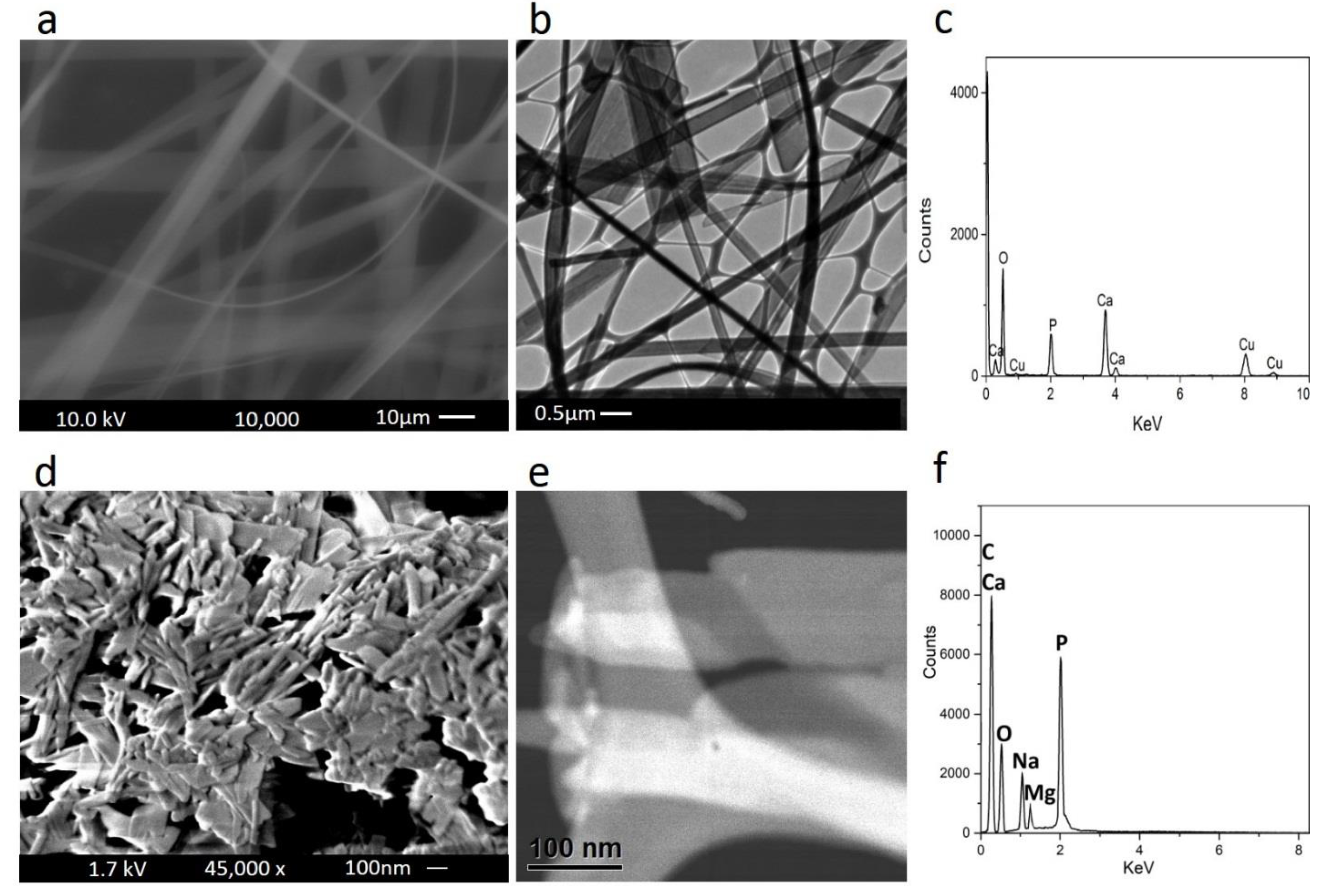
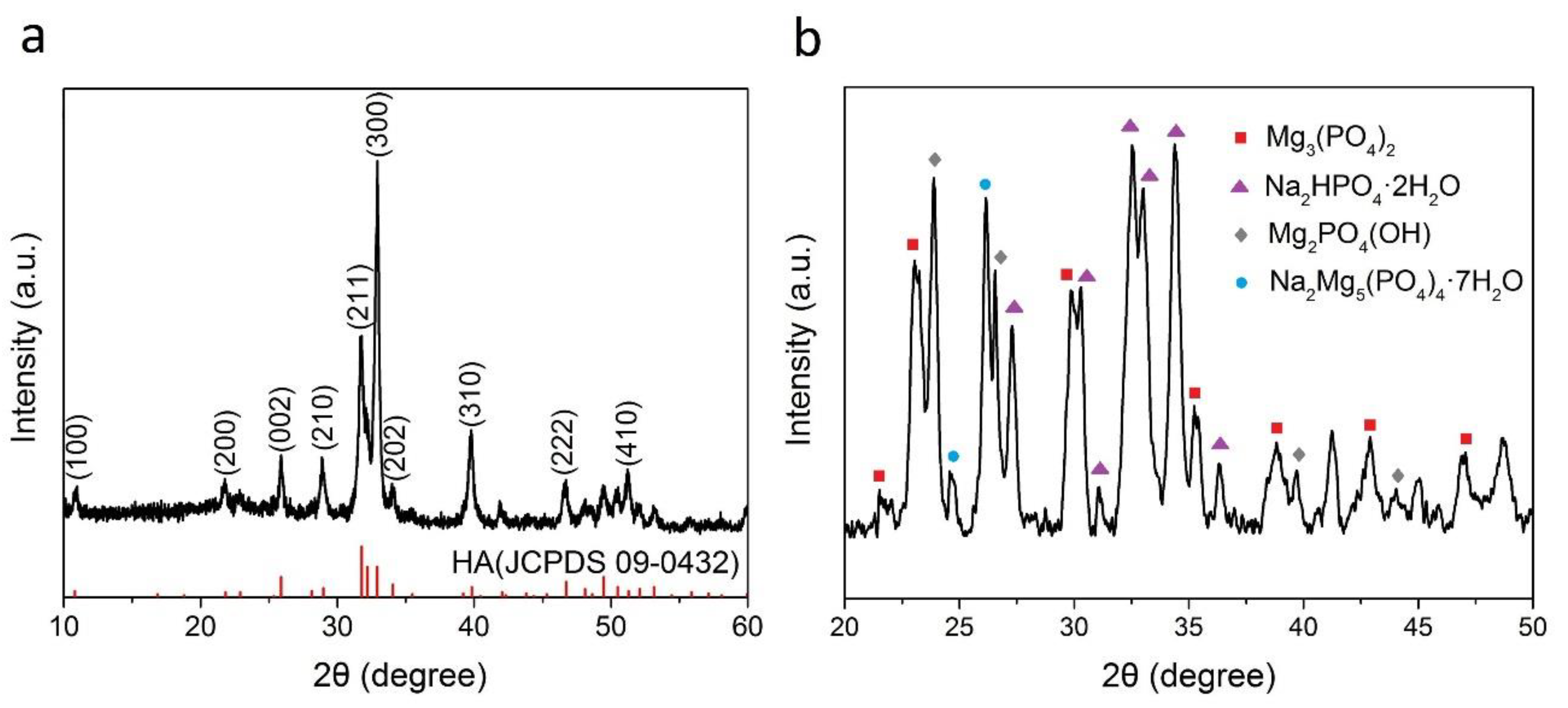
2.1. Synthesis of the HA Nanofibers
2.2. Synthesis of 2D MP Nanosheets
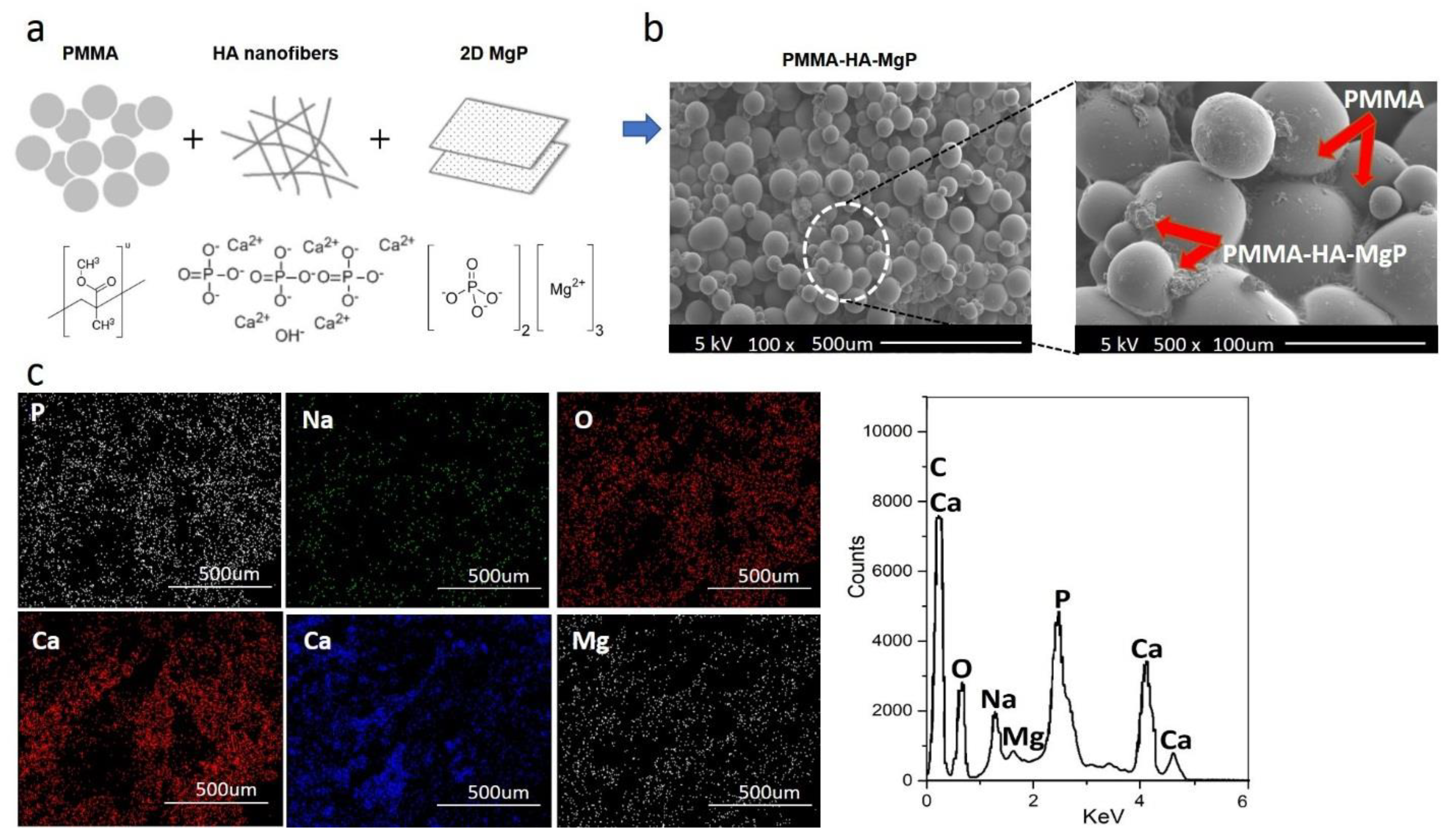
2.3. PMMA-HA-MgP Nanocomposite Preparation
2.4. Experimental Design
2.5. Compression Tests
2.6. Cytotoxicity Tests
2.7. Structural and Chemical Characterizations
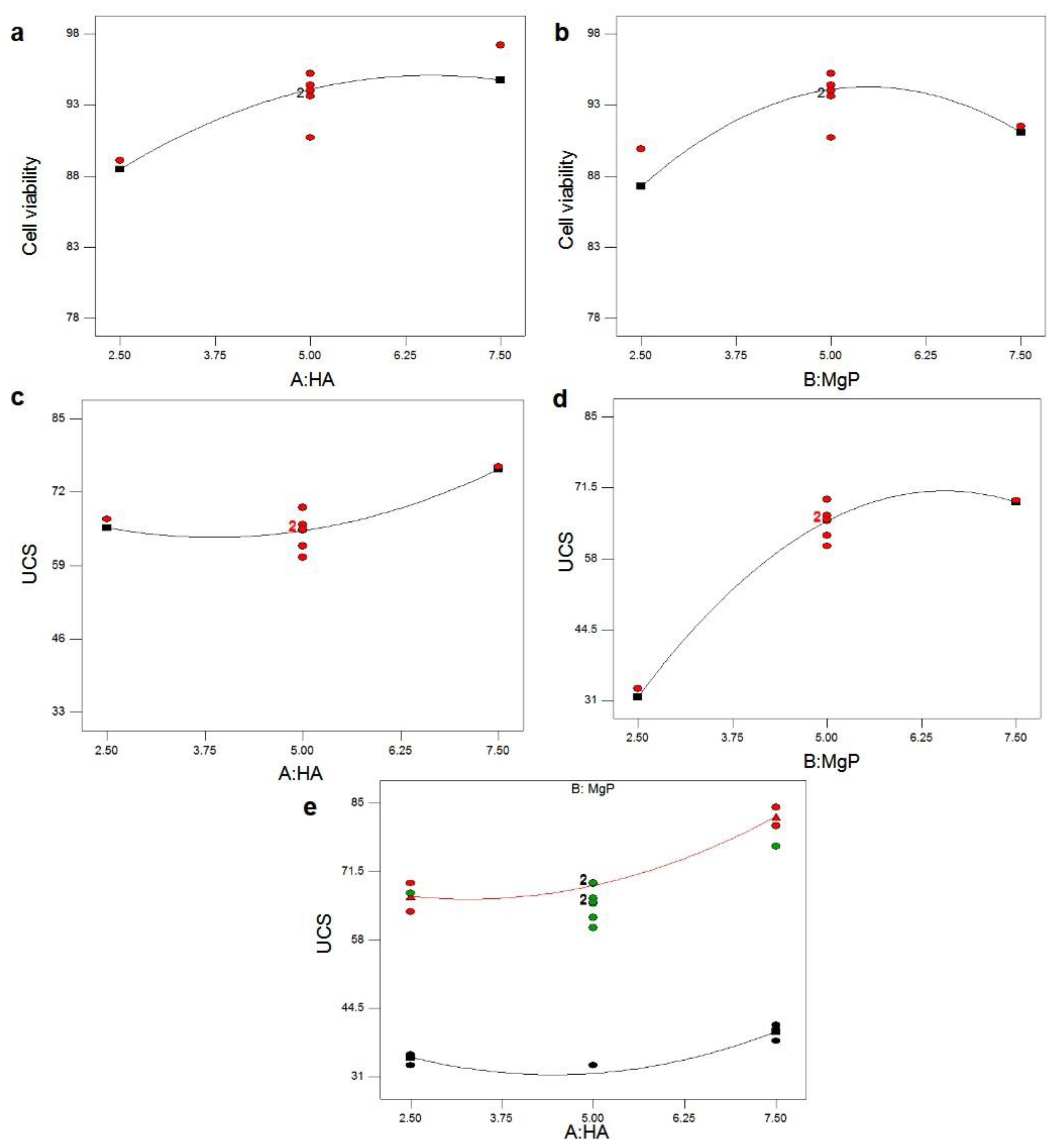
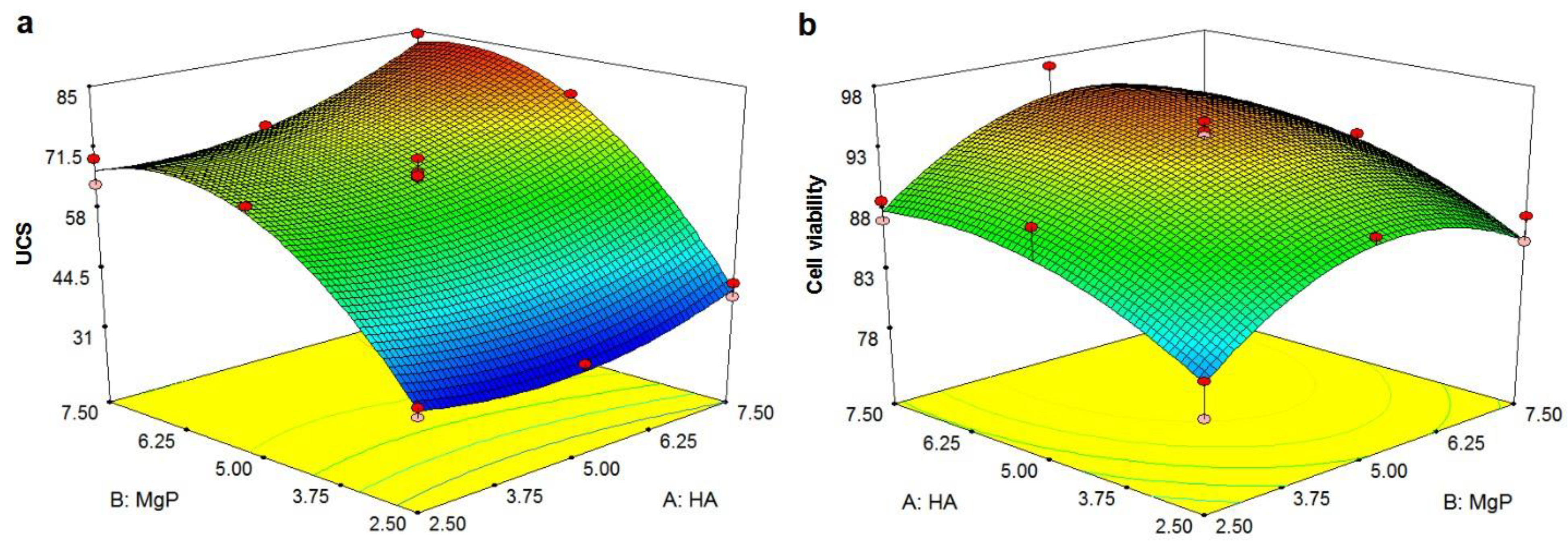
3. Results and Discussion
4. Conclusions
Author Contributions
Funding
Informed Consent Statement
Data Availability Statement
Acknowledgments
Conflicts of Interest
References
- Fottner, A.; Nies, B.; Kitanovic, D.; Steinbrück, A.; Hausdorf, J.; Mayer-Wagner, S.; Pohl, U.; Jansson, V. In vivo evaluation of bioactive PMMA-based bone cement with unchanged mechanical properties in a load-bearing model on rabbits. J. Biomater. Appl. 2015, 30, 30–37. [Google Scholar] [CrossRef] [PubMed] [Green Version]
- Anusavice, K.J.; Phillips, R.W. Phillips’ Science of Dental Materials; Saunders: Philadelphia, PA, USA, 2003. [Google Scholar]
- Vaishya, R.; Agarwal, A.K.; Tiwari, M.; Vaish, A.; Vijay, V.; Nigam, Y. Medical textiles in orthopedics: An overview. J. Clin. Orthop. Trauma 2017, 9, S26–S33. [Google Scholar] [CrossRef] [PubMed]
- Sayeed, Z.; Padela, M.T.; El-Othmani, M.M.; Saleh, K.J. 9—Acrylic bone cements for joint replacement. In Biomedical Composites; Woodhead Publishing Series in Biomaterials: Sawston, UK, 2017; pp. 199–214. [Google Scholar] [CrossRef]
- Wanh, H.; Maeda, T.; Miazaki, T. Effect of calcium acetate content on apatite-forming ability and mechanical property of PMMA bone cement modified with quaternary ammonium. Materials 2020, 13, 4998. [Google Scholar] [CrossRef]
- Kim, S.B.; Kim, Y.J.; Yoon, T.L.; Park, S.A.; Cho, I.H.; Kim, E.J.; Kim, I.A.; Shin, J.-W. The characteristics of a hydroxyapatite–chitosan–PMMA bone cement. Biomaterials 2004, 25, 5715–5723. [Google Scholar] [CrossRef] [PubMed]
- Barralet, J.E.; Gaunt, T.; Wright, A.J.; Gibson, I.R.; Knowles, J.C. Effect of porosity reduction by compaction on compressive strength and microstructure of calcium phosphate cement. J. Biomed. Mater. Res. 2002, 63, 1–9. [Google Scholar] [CrossRef] [Green Version]
- Arora, M. Polymethylmethacrylate bone cements and additives: A review of the literature. World J. Orthop. 2013, 4, 67. [Google Scholar] [CrossRef]
- Zhang, C.; Zhu, J.; Zhang, T.; Li, M.; Jiang, G.; Zhao, J. Small intestinal submucosa/polymethyl methacrylate composite bone cement for vertebral repair. Mater. Des. 2018, 154, 254–265. [Google Scholar] [CrossRef]
- Moursi, A.M.; Winnard, A.V.; Winnard, P.L.; Lannutti, J.J.; Seghi, R.R. Enhanced osteoblast response to a polymethylmethacrylate–hydroxyapatite composite. Biomaterials 2002, 23, 133–144. [Google Scholar] [CrossRef]
- Sugino, A.; Miyazaki, T.; Kawachi, G.; Kikuta, K.; Ohtsuki, C. Relationship between apatite-forming ability and mechanical properties of bioactive PMMA-based bone cement modified with calcium salts and alkoxysilane. J. Mater. Sci. Mater. Med. 2008, 19, 1399–1405. [Google Scholar] [CrossRef]
- Mann, K.A.; Miller, M.A.; Cleary, R.J.; Janssen, D.; Verdonschot, N. Experimental micromechanics of the cement–bone interface. J. Orthop. Res. 2008, 26, 872–879. [Google Scholar] [CrossRef] [Green Version]
- Vale, F.M.; Castro, M.; Monteiro, J.; Couto, F.S.; Pinto, R.; Gião Toscano Rico, J.M. Acrylic bone cement induces the production of free radicals by cultured human fibroblasts. Biomaterials 1997, 18, 1133–1135. [Google Scholar] [CrossRef]
- Kalteis, T.; Luring, C.; Gugler, G.; Zysk, S.; Caro, W.; Handel, M.; Grifka, J. Acute tissue toxicity of PMMA bone cements. Z. Orthop. Ihre Grenzgeb. 2004, 142, 666–672. [Google Scholar] [CrossRef] [PubMed]
- Almeida, T.; Leite Ferreira, B.J.M.; Loureiro, J.; Correia, R.N.; Santos, C. Preliminary Evaluation of the in vitro cytotoxicity of PMMA-co-EHA bone cement. Mater. Sci. Eng. C 2011, 31, 658–662. [Google Scholar] [CrossRef]
- Shi, Z.; Neoh, K.G.; Kang, E.T.; Wang, W. Antibacterial and mechanical properties of bone cement impregnated with chitosan nanoparticles. Biomaterials 2006, 27, 2440–2449. [Google Scholar] [CrossRef] [PubMed]
- Vallés, C.; Abdelkader, A.M.; Young, R.J.; Kinloch, I.A. The effect of flake diameter on the reinforcement of few-layer graphene-PMMA composites. Compos. Sci. Technol. 2015, 111, 17–22. [Google Scholar] [CrossRef]
- Du, G.; Wang, J.-J. The mechanical properties of surface treated UHMWPE fibers and TiO2 reinforced PMMA composite. Surf. Interface Anal. 2017, 49, 940–944. [Google Scholar] [CrossRef]
- Ayatollahi, M.R.; Mirmohammadi, S.A.; Shirazi, H.A. The tension-shear fracture behavior of polymeric bone cement modified with hydroxyapatite nano-particles. Arch. Civ. Mech. Eng. 2018, 18, 50–59. [Google Scholar] [CrossRef]
- Samad, H.A.; Jaafar, M.; Othman, R.; Kawashita, M.; Razak, N.H.A. New bioactive glass-ceramic: Synthesis and application in PMMA bone cement composites. Biomed. Mater. Eng. 2011, 21, 247–258. [Google Scholar] [CrossRef]
- Paz, E.; Forriol, F.; del Real, J.C.; Dunne, N. Graphene oxide versus graphene for optimisation of PMMA bone cement for orthopaedic applications. Mater. Sci. Eng. C 2017, 77, 1003–1011. [Google Scholar] [CrossRef]
- Ormsby, R.; McNally, T.; Mitchell, C.; Dunne, N. Incorporation of multiwalled carbon nanotubes to acrylic based bone cements: Effects on mechanical and thermal properties. J. Mech. Behav. Biomed. Mater. 2010, 3, 136–145. [Google Scholar] [CrossRef]
- Singh, M.K.; Shokuhfar, T.; de Gracio, J.J.A.; de Sousa, A.C.M.; Fereira, J.M.D.F.; Garmestani, H.; Ahzi, S. Hydroxyapatite Modified with Carbon-Nanotube-Reinforced Poly(methyl methacrylate): A Nanocomposite Material for Biomedical Applications. Adv. Funct. Mater. 2008, 18, 694–700. [Google Scholar] [CrossRef]
- Bacali, C.; Badea, M.; Maldovan, M.; Sarosi, C.; Nastase, V.; Baldea, I.; Chiorean, R.S.; Constantiniuc, M. The influence of graphene in improvement of physico-mechanical properties in PMMA denture base resins. Materials 2019, 12, 2335. [Google Scholar] [CrossRef] [Green Version]
- Hill, J.; Orr, J.; Dunne, N. In vitro study investigating the mechanical properties of acrylic bone cement containing calcium carbonate nanoparticles. J. Mater. Sci. Mater. Med. 2008, 19, 3327–3333. [Google Scholar] [CrossRef]
- Jiang, H.-J.; Xu, J.; Qiu, Z.-Y.; Ma, X.-L.; Zhang, Z.-Q.; Tan, X.-X.; Cui, Y.; Cui, Z.-F. Mechanical Properties and cytocompatibility improvement of vertebroplasty PMMA bone cements by incorporating mineralized collagen. Materials 2015, 8, 2616–2634. [Google Scholar] [CrossRef]
- Slane, J.; Vivanco, J.; Meyer, J.; Ploeg, H.-L.; Squire, M. Modification of acrylic bone cement with mesoporous silica nanoparticles: Effects on mechanical, fatigue and absorption properties. J. Mech. Behav. Biomed. Mater. 2014, 29, 451–461. [Google Scholar] [CrossRef]
- Alzarrug, F.A.; Dimitrijević, M.M.; Jančić Heinemann, R.M.; Radojević, V.; Stojanović, D.B.; Uskoković, P.S.; Aleksić, R. The use of different alumina fillers for improvement of the mechanical properties of hybrid PMMA composites. Mater. Des. 2015, 86, 575–581. [Google Scholar] [CrossRef]
- Gutiérrez-Mejía, A.; Herrera-Kao, W.; Duarte-Aranda, S.; Loría-Bastarrachea, M.I.; Canché-Escamilla, G.; Moscoso-Sánchez, F.J.; Cauich-Rodríguez, J.V.; Cervantes-Uc, J.M. Synthesis and characterization of core–shell nanoparticles and their influence on the mechanical behavior of acrylic bone cements. Mater. Sci. Eng. C 2013, 33, 1737–1743. [Google Scholar] [CrossRef] [PubMed]
- Yu, W.; Wang, X.; Tang, Q.; Guo, M.; Zhao, J. Reinforcement of denture base PMMA with ZrO2 nanotubes. J. Mech. Behav. Biomed. Mater. 2014, 32, 192–197. [Google Scholar] [CrossRef] [PubMed]
- Chen, Y.; Tan, C.; Zhang, H.; Wang, L. Two-dimensional graphene analogues for biomedical applications. Chem. Soc. Rev. 2015, 44, 2681–2701. [Google Scholar] [CrossRef] [PubMed]
- Brückner, T.; Gbureck, U. Nano-magnesium phosphate hydrogels: Efficiency of an injectable and biodegradable gel formulation towards bone regeneration. AME Med. J. 2017, 2, 1–4. [Google Scholar] [CrossRef]
- Qi, M.; Huang, Z.; Yao, W.; Long, F.; Cheng, M.; Song, B.; Banner, D.; Shahbazian-Yassar, R.; Lu, Y.; Shokuhfar, T. In situ visualization of the superior nanomechanical flexibility of individual hydroxyapatite nanobelts. CrystEngComm 2018, 20, 1031–1036. [Google Scholar] [CrossRef]
- Qi, M.; Huang, Z.; Phakatkar, A.; Yao, W.; Yuan, Y.; Foroozan, T.; Xiao, G.; Shahbazian-Yassar, R.; Lu, Y.; Shokuhfar, T. Facile hydrothermal synthesis of antibacterial multi-layered hydroxyapatite nanostructures with superior flexibility. CrystEngComm 2018, 20, 1304–1312. [Google Scholar] [CrossRef]
- Shirdar, M.R.; Golshan, A.; Izman, S.; Ghodsiyeh, D. The application of surface response methodology to the pretreatment of WC substrates prior to diamond coating. J. Mater. Eng. Perform. 2014, 23, 13–24. [Google Scholar] [CrossRef]
- Wang, A.; Li, Y.; Yang, B.; Xu, B.; Kong, L.; Liu, D. Process optimization for vacuum distillation of Sn–Sb alloy by response surface methodology. Vacuum 2014, 109, 127–134. [Google Scholar] [CrossRef]
- Noordin, M.Y.; Venkatesh, V.C.; Sharif, S.; Elting, S.; Abdullah, A. Application of response surface methodology in describing the performance of coated carbide tools when turning AISI 1045 steel. J. Mater. Process. Technol. 2004, 145, 46–58. [Google Scholar] [CrossRef] [Green Version]
- Gao, Y.; Xu, J.; Luo, X.; Zhu, J.; Nie, L. Experiment research on mix design and early mechanical performance of alkali-activated slag using response surface methodology (RSM). Ceram. Int. 2016, 42, 11666–11673. [Google Scholar] [CrossRef]
- Montgomery, D.C. Design and Analysis of Experiments; John Wiley & Sons: Hoboken, NJ, USA, 2008. [Google Scholar]
- Satheesh Raja, R.; Manisekar, K. Experimental and statistical analysis on mechanical properties of nano flyash impregnated GFRP composites using central composite design method. Mater. Des. 2016, 89, 884–892. [Google Scholar] [CrossRef]
- ISO 5833:2002. Implants for Surgery-Acrylic Resin Cements; ISO/TC 150/SC 1 Materials: Berlin, Germany, 2002. [Google Scholar]
- Laurenti, M.; Al Subaie, A.; Abdallah, M.-N.; Cortes, A.R.G.; Ackerman, J.L.; Vali, H.; Basu, K.; Zhang, Y.L.; Murshed, M.; Strandman, S.; et al. Two-Dimensional Magnesium Phosphate Nanosheets Form Highly Thixotropic Gels That Up-Regulate Bone Formation. Nano Lett. 2016, 16, 4779–4787. [Google Scholar] [CrossRef]
- Mostaan, H.; Shamanian, M.; Monirvaghefi, S.M.; Behjati, P.; Szpunar, J.A.; Sherafati, J. Electron beam assisted joining of nanograin-sized Fe–Co–V magnetic foils: Study and optimization of magnetic properties of weld joints. Vacuum 2014, 109, 148–156. [Google Scholar] [CrossRef]
- Shirdar, M.R.; Izman, S.; Taheri, M.M.; Assadian, M.; Abdul Kadir, M.R. Effect of Electrophoretic Deposition Parameters on the Corrosion Behavior of Hydroxyapatite-Coated Cobalt–Chromium Using Response Surface Methodology. Arab. J. Sci. Eng. 2015, 41, 591–598. [Google Scholar] [CrossRef]
- Rezazadeh Shirdar, M.; Taheri, M.M.; Moradifard, H.; Keyvanfar, A.; Shafaghat, A.; Shokuhfar, T.; Izman, S. Hydroxyapatite–Titania nanotube composite as a coating layer on Co–Cr-based implants: Mechanical and electrochemical optimization. Ceram. Int. 2016, 42, 6942–6954. [Google Scholar] [CrossRef]
- Assadian, M.; Shirdar, M.R.; Idris, M.H.; Izman, S.; Almasi, D.; Taheri, M.M.; Kadir, M.R.A. Optimisation of Electrophoretic Deposition Parameters in Coating of Metallic Substrate by Hydroxyapatite Using Response Surface Methodology. Arab. J. Sci. Eng. 2015, 40, 923–933. [Google Scholar] [CrossRef]
- Chan, L.C.; Lu, X.Z.; Yu, K.M. Multiscale approach with RSM for stress-strain behaviour prediction of micro-void-considered metal alloy. Mater. Des. 2015, 83, 129–137. [Google Scholar] [CrossRef]
- Bezerra, M.A.; Santelli, R.E.; Oliveira, E.P.; Villar, L.S.; Escaleira, L.A. Response surface methodology (RSM) as a tool for optimization in analytical chemistry. Talanta 2008, 76, 965–977. [Google Scholar] [CrossRef]
- Kooshki, S.; Pestehe, S.J.; Bozorgzadeh, H.R. Design of new tapered-bed Dielectric Barrier Discharge reactor for atmospheric-pressure plasma modification of starch. Vacuum 2018, 156, 224–232. [Google Scholar] [CrossRef]


| Levels | Factors | |
|---|---|---|
| HA Ratio (% wt) A | MgP Ratio (% wt) B | |
| High (1) | 7.5 | 7.5 |
| Low (−1) | 2.5 | 2.5 |
| Centre point (0) | 0 | 0 |
| Std. no. | Run | HA (% wt) | MgP (% wt) | UCS (MPa) | Cell Viability (%) |
|---|---|---|---|---|---|
| 1 | 6 | 2.50 | 2.50 | 33.3 | 81.8 |
| 2 | 13 | 2.50 | 2.50 | 35.4 | 78.9 |
| 3 | 10 | 7.50 | 2.50 | 38.1 | 88.7 |
| 4 | 12 | 7.50 | 2.50 | 41.2 | 87.1 |
| 5 | 4 | 2.50 | 7.50 | 69.2 | 85.4 |
| 6 | 17 | 2.50 | 7.50 | 63.6 | 87.5 |
| 7 | 14 | 7.50 | 7.50 | 84.2 | 91.1 |
| 8 | 3 | 7.50 | 7.50 | 80.5 | 89.9 |
| 9 | 16 | 2.50 | 5.00 | 67.2 | 89.1 |
| 10 | 2 | 7.50 | 5.00 | 76.5 | 97.2 |
| 11 | 5 | 5.00 | 2.50 | 33.3 | 89.9 |
| 12 | 8 | 5.00 | 7.50 | 69.1 | 91.5 |
| 13 | 15 | 5.00 | 5.00 | 65.2 | 94.4 |
| 14 | 7 | 5.00 | 5.00 | 62.4 | 95.2 |
| 15 | 11 | 5.00 | 5.00 | 60.4 | 93.6 |
| 16 | 9 | 5.00 | 5.00 | 66.2 | 90.7 |
| 17 | 18 | 5.00 | 5.00 | 69.3 | 94 |
| 18 | 1 | 5.00 | 5.00 | 65.4 | 93.6 |
| Source | Sum of Squares | Df | Mean Square | F Value | p-Value Prob > F | |
|---|---|---|---|---|---|---|
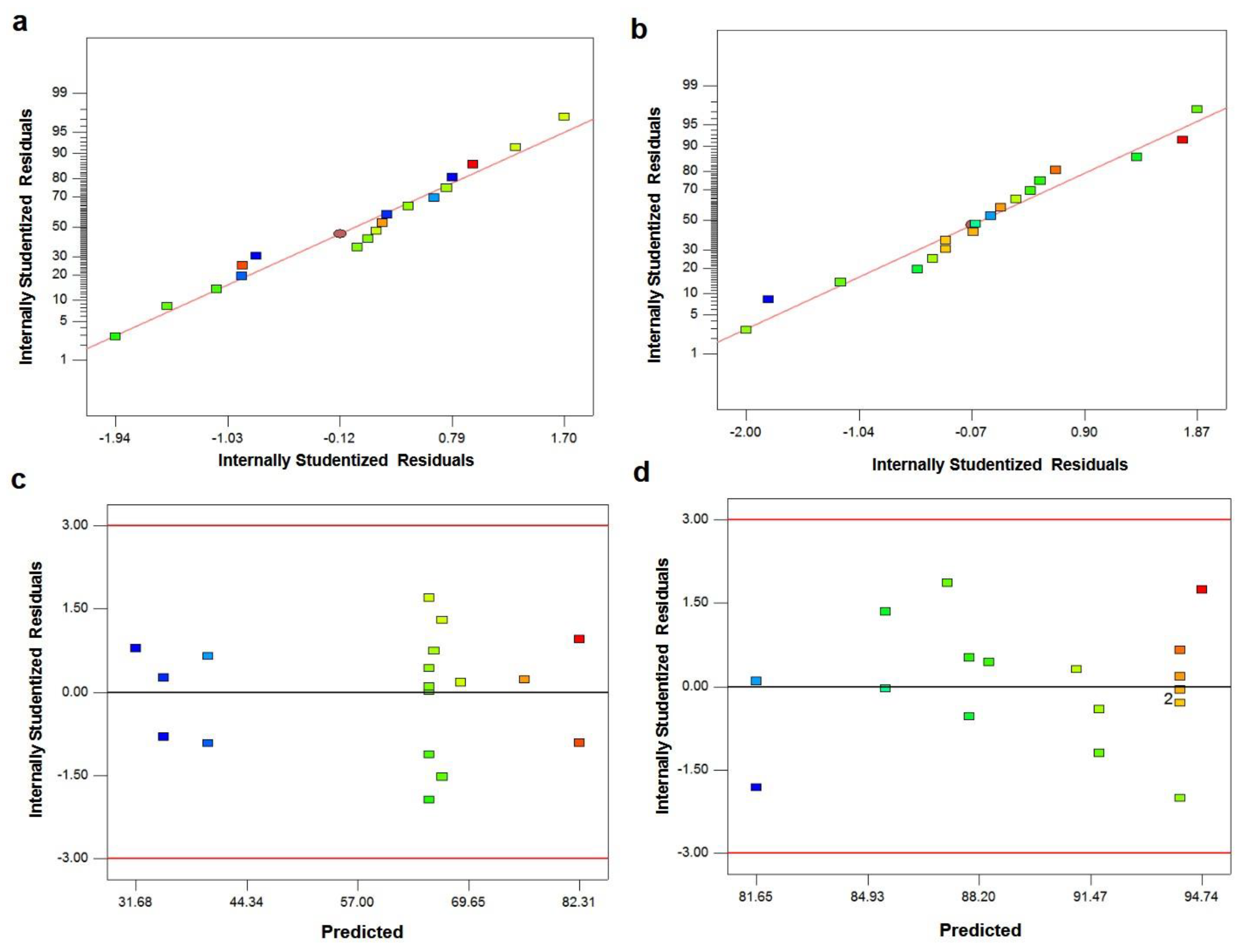
| Model | 4477.32 | 5 | 895.46 | 127.78 | <0.0001 | significant |
| A-HA | 268.32 | 1 | 268.32 | 38.29 | <0.0001 | |
| B-MgP | 3433.61 | 1 | 3433.61 | 489.96 | <0.0001 | |
| AB | 56.71 | 1 | 56.71 | 8.09 | 0.0148 | |
| A2 | 101.40 | 1 | 101.40 | 14.47 | 0.0025 | |
| B2 | 691.06 | 1 | 691.06 | 98.61 | <0.0001 | |
| Residual | 84.10 | 12 | 7.01 | |||
| Lack of Fit | 6.71 | 3 | 2.24 | 0.26 | 0.8523 | not significant |
| Pure Error | 77.38 | 9 | 8.60 | |||
| Cor Total | 4561.42 | 17 | ||||
| Std. Dev. | 2.65 | R-Squared | 0.9816 | |||
| Mean | 60.03 | Adj R-Squared | 0.9739 | |||
| C.V. % | 4.41 | Pred R-Squared | 0.9611 | |||
| PRESS | 177.33 | Adeq Precision | 33.121 |
| Source | Sum of Squares | df | Mean Square | F Value | p-Value Prob > F | |
|---|---|---|---|---|---|---|
| Model | 335.39 | 5 | 67.08 | 21.46 | <0.0001 | significant |
| A-HA | 97.97 | 1 | 97.97 | 31.35 | 0.0001 | |
| B-MgP | 36.10 | 1 | 36.10 | 11.55 | 0.0053 | |
| AB | 6.12 | 1 | 6.12 | 1.96 | 0.1868 | |
| A2 | 19.08 | 1 | 19.08 | 6.10 | 0.0295 | |
| B2 | 75.36 | 1 | 75.36 | 24.12 | 0.0004 | |
| Residual | 37.50 | 12 | 3.13 | |||
| Lack of Fit | 17.32 | 3 | 5.77 | 2.58 | 0.1187 | not significant |
| Pure Error | 20.18 | 9 | 2.24 | |||
| Cor Total | 372.89 | 17 | ||||
| Std. Dev. | 1.77 | R-Squared | 0.8994 | |||
| Mean | 89.98 | Adj R-Squared | 0.8575 | |||
| C.V. % | 1.96 | Pred R-Squared | 0.7578 | |||
| PRESS | 90.30 | Adeq Precision | 13.683 |
| Source | Sum of Squares | df | Mean Square | F Value | p-Value Prob > F | |
|---|---|---|---|---|---|---|
| Model | 329.27 | 4 | 82.32 | 24.53 | <0.0001 | significant |
| A-HA | 97.97 | 1 | 97.97 | 29.19 | 0.0001 | |
| B-MgP | 36.10 | 1 | 36.10 | 10.76 | 0.0060 | |
| A2 | 19.08 | 1 | 19.08 | 5.68 | 0.0330 | |
| B2 | 75.36 | 1 | 75.36 | 22.46 | 0.0004 | |
| Residual | 43.63 | 13 | 3.36 | |||
| Lack of Fit | 23.45 | 4 | 5.86 | 2.61 | 0.1064 | not significant |
| Pure Error | 20.18 | 9 | 2.24 | |||
| Cor Total | 372.89 | 17 | ||||
| Std. Dev. | 3.87 | R-Squared | 0.9920 | |||
| Mean | 51.63 | Adj R-Squared | 0.9891 | |||
| C.V. % | 7.50 | Pred R-Squared | 0.9808 | |||
| PRESS | 686.26 | Adeq Precision | 44.496 |
| No. | HA (% wt) | MgP (% wt) | Actual UCS (Mpa) | Predicted UCS (Mpa) | Residual | Error (%) |
|---|---|---|---|---|---|---|
| 1 | 2.5 | 7.5 | 69.2 | 66.6 | 2.6 | 3.7 |
| 2 | 7.5 | 5 | 76.5 | 76 | 0.5 | 0.6 |
| 3 | 2.5 | 2.5 | 35.4 | 34.8 | 0.6 | 1.6 |
| 4 | 3.5 | 4 | 58.8 | 54.9 | 3.9 | 6.6 |
| 5 | 4.5 | 3 | 39.6 | 40.3 | 0.7 | 1.7 |
| 6 | 6 | 6.5 | 81.5 | 75.5 | 6 | 7.3 |
| No. | HA (% wt) | MgP (% wt) | Actual Cell Viability (%) | Predicted Cell Viability (%) | Residual | Error (%) |
|---|---|---|---|---|---|---|
| 1 | 2.5 | 7.5 | 85.4 | 85.4 | 0 | 0 |
| 2 | 7.5 | 5 | 97.2 | 94.7 | 2.5 | 2.5 |
| 3 | 2.5 | 2.5 | 78.9 | 81.6 | 2.7 | 3.4 |
| 4 | 3.5 | 4 | 91.3 | 89.7 | 1.6 | 1.7 |
| 5 | 4.5 | 3 | 89.7 | 88.6 | 1.1 | 1.2 |
| 6 | 6 | 6.5 | 90.2 | 94.3 | 4.1 | 4.5 |
| No. | HA (% wt) | MgP (% wt) | UCS (Mpa) | Cell Viability | Desirability |
|---|---|---|---|---|---|
| 1 | 7.50 | 6.12 | 82.5307 | 94.6098 | 0.911 |
| 2 | 7.50 | 6.08 | 82.4215 | 94.6428 | 0.911 |
Publisher’s Note: MDPI stays neutral with regard to jurisdictional claims in published maps and institutional affiliations. |
© 2021 by the authors. Licensee MDPI, Basel, Switzerland. This article is an open access article distributed under the terms and conditions of the Creative Commons Attribution (CC BY) license (https://creativecommons.org/licenses/by/4.0/).
Share and Cite
Shirdar, M.R.; Taheri, M.M.; Qi, M.-L.; Gohari, S.; Farajpour, N.; Narayanan, S.; Foroozan, T.; Sharifi-Asl, S.; Shahbazian-Yassar, R.; Shokuhfar, T. Optimization of the Mechanical Properties and the Cytocompatibility for the PMMA Nanocomposites Reinforced with the Hydroxyapatite Nanofibers and the Magnesium Phosphate Nanosheets. Materials 2021, 14, 5893. https://0-doi-org.brum.beds.ac.uk/10.3390/ma14195893
Shirdar MR, Taheri MM, Qi M-L, Gohari S, Farajpour N, Narayanan S, Foroozan T, Sharifi-Asl S, Shahbazian-Yassar R, Shokuhfar T. Optimization of the Mechanical Properties and the Cytocompatibility for the PMMA Nanocomposites Reinforced with the Hydroxyapatite Nanofibers and the Magnesium Phosphate Nanosheets. Materials. 2021; 14(19):5893. https://0-doi-org.brum.beds.ac.uk/10.3390/ma14195893
Chicago/Turabian StyleShirdar, Mostafa Rezazadeh, Mohammad Mahdi Taheri, Mei-Li Qi, Soheil Gohari, Nasim Farajpour, Surya Narayanan, Tara Foroozan, Soroosh Sharifi-Asl, Reza Shahbazian-Yassar, and Tolou Shokuhfar. 2021. "Optimization of the Mechanical Properties and the Cytocompatibility for the PMMA Nanocomposites Reinforced with the Hydroxyapatite Nanofibers and the Magnesium Phosphate Nanosheets" Materials 14, no. 19: 5893. https://0-doi-org.brum.beds.ac.uk/10.3390/ma14195893