Article

Evaluation of Anti-Tyrosinase and Antioxidant Properties of Four Fern Species for Potential Cosmetic Applications

1
Department of Pharmacy, Faculty of Health Sciences, Universidad San Jorge, 50830 Villanueva de Gállego (Zaragoza), Spain
2
Department of Biochemistry and Physiology, Faculty of Pharmacy and Food Sciences, Universitat de Barcelona, 08028 Barcelona, Spain
3
Instituto Agroalimentario de Aragón-IA2, CITA-Universidad de Zaragoza, 50013 Zaragoza, Spain
*
Author to whom correspondence should be addressed.
Submission received: 4 January 2019 / Revised: 6 February 2019 / Accepted: 11 February 2019 / Published: 19 February 2019
(This article belongs to the Special Issue Forest, Foods and Nutrition)

Abstract

:
Ferns are poorly explored species from a pharmaceutical perspective compared to other terrestrial plants. In this work, the antioxidant and tyrosinase inhibitory activities of hydrophilic and lipophilic extracts, together with total polyphenol content, were evaluated in order to explore the potential cosmetic applications of four Spanish ferns collected in the Prades Mountains (Polypodium vulgare L., Asplenium adiantum-nigrum L., Asplenium trichomanes L., and Ceterach officinarum Willd). The antioxidant activity was evaluated using the 2,2-diphenyl-1-picrylhydrazyl (DPPH) radical, oxygen radical absorbance capacity (ORAC) and xanthine/xanthine oxidase (X/XO) assays. The potential to avoid skin hyperpigmentation was tested by inhibiting the tyrosinase enzyme, as this causes melanin synthesis in the epidermis. All ferns were confirmed as antioxidant and anti-tyrosinase agents, but interestingly hydrophilic extracts (obtained with methanol) were more potent and effective compared to lipophilic extracts (obtained with hexane). Polypodium vulgare, Asplenium adiantum-nigrum, and Ceterach officinarum methanolic extracts performed the best as antioxidants. Polypodium vulgare methanolic extract also showed the highest activity as a tyrosinase inhibitor.

Graphical Abstract

1. Introduction

The incidence of cutaneous disorders and melanoma has increased worldwide [1]; in fact, non-melanoma skin cancer has become the principal skin cancer among fair-skinned people [2]. Sun radiation is known to accelerate photodamage of the skin, and ultraviolet radiation is one of the main factors that causes skin hyperpigmentation and skin aging [3].
Melanin, which is obtained by irreversible tyrosine catalyzed reactions, is an important epidermal agent that blocks ultraviolet radiation [4]. It has been noted that melanocyte cultures from black skin-types increase melanogenesis and melanosis more than in fair skin-types [5]. Consequently, lighter and thinner skin is 6–33 times more susceptible to developing minimal perceptible erythema than darker and thicker skin [6,7]. Ectopic dermal melanocytes, a result of successive erythemas, are shown to be directly dependent on increased melanin in the epidermis (hypermelanosis), which can trigger sun spots [8].
Primary photoprotection, also called non-systemic photoprotection, has traditionally been considered the main strategy against the harmful effects of sun radiation [9]. This method is based on having healthy habits towards sun exposure and the use of physical photoprotective agents; however, some disadvantages have been described [10]. Antioxidant oral supplements from secondary metabolites of plants are an adjunctive to primary photoprotection [11]. This most recent strategy on photoprotection is known as secondary photoprotection, or systemic photoprotection [12,13], in which standardized aqueous extract from the fronds of Polypodium leucotomos L., which is marketed under the trade name Fernblock® (Cantabria Labs, Santander, Spain), has been one of the most popular systemic and topical photoprotective oral agents in cosmetic science [14,15]. The effectiveness and safety of the use of this fern is a consequence of its multiple pathways of action described by Palomino et al. [16].
Ferns (Polypodiopsida), formerly considered pteridophytes, have been reported as one of the least understood classes of tracheophyte plants from a phylogenetic perspective [17,18]. Recent reviews of the Polypodium genus have been published since the commercialization of Fernblock®, as shown in Berman et al. [19]. Other authors have recently published updated reviews on the phytochemistry and ethnopharmacology of ferns, highlighting the presence of polyphenols (particularly flavonoids), terpenoids, steroids, and alkaloids [20,21]. Most of these bioactive compounds are described as natural enzyme inhibitors in biomedical research drug discovery due to anticancer, antidiabetic, and antiaging properties [22,23,24]. The selected ferns in this study (Polypodium vulgare L., Asplenium adiantum-nigrum L., Asplenium trichomanes L., and Ceterach officinarum Willd) are some of the most common leptosporangiate ferns reported on the Prades Mountains (Spain, 41°18′43″ N 1°05′09″ E) [25].
Considering Fernblock® as a reference in skin photoprotection [26,27], the potential anti-aging and skin-whitening properties of four Spanish ferns collected in the Prades Mountains have been studied. In vitro antioxidant activities against different free radicals (2,2-diphenyl-1-picrylhydrazyl—DPPH, oxygen radical absorbance capacity—ORAC, and xanthine/xanthine oxidase—X/XO methods) and in vitro inhibition of the tyrosinase enzyme were evaluated.

2. Material and Methods

2.1. Chemicals and Reagents

All reagents used were of analytical grade. Methanol, hexane, tyrosinase, and L-dihydroxyphenylalanine (L-DOPA) were acquired through Vidrafoc® (Barcelona, Spain). Dimethyl sulfoxide (DMSO) was obtained from Fisher Scientific® (Madrid, Spain). The provider of 5-hydroxy-2-(hydroxymethyl) pyran-4-one (kojic acid) was Alfa Aesar® (Karlsruhe, Germany). The reagents used to determine the antioxidant activity such as 2,2-diphenyl-1-picrylhydrazyl (DPPH radical), 2,6-dihydroxypurine (xanthine), xanthine oxidase, and (±)-6-hydroxy-2,5,7,8-tetramethylchromane-2-carboxylic acid (trolox) were supplied by Sigma-Aldrich® (Madrid, Spain). Sodium carbonate anhydrous (Na2CO3) and nitrotetrazolium blue chloride (NBT) were purchased from Laboaragon® (Cartuja Baja, Spain) and Sumalsa® (Zaragoza, Spain), respectively. All aqueous solutions were prepared with ultra-pure water.

2.2. Plant Material

It was checked that the subject species had been described in The Plant List [28] and by Banco de Datos de Biodiversidad de Cataluña [29].
The whole fresh fronds of selected fern species were identified and collected from the Prades Mountains, in the province of Tarragona (Spain), in November 2016 by Adrià Farràs and Josep Ma Farràs using botanical keys [30]. The samples were dried in the shade at room temperature.
A dried voucher specimen has been deposited at the Herbarium of Universidad San Jorge, Zaragoza, Spain (Polypodium vulgare: voucher no. 003-2016; Asplenium adiantum-nigrum: voucher no. 004-2016; Asplenium trichomanes: voucher no. 005-2016; Ceterach officinarum voucher no. 006-2016). These examples were authenticated by Dr. J.A. Vicente Orellana from Universidad CEU San Pablo (Madrid, Spain).

2.3. Extracts Preparation

Hydrophilic (= polar) and lipophilic (= non polar) extracts were prepared using methanol or hexane, respectively. Dried fronds of the four species were powdered mechanically until obtaining 40 mg of each. The powdered fronds of each species were split equally into two erlenmeyers of 500 mL. Each 20 mg of fern powder was macerated with 250 mL of solvent (hexane or methanol) at room laboratory temperature for 24 h. The extract was filtered using Whatman N°4 filter paper, and the solvent was evaporated using rotatory evaporator with a thermostatic bath at 30 °C. This process was completed two more times until exhaustion of plant material; extracts were stored at −20 °C until further experiments. Yields were calculated in percentages from the dry weight of fronds used and the quantity of dry mass obtained by extraction.

2.4. Phytochemical Screening by Thin Layer Chromatography (TLC) and Total Phenolic Content (TPC)

Silica gel TLC plates coated with fluorescent indicator F254 were used in order to detect phenolic compounds (flavonoids and phenolic acids) in the samples. 10 µL of hexane and methanolic extracts of the samples at concentrations of 10 mg/mL were run on the plates with EtOAc/MeOH/H2O (65:15:5, v:v:v) as mobile phase. After eluting the samples, plates were dried, sprayed with the Natural Products polyethylene glycol (PEG) reagent, observed at 365 nm and retention factors (Rf) calculated [31].
TPC was quantified by the Folin Method as previously described using gallic acid for the standard calibration curve [32].

2.5. Determination of Antioxidant Activities

Antioxidant capacity was assessed by three complementary methods that were DPPH, ORAC, and superoxide radicals generated by X/XO.

2.5.1. DPPH Radical Scavenging Activity

The neutralization of DPPH radicals as antioxidant method was reported the first time by Blois et al. [33]. In this case, the assay was carried out according to the modifications described by Casedas et al. [34]. In 96-well microplates, each well contained 150 μL of extract and 150 μL of DPPH (0.04 mg/mL methanol solution). Antioxidant activity was determined measuring absorbance (Abs) at 515 nm after 30 min of dark incubation. Blank and control wells were also considered. The highest concentration of extracts tested was 1 mg/mL. Trolox, a water soluble derivate of vitamin E, was used as positive standard. Background interferences from solvents and samples were deducted from the activities prior to calculating radical scavenging capacity (RSC) as follows: RSC (%) = [(Abscontrol − Abssample)/Abscontrol] × 100.

2.5.2. ORAC Assay

ORAC assay was carried out to measure the capacity of extracts to scavenge peroxyl radicals. Samples and trolox were dissolved in PBS and methanol (50:50, v:v). Samples were incubated with fluorescein (0.07 µM) in 96-well plates for 10 min at 37 °C. Afterwards, AAPH (0.012 M) was supplemented and fluorescence was measured for 98 min at 485 nm of excitation and 520 nm of emission, in a FLUOstar Optima fluorimeter (BMG Labtech, Ortenberg, Germany) [35]. Results were expressed as µmol trolox equivalents (TE)/mg sample.

2.5.3. Superoxide Radicals Generated by Xanthine/Xanthine Oxidase (X/XO)

Xanthine oxidase and xanthine as substrate are responsible for the production of superoxide radicals [36]. The effects of fern extracts on superoxide radicals generated by X/XO were evaluated by measuring the formation of the NBT (nitrotetrazolium blue chloride)-radical superoxide complex [37] using a described procedure [34]. The reaction mixture was prepared every day as a consequence of reduced stability. This mixture was composed of 90 μM xanthine, 16 mM Na2CO3, and 22.8 μM NBT in phosphate buffer (pH 7.0). 240 μL of the reaction mixture in each well with 30 μL of extract solution and XO was incubated in the dark for 2 min at 37 °C and absorbance read at 560 nm. Blank and control wells were also considered, and background interferences from solvents and samples were also deducted from the activities previous to calculating the RSC (%). The reference substance (trolox) was the same used in DPPH and ORAC assays.

2.6. Inhibition of Tyrosinase Activity

The inhibition of tyrosinase was performed following a previous method [38]. Samples were mixed with 40 μL L-DOPA and 80 μL potassium phosphate buffer (pH 6.8). Finally, 40 μL of tyrosinase (200 U/mL) was added in the wells. L-DOPA and tyrosinase were solved in buffer. The inhibition of tyrosinase was determined at 475 nm. Methanolic extracts were dissolved in methanol, and hexane extracts were dissolved in DMSO. Kojic acid was the reference inhibitor substance. Background interferences from solvents and samples were previously deducted from the activities to calculate the percentage of enzymatic inhibition (compared to control activity). Control wells had the same mix except the sample/inhibitor, which was replaced by the solvents of these.

2.7. Statistical Analysis

All samples were analyzed in triplicates (n = 3), at least, on different days. Statistical significance was analyzed by using GraphPad Prism version 6, San Diego, CA, USA. Data are presented as mean +/- standard error. The half maximal inhibitory concentration (IC50) values were obtained by non-linear regression. Activities have been compared using a one-way analysis of variance (ANOVA). Statistical differences were considered as follows: p ≤ 0.05 (*), p ≤ 0.01 (**), and p ≤ 0.001 (***). Correlations were performed between TPC and ORAC values, and TPC and tyrosinase IC50 values; Pearson values were also obtained using GraphPad Prism version 6.

3. Results

3.1. Plant Material and Yields

Table 1 shows the scientific names and Spanish common names of the collected samples as well as yields of extraction in each case. The hydrophilic extracts obtained with methanol have higher yields (ranging from 16.27% to 29.55%) than lipophilic extracts obtained with hexane (ranging from 1.49% to 2.34%).

3.2. Polyphenol Content by Thin Layer Chromatography (TLC) and Folin Method

TLC plates sprayed with Natural Products-PEG reagent revealed the presence of flavonoids in methanolic samples obtained from Asplenium trichomanes (ATM, Rf = 0.97, 0.92, 0.86) and Ceterach officinarum (COM, Rf = 0.66, 0.5). Spots corresponding to phenolic acids were also detected with similar retention factor (Rf) values in the methanolic samples from Polypodium vulgare (PVM, Rf = 0.81), Asplenium adiantum-nigrum (AAM, Rf = 0.81), Ceterach officinarum (COM, Rf = 0.81), and hexane extract of Ceterach officinarum (COH, Rf = 0.81). An image of the TLC plate sprayed with Natural Products-PEG reagent can be downloaded from Supplementary Materials (Figure S1). Total Phenolic Content (TPC) was quantified using the Folin–Ciocalteu reagent; as observed in Table 1, methanolic extracts contained higher amounts of polyphenols, with PVM and COM showing the highest values. As predicted, methanol seems to be a better solvent to extract polyphenols.

3.3. Antioxidant Activity

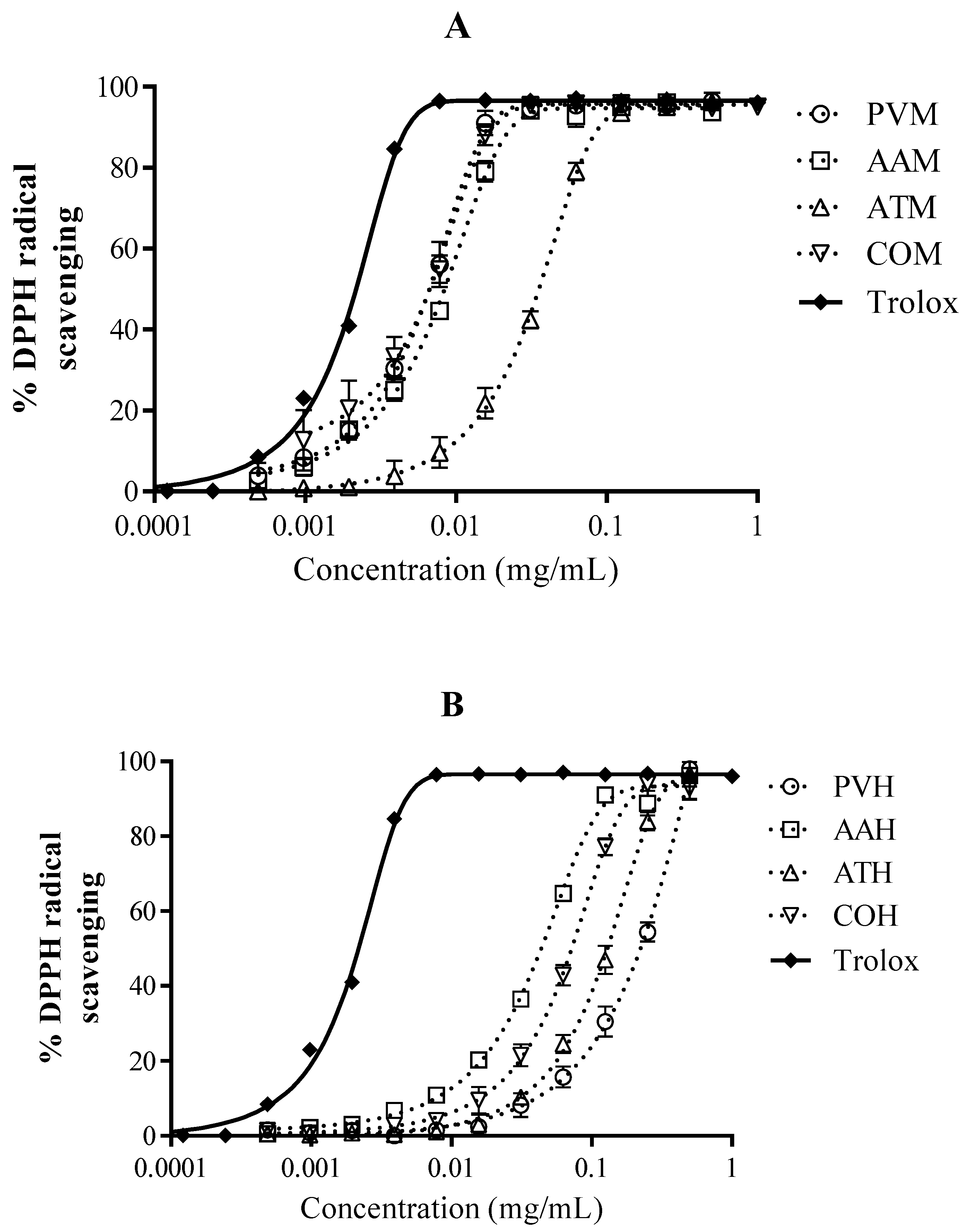
3.3.1. DPPH Radical Scavenging Activity

As seen in Figure 1, methanolic extracts had a more powerful capacity for DPPH reduction than hexane extract for each fern. Methanolic extracts had a very similar profile of antiradical activity reaching 100% of radical inhibition at concentrations between 0.01 and 0.1 mg/mL. PVM and COM had the lowest IC50 values and can therefore be considered as the best antioxidants. Extracts obtained with hexane were also antioxidants, but the concentrations needed to scavenge 100% of DPPH radicals were superior compared to samples extracted with methanol as solvent.

3.3.2. ORAC Assay

The ORAC assay is an internationally recognized method to measure antioxidant capacity. In Table 2, the ORAC values were also higher for methanolic extracts, particularly for PVM and COM, which is in accordance with data obtained in the DPPH assay. In Figure 2A, there is a positive correlation between ORAC values and TPC, which seems to indicate that the antioxidant activity may be mediated by polyphenols.

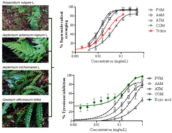
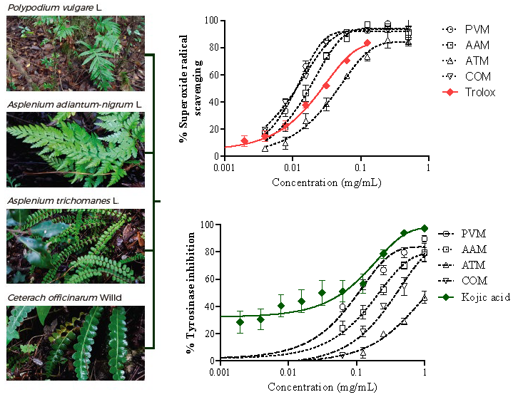
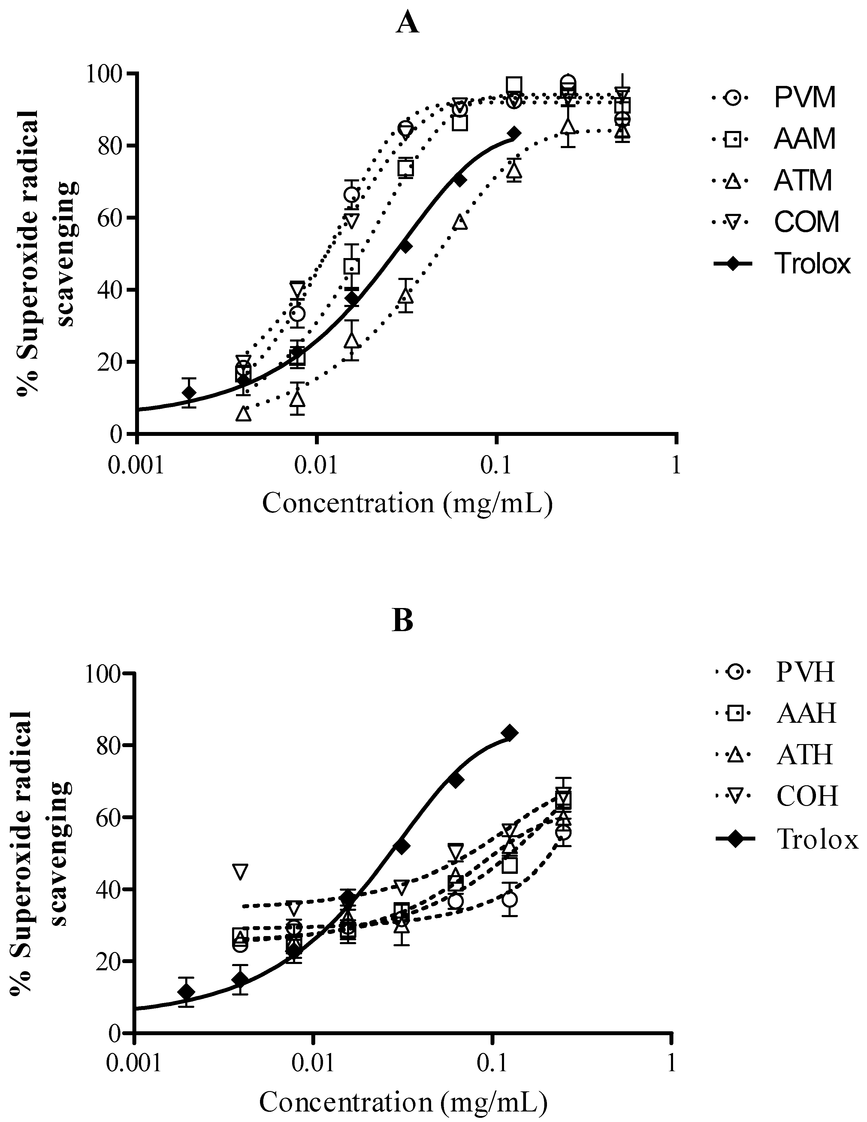
3.3.3. Superoxide Radicals Generated by Xanthine/Xanthine Oxidase (X/XO)

In order to determine if the extracts were able to scavenge physiological radicals like superoxide anion (O2) generated by X/XO, the extracts were tested using this methodology at various concentrations [15]. There were significant differences between methanolic and hexane extracts (Figure 3), but surprisingly, the activity of certain methanolic extracts was superior to the reference compound trolox (Figure 3A). Table 3 reveals that PVM, AAM, and COM showed lower IC50 values than trolox, which confirms their potential as antiradical agents.

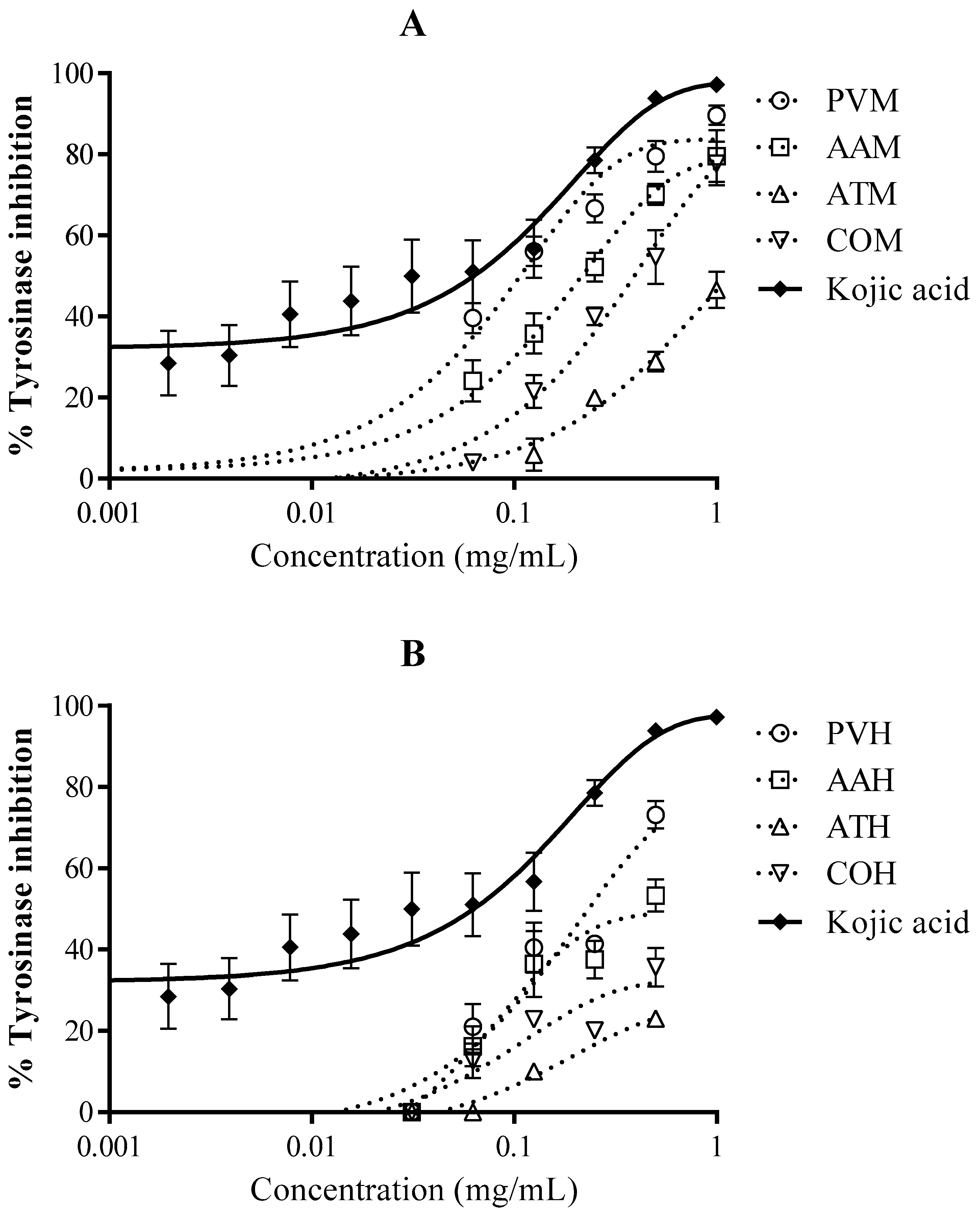
3.4. Tyrosinase Inhibition

Figure 4 shows that methanolic extracts were also better than hexane extracts as anti-tyrosinase agents. All methanolic samples presented IC50 values between 0.107 mg/mL and 1.175 mg/mL, whereas in the case of hexane samples, only PVH was able to reach the IC50 value (Table 3). According to the lowest IC50 values, PVM was the best sample as tyrosinase inhibitor, followed by AAM. In this case, tyrosinase inhibition was not correlated with total phenolic content (Figure 2B).

4. Discussion

Ferns have been used in traditional medicine in Central and South America. In fact, two of the selected species (P. vulgare and A. adiantum-nigrum) are known to possess anti-inflammatory and expectorant properties and are used in traditional medicine for colds [39]. In 2008, the European Medicines Agency (EMA) also approved the monograph of Polypodium vulgare rhizome for the treatment of cough and colds [40].
The antioxidant properties of ferns are not new; in fact, extracts of some ferns, such as Pityrogramma calomelanos and Polypodium leucotomos, were recently reported as antioxidants due to the presence of polyphenols and flavonoids [21,41]. Different types of flavonoids have been described on selected Aspleniaceae ferns [42] and in ferns of the Dryopteris genus [43]. It has also been described that the antioxidant mechanisms of flavonoids can be based on hydrogen atom transfer (HAT), single electron transfer (SET), and transition metals chelation (TMC) [44]; however, the antioxidant mechanisms for phenolic acids are predominantly HAT rather than SET [45]. The differing nature of the antioxidant methods tested in this article allows us to determine and characterize the antiradical activity [46,47].
All methanolic samples gave strong positive results in the three tested antioxidant methods. This fact suggests the presence of phenolic acids and flavonoids with hydroxyls groups in the B-ring in the samples [48]. However, the corresponding hexane extracts displayed higher IC50 values and weaker antioxidant properties in the DPPH, ORAC, and X/XO assays. The lower antioxidant activity of hexane extracts could be due to the lower polarity of the solvent system, indicating that the majority of phenol and flavonoid compounds are present in the methanolic extracts as determined by Folin and TLC analysis. Additionally, all methanolic extracts showed higher extraction yields compared to the corresponding hexane extract, indicating that the majority of phytoconstituents are hydrophilic molecules. ORAC is a method for antioxidant activity widely used in food sciences, but it is not the first time that certain ferns have also been evaluated using this methodology. The main advantage of this methodology is that the use of fluorescence in the ORAC assay avoids interference with the colored samples [49].
In the superoxide method, superoxide radicals are reduced by receiving one electron (SET mechanism). Flavonoids are known to possess antiradical activity by SET mechanisms; for this reason, the reported activity in this method may be due to flavonoids [50]. The successful results with IC50 values better than trolox demonstrate an exploitable antioxidant activity in line with previous results for Polypodium leucotomos [51].
The isolation of some phytoecdysteroids in certain ferns has been the focus for certain medicinal applications [52]. For example, phytoecdysteroids have already been isolated in Polypodium vulgare [53]. The antioxidative properties by singlet oxygen quenching (SET) and the promotion of differentiation of human keratinocytes of these components may be the reason for obtaining certain bioactivities in hexane extracts [54,55]. Contrary to expectation, all hexane extracts exerted antioxidant properties in the different tested methods. The content of terpenoids in ferns could be responsible for these results in relation to the antioxidant activity of the hydrofobic (hexane) extracts [43,56]. Additionally, carotenoids, which are tetraterpenoids, have been reported in a number of fern species [57].
According to tyrosinase inhibition, Selaginella tamariscina and Stenoloma chusanum are the only ferns that have been described as anti-tyrosinase agents, with flavonoids involved in this activity [42,58,59]. In our study, methanolic extracts also displayed the lowest IC50 values, displaying better anti-tyrosinase activity than hexane extracts. This might ascribe the activity to phenolic compounds; nevertheless, the Pearson values dismiss the positive correlation between IC50 values in the tyrosinase assay and TPC. Due to the fact that flavonoids have been described in certain species of the Polypodiaceae family [60,61], we might assume that anti-tyrosinase activity of methanolic and hexane extracts could be due to flavonoids; however, this is not completely in agreement with our results, and other authors have found that the inhibition of tyrosinase could be also due to cycloartanes derivatives isolated in PV [62,63,64].

5. Conclusions

Regarding the results obtained, particularly for the methanolic extracts, antioxidant and potential depigmenting activity has been reinforced for the species Polypodium vulgare, Asplenium adiantum-nigrum, Asplenium trichomanes, and Ceterach officinarum. Hydrophilic extracts of these species could be of interest to develop pharmaceutical or cosmetic products, but further studies are needed to better understand the properties and safety aspects of these species.

Supplementary Materials

The following are available online at https://0-www-mdpi-com.brum.beds.ac.uk/1999-4907/10/2/179/s1, Figure S1: TLC analysis of methanol and hexane extracts of the four fern samples.

Author Contributions

Conceptualization, V.L. and A.F.; Methodology, V.L. and A.F.; Software, V.L. and F.L.; Validation, V.L. and F.L.; Formal Analysis, V.L.; Investigation, A.F., G.C. and F.L.; Resources, V.L. and E.M.T.; Data Curation, A.F., F.L. and V.L; Writing—Original Draft Preparation, A.F.; Writing—Review & Editing, V.L.; Visualization, V.L.; Supervision, V.L., E.M.T. and M.M.; Project Administration, V.L.; Funding Acquisition, V.L. and E.M.T.

Funding

This research received no external funding.

Acknowledgments

We acknowledge Universidad San Jorge for technical assistance and laboratory facilities.

Conflicts of Interest

The authors declare that they do not have any conflict of interest.

Abbreviations

AAAsplenium adiantum-nigum
AAPH2,2’-azobis(2-amidinopropane) dihydrochloride
ATAsplenium trichomanes
COCeterach officinarum
DMSOdimethyl sulfoxide
DPPH2,2-diphenyl-1-picrylhydrazyl
GAEgallic acid equivalents
NBTnitrotetrazolium blue chloride
ORACoxygen radical absorbance capacity
PVPolypodium vulgare
TEtrolox equivalents
TLCthin layer chromatography
TPCtotal phenolic content
X/XOxanthine/xanthine oxidase

References

  1. Leiter, U.; Keim, U.; Eigentler, T.; Katalinic, A.; Holleczek, B.; Martus, P.; Garbe, C. Incidence, Mortality, and Trends of Nonmelanoma Skin Cancer in Germany. J. Investig. Dermatol. 2017, 137, 1860–1867. [Google Scholar] [CrossRef] [PubMed]
  2. Tran, T.N.T.; Schulman, J.; Fisher, D.E. UV and pigmentation: Molecular mechanisms and social controversies. Pigment Cell Melanoma Res. 2008, 21, 509–516. [Google Scholar] [CrossRef] [PubMed]
  3. Kimlin, M.G.; Guo, Y.M. Assessing the impacts of lifetime sun exposure on skin damage and skin aging using a non-invasive method. Sci. Total Environ. 2012, 425, 35–41. [Google Scholar] [CrossRef] [PubMed]
  4. Jansen, R.; Wang, S.Q.; Burnett, M.; Osterwalder, U.; Lim, H.W. Photoprotection Part I. Photoprotection by naturally occurring, physical, and systemic agents. J. Am. Acad. Dermatol. 2013, 69, 12. [Google Scholar]
  5. Smit, N.P.M.; Kolb, R.M.; Lentjes, E.; Noz, K.C.; van der Meulen, H.; Koerten, H.K.; Vermeer, B.J.; Pavel, S. Variations in melanin formation by cultured melanocytes from different skin types. Arch. Dermatol. Res. 1998, 290, 342–349. [Google Scholar] [CrossRef]
  6. Gloster, H.M.; Neal, K. Skin cancer in skin of color. J. Am. Acad. Dermatol. 2006, 55, 741–760. [Google Scholar] [CrossRef] [PubMed]
  7. Sklar, L.R.; Almutawa, F.; Lim, H.W.; Hamzavi, I. Effects of ultraviolet radiation, visible light, and infrared radiation on erythema and pigmentation: A review. Photochem. Photobiol. Sci. 2013, 12, 54–64. [Google Scholar] [CrossRef]
  8. Ortonne, J.P.; Passeron, T. Melanin pigmentary disorders: Treatment update. Dermatol. Clin. 2005, 23, 209–226. [Google Scholar] [CrossRef]
  9. Gonzalez, S.; Fernandez-Lorente, M.; Gilaberte-Calzada, Y. The latest on skin photoprotection. Clin. Dermatol. 2008, 26, 614–626. [Google Scholar] [CrossRef]
  10. Jansen, R.; Osterwalder, U.; Wang, S.Q.; Burnett, M.; Lim, H.W. Photoprotection Part II. Sunscreen: Development, efficacy, and controversies. J. Am. Acad. Dermatol. 2013, 69, 14. [Google Scholar]
  11. Chen, A.C.; Halliday, G.M.; Damian, D.L. Non-melanoma skin cancer: Carcinogenesis and chemoprevention. Pathology 2013, 45, 331–341. [Google Scholar] [CrossRef] [PubMed]
  12. Saewan, N.; Jimtaisong, A. Natural products as photoprotection. J. Cosmet. Dermatol. 2015, 14, 47–63. [Google Scholar] [CrossRef] [PubMed]
  13. Nichols, J.A.; Katiyar, S.K. Skin photoprotection by natural polyphenols: Anti-inflammatory, antioxidant and DNA repair mechanisms. Arch. Dermatol. Res. 2010, 302, 71–83. [Google Scholar] [CrossRef] [PubMed]
  14. Del Rosso, J.Q. Use of Polypodium leucotomas Extract in Clinical Practice: A Primer for the Clinician. J. Clin. Aestheti. Dermatol. 2016, 9, 37–42. [Google Scholar]
  15. Murbach, T.S.; Beres, E.; Vertesi, A.; Glavits, R.; Hirka, G.; Endres, J.R.; Clewell, A.E.; Szakonyine, I.P. A comprehensive toxicological safety assessment of an aqueous extract of Polypodium leucotomos, Fernblock, R)). Food Chem. Toxicol. 2015, 86, 328–341. [Google Scholar] [CrossRef]
  16. Palomino, O.M. Current knowledge in Polypodium leucotomos effect on skin protection. Arch. Dermatol. Res. 2015, 307, 199–209. [Google Scholar] [CrossRef]
  17. Wolf, P.G.; Pryer, K.M.; Smith, A.R.; Hasebe, M. Phylogenetic studies of extant pteridophytes. In Molecular Systematics of Plants, II: DNA Sequencing; Soltis, D.E., Soltis, P.S., Doyle, J.J., Eds.; Kluwer Academic Publishers: Dordrecht, The Netherlands, 1998; pp. 541–556. [Google Scholar]
  18. Christenhusz, M.J.M.; Chase, M. Trends and concepts in fern classification. Ann. Bot. 2014, 113, 571–594. [Google Scholar] [CrossRef] [Green Version]
  19. Berman, B.; Ellis, C.; Elmets, C. Polypodium Leucotomos—An Overview of Basic Investigative Findings. J. Drugs Dermatol. 2016, 15, 224–228. [Google Scholar]
  20. Cao, H.; Chai, T.T.; Wang, X.; Morais-Braga, M.F.B.; Yang, J.H.; Wong, F.C.; Wang, R.B.; Yao, H.K.; Cao, J.G.; Cornara, L.; et al. Phytochemicals from fern species: Potential for medicine applications. Phytochem. Rev. 2017, 16, 379–440. [Google Scholar] [CrossRef]
  21. Amoroso, V.B.M.; Rainear, A.; Villalobos, A.P. Bringing back the lost value of Philippine edible ferns: Their antioxidant, proteins and utilization. Int. J. Adv. Res. 2017, 5, 757–770. [Google Scholar] [CrossRef]
  22. Tomsik, P. Ferns and Lycopods—A Potential Treasury of Anticancer Agents but Also a Carcinogenic Hazard. Phytother. Res. 2014, 28, 798–810. [Google Scholar] [CrossRef] [PubMed]
  23. Nguyen, P.H.; Zhao, B.T.; Ali, M.Y.; Choi, J.S.; Rhyu, D.Y.; Min, B.S.; Woo, M.H. Insulin-Mimetic Selaginellins from Selaginella tamariscina with Protein Tyrosine Phosphatase 1B, PTP1B, Inhibitory Activity. J. Nat. Prod. 2015, 78, 34–42. [Google Scholar] [CrossRef] [PubMed]
  24. Zheng, X.K.; Wang, W.W.; Zhang, L.; Su, C.F.; Wu, Y.Y.; Ke, Y.Y.; Hou, Q.W.; Liu, Z.Y.; Gao, A.S.; Feng, W.S. Antihyperlipidaemic and antioxidant effect of the total flavonoids in Selaginella tamariscina, Beauv., Spring in diabetic mice. J. Pharm. Pharmacol. 2013, 65, 757–766. [Google Scholar] [CrossRef] [PubMed]
  25. Saiz, J.C.M.; Pataro, L.; Sotomayor, S.P. Atlas of the pteridophytes of the Iberian Peninsula and the Balearic Islands. Acta Bot. Malacit. 2015, 40, 5–55. [Google Scholar]
  26. Do, Q.T.; Bernard, P. Reverse pharmacognosy: A new concept for accelerating natural drug discovery. In Lead Molecules from Natural Products: Discovery and New Trends; Khan, M.T.H., Ather, A., Eds.; Elsevier: Amsterdam, The Netherlands, 2006; pp. 1–20. [Google Scholar]
  27. Tanew, A.; Radakovic, S.; Gonzalez, S.; Venturini, M.; Calzavara-Pinton, P. Oral administration of a hydrophilic extract of Polypodium leucotomos for the prevention of polymorphic light eruption. J. Am. Acad. Dermatol. 2012, 66, 58–62. [Google Scholar] [CrossRef] [PubMed]
  28. The Plant List. Available online: http://www.theplantlist.org/ (accessed on 16 November 2016).
  29. Banco de Datos de Biodiversidad de Cataluña, Generalidad de Cataluña. Available online: http://biodiver.bio.ub.es/biocat/index.jsp (accessed on 16 November 2016).
  30. Bonnier, G.; de Layens, G. Claves Para la determinación de Plantas Vasculares, 1st ed.; Ediciones Omega, S.L.: Barcelona, Spain, 1988. [Google Scholar]
  31. Wagner, H.; Bladt, S. Plant Drug Analysis: A Thin Layer Chromatography Atlas, 2nd ed.; Springer: Berlin, Germany, 1996. [Google Scholar]
  32. Singleton, V.L. Citation classic–Colorimetry of total phenolics with phosphomolybdic-phosphotungstic acid reagents. Curr. Contents Agric. Biol. Environ. Sci. 1985, 48, 18. [Google Scholar]
  33. Blois, M.S. Antioxidant determinations by the use of a stable free radical. Nature 1958, 181, 1199–1200. [Google Scholar] [CrossRef]
  34. Cásedas, G.; Les, F.; Gómez-Serranillos, M.P.; Smith, C.; López, V. Bioactive and functional properties of sour cherry juice (Prunus cerasus). Food Funct. 2016, 7, 4675–4682. [Google Scholar] [CrossRef]
  35. Davalos, A.; Gomez-Cordoves, C.; Bartolome, B. Extending applicability of the oxygen radical absorbance capacity (ORAC-fluorescein, assay. J. Agric. Food Chem. 2004, 52, 48–54. [Google Scholar] [CrossRef]
  36. Fridovich, I. Quantitative aspects of the production of superoxide anion radical by milk xanthine oxidase. J. Biol. Chem. 1970, 245, 4053–4057. [Google Scholar]
  37. Thayer, W.S. Superoxide-Dependent and Superoxide-Independent Pathways for Reduction of Nitroblue Tetrazolium in Isolated Rat Cardiac Myocytes. Arch. Biochem. Biophys. 1990, 276, 139–145. [Google Scholar] [CrossRef]
  38. Senol, F.S.; Orhan, I.E.; Ozgen, U.; Renda, G.; Bulut, G.; Guven, L.; Karaoglan, E.S.; Sevindik, H.G.; Skalicka-Wozniak, K.; Caliskan, U.K.; et al. Memory-vitalizing effect of twenty-five medicinal and edible plants and their isolated compounds. S. Afr. J. Bot. 2016, 102, 102–109. [Google Scholar] [CrossRef]
  39. Ho, R.; Teai, T.; Bianchini, J.P.; Lafont, R.; Raharivelomanana, P. Ferns: From Traditional Uses to Pharmaceutical Development, Chemical Identification of Active Principles, Working with Ferns: Issues and Applications; Springer: New York, NY, USA, 2010; pp. 321–346. [Google Scholar]
  40. European Medicines Agency (EMA). Assessment Report on Polypodiumvulgare L.; Rizoma European Medicines Agency: London, UK, 2008; p. 22.
  41. Garcia, F.; Pivel, J.P.; Guerrero, A.; Brieva, A.; Martinez-Alcazar, M.; Caamano-Somoza, M.; Gonzalez, S. Phenolic components and antioxidant activity of Fernblock (R), an aqueous extract of the aerial parts of the fern Polypodium leucotomos. Methods Find. Exp. Clin. Pharmacol. 2006, 28, 157–160. [Google Scholar] [CrossRef] [PubMed]
  42. Iwashina, T.; Matsumoto, S. Flavonoid Properties of Six Asplenium Species in Vanuatu and New Caledonia, and Distribution of Flavonoid and Related Compounds in Asplenium, Bulletin of the National Museum of Nature and Science; Series B; Botany/National Museum of Nature and Science: Tokyo, Japan, 2011; p. 13. [Google Scholar]
  43. Zhang, M.; Cao, J.G.; Dai, X.L.; Chen, X.F.; Wang, Q.X. Flavonoid Contents and Free Radical Scavenging Activity of Extracts from Leaves, Stems, Rachis and Roots of Dryopteris erythrosora. Iran. J. Pharm. Res. 2012, 11, 991–997. [Google Scholar]
  44. Prochazkova, D.; Bousova, I.; Wilhelmova, N. Antioxidant and prooxidant properties of flavonoids. Fitoterapia 2011, 82, 513–523. [Google Scholar] [CrossRef] [PubMed]
  45. Robbins, R.J. Phenolic acids in foods: An overview of analytical methodology. J. Agric. Food Chem. 2003, 51, 2866–2887. [Google Scholar] [CrossRef] [PubMed]
  46. Carocho, M.; Ferreira, I. A review on antioxidants, prooxidants and related controversy: Natural and synthetic compounds, screening and analysis methodologies and future perspectives. Food Chem. Toxicol. 2013, 51, 15–25. [Google Scholar] [CrossRef] [PubMed] [Green Version]
  47. Karadag, A.; Ozcelik, B.; Saner, S. Review of Methods to Determine Antioxidant Capacities. Food Anal. Methods 2009, 2, 41–60. [Google Scholar] [CrossRef]
  48. Roginsky, V.; Lissi, E.A. Review of methods to determine chain-breaking antioxidant activity in food. Food Chem. 2005, 92, 235–254. [Google Scholar] [CrossRef]
  49. Kamisan, F.H.; Yahya, F.; Mamat, S.S.; Kamarolzaman, M.F.F.; Mohtarrudin, N.; Kek, T.L.; Salleh, M.Z.; Hussain, M.K.; Zakaria, Z.A. Effect of methanol extract of Dicranopteris linearis against carbon tetrachloride-induced acute liver injury in rats. BMC Complement. Altern. Med. 2014, 14, 10. [Google Scholar] [CrossRef]
  50. Robak, J.; Gryglewski, R.J. Flavonoids are scavengers of superoxide anions. Biochem. Pharmacol. 1988, 37, 837–841. [Google Scholar] [CrossRef]
  51. Gomes, A.J.; Lunardi, C.N.; Gonzalez, S.; Tedesco, A.C. The antioxidant action of Polypodium leucotomos extract and kojic acid: Reactions with reactive oxygen species. Braz. J. Med Biol. Res. 2001, 34, 1487–1494. [Google Scholar] [CrossRef]
  52. Thiem, B.; Kikowska, M.; Malinski, M.P.; Kruszka, D.; Napierala, M.; Florek, E. Ecdysteroids: Production in plant in vitro cultures. Phytochem. Rev. 2017, 16, 603–622. [Google Scholar] [CrossRef]
  53. Arai, Y.; Shiojima, K.; Ageta, H. Fern constituents: Cyclopodmenyl acetate, a cycloartanoid having a new 33-carbon skeleton, isolated from Polypodium vulgare. Chem. Pharm. Bull. 1989, 37, 560–562. [Google Scholar] [CrossRef]
  54. Lafont, R.; Dinan, L. Practical uses for ecdysteroids in mammals including humans: And update. J. Insect Sci. 2003, 3, 30. [Google Scholar] [CrossRef]
  55. Mamadalieva, N.Z. Phytoecdysteroids from Silene plants: Distribution, diversity and biological (antitumour, antibacterial and antioxidant, activities. Bol. Latinoam. Y Del Caribe De Plantas Med. Y Aromat. 2012, 11, 474–497. [Google Scholar]
  56. Graßmann, J. Terpenoids as Plant Antioxidants; Gerald, L., Ed.; Vitamins & Hormones, Academic Press: Cambridge, MA, USA, 2005; pp. 505–535. [Google Scholar]
  57. Takaichi, S. Tetraterpenes: Carotenoids. In Natural Products; Ramawat, K.G., Mérillon, J.-M., Eds.; Springer: Berlin, Germany, 2013; pp. 3251–3283. [Google Scholar]
  58. Nguyen, P.H.; Ji, D.J.; Han, Y.R.; Choi, J.S.; Rhyu, D.Y.; Min, B.S.; Woo, M.H. Selaginellin and biflavonoids as protein tyrosine phosphatase 1B inhibitors from Selaginella tamariscina and their glucose uptake stimulatory effects. Bioorg. Med. Chem. 2015, 23, 3730–3737. [Google Scholar] [CrossRef]
  59. Wu, S.Q.; Li, J.; Wang, Q.X.; Cao, J.G.; Yu, H.; Cao, H.; Xiao, J.B. Chemical composition, antioxidant and anti-tyrosinase activities of fractions from Stenoloma chusanum. Ind. Crop. Prod. 2017, 107, 539–545. [Google Scholar] [CrossRef]
  60. Su, W.; Li, P.Y.; Huo, L.N.; Wu, C.Y.; Guo, N.N.; Liu, L.Q. Phenolic content and antioxidant activity of Phymatopteris hastate. J. Serb. Chem. Soc. 2011, 76, 1485–1496. [Google Scholar] [CrossRef]
  61. Chai, T.T.; Quah, Y.; Ooh, K.F.; Ismail, N.I.M.; Ang, Y.V.; Elamparuthi, S.; Yeoh, L.Y.; Ong, H.C.; Wong, F.C. Anti-Proliferative, Antioxidant and Iron-Chelating Properties of the Tropical Highland Fern, Phymatopteris triloba (Houtt, Pichi Serm (Family Polypodiaceae). Trop. J. Pharm. Res. 2013, 12, 747–753. [Google Scholar] [CrossRef]
  62. Ageta, H.; Arai, Y. Chemotaxonomy of ferns 3. triterpenoids from Polypodiumpolypodioides. J. Nat. Prod. 1990, 53, 325–332. [Google Scholar] [CrossRef]
  63. Arai, Y.; Yamaide, M.; Yamazaki, S.; Ageta, H. Fern constituents–Triterpenoids isolated from Polypodium-vulgare, Polypodium-fauriei and Polypodium-virginianum. Phytochemistry 1991, 30, 3369–3377. [Google Scholar] [CrossRef]
  64. Khan, M.T.H.; Khan, S.B.; Ather, A. Tyrosinase inhibitory cycloartane type triterpenoids from the methanol extract of the whole plant of Amberboa ramosa Jafri and their structure-activity relationship. Bioorg. Med. Chem. 2006, 14, 938–943. [Google Scholar] [CrossRef] [PubMed]
Figure 1. Antioxidant activity against DPPH radicals of methanol extracts (A) and hexane extracts (B) using trolox as a reference.
Forests 10 00179 g001
Figure 2. Correlation studies between polyphenol content and antioxidant activity (A) and between polyphenol content and tyrosinase inhibition (B). Pearson r values confirm that there is a correlation between polyphenol content and antioxidant activity measured by the ORAC method, whereas no correlation exists between polyphenol content and the inhibition of the tyrosinase enzyme.
Forests 10 00179 g002
Figure 3. Antioxidant activity of fern methanolic (A) and hexane (B) extracts against superoxide radicals generated by xanthine/xanthine oxidase using trolox as a reference. *** p < 0.001 versus the hexane extract in the same species.
Forests 10 00179 g003
Figure 4. Inhibition of tyrosinase by methanolic (A) and hexane (B) extracts. Kojic acid was used as reference.
Forests 10 00179 g004
Table 1. Fern samples, botanical names, yields of extraction, and total polyphenol content (TPC).
SpeciesSpanish Common NameMethanol ExtractHexane Extract
CodeYield (%) aTPC (µGAE/mg)CodeYield (%) aTPC (µGAE/mg)
Polypodium vulgare L. (Polypodiaceae)“Polipodio”PVM23.53172.8 ± 3.8PVH1.4974.7 ± 5.8
Asplenium adiantum-nigrum L. (Aspleniaceae)“Culantrillo negro”AAM16.27113.5 ± 5.8AAH1.5796.0 ± 3.8
Asplenium trichomanes L. (Aspleniaceae)“Culantrillo rojo”ATM29.55100.4 ± 0.7ATH2.0170.3 ± 6.2
Ceterach officinarum Willd (Aspleniaceae)“Doradilla”COM28.04193.2 ± 3.8COH2.3470.3 ± 7.6
a The yield is a relation between the weight of the dried extract and the weight of dried plant material expressed as percentage (%). GAE: gallic acid equivalents; PVM: P. vulgare methanol extract; AAM: A. adiantum-nigum methanol extract; ATM: A. trichomanes methanol extract; COM: C. officinarum methanol extract; PVH: P. vulgare hexane extract; AAH: A. adiantum-nigum hexane extract; ATH: A. trichomanes hexane extract; and COH: C. officinarum hexane extract.
Table 2. ORAC values of methanolic and hexane extracts of fern species.
FernsORAC (µmol Trolox Equivalents/mg Sample)
Methanolic ExtractHexane Extract
Polypodium vulgare (PV)2.34 ± 0.040.38 ± 0.02
Asplenium adiantum-nigrum (AA)2.25 ± 0.030.34 ± 0.11
Asplenium trichomanes (AT)2.25 ± 0.140.44 ± 0.01
Ceterach officinarum (CO)2.93 ± 0.23 *0.84 ± 0.06 #
* p < 0.05 versus PV, AA, and AT methanol extracts. # p < 0.05 versus PV, AA, and AT hexane extracts. Data analyzed using a one-way ANOVA and Tukey post-hoc test.
Table 3. Summary of IC50 values of methanolic and hexane extracts in the DPPH, xanthine/xanthine oxidase, and tyrosinase assays.
SpeciesIC50 (mg/mL) a
DPPH RadicalO2 RadicalTyrosinase Inhibition
Methanol ExtractHexane ExtractMethanol ExtractHexane ExtractMethanol ExtractHexane Extract
Polypodium vulgare (PV)0.0070.2330.0110.2010.1070.233
Asplenium adiantum-nigrum (AA)0.0080.0440.0110.1280.216ND
Asplenium trichomanes (AT)0.0360.1290.0470.0901.175ND
Ceterach officinarum (CO)0.0070.0720.0120.0730.392ND
Kojic acid--0.063
Trolox0.0020.026-
a Each value is expressed as the mean of at least three independent measurements. ND: Not determined at assayed concentration (consequence of low activity). IC50 value is defined as the effective concentration of extract at which 50% DPPH radicals, 50% of superoxide radicals generated by xanthine/xanthine oxidase, or 50% of the tyrosinase enzyme are inactivated. IC50 value was obtained by interpolation from non-linear regression analysis using GraphPad Prism version 6.

