Article

Cloning, Characterization and Expression Analysis of the Phosphate Starvation Response Gene, ClPHR1, from Chinese Fir

1
Forestry College, Fujian Agriculture and Forestry University, Fuzhou 350002, China
2
Southern Swedish Forest Research Center, Faculty of Forest Science, Swedish University of Agricultural Sciences, P.O. Box 49, SE-230 53 Alnarp, Sweden
3
Fujian Provincial Colleges and University Engineering Research Center of Plantation Sustainable Management, Fuzhou 350002, China
*
Author to whom correspondence should be addressed.
Submission received: 13 December 2019 / Revised: 9 January 2020 / Accepted: 11 January 2020 / Published: 14 January 2020
(This article belongs to the Section Forest Ecophysiology and Biology)

Abstract

:
The study on the function and sequence of PHR1 (Phosphate Starvation Response gene 1) gene, which plays a central role in plant phosphorus (Pi) signal regulatory network, is of great significance to further study response mechanisms to Pi deficiency. In this work, the previously selected Pi-efficient Chinese fir clone M32 was used as research material to obtain the full-length sequence of ClPHR1 transcription factors in Chinese fir by RACE (Rapid Amplification of cDNA Ends) full-length cloning technique, and the structure, function and subcellular localization of ClPHR1 gene encoding protein were analyzed. The temporal and spatial expression characteristics of ClPHR1 transcription factors in Chinese fir under low Pi stress were also analyzed, and the overexpression of ClPHR1 gene in transgenic Arabidopsis thaliana was obtained to verify the function of ClPHR1 gene under low Pi stress. The results showed that the length of the ClPHR1 gene obtained by rapid amplification of cDNA ends technique was 1954 bp, of which 1512 bp was an open reading frame. ClPHR1 was predicted to be an unstable hydrophilic protein with only one possible transmembrane domain. The ClPHR1 gene had a highly conserved MYB-CC domain, which is similar to the PHR1 gene of other plants. Phylogenetic tree analysis showed that the sequence had high homology with PHR1 genes in the Prunus species. The ClPHR1 was expressed in all organs of Chinese fir, with the highest expression in the roots, followed by the leaves with the lowest expression in stems. ClPHR1 expression in roots was reduced dramatically at the beginning of Pi stress treatment and followed by an increase at 7days; in leaves, it increased dramatically at the beginning of Pi starvation treatment and showed a decreasing trend after 3 days; in stems, the expression level of ClPHR1 increased after 7 days of Pi stress treatment. The transient expression vector was introduced into plant cells, and it was found that ClPHR1 was located in the nucleus and was a MYB-CC transcription factor expressed in the cell nucleus. The ClPHR1 overexpression vector was constructed, and then introduced into Arabidopsis thaliana by agrobacterium infection inflorescence method. The expressions of Pi transporter genes, AtPHT1;1, AtPHT1;2, AtPHT1;8 and AtPHT1;9, was significantly higher in the overexpressing strain than that in the wild type strain. The results suggest that the ClPHR1 transcription factor could regulate the regulation of downstream Pi transporter gene and increase Pi utilization efficiency of the Chinese fir under Pi stress.

1. Introduction

Plant-available phosphorus (Pi) is deficient worldwide, particularly in extremely old and highly weathered soils, as Pi can be easily immobilized into insoluble phosphate by calcium, iron, aluminum and other metal ions or adsorbed by soil colloids as closed-form phosphorus [1,2,3]. The shortage of Pi in soil is an important factor affecting the yield of Chinese fir (Cunninghamia lanceolate) plantation, which is an important fast-growing timber species in China [4,5,6]. In addition, the continuous cropping pattern of planting on the same site aggravates the lack of Pi in forest soil [7,8]. Liming and the application of Pi-containing fertilizers are usually recommended to amend soil acidity and enhance Pi availability; however, its high cost and sustained availability coupled with eutrophication and hypoxia of lakes and marine estuaries are limiting its wider application [9,10,11]. Thus, screening Pi-efficient genotypes that are efficient in acquiring Pi from the soil and its subsequent utilization are the most sustainable method of Pi management [12,13,14].
A variety of adaptive strategies have been developed in the long-term adaptation of plants to low Pi environments [15,16]. Transcription factors, such as WRKY75 [17,18], ZAT6 [19,20], MYB62 [21], BHLH32 [22,23], AtPHR1 [24,25] and PHR1-LIKE1 are the major regulators of Pi homeostasis [26,27]. AtPHR1, a member of MYB-CC family, is the first identified transcription factor involved in low phosphorus response in Arabidopsis thaliana, which itself is not regulated by environmental phosphorus levels [28,29]. The AtPHR1 binding site was found in the promoter region of most PSI (phosphorus deficiency induced) genes in Arabidopsis thaliana. The majority of PSI genes were downregulated in the PHR1 mutants [30,31]. Comparing to the Arabidopsis wild type, the expression of multiple Pi starvation responsive genes in PHR1 mutants decreased significantly [28,30], while the transcription of Pi starvation responsive genes increased in AtPHR1 overexpressor Arabidopsis [29]. A large number of studies have shown that the PHR1 transcription factor and its homologous genes play a central role in the phosphorus signal regulatory network. Study on the function and sequence characteristics of PHR1 gene plays an important role in understanding the response mechanism of plants to Pi deficiency and in screening and breeding phosphorus efficient plants [32].
In this study, a Chinese fir transcription factor, ClPHR1, was isolated and analyzed for its structure, function and expression characteristics. The functional characterization in transgenic Arabidopsis plants was also examined, which showed an upregulation to phosphorus starvation inducible genes. It resulted in reduced sensitivity to low Pi stress, improved plant growth and Pi content in plants in lower Pi condition. The results will be helpful to our understanding to improving phosphorus utilization efficiency of Chinese fir by genetic engineering.

2. Materials and Methods

2.1. Pi Treatment and Growth Conditions

The previously selected Pi-efficient Chinese fir Clone M32 was used as research material in this experiment [33]. One-year-old seedlings were cultured in sandy medium with 1/3 Hoagland nutrient solution (pH = 5.6) for 30 days. The phosphorus stress test was carried out on the seedlings with similar growth vigor, and Pi starvation (NP, 0 mmol·L−1 KH2PO4 and 1.0 mmol·L−1 KCl), low Pi supply (LP, 0.5 mmol·L−1 KH2PO4 and 0.5 mmol·L−1 KCl) and high Pi supply treatments (HP, 1.0 mmol·L−1 KH2PO4) were set up. The basal nutrient solution contained 5.0 mmol·L−1 KNO3, 2.0 mmol·L−1 MgSO4·H2O, 5.0 mmol·L−1 Ca(NO3)2·4H2O, 1 ml·L−1 Fe-EDTA, 46.3 μmol·L−1 H3BO3, 0.3 μmol·L−1 CuSO4·5H2O, 0.8 μmol·L−1 ZnSO4·7H2O, 9.1 μmol·L−1 MnCl2·4H2O and 0.4 μmol·L−1 H2MoO4·4H2O. Roots, stems, and leaves from three seedlings (n = 3 biological replicates) were harvested at 0, 1, 3, and 7 days each after Pi stress treatments, frozen in liquid nitrogen and then stored at –80 °C for further analyses.

2.2. RNA Extraction and Synthesis of First-Strand cDNA

The frozen tissue samples (50–100 mg) were ground into a fine powder in liquid nitrogen. Total RNA was then extracted from various tissues using EASY spin Plus Complex Plant RNA Kit (AidlabBiotech, Peking, China) according to the manufacturer’s instructions. The quality and quantity of RNA were confirmed and determined by 1% agarose gel electrophoresis and BioPhotometer (Eppendorf, Hamburg, Germany). The A260/A280 ratio of RNA between 1.8 and 2.0 was considered to be the required quality for further experiments. The complementary DNA (cDNA) was synthesized using GoScriptTM Reverse Transcription System (Promega, Madison, WI, USA). The reverse transcription system was based on 2 µg RNA in a final volume of 20 µL in reaction volume. Reverse transcription was performed at 42 °C for 60 min with a final denaturation at 70 °C for 5 min. The resulting cDNA was diluted to 100 ng/μL with nuclease-free water and stored at −20 °C.

2.3. Intermediate Conservative Region of ClPHR1Gene and the Full-Length cDNA of ClPHR1 Gene

According to the conserved regions of PHR1 gene in soybean (GenBank: JN201526.1), wheat (GenBank: KC218925.1), and European rape (GenBank: JN806156.1), degenerate primers (Forward: 5′-GHATGCGBTGGACDCCDGARC-3′ and Reverse: 5′-CYTSCCYTGTTCYTCWATYCG-3′, R = A/G,Y = C/T,S = C/G,W = A/T,H = A/C/T,B = C/G/T,D = A/G/T) were designed by Clustal X 2.0. PCR amplification of the cDNA of Chinese fir was performed at 94 °C for 2 min followed by 35 cycles of 94 °C for 30 s, 55 °C for 30 s and 72 °C for 2 min. The PCR products were electrophoretized on 1.0% agarose gel. The target fragments were recovered and purified by the Gel Extraction Kit (OMEGA Bio-Tek, Norcross, GA, USA) and sequenced by SangonBiotech (Shanghai, China). Rapid amplification of cDNA ends (RACE amplification) was performed based on the core fragment of PHR1 gene and obtained the sequence information of the 5′RACE segment (Primer: 5′RACE-1: 5′-CTCTGGTCTGTACCTAGC-3′; 5′RACE-2: 5′-AGTCAAGCTTTCAACTTTCAT-3′; 5′RACE-3: 5′-TTAGCACACCCTTAGGAGTTG-3′) and the 3′RACE segment (Primer: 3′RACE-1: 5′-AGTCATCTGAAGGGTCCTCGGAGAAGA-3′; 3′RACE-2: 5′-AAACGGGTATCGAGATCACTGAAGC-3′). The full-length cDNA sequence of ClPHR1 gene was obtained by splicing the sequences from rapid amplification of cDNA end experiment results with the software Vector NTI 10.3.0. Primer synthesis (https://vector-nti.updatestar.com/en) and positive structure sequencing were carried out by SangonBiotech (Shanghai, China).

2.4. Bioinformatics Analysis

Analysis of the open reading frame of genes was conducted with ORF Finder (https://0-www-ncbi-nlm-nih-gov.brum.beds.ac.uk/orffinder). Sequence analysis and amino acid translation were conducted with DNAMAN 8.0 software (Lynnon Corporation, Vaudreuil-Dorion, QC, Canada). Conserved domains in the protein were found using the NCBI Conserved Domain Search server (https://0-www-ncbi-nlm-nih-gov.brum.beds.ac.uk/Structure/cdd/wrpsb.cgi). The amino acid homology was analyzed by the NCBI BLAST tool (https://blast.ncbi.nlm.nih.gov/Blast.cgi). Multiple sequence alignment and phylogenetic analysis were conducted with the Clustal X2.0 and MEGA 5.0 software, respectively. ProtParam (http://web.expasy.org/protparam/) was used to analyze the physicochemical properties and amino acid composition of proteins. ProtScale (http://web.expasy.org/protscale/) was used to predict the hydrophobicity of proteins. SignalP4.1 (http://www.cbs.dtu.dk/services/SignalP/) was used to predict the signal peptide encoding proteins. NetPhos2.0 (http://www.cbs.dtu.dk/services/NetPhos-2.0/) was used to predict the phosphorylation sites of proteins. TMHMM (http://www.cbs.dtu.dk/services/TMHMM/) was used to predict the transmembrane domain of the protein. The secondary structure of the protein was predicted by SOPMA (https://npsa-prabi.ibcp.fr/cgi-bin/npsa_automat.pl?page=npsa_sopma.html). Homology modelling was constructed by SWISS-MODEL (https://swissmodel.expasy.org/).

2.5. Construction of ClPHR1 Transient Expression Vector and Subcellular Localization

The ClPHR1 gene was linked to the EcoRI-SalI site of the vector PGReenII-62-SK-EGFP to form a fusion expression vector and the primers (P2232F:5′-CGGAATTCATGGAGGCACGCCCTGCTTTGTC-3′, P2232R:5′-CCGACGTCGACTTCTTTTATTTTGGCACGCTCTG-3′) were synthesized. The plasmid PGReenII-62-SK-PHR1-EGFP was transfected into Arabidopsis thaliana protoplasts by PEG [34]. The fluorescence images were taken using a laser confocal scanning microscope at a wavelength of 405 mm and 488 nm.

2.6. Expression of the ClPHR1 Gene in Roots, Stems and Leaves of Chinese Fir Seedlings

Real-time fluorescent quantitative PCR(RT-qPCR) was conducted to analyze the expression of the ClPHR1 gene in roots, stems and leaves of Chinese fir seedlings at various stress time points (0, 1, 3 and 7 days after SP, LP, NP-treatment). Quantitative real-time PCR was performed and analyzed using GoTag qPCR Master Mix (Promega) and a StepOnePlus™ real-time fluorescence quantitative PCR instrument (Corbett Research, Australia). β-Actin1 was used as a reference gene to normalize expression data, as this gene was found to be stable [35]. The primers for amplifying β-Actin1 were 5′-CTCTCTCAGCACCTTCGAGCAG-3′ (Forward) and 5′-TCCACATACAACCGCTCCACTG-3′ (Reverse). ClPHR1 gene-specific primers were 5′-GCAACTCCTAAGGGTGTGCT-3′ (Forward) and 5′-GCACCTTCTGCTTCCCTGAT-3′ (Reverse). The qPCR was performed in a final volume of 20 μL in a mixture containing 10 μL 2× SYBR Premix Ex Taq™, 0.5 μL each of specific forward and reverse primers and 2 μL cDNA. The amplification program was 95 °C for 3 min followed by 40 cycles of 95 °C for 20 s, 60 °C for 30 s, and 72 °C for 30 s. The specificity of the amplification was controlled by melting curve analysis (from 65 to 95 °C). The relative expression of ClPHR1 gene in different Pi treatments over time was calculated by 2−ΔΔCt method, which is applicable when the amplification efficiencies of the target and reference genes are approximately equal [36].

2.7. Transformation of Arabidopsis and Identification of Transgenic Plants

ClPHR1 gene was linked to the KpnI-SalI site of the vector pCAMBIA-1300-GFPto form a fusion expression vector, and the primers (P2021F:5′-GGGGTACCATGGAGGCACGCCCTGCTTTGTCC-3′, P2021R:5′-CCGACGTCGACTTCTTTTATTTTGGCACGCTCTG-3′) were synthesized. The constructed vector was transferred into Agrobacterium tumefactions strain GV3101 using the freeze-thaw method and was introduced into Arabidopsis thaliana using the floral dip method [37]. Arabidopsis thaliana (Ecotype Columbia) was planted in peat and vermiculite at a ratio of 2:1, grown at 25 °C at 40–60% humidity, under a 14 h light/10 h dark photoperiod at 2000–3000 lx light intensity. About 200 seeds (40 mg) were vernalized on 1/2 MS medium containing 16 μg/ml hygromycin for 2 days, and then cultured for 7–10 days under continuous illumination. Transgenic seeds was screened and cultivated on MS+ Hygromycin for 2 weeks, and the positive plants were transferred into soil for further culture. After the seeds were mature, the seeds of the T2 generation of the single transgenic plant were harvested. The total DNA was extracted from the leaves of the positive plants and identified by PCR using the sequence primers of the target gene and the sequence primers on the vector. Primers P-CheckF (5′-ACAAGGCAAGTATCTTC-3′) and P-CheckR (5′-CCGACGTCGACTTCTTTTATTTTGGCACGCTCTG-3′) were used to verify the target gene sequence by PCR, and primers P-CheckF and P-VECTER-R1 (5′-AGAGGCCACGATTTGACACA-3′) were used for verification of gene sequences on vector. The T2 generation seeds were screened for hygromycin resistance, and the T3 generation without 3:1 resistance was considered as a homozygous line and used in the following experiments.

2.8. Treatment of ClPHR1 Transgenic and Wild-type Arabidopsis and Expression Analysis of Phosphorous Starvation Response Genes

Arabidopsis seeds of wild-type and overexpressed ClPHR1 genotype were germinated and seedlings grown to the four-leaf stage were transplanted in a petri dish containing 1/3 Hoagland low-Pi nutrient solution (Low P, 0.5 mmol/L KH2PO4, 0.5 mmol/L KCl) and high-Pi nutrient solution (High P, 1.0 mmol/L KH2PO4) and cultured in an artificial climate chamber. The roots of Arabidopsis thaliana seedlings cultured for 7 days were frozen in a freezer at −80 °C after liquid nitrogen quick-freezing for qPCR. The expression of Pht1;1, Pht1;2, Pht1;4, Pht1;5, Pht1;8 and Pht1;9 were analyzed. The specific primers for different genes are listed in Table 1. The above genes were amplified and the relative expression levels of the target genes were measured as described before.

3. Results

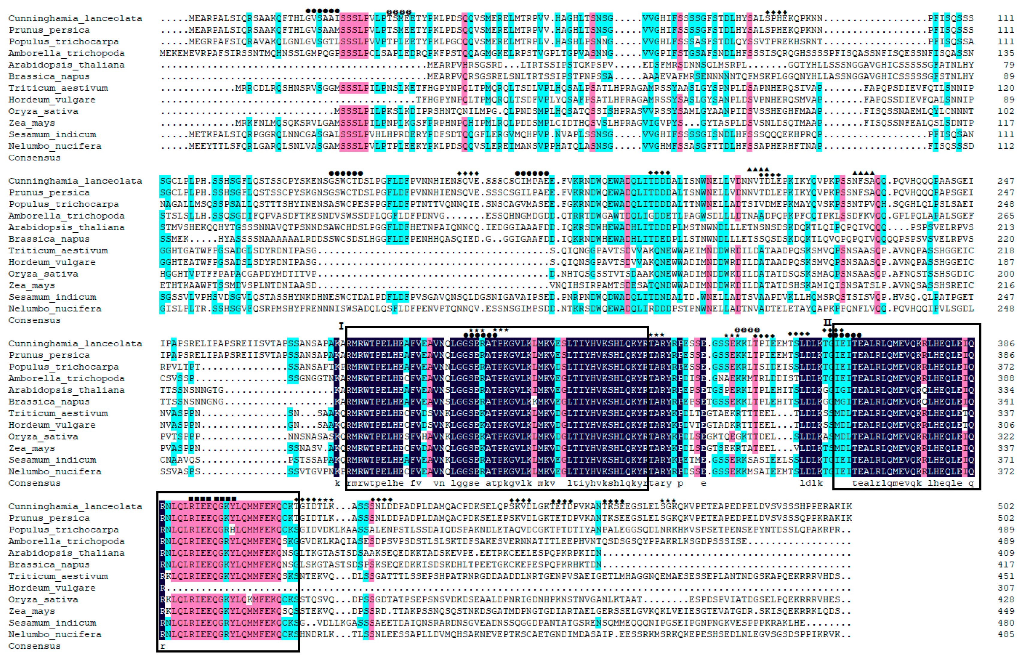
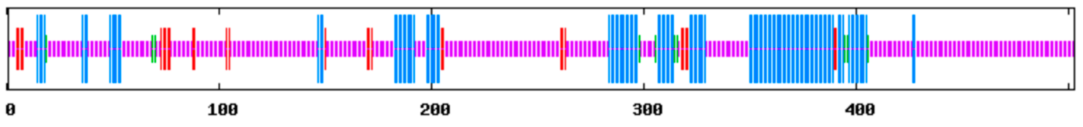
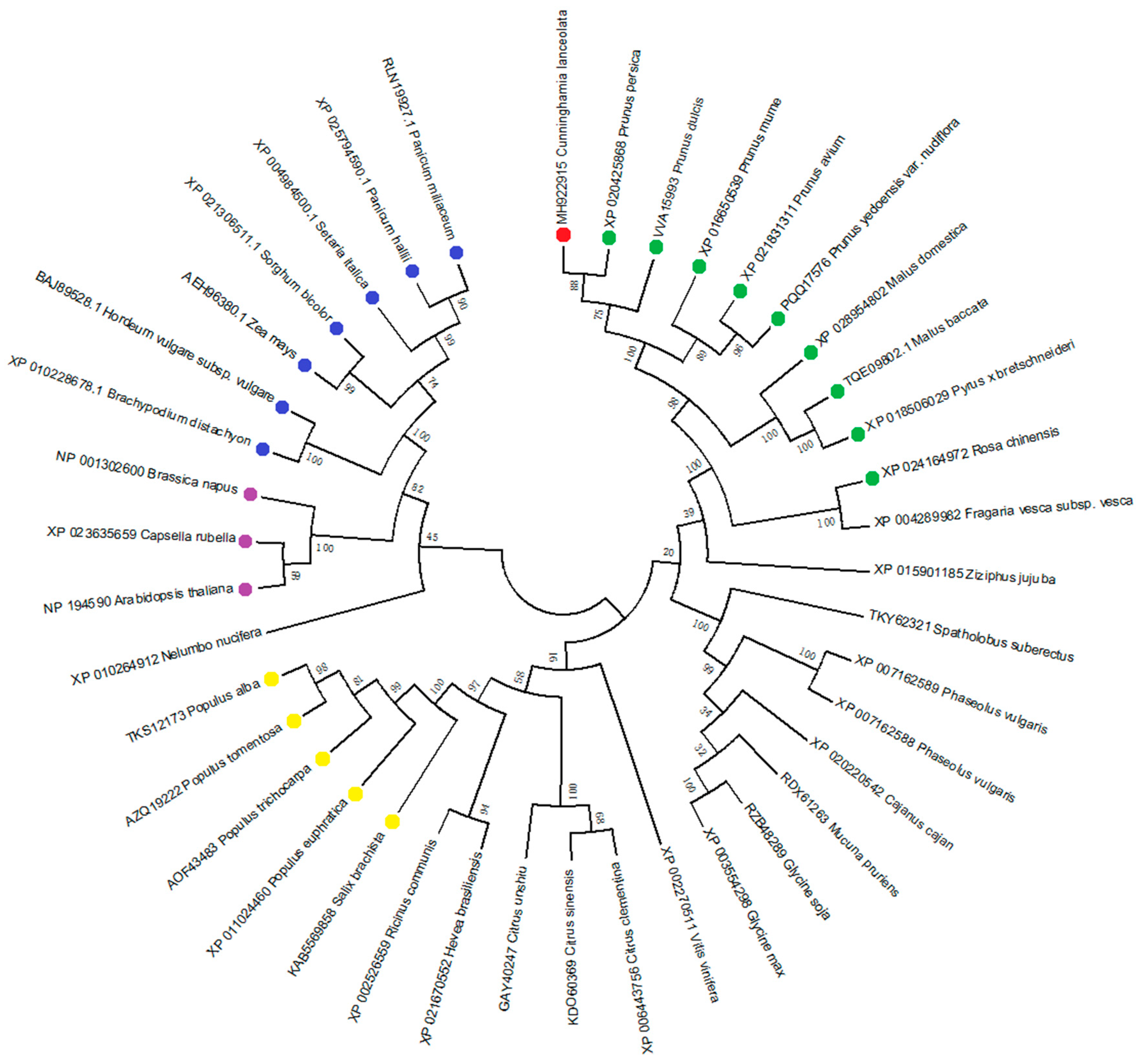
3.1. Cloning and Sequence Analysis of ClPHR1

To isolate the putative PHR1 gene from Cunninghamia lanceolate, we compared soybean, wheat, and European rape conserved sequences to design the degenerate primer, and a 314 bp intermediate conserved fragment was obtained by PCR amplification using one-year-old Chinese fir root cDNA as a template. Based on the conserved cDNA sequence of the target gene, after 5′-RACE and 3′-RACE amplification and sequencing, the partial cDNA fragments, 5′-RACE and 3′-RACE were spliced, and a complete cDNA sequence with a length of 1954 bp and the newly identified gene, ClPHR1, was designated and deposited with GenBank under the accession number MH922915.
Sequence analysis revealed an open reading frame (ORF) of 1512 bp, which encoded a predicted protein of 504 amino acids with a calculated molecular weight of 55.40 kDa and theoretical pI value of 5.37, which indicated that the number of acidic amino acid was higher than that of basic amino acids. The molecular formula of the protein was C2406H3830N672O791S18, and the instability coefficient was 64.35, which suggests that the protein is an unstable protein. The hydrophilicity of the protein encoded by ClPHR1 gene was identified as being hydrophobic as evaluated by Hphob/Kyte–Doolittle method [38]. The protein had no signal peptide and was not a secretory protein as calculated with SignalP4.1. The ClPHR1 gene had a common MYB-CC conserved domain and a conserved amino acid site in the PHR gene family. Using the software NetPhos2.0, 46 sites of Serine, 21 sites of Threonine and 4 sites of Tyrosine were detected for the phosphoprotein encoded in the ClPHR1 gene. Using TMpred server online software to predict the transmembrane structure of the protein encoded by the ClPHR1 gene, the results showed that there might be a transmembrane structure between 10–30 amino acid residues of the protein.
The amino acid sequence of ClPHR1 gene in Chinese fir was searched and compared using the BLASTP tool in NCBI, and it was found that the amino acid sequence of ClPHR1 gene in Chinese fir was consistent with that of a peach (Prunus persica), up to 99%. The ClPHR1 gene had two domains, the MYB domain and CC domain, and belonged to the MYB-CC family. BLAST analysis revealed that ClPHR1 had high identity with other reported plant PHR1 in GenBank, sharing 98.61% identity with the PHR gene in Prunus persica (XP_020425868.1), 98.01% identity with Prunus dulcis (VVA15993.1), and 96.22% identity with Prunus avium (XP_021831311.1). The PHR family protein sequences of the known species such as Arabidopsis thaliana, Oryza sativa, Zea mays, Brassica napus, Prunus persica, Populustrichocarpa, Amborellatrichopoda, Triticum aestivum, Hordeum vulgare, Sesamumindicum and Nelumbo nucifera were downloaded from NCBI and did multiple alignments with ClPHR1 (Figure 1). The protein might have six functional protein sites: N-glycosylation site, cAMP- and cGMP-dependent protein kinase phosphorylation site, protein kinase C phosphorylation site, casein kinase II phosphorylation site, tyrosine kinase phosphorylation site, and N-myristoylation site. The results also indicated that these amino acid sequences were highly conserved in both the MYB DNA-binding domain and the coiled-coil domain. The secondary structure of the ClPHR1 protein was predicted by the SOPMA tool (Figure 2). The results indicated that ClPHR1 consisted mainly of random coils (68.99%) and α-helix (23.46%), as well as a few extended strands (5.57%) and β-turns (1.99%). The conserved domains MYB (278–332 amino acids in ClPHR1) and CC (363–408 amino acids in ClPHR1) of the PHR1 family were mainly composed of α-helix. It also implied that the 68.99% random coils would play an important role in the specific functional three-dimensional structure that constituted the structure and function of ClPHR1 protein.
To explore the evolutionary relationship between ClPHR1 and PHRs from other plants, phylogenetic analysis was performed based on the neighbor-joining method and bootstrap analysis (n = 1000) using 41 PHR1 sequences, including the sequences of the PHR1 genes used in multiple alignment (Figure 3). They were separated into two main groups. The ClPHR1 from Cunninghamia lanceolate and the PHR1s from Rosaceae species were clustered together. The result grouped ClPHR1 with other PHR1s from Rosaceae species; a close relationship to PpPHR1 from Prunus Persica was also indicated; more distantly related to monocot species.

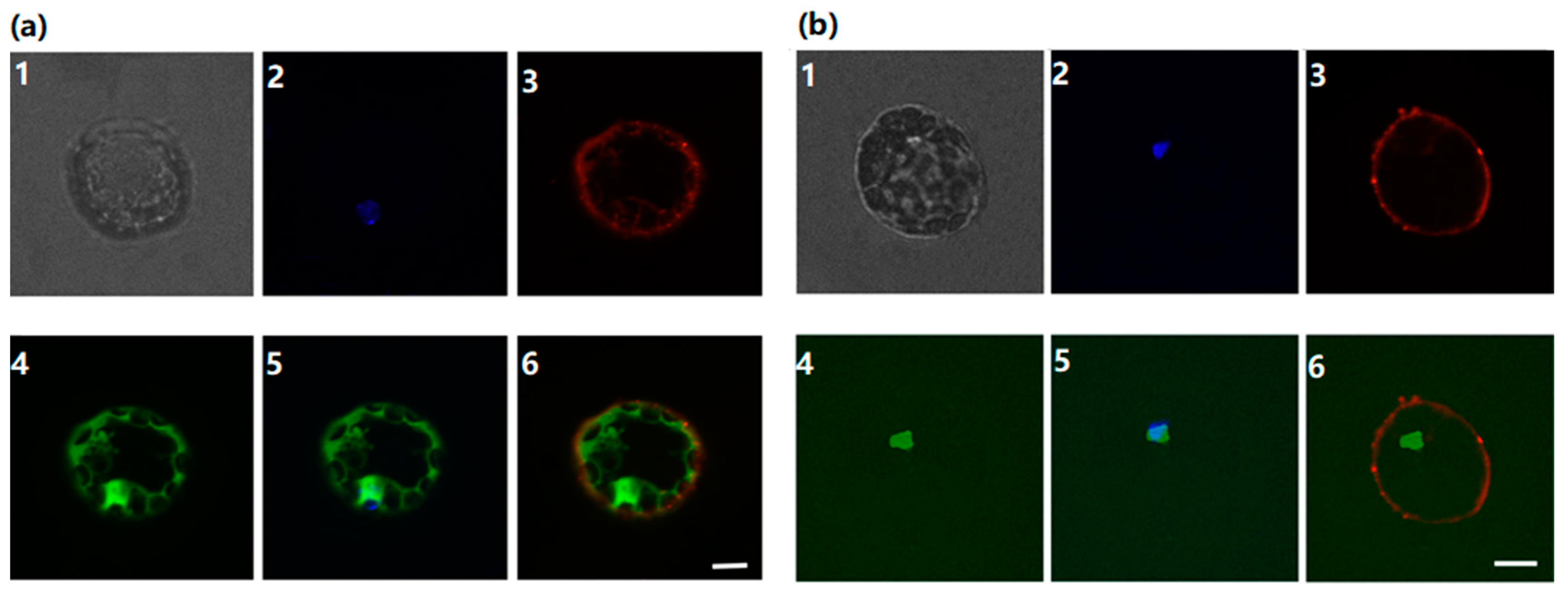
3.2. Subcellular Localization of ClPHR1 Gene

In order to determine whether ClPHR1 encodes a transcription factor protein similar to AtPHR1, the ClPHR1∷GFP fusion protein construct (Vector PGReenII-62-SK-PHR1-EGFP) was generated by fusing the ClPHR1 coding sequence to the 5′ end of the GFP gene (Figure 4). The GFP fluorescence signal was distributed throughout all of the control cells that were transformed with the GFP vector (Figure 4a). In contrast, the fluorescence signal was located predominately in the nuclei of ClPHR1∷GFP transformed cells (Figure 4b). The results demonstrated that ClPHR1 was a nuclear protein.

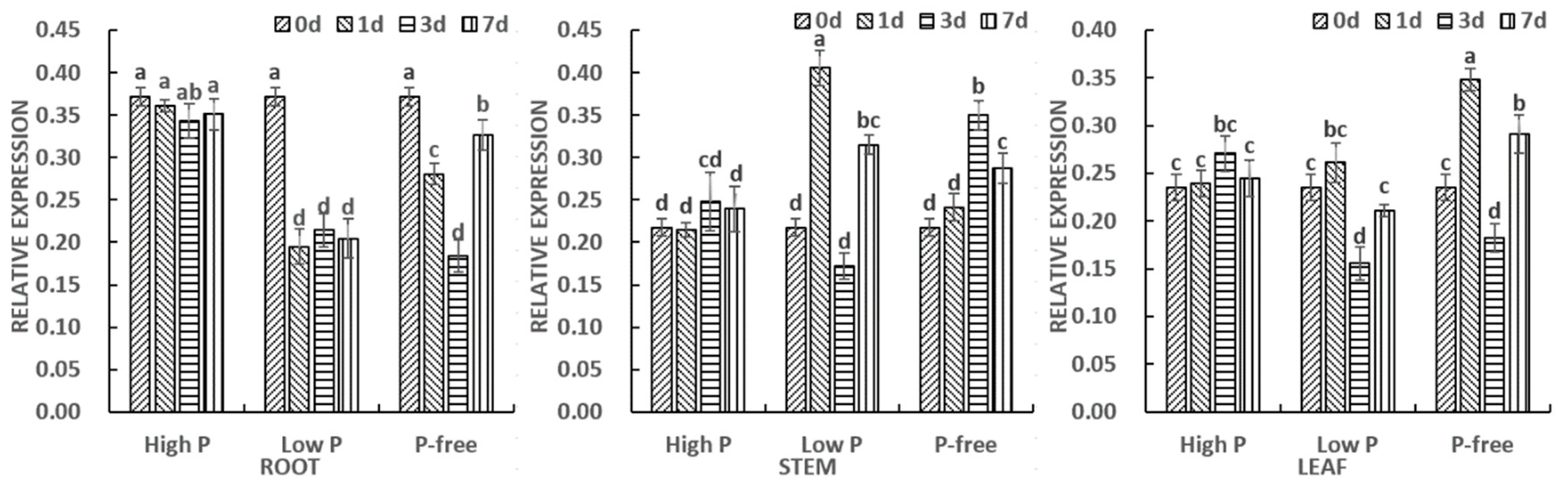
3.3. Expression Analysis of ClPHR1

To investigate the spatial and temporal expression patterns of ClPHR1 in Chinese fir under different Pi conditions, qPCR analysis was performed on different seedling tissues 0, 1, 3, 7 days after Pi treatment, respectively (Figure 5). Under different Pi conditions, ClPHR1 showed a constitutive expression pattern in all three tissues. As a whole, the expression of ClPHR1 was higher in roots, followed by leaves and the lowest being in stems. Expression levels of ClPHR1 were quantified in various tissues at different time points (0, 1, 3 and 7 days) during Pi-starvation treatment. ClPHR1 expression in roots was reduced dramatically at the beginning of low P and P starvation treatment, followed by an increase after 7 days to approximately 90 percent of the level observed at day 0 in P-starved treatment. By contrast, ClPHR1 expression in leaves increased dramatically at the beginning of P starvation treatment, and both showed a decrease at 3 days to approximately 70 percent of the level observed at day 0 in low P and P starvation treatment. And in stems, the expression level of ClPHR1 increased by approximately 1.4-fold after 7 days of Pi stress treatment.

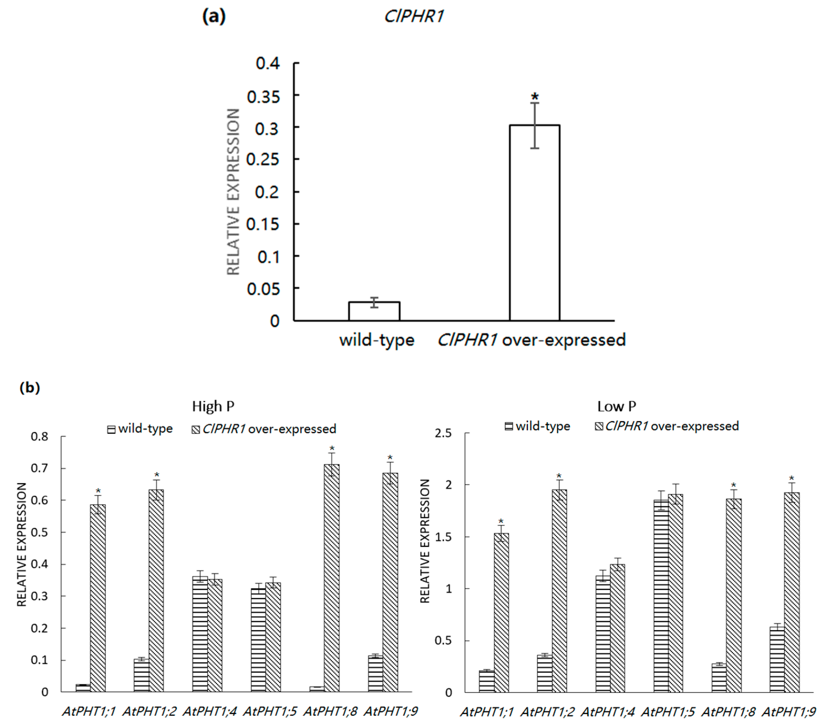
3.4. Expression of Pi Transporter in ClPHR1-Overexpressing Transgenic Plants

To confirm ClPHR1 overexpressed in ClPHR1 transgenic Arabidopsis, the expression of ClPHR1 in wild-type and transgenic Arabidopsis under high P conditions was detected by qPCR. The results showed that the overexpression of ClPHR1 in ClPHR1-overexpressor was 10.82-fold higher than that in wild-type Arabidopsis (Figure 6a). To investigate whether the Pi transport system is regulated by ClPHR1, the expression levels of six high-affinity Pi transporter genes (AtPHT1;1, AtPHT1;2, AtPHT1;4, AtPHT1;5 AtPHT1;8 and AtPHT1;9) were analyzed by qPCR in wild-type and transgenic Arabidopsis plants cultured for 7 days with low and high Pi supply (Figure 6b). The results showed that the expressions of AtPHT1;1, AtPHT1;2, AtPHT1;8 and AtPHT1;9 were significantly higher in the young roots of overexpressing plants than those of wild type plants grown in both high and low Pi supply levels, but there were no significant differences in the expression of AtPHT1;4 and AtPHT1;5 between overexpressing lines and wild-type lines in both high and low Pi supply levels. The expression of Pi transporter genes in Arabidopsis thaliana under low Pi supply was higher than that under high Pi supply, which indicated that low Pi culture induced the expression of Pi transporter genes in Arabidopsis thaliana roots to enhance the ability to absorb and transport Pi from the medium. Thus, ClPHR1 might have upregulated the expression of AtPHT1;1, AtPHT1;2, AtPHT1;8 and AtPHT1;9 in roots under low Pi supply. The lack of significant difference in expression of AtPHT1;4 and AtPHT1;5 in the roots of both genotypes implies that ClPHR1 gene might not regulate expression of these genes.

4. Discussion

Transcription factors play an important role in various stages of plant growth and development and in different metabolic pathways, especially in regulating plant adaptation to biotic and abiotic stresses. At present, the main transcription factors of Pi transport in plants are MYB family, bHLH family, and WRKY family [39,40]. PHR1 is located in the center of plant Pi signal regulatory network, and the research on the function and mechanism of PHR1 is a key to our understanding of plant Pi signal regulatory network. AtPHR1 is the first identified transcription factor involved in Arabidopsis’s low Pi response, which is not itself regulated by environmental Pi levels [24,41]. OsPHR1 and OsPHR2 were expressed in the roots, stems and leaves of rice, and the subcellular localization was in the nucleus [42]. ZmPHR1 of maize was also a transcription factor with the MYB-CC domain. The overexpression of several PSI genes in ZmPHR1 of Arabidopsis thaliana was upregulated, and the accumulation of Pi in the overexpression strain of ZmPHR1 was higher than that of the wild type under low Pi conditions [42,43].
In this study, we compared the conserved PHR1 gene sequences of known plants, analyzed the sequence similarity, designed and obtained the primers of the putative PHR1 gene from Chinese fir, and used the full-length cloning method and PCR technique. The prediction of the domain in ClPHR1 indicated that the protein has a highly conserved MYB-CC domain shared by other PHR family genes and is a member of the MYB-CC transcription factor family. Blast and multiple alignment analysis showed that the ClPHR1 protein sequence has high similarity with that of other reported PHR1 proteins, indicating that the PHR1 protein has been highly conserved during evolution. The homology of ClPHR1 gene to Prunus persica and Prunus dulcis was higher than that of other plants, which indicated a closer phylogenetic relationship. It is relatively distant evolutionary relationship with the genes of ClPHR1 in Chinese fir and PHR1 in monocots like maize and rice and Brassicaceae species like Arabidopsis and Brassica napus. All of the reported PHR1 transcription factors have MYB-CC domains and are verified to encode a single nucleoprotein, while the phylogenetic tree analysis based on the PHR1 gene revealed the evolutionary relationship between of Chinese fir and different plant species.
Expression vectors, including plasmid vectors, bacteriophage vectors and viral vectors, are often used to study the function of target genes. The proteins encoded by plant genes are targeted to specific parts of the cell by precise guidance mechanisms based on the location information contained in their amino acid sequences. The newly synthesized protein must be in the proper subcellular position to perform its function properly. Therefore, the subcellular localization of plant genes is one of the important bases for understanding the biological function of the encoded protein. The AtPHR1 gene has been reported to be localized in the Arabidopsis nucleus [29]. ZmPHR1 of maize and OsPHR1/2/3 of rice were fused with GFP, and then transformed into onion epidermal cells respectively, and the subcellular localization was confirmed in the nucleus [43]. In this experiment, PGReenII-62-SK-PHR1-EGFP was used as the final vector, the amplified primers were designed, the target gene ClPHR1 was linked with the expression vector, and the recombinant plasmid was obtained as the transient expression vector. The recombinant plasmid vector was constructed by the PEG method and the subcellular localization of ClPHR1 gene was identified based on the transient transfection technique. The fluorescence signal of EGFP-PHR1 protein transfected by PGReenII-62-SK-PHR1-EGFP was concentrated in the nucleus, that is, ClPHR1 gene is located in the nucleus and encodes a nuclear protein.
By using real-time fluorescent quantitative PCR, we designed a low-Pi stress test to analyze the temporal and spatial expression of ClPHR1 gene in Chinese fir. ClPHR1 gene was expressed in different organs of Chinese fir depending on the Pi concentrations. Among different organs of Chinese fir seedlings, the relative expression of ClPHR1 was the highest in roots, the second in leaves and the lowest in stems. A previous study has shown that the expression of PHR1 gene in leaves was highest in rice and maize, followed by roots and stems, and BnPHR1 gene in Brassica napus was highest in roots, which is the same as ClPHR1 gene in Chinese fir [44]. Studies have shown that plants growing in Pi deficient environment display an increased expression of high affinity Pi transporters in the roots to optimize Pi uptake from the soil [45] and several genes have been expressed and involved in the P starvation rescue system [46]. The expression of ClPHR1 gene in leaves and stems under low Pi supply might be related to redistribution of Pi among different organs to maintain internal Pi homeostasis, as observed in previous study [47].
An expression vector carrying the ClPHR1 gene was introduced into Arabidopsis thaliana to obtain stable inheritance and overexpress ClPHR1 gene in Arabidopsis [43,48]. High affinity phosphate transporter PHT1 family plays an important role in low phosphorus condition regulated by PHR1. The AtPHT1;1, AtPHT1;2, AtPHT1;4, AtPHT1;5, AtPHT1;8 and AtPHT1;9 genes in Arabidopsis thaliana PHT1 family may be regulated by AtPHR1 gene [9]. To assess the function and its possible regulatory role of ClPHR1 on AtPHT1 gene expression, we analyzed the transcript accumulation pattern of six ATHPT1 genes. It was found that overexpression of ClPHR1 in Arabidopsis resulted in significant accumulation of AtPHT1;1, AtPHT1;2, AtPHT1;8 and ATPHT1;9 transcripts in the root tissues, while no significant change was observed in expression of AtPHT1;4 and AtPHT1;5, which suggests no regulatory connection of ClPHR1 to the latter PHT1s. Phenotypically, both wild-type and overexpression plants were less vigorous when cultivated under low Pi supply level than those cultivated under high phosphorus condition. However, under low and high Pi supply levels, the overexpressing plants were superior to the wild-type plants in most morphological traits; notably in number of leaves (data not shown). The functional study of rice OsPHR2 gene showed that OsPHR2 upregulated the expression of OsPT9 gene, which was closely related to AtPHT1;8, AtPHT1;9 [40] while the ZmPHR1 gene regulates the expression of AtPHT1;1, AtPHT1;8 and AtPHT1;9 in maize roots [42]. Thus, it is quite plausible that in Chinese fir, we have operation of a similar regulatory module between ClPHR1 and PHT1.
As the genetic transformation system of Chinese fir has not been well established, this experiment can only overexpress the ClPHR1 gene in Arabidopsis thaliana. Although it is not clear whether the expression of transcription factor has an effect on the morphological and physiological aspects of Chinese fir, the result of the Pi response gene of Arabidopsis thaliana overexpressed by ClPHR1 suggests that the expression of transcription factor may have an effect on the morphological and physiological responses of Chinese fir to Pi starvation. Increasing the expression of ClPHR1 may be a feasible approach for Pi efficient genetic improvement of Chinese fir. In order to reveal the function of ClPHR1 transcription factor in Chinese fir, more studies on genetic transformation and overexpression analysis in Chinese fir need to be carried out in the future, and analysis of the mechanism of ClPHR1 regulating PSI genes in Chinese fir also needs further investigation.

5. Conclusions

The phosphate starvation response gene, ClPHR1, in Chinese fir was successfully obtained by rapid amplification of cDNA ends (RACE) technique. Bioinformatics analysis showed that ClPHR1 showed a high degree of homology and structural similarity with other plant PHR1 proteins that have been reported. The expression analysis of ClPHR1 gene shows that ClPHR1 was expressed in different organs of Chinese fir, with the highest expression in roots, followed by leaves and the lowest expression in stems. In the ClPHR1-overexpressing transgenic Arabidopsis thaliana line, the expressions of Pi transporter genes AtPHT1;1, AtPHT1;2, AtPHT1;8 and AtPHT1;9 were significantly higher than that in the wild type line. The overexpression of ClPHR1 may have some effects on metabolic pathways in Arabidopsis plants, as overexpression of this gene stimulated Pi-starvation signaling and downstream Pi transporter genes, which triggered the Pi starvation response. It is not clear whether ClPHR1 expression has any other direct or indirect effect on metabolism, but our results suggest that increasing ClPHR1 expression may be a viable way to improve the Pi utilization efficiency of Chinese fir.

Author Contributions

Conceptualization, X.M. and M.L.; Formal analysis, W.C. and R.C.; Funding acquisition, M.L.; Investigation, W.C., R.C., Y.Z., J.L.; Methodology, W.C., R.C., Y.Z., J.L., X.M. and M.L.; Supervision, M.L.; Validation, M.T.; Visualization, Y.Z. and W.C.; Writing—original draft, W.C.; Writing—review & editing, M.T. All authors have read and agreed to the published version of the manuscript.

Funding

This work was financially supported by the Innovation and Industrialization Project of Advanced Generations of Chinese Fir in Fujian Forestry Department (ZYCX-LY-2017004) and National Natural Science Foundation of China (U1405211).

Acknowledgments

We are grateful for Shuoshuo Su, Wenjie Gao, Pengfei Wu and Peng Shuai for valuable discussion and their assistance during the lab work.

Conflicts of Interest

The authors declare no conflicts of interest.

References

  1. Bian, L.M.; Zheng, R.H.; Su, S.D.; Lin, H.Z.; Xiao, H.; Wu, H.X.; Shi, J. Spatial analysis increases efficiency of progeny testing of Chinese fir. J. For. Res. 2017, 28, 445–452. [Google Scholar] [CrossRef] [Green Version]
  2. Zhang, X.Q.; Chhin, S.; Fu, L.Y.; Lu, L.L.; Duan, A.G.; Zhang, J.G. Climate-sensitive tree height–diameter allometry for Chinese fir in southern China. For. Int. J. For. Res. 2019, 92, 167–176. [Google Scholar] [CrossRef]
  3. Wang, C.Q.; Xue, L.; Dong, Y.H.; Hou, L.Y.; Wei, Y.H.; Jiao, R.Z. Responses of soil microbial community structure to stand densities of Chinese fir plantations. J. For. Res. 2019, 24, 162–167. [Google Scholar] [CrossRef]
  4. Ma, X.Q.; Heal, K.V.; Liu, A.Q.; Jarvis, P.G. Nutrient cycling and distribution in different-aged plantations of Chinese fir in southern China. For. Ecol. Manag. 2007, 243, 61–74. [Google Scholar] [CrossRef]
  5. Lynch, J.P. Roots of the second green revolution. Aust. J. Bot. 2007, 55, 493–512. [Google Scholar] [CrossRef]
  6. Niu, Y.F.; Chai, R.S.; Jin, G.L.; Wang, H.; Tang, C.X.; Zhang, Y.S. Responses of root architecture development to low phosphorus availability: A review. Ann. Bot. 2013, 112, 391–408. [Google Scholar] [CrossRef] [PubMed]
  7. Farooq, T.H.; Wu, W.; Tigabu, M. Growth, Biomass Production and Root Development of Chinese fir in Relation to Initial Planting Density. Forests 2019, 10, 236. [Google Scholar] [CrossRef] [Green Version]
  8. Wang, C.Q.; Xue, L.; Dong, Y.H.; Hou, L.Y.; Wei, Y.H.; Chen, J.Q.; Jiao, R.Z. The development of Chinese fir plantations undergo significant changes in soil microbial metabolic function and enzyme activities. J. For. Res. 2019, 24, 261–265. [Google Scholar] [CrossRef]
  9. Li, L.; Luke, M.M.; Chen, F.S.; Wang, H.M.; Ma, Z.Q.; Guo, D.L. Different responses of absorptive roots and arbuscular mycorrhizal fungi to fertilization provide diverse nutrient acquisition strategies in Chinese fir. For. Ecol. Manag. 2019, 433, 64–72. [Google Scholar] [CrossRef]
  10. Bumb, B.L.; Baanante, C.A. The Role of Fertilizer in Sustaining Food Security and Protecting the Environment to 2020; Food, agriculture and the environment discussion paper 17; International Food Policy Research Institute Research Institute: Washington, DC, USA, 1996. [Google Scholar]
  11. Runge-Metzger, A. Closing the Cycle: Obstacles to Efficient P Management for Improved Global Food Security. In Phosphorus in the Global Environment: Transfers, Cycles and Management; Tiessen, H., Ed.; John Wiley and Sons Ltd.: Chichester, UK, 1995; pp. 27–42. [Google Scholar]
  12. Chen, Y.H.; Nguyen, T.H.N.; Qin, J.J.; Jiao, Y.; Li, Z.L.I.; Ding, S.; Lu, Y.; Liu, Q.F.; Luo, Z.B. Phosphorus assimilation of Chinese fir from two provenances during acclimation to changing phosphorus availability. Environ. Exp. Bot. 2018, 153, 21–34. [Google Scholar] [CrossRef]
  13. Wang, Y.; Deng, L.Y.; Chen, S.Q.; Chen, Z.J.; Fan, Y.H. Study on Modification of Fast-growing Chinese Fir Stumpage. IOP Conf. Ser. Earth Environ. Sci. 2018, 108. [Google Scholar] [CrossRef]
  14. Zheng, H.Q.; Hu, D.H.; Wei, R.P.; Yan, S.; Wang, R.H. Chinese Fir Breeding in the High-Throughput Sequencing Era: Insights from SNPs. Forests 2019, 10, 681. [Google Scholar] [CrossRef] [Green Version]
  15. Plaxton, W.C.; Tran, H.T. FOCUS ISSUE ON PHOSPHORUS PLANT PHYSIOLOGY: Metabolic Adaptations of Phosphate-Starved Plants. Plant Cell 2011, 156, 1006–1015. [Google Scholar]
  16. Wang, F.; Deng, M.J.; Xu, J.M.; Zhu, X.L.; Mao, C.Z. Molecular mechanisms of phosphate transport and signaling in higher plants. Semin. Cell Dev. Biol. 2018, 74, 114–122. [Google Scholar] [CrossRef] [PubMed]
  17. Devaiah, B.N.; Karthikeyan, A.S.; Raghothama, K.G. WRKY75 transcription factor is a modulator of phosphate acquisition and root development in Arabidopsis(Article). Plant Physiol. 2007, 143, 1789–1801. [Google Scholar] [CrossRef] [PubMed] [Green Version]
  18. Rishmawi, L.; Pesch, M.; Juengst, C.; Schauss, A.; Schrader, A.; Huelskamp, M. Non-cell-autonomous regulation of root hair patterning genes by WRKY75 in Arabidopsis thaliana. Plant Physiol. 2014, 165, 186–195. [Google Scholar] [CrossRef] [Green Version]
  19. Devaiah, B.N.; Nagarajan, V.K.; Raghothama, K.G. Phosphate homeostasis and root development in Arabidopsis are synchronized by the zinc finger transcription factor ZAT6(Article). Plant Physiol. 2007, 145, 147–159. [Google Scholar] [CrossRef] [Green Version]
  20. Liu, X.; Nguyen, X.C.; Kim, K.E.; Han, H.J.; Yoo, J.; Lee, K.; Kim, M.C.; Yun, D.; Chung, W.S. Phosphorylation of the zinc finger transcriptional regulator ZAT6 by MPK6 regulates Arabidopsis seed germination under salt and osmotic stress. Biochem. Biophys. Res. Commun. 2013, 430, 1054–1059. [Google Scholar] [CrossRef]
  21. Devaiah, B.N.; Madhuvanthi, R.; Karthikeyan, A.S.; Raghothama, K.G. Phosphate Starvation Responses and Gibberellic Acid Biosynthesis Are Regulated by the MYB62 Transcription Factor in Arabidopsis. Mol. Plant 2009, 2, 43–58. [Google Scholar] [CrossRef] [Green Version]
  22. Wu, P.; Ma, L.G.; Hou, X.L.; Wang, M.Y.; Wu, Y.R.; Liu, F.Y.; Wang, D.X. Phosphate starvation triggers distinct alterations of genome expression in Arabidopsis roots and leaves. Plant Physiol. 2003, 132, 1260–1271. [Google Scholar] [CrossRef] [Green Version]
  23. Chen, Z.H.; Jenkins, G.I.; Nimmo, H.G. Identification of an F-box protein that negatively regulates P(i) starvation responses. Plant Cell Physiol. 2008, 49, 1902–1906. [Google Scholar] [CrossRef] [PubMed] [Green Version]
  24. Bournier, M.; Tissot, N.; Mari, S.; Boucherez, J.; Lacombe, E.; Briat, J.F.; Gaymard, F. Arabidopsis Ferritin 1 (AtFer1) Gene Regulation by the Phosphate Starvation Response 1 (AtPHR1) Transcription Factor Reveals a Direct Molecular Link between Iron and Phosphate Homeostasis. J. Biol. Chem. 2013, 288, 22670–22680. [Google Scholar] [CrossRef] [PubMed] [Green Version]
  25. Li, X.H.; Wang, Y.J.; Wu, B.; Kong, Y.B.; Li, W.L.; Chang, W.S.; Zhang, C.Y. GmPHR1, a Novel Homolog of the AtPHR1 Transcription Factor, Plays a Role in Plant Tolerance to Phosphate Starvation. Agric. Sci. China 2014, 13, 2584–2593. [Google Scholar] [CrossRef] [Green Version]
  26. Chiou, T.J.; Lin, S.I. Signaling Network in Sensing Phosphate Availability in Plants. Annu. Rev. Plant Biol. 2010, 62, 185–206. [Google Scholar] [CrossRef] [PubMed] [Green Version]
  27. Yang, X.; Finnegan, P.M. Regulation of phosphate starvation responses in higher plants. Ann. Bot. 2010, 105, 513–526. [Google Scholar] [CrossRef] [PubMed]
  28. Rubio, V.; Linhares, F.; Solano, R.; Martín, A.C.; Iglesias, J.; Leyva, A.; Paz-Ares, J. A conserved MYB transcription factor involved in phosphate starvation signaling both in vascular plants and in unicellular algae. Genes Dev. 2001, 15, 2122–2133. [Google Scholar] [CrossRef] [PubMed] [Green Version]
  29. Nilsson, L.; MÜller, R.; Nielsen, T.H. Increased expression of the MYB-related transcription factor, PHR1, leads to enhanced phosphate uptake in Arabidopsis thaliana(Article). Plant Cell Environ. 2007, 30, 1499–1512. [Google Scholar] [CrossRef]
  30. Bari, R.; Pant, B.D.; Stitt, M.; Scheible, W.R. PHO2, microRNA399, and PHR1 define a phosphate-signaling pathway in plants(Article). Plant Physiol. 2006, 141, 988–999. [Google Scholar] [CrossRef] [Green Version]
  31. Tesfaye, M.; Liu, J.Q.; Allan, D.L.; Vance, C.P. FOCUS ISSUE ON LEGUME BIOLOGY: Genomic and Genetic Control of Phosphate Stress in Legumes. Plant Cell 2007, 144, 594–603. [Google Scholar]
  32. Motte, H.; Beeckman, T. PHR1 Balances between Nutrition and Immunity in Plants (Short Survey). Dev. Cell 2017, 41, 5–7. [Google Scholar] [CrossRef] [Green Version]
  33. Wu, P.F.; Ma, X.Q.; Chen, Y.L.; Lin, W.J.; Huang, S.Y.; Liu, L.Q. Comparison of phosphorus use efficiency among clonal test plantations of Chinese fir. J. Fujian Agric. For. Univ. 2012, 41, 40–45. [Google Scholar]
  34. Maas, C.; Werr, W. Mechanism and optimized conditions for PEG mediated DNA transfection into plant protoplasts. Plant Cell Rep. 1989, 8, 148–151. [Google Scholar] [CrossRef]
  35. Chen, R.; Chen, W.; Tigabu, M.; Zhong, W.; Li, Y.; Ma, X.; Li, M. Screening and Evaluation of Stable Reference Genesfor Quantitative Real-Time Polymerase ChainReaction (qRT-PCR) Analysis in Chinese Fir Rootsunder Water, Phosphorus, and Nitrogen Stresses. Forests 2019, 10, 1087. [Google Scholar] [CrossRef] [Green Version]
  36. Livak, K.J.; Schmittgen, T.D. Analysis of relative gene expression data using real-time quantitative PCR and the 2-ΔΔCT method. Methods 2001, 25, 402–408. [Google Scholar] [CrossRef] [PubMed]
  37. Clough, S.J.; Bent, A.F. Floral dip: A simplified method forAgrobacterium-mediated transformation ofArabidopsis thaliana. Plant J. Cell Mol. Biol. 1998, 16, 735–743. [Google Scholar] [CrossRef] [Green Version]
  38. Kyte, J.; Doolittle, R.F. A Simple Method for Displaying the Hydropathic Character of a Protein. J. Mol. Biol. 1982, 157, 105–132. [Google Scholar] [CrossRef] [Green Version]
  39. Thomas, B.E.A. Regulation of Phosphate Starvation Response in Arabidopsis. Master’s Thesis, Texas A&M University, College Station, TX, USA, 2006. [Google Scholar]
  40. Dolores, G.A.; Odilón, O.R.J.; Lenin, Y.V.; Luis, C.T.; Luis, H.E. Adaptation to Phosphate Scarcity: Tips from Arabidopsis Roots. Trends Plant Sci. 2018, 23, 721–730. [Google Scholar]
  41. Osorio, M.B.B.; Ng, S.; Berkowitz, O.; Clercq, I.D.; Mao, C.; Shou, H.; Whelan, J.; Jost, R. SPX4 Acts on PHR1-Dependent and -Independent Regulation of Shoot Phosphorus Status in Arabidopsis. Plant Physiol. 2019, 181, 332–352. [Google Scholar] [CrossRef] [PubMed] [Green Version]
  42. Wu, P.; Wang, X.M. Role of OsPHR2 on phosphorus homeostasis and root hairs development in rice (Oryza sativa L.). Plant Signal. Behav. 2008, 3, 674–675. [Google Scholar] [CrossRef] [Green Version]
  43. Wang, X.H.; Bai, J.R.; Liu, H.M.; Sun, Y.; Shi, X.Y.; Ren, Z.Q. Overexpression of a Maize Transcription Factor ZmPHR1 Improves Shoot Inorganic Phosphate Content and Growth of Arabidopsis under Low-Phosphate Conditions. Plant Mol. Biol. Report. 2013, 31, 665–677. [Google Scholar] [CrossRef]
  44. Ren, F.; Guo, Q.Q.; Chang, L.L.; Chen, L.; Zhao, C.Z.; Zhong, H.; Li, X.B. Brassica napus PHR1 Gene Encoding a MYB-Like Protein Functions in Response to Phosphate Starvation. PLoS ONE 2012, 7, e44005. [Google Scholar] [CrossRef] [PubMed] [Green Version]
  45. Raghothama, K.G. Phosphate acquisition. Annu. Rev. Plant Physiol. Plant Mol. Biol. 1999, 50, 665–693. [Google Scholar] [CrossRef] [PubMed]
  46. Li, L.H.; Qiu, X.H.; Li, X.H.; Wang, S.P.; Lian, X.M. The expression profile of genes in rice roots under low phosphorus stress. Sci. China Ser. C Life Sci. 2009, 52, 1055–1064. [Google Scholar] [CrossRef] [PubMed]
  47. Wu, P.; Ma, X.; Tigabu, M.; Chen, W.; Liu, A.Q.; Odén, P.C. Root morphological plasticity and biomass production of two Chinese fir clones with high phosphorus efficiency under low phosphorus stress. Can. J. Res. 2011, 41, 228–234. [Google Scholar] [CrossRef]
  48. Chen, F.J.; Liu, Q.; Vogel, J.P.; Wu, J.J. Agrobacterium-Mediated Transformation of Brachypodiumdistachyon. Curr. Protoc. Plant Biol. 2019, 4, e20088. [Google Scholar] [CrossRef]
Figure 1. Amino acid sequences between ClPHR1 gene and some PHR family genes in other plants. I: the MYB domain (myb_SHAQKYF, TIGR01557); II: the CC domain (Myb_CC_LHEQLE, pfam14379); ●: N-myristoylation site; ★: Protein kinase C phosphorylation site; ◆: Casein kinase II phosphorylation site; ■: Tyrosine kinase phosphorylation site; ▲: N-glycosylation site; ◎: cAMP- and cGMP-dependent protein kinase phosphorylation site.
Forests 11 00104 g001
Figure 2. Prediction of ClPHR1 structure. Prediction of ClPHR1 secondary structure; blue: α-helix; red: extended strand; green: β-turn; purple: random coil.
Forests 11 00104 g002
Figure 3. Tree of the ClPHR1 gene. The monocot species are represented with blue dots; the dicotyledon species, Brassicaceae species are indicated with purple dots; Salicaceae species with yellow dots; Rosaceae species with green dots and Cunninghamia lanceolate with a red dot.
Forests 11 00104 g003
Figure 4. Subcellular localization analysis of the PHR1-GFP protein in Arabidopsis protoplasts. Subcellular localization was investigated in Arabidopsis protoplasts transfected by vector PGReenII-62-SK-EGFP (a) and PGReenII-62-SK-PHR1-EGFP (b) using a confocal microscope. The images of bright-field, dapi (blue), the GFP fluorescence (green) only, the Dil (red) only and combined ones are shown. The epidermal cells selected for the confocal images (2, 3, 4, 5 and 6 in (a) and (b)) are the same as the images (a-1 and b-1) taken with transmitted light, respectively. Images a-2 and b-2 are images taken with the cells after DAPI staining to nuclei, and images a-3 and b-3 with the cells after Dil staining to membranes. The green fluorescence of ClPHR1∷GFP was detected in the nucleus (b-4) and that of GFP was detected in the cytomembrane and nucleus (a-4). Images a-5 and b-5 are images merged by a-2 and a-4, b-2 and b-4, respectively. Images a-6 and b-6 are images merged by a-3 and a-4, b-3 and b-4, respectively. Scale bars indicate 10 μm.
Forests 11 00104 g004
Figure 5. Expression of ClPHR1 in different organs of the Chinese fir under different culture times and phosphorus concentrations. Chinese fir seedlings were grown in high Pi supply (High P, 1.0 mM Pi), low Pi supply (Low P, 0.5 mM Pi) and Pi starvation treatments (P-free, 0 mM Pi) for 0, 1, 3 and 7 days. Expression of ClPHR1 was detected by RT-PCR analysis. β-Actin1 was used as a reference gene. The expression of ClPHR1 was detected in roots, stems and leaves, and roots exhibited the highest levels of ClPHR1 transcripts among the three organs in High P treatment. The experiments were repeated three times on three seedlings (n = 3) in each experiment. ANOVA was conducted to determine significant differences over time and bars followed by different letter(s) are significantly different. The data are presented as mean and standard deviation.
Forests 11 00104 g005
Figure 6. Expression of ClPHR1 and phosphorus transporter genes in transgenic Arabidopsis. (a): The relative expression level of ClPHR1 in roots ofwild-type and ClPHR1-overexpressing Arabidopsis under high P (1 mM Pi) conditions; (b): The expression of phosphorus transporter genes (AtPHT1;1, AtPHT1;2, AtPHT1;4, AtPHT1;5, AtPHT1;8 and AtPHT1;9) in roots of wild-type and ClPHR1-overexpressing Arabidopsis grown in Low P (0.5 mM Pi) and High P (1 mM Pi) conditions, respectively. In both (a) and (b), AtActin was used as a reference gene. The experiments were repeated three times on three seedlings (n = 3) in each experiment. The data are presented as mean and standard deviation, and bars with asterisks (*) are significantly different between wild-type and the ClPHR1 overexpressing line by using a t-test.
Forests 11 00104 g006
Table 1. Primer sequences of the phosphorous transporters.
Gene NameArabidopsis TAIR Gene IdentifierPrimer Sequence (5′ to 3′)
Pht1;1AT5G43350F: AACGCCTCCTCAAGTTGACTAC
R: TGTTCTTGGCAACCAAAGCG
Pht1;2AT5G43370F: TTAGCGCACAACGGAAAGAC
R: ACGCATCGGTAAAGAAACCC
Pht1;4AT2G38940F: TTGGCTTGGATTTGGCATCG
R: AAAACCGCAGAGACAAAGGC
Pht1;5AT2G32830F: TGCGACGACGTTTGTTGTTC
R: ACCCAAACGCTCCAACAATC
Pht1;8AT1G20860F: TCTTGGTGGCGTTTGCATTG
R: TTGCCCGTCAACAATCTGTG
Pht1;9AT1G76430F: AGTTTGGTGCGATTGTTGGC
R: ACGCGATTCTCACACGTTTC

