Article

Chemical Components of Fungus Comb from Indo-Malayan Termite Macrotermes gilvus Hagen Mound and Its Bioactivity against Wood-Staining Fungi

by
Dodi Nandika
1,*,
Lina Karlinasari
1,
Arinana Arinana
1,
Irmanida Batubara
2,
Putri Sari Sitanggang
1,
Djoko Santoso
3,
Lucia Dhiantika Witasari
4,
Yanti Rachmayanti
5,
Dikhi Firmansyah
5,
I Ketut Sudiana
3 and
Desca Medika Hertanto
3
1
Department of Forest Products, Faculty of Forestry and Environment, Darmaga Campus, IPB University, Bogor 16680, Indonesia
2
Department of Chemistry, Faculty of Mathematics and Natural Sciences, and Tropical Biopharmaca Research Center, Darmaga Campus, IPB University, Bogor 16680, Indonesia
3
Faculty of Medicine, Campus A Universitas Airlangga, Surabaya 60132, Indonesia
4
Faculty of Agricultural Technology, Bulaksumur Campus, Gadjah Mada University, Yogyakarta 55281, Indonesia
5
Division of Biochemistry, Institut Teknologi Bandung, Jl. Ganesha no. 10, Bandung 40132, Indonesia
*
Author to whom correspondence should be addressed.
Submission received: 13 October 2021 / Revised: 11 November 2021 / Accepted: 15 November 2021 / Published: 18 November 2021
(This article belongs to the Special Issue Evaluation and Protection of Wood and Wood Products)

Abstract

:
Recently, the architectural and physical properties of the fungus comb from subterranean termite Macrotermes gilvus Hagen (Isoptera: Termitidae) mounds had been studied and it is important to determine its chemical profile as well as to evaluate its anti-staining-fungi activity. The results showed that fungus comb of M. gilvus has a high crude ash (30.57%), fiber (25.46%), starch (7.76%), protein (5.80%, 5.53% amino acid), acid-insoluble ash (3.45%), and fat (0.73%). It also contained phenol hydroquinone, steroids, terpenoids, and saponin compounds. Seventeen amino acids were identified via high-performance liquid chromatography analysis, of which arginine, leucine, glutamate, and aspartic acid were the majority. According to gas chromatography-mass spectrometry analysis, the n-hexane extract consists of several types of fatty acid derivatives. Meanwhile, the ethyl acetate (EtOAc) extracts were primarily phenol groups with 1,2,3-propanetriol (glycerol) at the highest relative concentration. Four fungus-comb extracts (n-hexane, EtOAc, MeOH, and water) inhibited the Aspergillus foetidus fungus, with inhibition rates ranging from 24.17% to 100% and EtOAc extract as the most active extract. It appears that EtOAc extracts from the M. gilvus fungus comb can be considered an active ingredient source of novel organic fungicide in preventing wood-staining fungi attacks on susceptible wood.

1. Introduction

Indonesia is a tropical country that offers a suitable environment for a huge number of termite species. Of the 3106 species of termite that have been recorded worldwide, 300 species (11.5% of them) were discovered in this country [1,2]. In this circumstance, the Indo-Malayan termite Macrotermes gilvus Hagen (Isoptera: Termitidae) has the most extensive geographical distribution out of the termite species in the country [3]. The existence of this termite species has been reported in almost all parts of the country, including all districts of Jakarta, the capital city of Indonesia [2,4]. Even within the South Jakarta landscape, the existence of this species was more dominant than other termite species. A colony of this termite species lives in nest systems in the form of a mound, with a complex network of tunnels inside. This serves to protect the colony from the influence of extreme external environmental conditions [5]. Subterranean termites of the subfamily Macrotermitinae, particularly Macrotermes gilvus Hagen (Isoptera: Termitidae), have the most extensive geographical distribution in Indonesia [3]. These termites create fungus combs or fungus gardens inside their nest as a food source for the colony members [6]. Therefore, this termite, as well as the other members of the subfamily Macrotermitinae, is known as a fungus-cultivating termite.
The fungus comb is a special structure created by termite colonies from the sub-family Macrotermitinae (Isoptera: Termitidae) in their nests as a substrate for the growth of the fungus Termitomyces sp. [7]. This special structure is in the form of a mammalian brain with a volume of 44.17 ± 7.36 cm and has interconnected burrows from the surface to the bottom [5]. Architecturally, the fungus comb consists of two structural parts: the fresh comb at the top and the old comb at the bottom. The density of old combs is higher compared to the fresh comb, which functions as a strong foundation for fungus-comb structure [5]. Singh et al. [8] reported that the structure of a fungus comb, which is ventilated and connected to the nest wall by particular tunnels, allows the exchange of CO2 with the surrounding atmosphere, therefore maintaining a stable temperature inside the fungus comb throughout the year. Fungus comb has abundant clusters of nodules, small (about 1 mm in diameter) white spherules of Termitomyces species [7]. Fungal mycelium grows in these fungus combs and produces Termytomyces nodules as a food source for the Macrotermitinae colonies. The nodules are the asexual phase composed of a hyphae collection with short cells, conidium, and sphaerocyst [9]. When the fungus comb fails to be created by the termite colony, they eventually lack food and are unable to survive. However, Termitomyces fungus does not always produce fruiting bodies, as termites consume the fungi in their nodule form [10].
The termite colony creates conditions in which there is only one genus of fungus, i.e., Termitomyces, that can grow in the fungus comb [11,12], including in the fungus comb of Macrotermes sp., Odontotermes sp., and Ancistrotermes sp. [13] In addition, Batra and Batra [14] reveal that termite saliva has antifungal activity that inhibits the growth of other fungus genera, except Termytomyces. Moreover, Thomas [15] reported that the micro climate inside the termite mound was steadily maintained, including a temperature around 30 °C, which is very suitable for the growth of Termytomyces. However, until today, there has been no comprehensive scientific information available regarding the mechanism of how Termitomyces sp. became the only species that grows in fungus combs. Thus, the authors predict that chemical properties of the fungus comb are also responsible for stimulating the growth of Termitomyces fungus species and suppressing others. Based on this proposition, M. gilvus mounds can be potentially considered as a source of particular active compounds for an environmentally friendly organic fungicide. With such curiosity, the chemical properties of fungus combs and their potential as an active source of antifungal ingredients are or great research interest. Recently, the authors studied the architectural and physical properties of fungus combs from Macrotermes gilvus mounds to understand their shape, volume, structure, the dimension of their burrows, texture, color, and density, as well as the existence of the Termitomyces nodules [5]. In addition, Anwar et al. [16] successfully demonstrated fungus comb establishment under in vitro conditions using Termitomyces cylindricus (Basidiomycota: Agaricales) basidiospores. However, the bioactivity of fungus-comb extract from Indo-Malayan termite M. gilvus mounds as an antifungal has not been reported. Therefore, this research will be a pioneering study to determine the chemical composition of fungus comb from Indo-Malayan termite M. gilvus mounds and will hopefully offer considerable contributions to the development of environmentally friendly organic fungicides, which are needed to prevent wood-staining fungi attacks on various wood species used in Indonesian wood industries.
Stain fungi are filamentous fungi that cause blue, greyish, green, and black discoloration on wood surfaces. Colonization of the fungi initiates a mineralization process occurs on the wood surface, leading to discoloration. The is discoloration influenced by melanin crystallization around hyphae or from secretion of extracellular material [17]. Salman et al. [18] reported that Aspergillus foetidus caused the worst discoloration on air-dried rubberwood, which is processed for furniture products in West Java Province, Indonesia. The growth rate of fungus on the wood surface reached 90.5%. Meanwhile, Oldertrøen et al. [19] found three stain-fungi species on rubberwood: Aspergillus niger, Aspergillus flavus, and Penicillium citrinum.
According to George [20] wood-staining fungi can seriously attack rubberwood within one day of felling. As a result of fungi attacks, rubberwood experiences discoloration, causing a significant decrease in its aesthetic value. An example of stain fungi is Botryodiplodia theobromae, which occurs together with the surface molds Aspergillus sp. and Penicillium spp., which also cause considerable loss in aesthetic value of the attacked wood. The susceptibility of rubberwood to stain fungi and mold colonization on its surface is considered a major concern for furniture and wooden-toy manufacturers. The tropical climate in Indonesia, characterized by temperatures above 25 °C and humidity of 80–100%, imposes challenging conditions for the protection of rubberwood from stain fungi attack. Such environmental conditions are perfect for the growth of stain fungi and therefore facilitate the colonization of stain fungi [21,22]. Particularly in the rainy season, rainwater containing nutrients wets the wood, thus promoting biodeterioration activities of fungi [23]. In 2018, economic loss due to stain fungi attacks on rubberwood raw material in Indonesian furniture industry alone reached almost USD 15,246 million [18]. In fact, this economic loss is higher than the value since others light-color wood species used in the furniture industry were also susceptible to stain fungi. Even still, it is expected that this value will increase in the future if there are no sufficient control techniques developed.
The prevention wood-staining fungi attacks is critical, considering that 85% of the log production for Indonesian wood industries (47.9 million m3/year) comes from plantation forests [24]. Unfortunately, most of the wood from the plantation forests is highly susceptible to biodeterioration, including susceptibility to wood-staining fungi attacks [25]. Such attacks contribute to the discoloration of the wood surface, i.e., changing it to blue, grayish, green, or black, considerably lessening its aesthetic value [18]. Valiante et al. [26] argued that discoloration is caused by pigmentation on the hyphae or melanin synthesis on the conidia during the sporulation process.
One of the most widely found wood-staining fungi that attacks wooden raw materials used in the Indonesian wood industry, including rubberwood (Hevea brasiliensis Muell. Arg.), is Aspergillus spp. [18]. This situation requires multiple approaches, including the exploration of organic fungicide that is safe for humans and the environment, the active ingredients of which are originally produced from indigenous natural resources.

2. Materials and Methods

2.1. Termite-Mound Survey and Fungus-Comb Collection

The M. gilvus mounds in the Yanlappa Experimental Forest, Bogor, West Java Province were searched for in five continuous observation plots measuring 150 m × 250 m in a rectangular shape, leading north-south between the coordinates of 6°25′3.05″ S–106°29′59.36″ E and 6°25′21.00″ S–106°29′46.20″ E. Contour and boundary maps were needed as a reference for determining the zero point in plot making. When a termite mound was found in the observation plot, its location was immediately recorded using the Garmin® eTrex 10 global positioning system. Fungus combs were randomly taken from six of eighteen of the mounds that were found in the observation plot (as shown in Figure 1). From each mound, nine fungus combs were taken, then wrapped in aluminum foil separately and placed in a sterilized cooler box. From each of these mounds, five specimens of the termite soldier caste were also taken and placed in a collection bottle containing 80% alcohol for identification purposes.

2.2. Termite-Species Identification

Specimens of the termite soldier caste from each M. gilvus mound were identified based on key identification characteristics outlined by Muzaffer [27] and Tho [28] using a Leica® M205 C microscope with a 40× magnification. The size of the body, mandible length of the head, and the whole antennae of the termites were measured with the help of ImageJ® (open access) software.

2.3. Analysis of the Chemical Content of the Fungus Combs

Several assessments were conducted to determine the chemical content of the fungus comb. First was a proximate analysis, continuing by determining the amino-acid content, extractive-substance contents, and determining the chemical content of selected extracts. Proximate analysis was performed using the standard methods outlined by some Indonesian national standards (SNI) to determine crude fat (SNI 2891-1992 point 8.1), total ash (SNI 01-3709-1995 point 6.3), acid-insoluble ash (SNI 01-3709-1995 point 6.4), crude fiber (SNI 2891-1992), and carbohydrate (SNI 2891-1992 point 9.5) levels of the fungus comb, while Kjeldahl’s methods were used to determine crude protein content [29]. The amino-acid content was determined using high-performance liquid chromatography (HPLC). In addition, phytochemical analysis was carried out, including tests for alkaloids, flavonoids, phenol hydroquinone, steroids, triterpenoids, tannins, and saponins according to the procedure outlined by Harborne [30].

2.3.1. Detection of Alkaloids

Approximately 1 g of the fungus-comb sample was added to 10 mL of chloroform and a few drops of ammonia. The chloroform fraction was separated and acidified with 10 drops of concentrated H2SO4. The acid fraction was divided into three parts, to which Dragendorf, Meyer, and Wagner reagents were added, respectively. The presence of alkaloids was indicated with the formation of a white precipitate by the Meyer reagent, a red precipitate by the Dragendorf reagent, and a brown precipitate by the Wagner’s reagent.

2.3.2. Detection of Phenol Hydroquinone

Approximately 1 g of the fungus-comb sample was extracted with 20 mL of 70% ethanol. Then, 2 drops of 5% FeCl3 were added to 1 mL of the extract. The formation of a green or blue-green color indicated the presence of phenolic compounds in the sample.

2.3.3. Detection of Steroids and Terpenoids

An total of 1 g of the fungus-comb sample was dissolved in 25 mL of hot ethanol (50 °C), then filtered into a porcelain dish and evaporated until dry. The residue was dissolved with ether and transferred into a test tube, and then 3 drops of anhydrous acetic acid and 1 drop of concentrated H2SO4 were added (Lieberman Burchard Test). Red or purple colors indicate the presence of terpenoids, while green or blue colors indicate the presence of steroids.

2.3.4. Tannin Detection

Approximately 1 g of the fungus-comb sample was added to 10 mL of distilled water and then boiled for 5 min. After cooling, 5 mL of 1% (w/v) FeCl3 was added to the filtrate. If the color changed to dark blue, it meant that the sample contained tannins.

2.3.5. Detection of Saponins

Approximately 1 g of the fungus-comb sample was put into a beaker, and 10 mL of distilled water was added. Then, it was boiled for 5 min, filtered, and the filtrate was used for testing. The saponin test was carried out by shaking 10 mL of the filtrate in a closed tube for 10 min. The appearance of foam for an interval of 10 min, i.e., stable foam, indicated the presence of saponins.

2.3.6. Solvent Extraction

Analysis was continued on the fungus-comb extractive substances in terms of the different polarity of the solvents. The extraction was performed for 1 g of fungus comb with 10 mL of solvent. The fungus-comb sample was first extracted with n-hexane to obtain the nonpolar extractive substances. The extraction was continued with the residue of the n-hexane extraction using ethyl acetate in order to obtain the semipolar extractive substances, i.e., ethyl-acetate extract. The extraction was continued again using methanol to obtain the methanol extract, followed by distilled water to obtain the water extract. Both the methanol and water extracts contained polar-extractive substances. The extraction yield was calculated by the difference between the initial and final dry weight after extraction, divided by the initial again, and expressed as a percentage. Each step was performed five times.

2.3.7. Extract Composition

The chemical compositions of the n-hexane and ethyl-acetate extracts were determined using a GC-MS® Agilent Technologies 6890N series with a helium (He) carrier gas. A total of 6 µL of the extract was placed into the GC-MS inlet. The compound separation and quantitative analysis via GC-MS used a capillary column with a diameter of 0.25 mm and a length of 60 m. An initial temperature of 70 °C was applied, with an increase of 15 °C/min until 290 °C was reached, with a final time of 20 min. The spectrum data on each peak were compared with the data in the WILEY 9th library.

2.4. Bioactivity Test of the Fungus-Comb Extracts

Measurement of the bioactivity of the fungus-comb extracts on wood-staining fungus Aspergillus foetidus (IPBCC 19 1482) was conducted on potato dextrose agar (PDA) media according to the procedure outlined by Liu et al. [31]. The ethyl acetate, methanol, and distilled-water extracts of the fungus comb were dissolved into 5 mL of 5% dimethyl sulfoxide (DMSO, Sigma Aldrich, St. Louis, MO) solvent to get concentrations of 2%, 4%, and 6% (w/v), while for the n-hexane extract were prepared to get concentrations of 0.3%, 0.4%, and 0.5% due to the lower solubility of this extract in the medium. Metilen bis-thyocyanate (MBT) as a wood preservative formulation [32] was dissolved in distilled water to get concentrations of 0.02%, 0.03%, 0.6%, 0.13%, 0.25%, 0.50%, 1%, and 2%, respectively, as positive control solutions. Each extract solution, as well as the positive control solutions, was then passed via syringe through a 0.45 µm Microsolve filter to get sterile extract, which was poured into a glass vial.
The media were sterilized in an autoclave at a temperature of 120 °C for 15 min. Sterile PDA medium (20 mL) was added with 5 mL of sterile extract and positive control separately in a petri dish (9 cm in diameter). Aspergillus foetidus fungus mycelia with a diameter of 0.6 cm were introduced into the center of the media surface in the petri dish. The fungus mycelia were also grown on non-extract PDA as a negative control. The inoculated media were incubated at room temperature (28 °C ± 2 °C) for 7 d. Each test had five replications. The inhibition rate of each extract solution, as well as the control solution, on the growth of A. foetidus fungus was calculated using Equation (1),
G r o w t h   I n h i b i t i o n   ( % ) = D 1 D 2 D 1 × 100
where Growth Inhibition is the inhibition of the extract solution on the fungus radial growth, D1 is the fungus-colony diameter in the negative control petri dish (cm), and D2 is the fungus-colony diameter in the extract-treated petri dish (cm).

2.5. Data Analysis

The data of chemical content of the fungus comb were reported as the means value. The data on bioactivity of fungus-comb extracts on A. foetidus were analyzed by two-way analysis of variance (ANOVA) at a 5% level of significance using IBM SPSS Statistics 22 software, continued with Duncan Test for further analysis.

3. Results and Discussion

3.1. Termite Species

The identification of the termite specimens collected from the Yanlappa Experimental Forest was conducted according to the procedures outlined by Muzaffer [27] and Tho [28], revealing that all of the termite specimens were Macrotermes gilvus Hagen (Isoptera: Termitidae). There are two types of soldier castes, i.e., major and minor (dimorphism). The morphological characteristics included mandibles extending forward with a length of 1.69 mm ± 0.06 mm in the major and 1.22 mm ± 0.07 mm in the minor soldiers that were slightly symmetrical. The body length of the major soldier termites, including the mandible, was 10.31 mm ± 0.58 mm. Meanwhile, the body length of the minor soldiers, including the mandibles, was 6.59 mm ± 0.60 mm. The head of the soldier was red-brown with a few hairs on its surface and 17 antenna segments. The average length of the head of a major and minor soldier was 4.63 mm ± 0.26 mm and 2.85 mm ± 0.23 mm, respectively.

3.2. General Analytical Results

The analytical figure of the fungus combs from the M. gilvus mounds is shown in Table 1. The fungus comb had a high moisture content (25.94%). The high moisture content of the fungus comb was related to the termite mound’s geographical position, i.e., at an altitude of 200 m to 300 m above sea level with air relative humidity range of 75% to 90% and an average rainfall of 3282 mm/year [5,33]. In other words, the fungus comb was collected from a very humid area; therefore, it has a high moisture content.
The data also show that the fungus comb has a high crude-fiber content (25.46%), which represents a high level of C-organic in the fungus comb. However, the crude protein content, which represents the N content, was only 5.80%. This is related to a mixture of changed plant material and fungus nodules, as well as soil particles contained in the fungus comb. This situation provides optimal medium for Termitomyces growth (Otani et al.) [34]. In line with that statement, Grasse [35] and Johnson [36] revealed that not all fungus combs are built with the same materials. The typical forest Macrotermes, such as M. muelleri and M. ivorensis, exclusively use discs of dead or green leaves, whereas the savanna Macrotermes, such as M. bellicosus and M. subhyalinus, collect dry wood or grass stalks. In Pseudacanthotermes, Odontotermes, Acanthotermes, Ancistrotermes and Microtermes, the fragments collected are derived from all parts of the plant i.e., leaves, stalks, and roots. While carbohydrates (7.76%) and proteins (5.8%) were identified as a relatively major part of the fungus comb, acid-insoluble ash (3.45%) and crude fat (0.73%) were found in limited amounts. Different results can be seen in the moisture and protein values extracted from the fungus comb Odontotermes formosus. The moisture value obtained was lower, at 25.94%, while the previous research showed 41.37%. However, this fungus comb has shown a higher protein value, at 5.80%, while in the previous study, the protein value was 2.66% [37]. Moreover, it should be noted that Termytomyces fungus is known to be rich in nitrogen, which is needed by termites as a source of nutrients to stay alive and to reproduce [38]. A fairly high nitrogen value also described the conditions of termite nests that were close to plant-vegetation cover and piles of organic matter [39]. The presence of fungi in the fungus comb increased the C-to-N ratio and allowed the termites to explore a more diverse source of cellulose [40]. The high C-to-N ratio results in the fungus comb taking a longer time to decompose. It should be realized that the given figure may vary within the colony, depending on the different stages of maturity.

3.3. Secondary Metabolite Content

The result show that the fungus combs from M. gilvus mounds contained phenol hydroquinone, steroids, terpenoids, and saponins. However, alkaloids, flavonoids, and tannins were not detected. The steroid, terpenoids, and phenol hydroquinone could be dissolved in semipolar solvents, e.g., ethyl acetate, as well as be dissolved in nonpolar solvents, e.g., n-hexane.

3.4. Amino Acids

The analysis using HPLC determined that the fungus combs from the M. gilvus termite mounds contained 17 amino acids, with a total content of 6.74% (w/w). The total amino-acid content was higher than the protein content, according to the proximate analysis. The different results occurred due to the different analysis methods, since the HPLC analysis results are more accurate than the Kjeldahl analysis. The most dominant types of amino acids were arginine (1.55%), leucine (0.64%), glutamate (0.44%), and aspartic acid (0.35%) (Table 2). Our results were different from previous a report using fungus combs of Odontotermes formosanus, which are mainly dominated by aspartic acid or glutamic acid. The fungus comb consists of aspartic acid (0.21%) as the main amino acid, followed by glutamic acid (0.19%), serine (0.10%), threonine (0.06%), and alanine (0.09%) [37]. Meanwhile Chiu et al. [41] reported that fungus comb from the same termite species consists of glutamic acid and aspartic acid as the amino acid, followed by arginine and other amino acids. The difference could be caused by different species of termite, different species of fungus, and also the environmental conditions.
The high arginine content in the fungus combs could be related to many biological functions. For example, arginine-rich cyclic cationic peptide has been commercialized for antibacterial and antifungal use [42]. Like the essential amino acids, arginine is involved in protein synthesis, ureagenesis, immune function, antioxidant activity, stress responses, and ammonia detoxification in fish [43]. Arginine as a supplement is reported to prevent hypertension and atherosclerosis evolution related to cardiovascular disorders [44]. Interestingly, L-Arginine from the marine bacterium Pseudoalteromonas flavipulchra is also predicted to be an antiaging compound [45]. As with arginine, the leucine content in the fungus combs could be related to antiaging since this amino acid could modulate mitochondrial dysfunction [46]. Supplementing leucine for adults and adolescents could improve muscle strength and volume by reducing inflammation [47]. These results could provide more opportunity to explore wider bioprospective potency of fungus comb.

3.5. Extractives Content

Based on the detected extractive substance, it was revealed that polar substances were more dominant in the fungus combs, as shown by the extraction yield of the distilled water (4.61%) and methanol (2.53%) extracts (as shown in Table 3). The minor content was found in the n-hexane extract, followed by in the ethyl-acetate extract. This means that the nonpolar and semipolar substances in the fungus comb were limited. The nonpolar compounds (n-hexane extract) are mostly lipids, while there were lipids and some semipolar compounds in the ethyl-acetate extract.
GC-MS analysis was performed on the n-hexane and ethyl-acetate extracts to obtain information related to the nonpolar and semipolar compounds. The polar extracts, such as in methanol and distilled-water extracts could not be separated and identified using GC-MS due to the difficulties of vaporizing polar compounds. In the n-hexane extract, an extract containing nonpolar compounds, 18 types of compounds were detected, 6 of which were the dominant compounds (as shown in Table 4). The compound that had the highest relative concentration was Bis (2-ethylhexyl) phthalate, or DEHP (69.43%), which is an ester of phthalic acid that has been used as a plasticizer in many materials, including in PVC, paints, adhesives, cosmetics, and packaging, among others [48,49]. These compounds easily migrate to the environment; therefore, they are present in any environment, e.g., in plants, soils, water, and sediments, and are classified as pollutants [49,50]. However, recently, it has been found that the DEHP compound is synthesized by many organisms e.g., plants, bacteria, or fungi, and several studies have demonstrated different biological activities for this compound [51]. For example, DEHP isolated from marine fungus Cladosporium sp. F14 is reported to display antibacterial activity [52,53].
In addition to DEHP, there were types of fatty-acid derivatives in the fungus-comb n-hexane extract, i.e., methyl palmitate, benzenepropanoic acid 3,5-bis (1,1 dimethylethyl) -4-hydroxy-, methyl ester, methyl linolelaidate, and methyl oleate. Methyl palmitate is reported to display nematicidal activity by inhibiting egg hatching and reducing egg masses, as well as herbicidal activity [13,54]. Benzenepropanoic acid, 3,5-bis (1,1 dimethylethyl)-4-hydroxy-methyl ester is reported as an antifungal agent as well as a natural antioxidant [55]. The other compounds, i.e., methyl linolelaidate and methyl oleate, have reported use as an emollient or skin conditioner [56].
The results of the GC-MS analysis showed that the ethyl acetate fraction of the fungus comb contained 14 types of compounds, four of which were the dominant compounds (as shown in Table 5). The compounds found in the ethyl-acetate extract of the fungus comb generally belonged to the phenol group. The compound 1,2,3-Propanetriol (glycerol) was the compound with the highest estimated relative concentration. Traditionally, glycerol is used in personal care products, e.g., soap and cosmetics, as well as in pharmaceuticals and food and feed products [57]. Glycerol is widely used for many applications since it plays a role as a humectant [58].
Phenol, 2-methoxy- and Phenol, 2,6-dimethoxy-, isolated from Pinus densilora and Quercus serrata extracts, are phenolic compounds that display antifungal activity [60]. Oramahi et al. [60] supported the statement that the two compounds isolated from liquid smoke of acacia wood (Acacia mangium) dust and Laban wood (Vitex pubescens) had antifungal activity. These compounds were thought to play an important role in antifungal activity against wood-rot fungi.

3.6. Bioactivity of Fungus-Comb Extract against Wood-Staining Fungi

The results show that the inhibition rate of the ethyl acetate, methanol, and water extracts on A. foetidus fungus growth ranged from 46.48% to 52.78%, 44.44% to 100%, 30.37% to 37.96%, and 24.17% to 57.50%, respectively. However, the negative control solution (0%, without any extracts) did not inhibit the growth of the fungus (as shown in Figure 2). In addition, the inhibition rate of the n-hexane extracts on fungus growth ranged from 46.48% to 52.78% (as shown in Figure 3).
It was revealed that the ethyl acetate, methanol, and water extracts at concentrations of 2%, 4%, and 6%, as well as the n-hexane extracts at concentration of 0.3%, 0.4%, and 0.5%, showed bioactivity in terms of inhibiting the growth of A. foetidus. Analysis of variance (ANOVA) followed by a Duncan’s test showed that the inhibition rates of any extracts tested on the growth of A. foetidus were mostly significantly different. However, the inhibition rate of the methanol extracts and water extracts did not show a significant difference (p-value less than or equal to 0.05). This was predicted to be due to the similarity of the metabolite compounds contained in both extracts. However, the inhibition rate caused by both extracts was lower than the that of ethyl-acetate extract at the same concentration. In other words, at the same concentration, the ethyl-acetate extracts had the highest inhibition rate on the growth of A. foetidus fungus compared to the other extracts. In addition, the ethyl-acetate extract with a 6% concentration, as well as the n-hexane extract with 0.5% concentration, showed the highest inhibition rates on the growth of A. foetidus fungus. Visually, it was also observed that the diameter of the A. foetidus colony on the PDA media treated by 6% ethyl-acetate extract, as well as by 0.5% n-hexane extract, after 7 d of incubation were the smallest compared to the others (as shown in Figure 4).
This study also revealed that the inhibition growth of the fungus-comb extract on A. foetidus was affected by the concentration of the extract solution added to the PDA media. The higher the concentration of the fungus-comb extract solution, the higher the inhibition growth of the extracts. This was due to the higher secondary metabolites contained in the media. According to Sitepu et al. [61], the greater the amount of extract in the test medium, the more the extracts will diffuse into the fungal cells, causing disruption of fungal growth. It was revealed that the 6% ethyl-acetate extract showed the highest bioactivity in terms of inhibiting the growth of A. foetidus fungus (the inhibiting growth reached 100%). In addition, its bioactivity was similar to 0.5% methylene bis-thiocyanate (MBT), used as a positive control solution (as shown in Figure 5). It is widely known that MBT is an active ingredient in synthetic wood preservatives commercially used for preventing wood-staining fungi in wood industries [32]. The high inhibition rate of fungal growth by MBT compounds at low concentrations is influenced by their antifungal mechanism, by blocking electron transfer in the fungus and preventing oxidation/reduction mechanisms [62].
At a concentration of 0.5%, the MBT had totally inhibited the fungus growth (the inhibition reached 100%). This fact led to the conclusion that a 0.5% concentration is the minimum inhibition concentration (MIC) of MBT against A. foetidus fungus (Table 6). According to Balouiri et al. [63], the MIC is the lowest concentration of a particular active solution or compound that could completely inhibit the growth of a particular fungus. The analysis of variance (ANOVA) showed that the concentration of MBT had a significant effect on its inhibition rate against A. foetidus growth (p-value less than or equal to 0.05). In addition, the Duncan’s test reveals those concentrations of 0.5%, 1%, and 2% had the highest inhibition rate on the growth of A. foetidus fungus and were significantly higher than the inhibition rates of the other concentrations.
The MIC data of all extracts and the positive control are summarized in Table 6. The most active extract is the ethyl-acetate extract since it has the lowest MIC value (6.0%), but this activity is not as good as MBT as positive control. The MIC value decreases when the active compounds are isolated. The compounds detected in GC-MS (Table 6), like 2-methoxy-phenol, 2,6-dimethoxyphenol, or other compounds when it is isolated would give afford MIC values. The MIC value of n-hexane extract could be better than ethyl-acetate extract, but since the extract is not soluble in the medium, it is impossible to do the test in higher concentrations than 5000 mg/L. In addition, the identified compounds in the n-hexane extract (Table 5) are also promising to have antifungal activity againts A. foetidus. The methanol extract and the distilled-water extract, even though not as good as ethyl-acetate extract to inhibit A. foetidus growth, were suspected to contain several secondary metabolite compounds that have antifungal properties, e.g., saponins, fatty acids, and terpenoids.
Antifungal compounds have several mechanisms for inhibiting fungal growth, e.g., inhibiting fungal cell-wall synthesis, disrupting fungal cell membranes, activating metabolic enzymes, and inhibiting nucleic acid and protein synthesis [63]. According to Padmini et al. [64], hexadecanoic acid (methyl palmitate) is one of the fatty acids that can inhibit fungal growth by damaging the structure of fungal cell walls and membranes through a synergistic mechanism with various other active compounds. Nazarudin et al. [65] reported that Benzenepropanoic acid 3,5-bis (1.1 dimethylethyl)-4-hydroxy-,methyl ester showed antifungal and antioxidant activity. According to Rawal and Sonawani [66], 1,2,3-Propanetriol (glycerol) has bioactivity as an antimicrobial. In addition, Bis(2-ethylhexyl) phthalate is a compound from the benzene group that can inhibit the growth of various microorganisms, including fungi. The mechanism of the antifungal properties of these compounds is to react with the hydrophobic groups on the cell membrane, causing disruption of cell-membrane permeability [67]. Phenolic compounds, i.e., 2-methoxy- and phenol,2,6-dimethoxy-, are phenolic compounds that display antifungal activity [59]. The antifungal mechanism possessed by these phenolic compounds is to damage the fungal cell membrane by forming pores in the cell membrane so that the chemical components in fungal cells, e.g., amino acids, carboxylic acids, inorganic phosphates, and phosphate esters, leave the cells and cause fungal death [68]. The antifungal mechanism of saponin compounds is to increase the permeability of fungal cells, causing disruption of the absorption of substances needed by the fungi for the growth process [68]. In addition, the antifungal mechanism of terpenoid secondary metabolites is to inhibit fungal growth by interfering with the process of forming fungal cell-wall membranes [69].

4. Conclusions

In addition to having a high enough moisture content (25.94%), the fungus combs from Indo-Malayan termite M. gilvus mounds also had a high crude-ash (30.57%) and crude-fiber (25.46%) content and was enriched with adequate carbohydrates (7.76%) and proteins (5,80%). In addition, the fungus combs contained 17 amino acids, with a total content of 5.53%, of which arginine, leucine, glutamate, and aspartic acid were the majority. Compared to other studies, protein the content of fungus comb from Indo-Malayan termite M. gilvus was much higher.
The fungus combs were rich in secondary metabolites, particularly from the phenol hydroquinone, steroid, terpenoids, and saponin groups. In addition, extractive substances from the fungus combs were dominated by nonpolar compounds in which the n-hexane extract consisted of 18 compounds, with 1,2,3-trimethyl-benzene, methyl palmitate, benzenepropanoic acid, 3,5-bis (1,1 dimethylethyl)-4-hydroxy-,methyl ester, methyl linolelaidate, methyl oleate, and bis(2-ethylhexyl) phthalate were identified as the dominant compounds. In the ethyl-acetate extract, 14 compounds were found, generally in the phenol group. Among the extractive substances, four dominant compounds, i.e., 2-methoxy-phenol, 2,6-dimethoxy-phenol, and benzenepropanoic acid, 3,5-bis (1,1 dimethylethyl)-4-hydroxy-,methyl ester were reported to display antifungal activities.
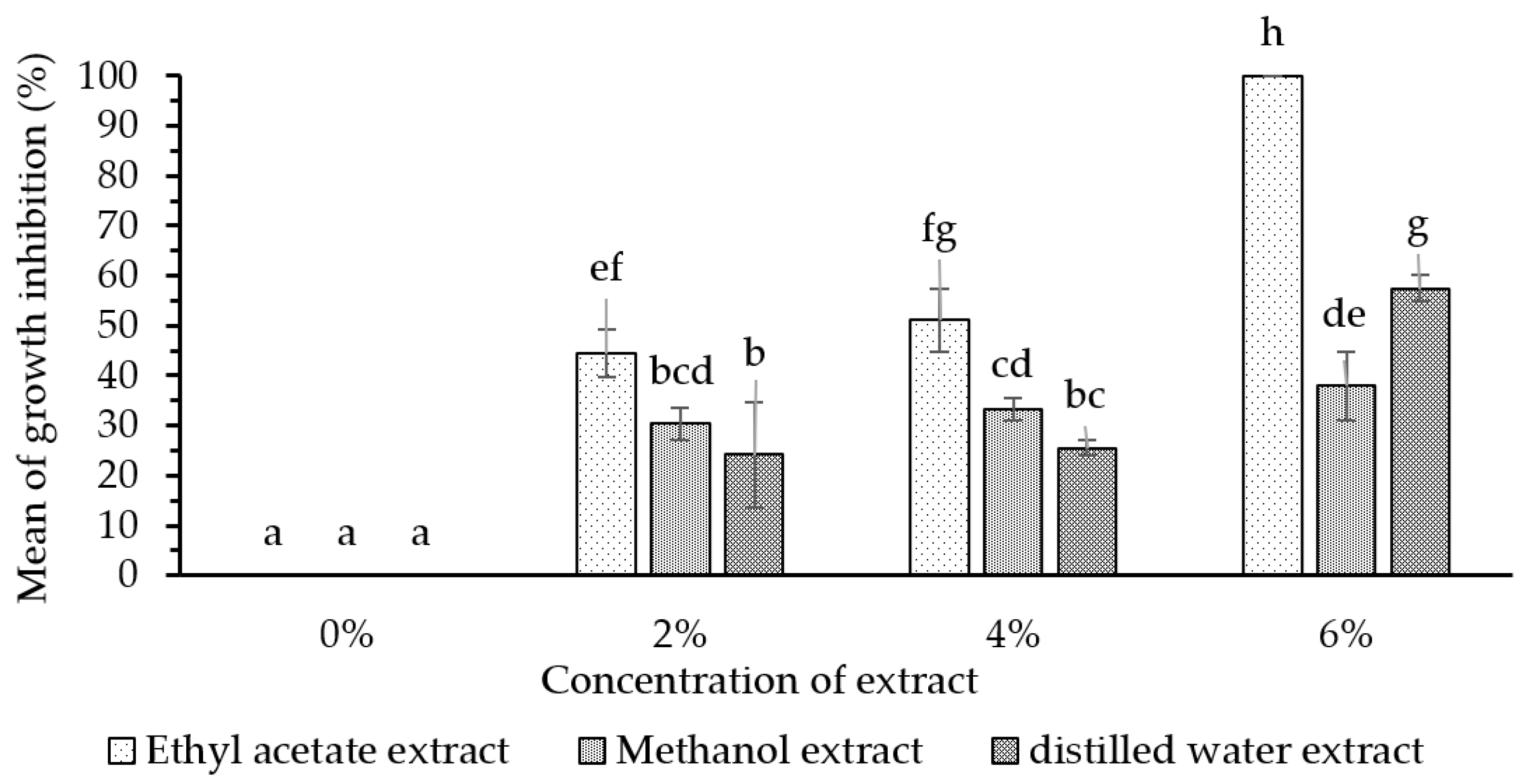
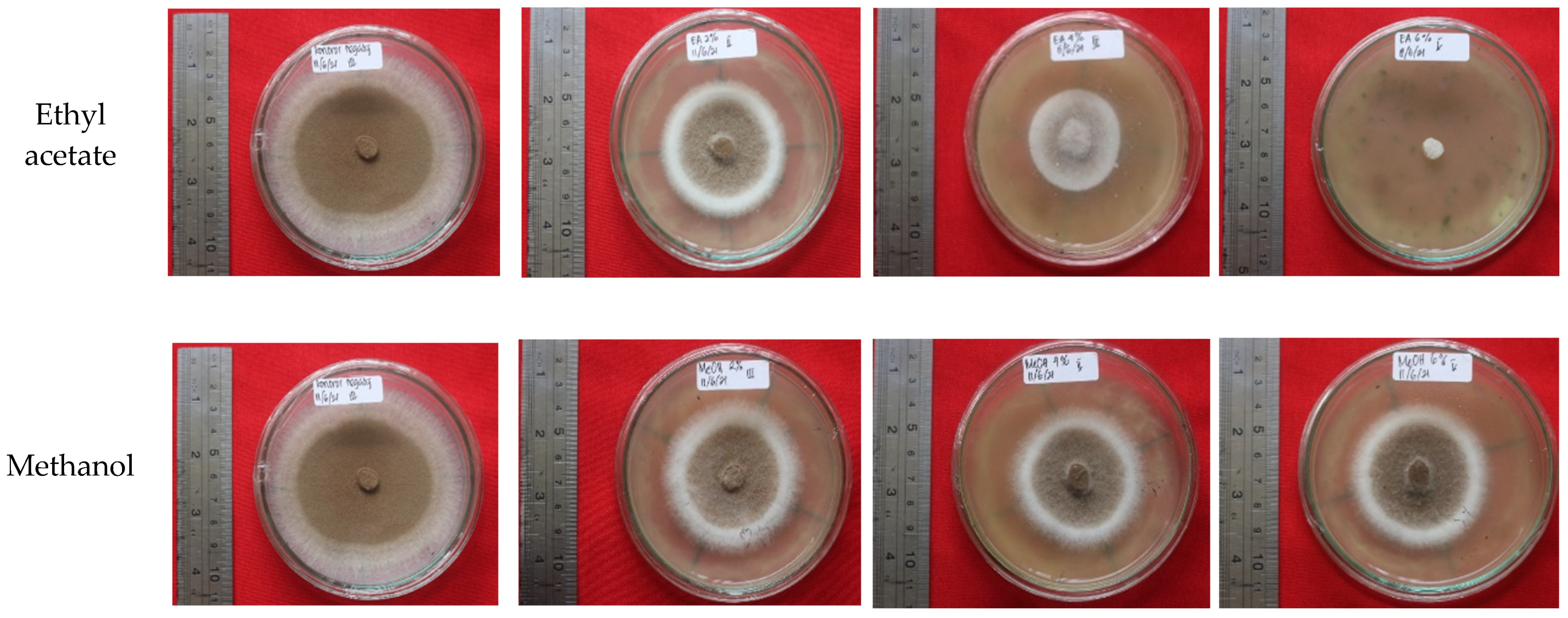
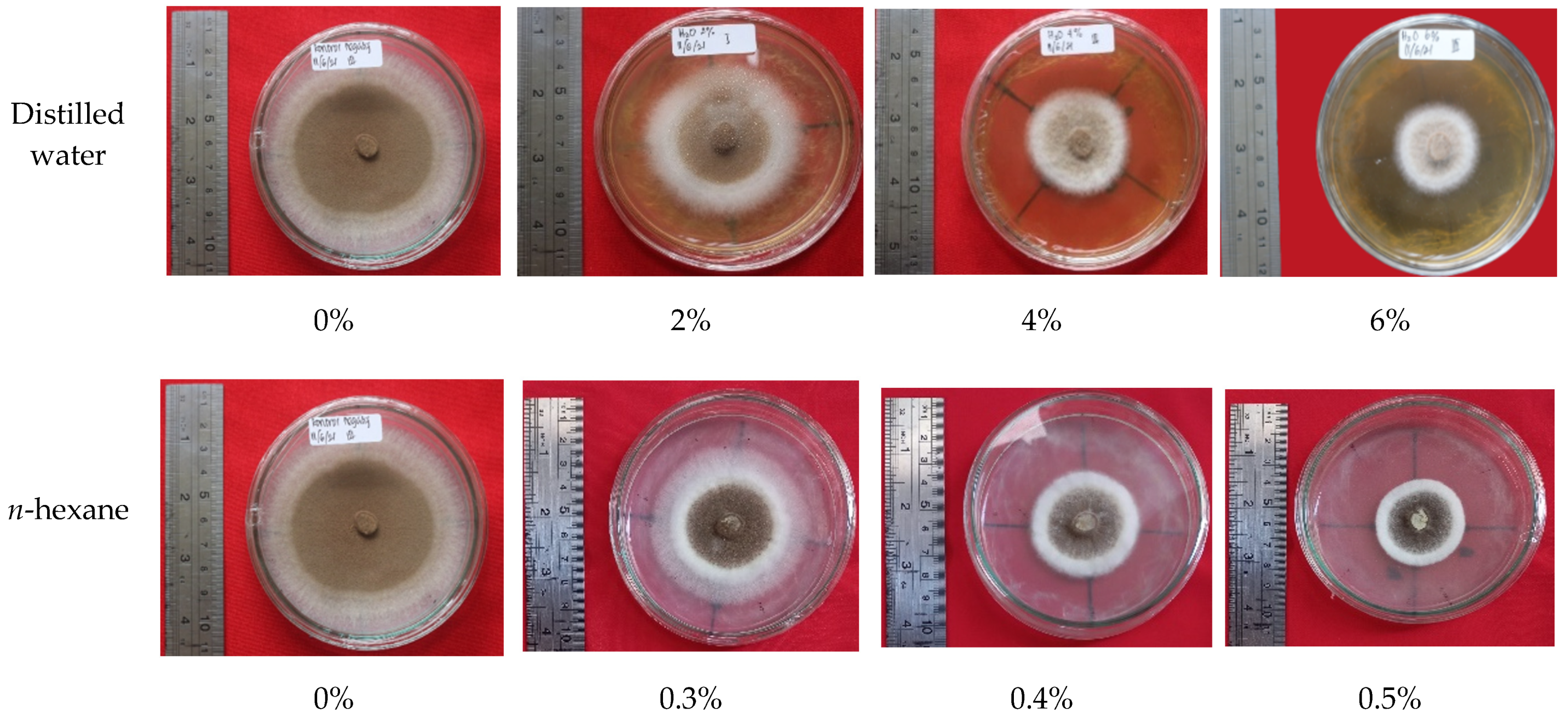
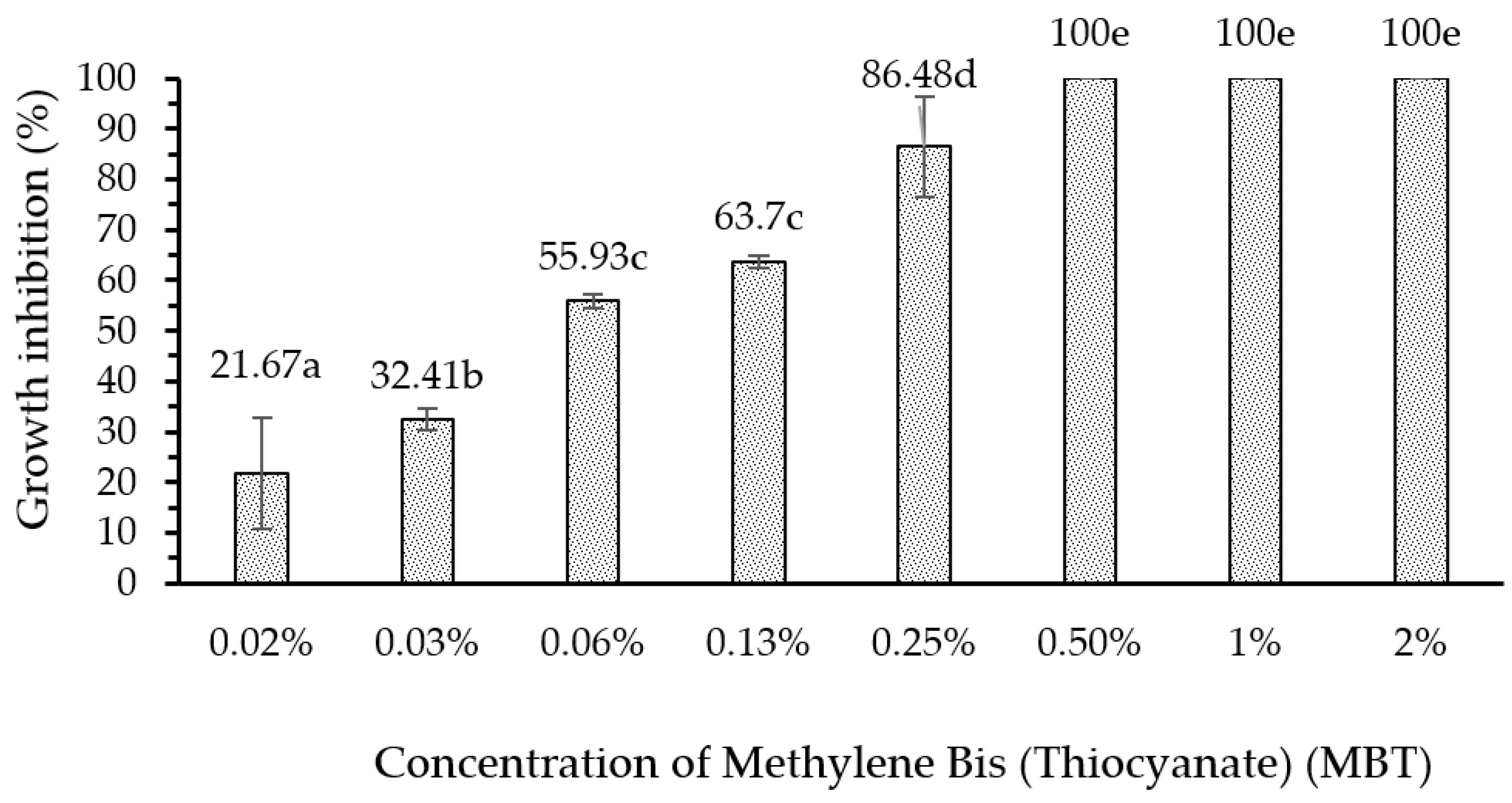
Ethyl-acetate extract and n-hexane extract from the fungus combs, with concentrations of 6% and 0.5%, respectively, showed remarkable bioactivity in terms of inhibiting wood-staining fungus A. foetidus. In addition, the bioactivity of the 6% ethyl-acetate extract showed a maximum inhibiting rate (100%) on wood-staining fungus growth equal to the efficacy of methylene bis-thiocyanate (MBT), which is widely used as an active ingredient in commercial synthetic wood preservatives. Further investigations are needed to identify the chemical structure of the active compounds in the fungus-comb extracts responsible for wood-staining-fungi-inhibition.

Author Contributions

Conceptualization, D.N.; methodology, D.N., I.B., A.A. and L.K.; termite-mound surveys and sample collection, A.A., L.K., D.N. and P.S.S.; chemical analysis, I.B., L.K. and A.A.; bioactivity test, P.S.S. and D.N.; resources, L.K. and A.A.; writing—first draft, D.N.; review and editing, D.N., I.B., L.K., A.A., D.S., Y.R., D.F., L.D.W., IK.S. and D.M.H. All authors have read and agreed to the published version of the manuscript.

Funding

This research was funded by university grants from IPB University, Gadjah Mada University, Institut Teknologi Bandung, and Airlangga University of the Republic of Indonesia, under Riset Kolaborasi Indonesia/Indonesian Collaborative Research Scheme FY 2021 (contract number 1074/IT3.L1/PN/2021).

Institutional Review Board Statement

Not applicable.

Informed Consent Statement

Not applicable.

Data Availability Statement

Not applicable.

Acknowledgments

The authors gratefully acknowledge the financial support of this research from IPB University, Gajah Mada University, Institut Teknologi Bandung, and Airlangga University of the Republic of Indonesia, under the Indonesian Collaborative Research Scheme FY 2021 (contract No. 1074/IT3.L1/PN/2021).

Conflicts of Interest

The authors declare no conflict of interest.

References

  1. Krishna, K.; Grimaldi, D.A.; Krishna, V.; Engel, M.S. Treatise on the Isoptera of the World: 1 Introduction (Bulletin of the American Museum of Natural History); American Museum of Natural History: New York, NY, USA, 2013; Volume 377, p. 1. [Google Scholar]
  2. Nandika, D.; Rismayadi, Y.; Diba, F. Rayap: Biologi Dan Pengendaliannya [Termite: Biology and Its Control], 2nd ed.; Universitas Muhamadiyah: Surakarta, Indonesia, 2015. [Google Scholar]
  3. Subekti, N. Distribution Patterns, Architecture and Population Density of Subterranean Termites Macrotermes gilvus Hagen (Blattodea: Termitidae) in Yanlappa Nature Reserve, West Java. Ph.D. Thesis, Faculty of Forestry, IPB University, Bogor, Indonesia, 2010. [Google Scholar]
  4. Hadi, Y.S.; Massijaya, M.Y.; Arinana, A. Subterranean termite resistance of polystyrene-treatedwood from three tropicalwood species. Insects 2016, 7, 37. [Google Scholar] [CrossRef] [Green Version]
  5. Kusumawardhani, D.T.; Nandika, D.; Karlinasari, L.; Arinana, A.; Batubara, I. Architectural and physical properties of fungus comb from subterranean termite macrotermes gilvus (Isoptera: Termitidae) mound. Biodiversitas 2021, 22, 1627–1634. [Google Scholar] [CrossRef]
  6. Arinana, A.; Aldina, R.; Nandika, D.; Rauf, A.; Harahap, I.S.; Sumertajaya, I.M.; Bahtiar, E.T. Termite diversity in urban landscape, South Jakarta, Indonesia. Insects 2016, 7, 20. [Google Scholar] [CrossRef] [PubMed] [Green Version]
  7. Arshad, M.A.; Schnitzer, M. The chemistry of a termite fungus comb. Plant Soil 1987, 98, 247–256. [Google Scholar] [CrossRef]
  8. Singh, K.; Muljadi, B.P.; Raeini, A.Q.; Jost, C.; Vandeginste, V.; Blunt, M.J.; Theraulaz, G.; Degond, P. The architectural design of smart ventilation and drainage systems in termite nests. Sci. Adv. 2019, 5, 8520. [Google Scholar] [CrossRef] [PubMed] [Green Version]
  9. De Fine Licht, H.H.; Boomsma, J.J.; Aanen, D.K. Presumptive horizontal symbiont transmission in the fungus-growing termite Macrotermes natalensis. Mol. Ecol. 2006, 15, 3131–3138. [Google Scholar] [CrossRef]
  10. Schisler, L.C.; Chang, S.T.; Quimio, T.G. Tropical Mushrooms: Biological Nature and Cultivation Methods. Mycologia 2005, 76, 383. [Google Scholar] [CrossRef]
  11. Rouland-Lefèvre, D.; Inoue, T.; Johjima, T. Termitomyces/Termite Interactions. In Intestinal Microorganisms of Termites and Other Invertebrates; Springer: Berlin/Heidelberg, Germany, 2006; pp. 335–350. [Google Scholar]
  12. Qian, G.; Li, S.; Wen, H. Fungal diversity of fungus comb in termite nests. Mycosystema 2011, 30, 556–565. [Google Scholar]
  13. Shaleh, I.; Jannual, N.; Hasin, S.; Kaewgrajang, T.; Raffiudin, R.; Nipitwattanaphon, M. Identification of fungus-growing termites and mutualistic Termitomyces from two provinces in Thailand. Int. J. Trop. Insect Sci. 2020, 41, 1555–1566. [Google Scholar] [CrossRef]
  14. Batra, L.R.; Batra, S.W.T. Fungus growing termites of tropical India and associated fungi. J. Kans. Entomol. Soc. 1966, 39, 725–738. [Google Scholar]
  15. Thomas, R.J. Factors affecting the distribution and activity of fungi in the nests of Macrotermitinae (Isoptera). Soil Biol. Biochem. 1987, 19, 343–349. [Google Scholar] [CrossRef]
  16. Anwar, T.; Qureshi, H.; Mahnashi, M.H.; Kabir, F.; Parveen, N.; Ahmed, D.; Afzal, U.; Batool, S.; Awais, M.; Alyami, S.A.; et al. Bioherbicidal ability and weed management of allelopathic methyl esters from Lantana camara. Saudi J. Biol. Sci. 2021, 28, 4365–4374. [Google Scholar] [CrossRef]
  17. Daniel, G. Fungal Degradation of Wood Cell Walls. In Secondary Xylem Biology: Origins, Functions, and Applications; Kim, Y.S., Funada, R., Singh, A.P., Eds.; Academic Press: London, UK, 1986; pp. 139–141. [Google Scholar]
  18. Salman, A.B.A.; Sudirman, S.L.; Nandika, D. Selection of stain fungi on rubberwood (Hevea brasiliensis) and its growth response against chitosan. Biodiversitas 2020, 21, 4501–4508. [Google Scholar] [CrossRef]
  19. OldertrØen, K.; H-Kittikun, A.; Phongpaichit, S.; Riyajan, S.; Teanpaisal, R. Treatment of rubberwood (Hevea brasiliensis) (Willd. ex. a juss) Muell. Arg. with maleic anhydride to prevent moulds. J. Forensic Sci. 2016, 62, 314–321. [Google Scholar]
  20. George, J. Preservative treatment of bamboo, rubberwood and coconut palm—Simple methods for treating building timbers. In Preservation of Timber in the Tropics; Findlay, W.P.K., Ed.; Kluwer Academic: Norwell, MA, USA, 1985; pp. 233–248. [Google Scholar]
  21. Drewello, R.; Weißmann, R. Microbiology influenced corrosion of glass. Appl. Microbiol. Biotechnol. 1997, 47, 337–346. [Google Scholar] [CrossRef]
  22. Müller, E.; Drewello, U.; Drewello, R.; Weißmann, R.; Wuertz, S. In situ analysis of biofilms on historic window glass using confocal laser scanning microscopy. J. Cult. Herit. 2001, 2, 31–42. [Google Scholar] [CrossRef]
  23. Pinto, C.; Palomar, T.; Alves, L.C.; da Silva, S.H.M.; Monteiro, R.C.; Macedo, M.F.; Villarigues, M.G. Fungal biodeterioration of stained-glass windows in monuments from Belem do Para (Brazil). Int. Biodeterior. Biodegrad. 2019, 138, 106–113. [Google Scholar] [CrossRef]
  24. Badan Pusat Statistik. Statistik Produksi Kehutanan 2018; Badan Pusat Statistik: Jakarta, Indonesia, 2019.
  25. Hadi, Y.S.; Massijaya, M.Y.; Abdillah, I.B.; Pari, G.; Arsyad, W.O.M. Color change and resistance to subterranean termite attack of mangium (Acacia mangium) and sengon (Falcataria moluccana) smoked wood. J. Korean Wood Sci. Technol. 2020, 48, 1–11. [Google Scholar]
  26. Valiante, V.; Baldin, C.; Hortschansky, P.; Jain, R.; Thywiben, A.; Starbburger, M.; Shelest, E.; Heinekamp, T.; Brakhage, A.A. The Aspergillus fumigatus conidial melanin production is regulated by the bifunctional bHLH Devr and MADS-box RlmA transcription factor. Mol. Microbiol. 2016, 102, 321–335. [Google Scholar] [CrossRef] [Green Version]
  27. Muzaffer, A. Key to the Indomalayan termites. Biologia 1958, 4, 33–198. [Google Scholar]
  28. Tho, Y.P.; Kirton, L.G.L. Termites of Peninsular Malaysia (Malayan Forest Records No. 36); Forest Research Institute Malaysia: Kuala Lumpur, Malaysia, 1992.
  29. Lou, J.; Zhang, J.; Xu, S.; Wang, D.; Fan, X. New Method to Evaluate the Crosslinking Degree of Resin Finishing Agent with Cellulose Using Kjeldahl Method and Arrhenius Formula. Processes 2021, 9, 767. [Google Scholar] [CrossRef]
  30. Harborne, J. Metode Fitokimia Penuntun Cara Modern Menganalisa Tumbuhan. In Phytochemical Methods: A Guide to Modern Techniques of Plant Analysis (In English), 2nd ed.; Padmawinata, K., Soediro, I., Eds.; ITB: Bandung, Indonesia, 1984; pp. 47–102, 152–153. [Google Scholar]
  31. Liu, Y.; Benohoud, M.; Yamdeu, J.H.G.; Gong, Y.Y.; Orfila, C. Green extraction of polyphenols from citrus peel by-products and their antifungal activity against Aspergillus Flavus. Food Chem. X 2021, 12, 100144. [Google Scholar] [CrossRef] [PubMed]
  32. Singh, T. Migration of methylene bis thiocyanate in wood and its effect on a wood degrading fungus. Eur. J. For. Res. 2008, 127, 195–202. [Google Scholar] [CrossRef]
  33. Subekti, N.; Solihin, D.D.; Nandika, D.; Surjokusumo, S.; Anwar, S. Distribution and morphology characteristic of Macrotermes gilvus Hagen in the natural habitat. J. Ilmu Dan Teknol. Has. Hutan 2008, 1, 27–33. [Google Scholar]
  34. Otani, S.; Challinor, V.L.; Kreunzenbeck, N.B.; Kildgaard, S.; Cristensen, S.K.; Larsen, L.L.M.; Aanen, D.K.; Rasmussen, S.A.; Beemelmanns, C.; Poulsen, M. Disease-free monoculture farming by fungus-growing termites. Sci. Rep. 2019, 9, 8819. [Google Scholar]
  35. Grasse, P.P. Termitologia; Masson: Paris, France, 1982; Volume I. [Google Scholar]
  36. Johnson, R.A. Colony development and establishment of fungus comb in Microtermes sp. nr. umbaricus Sjöstedt (Isoptera, Macrotermitinae) from Nigeria. J. Nat. Hist. 1981, 32, 3–12. [Google Scholar]
  37. Yang, G.; Ahmad, F.; Zhou, Q.; Guo, M.; Liang, S.; Gaal, H.A.; Mo, J. Investigation of physicochemical indices and microbial communities in temite fungus-combs. Front. Microbiol. 2021, 11, 581219. [Google Scholar] [CrossRef]
  38. Sawhasan, P.; Worapong, J.; Flegel, T.W.; Vinijsanun, T. Fungal partnerships stimulate growth of Termitomyces clypeatus stalk mycelium in vitro. World J. Microbiol. Biotechnol. 2012, 28, 2311–2318. [Google Scholar] [CrossRef]
  39. Arifin, Z.; Dahlan, Z.; Irsan, C.S.; Hartono, Y. Impact of the presence of subterranean termites Macrotermes gilvus (Termitidae) to physico-chemical soil modification on the rubber plantation land. J. Ecol. Nat. Environ. 2016, 8, 13–19. [Google Scholar]
  40. Korb, J. Termite mound architecture, from function to construction. In Biology of Termites: A Modern Synthesis; Springer: Dordrecht, The Netherlands; Heidelberg, Germany; London, UK; New York, NY, USA, 2011; pp. 349–373. [Google Scholar]
  41. Chiu, C.; Ou, J.; Chen, C.; Li, H. Odontotermes formosanus regulate α-amino acid and fatty acids with their symbiont fungus. In Proceedings of the 12th Pasific Rim Termite Research Group Conference, Yogyakarta, Indonesia, 21–22 March 2018; pp. 28–33. [Google Scholar]
  42. De Ullivarri, M.F.; Arbulu, S.; Garcia-Gutierrez, E.; Cotter, P.D. Antifungal Peptides as Therapeutic Agents. Front. Cell. Infect. Microbiol. 2020, 17, 105. [Google Scholar] [CrossRef]
  43. Hoseini, S.M.; Ahmad, K.M.; Yousefi, M.; Costas, B. Roles of arginine in fish nutrition and health: Insights for future researches. Rev. Aquac. 2020, 12, 2091–2108. [Google Scholar] [CrossRef]
  44. Gambardella, J.; Khondkar, W.; Morelli, M.B.; Wang, X.; Santulli, G.; Trimarco, V. Arginine and endothelial function. Biomedicines 2020, 8, 277. [Google Scholar] [CrossRef] [PubMed]
  45. Prastya, M.E.; Astuti, R.I.; Batubara, I.; Takagi, H.; Wahyudi, A.T. Natural extract and its fractions isolated from the marine bacterium Pseudoalteromonas flavipulchra STILL-33 have antioxidant and antiaging activities in Schizosaccharomyces pombe. FEMS Yeast Res. 2020, 20, 3. [Google Scholar] [CrossRef] [PubMed]
  46. Zhang, L.; Li, F.; Guo, Q.; Duan, Y.; Wang, W.; Zhong, Y.; Yang, Y.; Yin, Y. Leucine supplementation: A novel strategy for modulating lipid metabolism and energy homeostasis. Nutrients 2020, 12, 1299. [Google Scholar] [CrossRef]
  47. Theis, N.; Brown, M.A.; Wood, P.; Waldron, M. Leucine Supplementation Increases Muscle Strength and Volume, Reduces Inflammation, and Affects Wellbeing in Adults and Adolescents with Cerebral Palsy. J. Nutr. 2021, 151, 59–64. [Google Scholar] [CrossRef] [PubMed]
  48. Fierens, T.; Servaes, K.; Van Holderbeke, M.; Geerts, L.; De Henauw, S.; Sioen, I.; Vanermen, G. Analysis of phthalates in food products and packaging materials sold on the Belgian market. Food Chem. Toxicol. 2012, 50, 2575–2583. [Google Scholar] [CrossRef]
  49. Ortiz, A.; Sansinenea, E. Di-2-ethylhexylphthalate May Be a Natural Product, Rather than a Pollutant. J. Chem. 2018, 2018, 6040814. [Google Scholar] [CrossRef]
  50. Ma, T.T.; Luo, Y.M.C.; Teng, Y. Phthalate esters contamination in soil and plants on agricultural land near an electronic waste recycling site. Environ. Geochem. Health 2013, 35, 465–476. [Google Scholar] [CrossRef] [PubMed]
  51. El-Sayed, M.H. Di-(2-ethylhexyl) phthalate, a major bioactive metabolite with antimicrobial and cytotoxic activity isolated from the culture filtrate of newly isolated soil streptomyces (Streptomyces mirabilis) Strain NSQu-25. World Appl. Sci. J. 2012, 20, 1202–1212. [Google Scholar]
  52. Qi, S.H.; Xu, Y.; Xiong, H.R.; Qian, P.Y.; Zhang, S. Antifouling and antibacterial compounds from a marine fungus Cladosporium sp. F14. World J. Microbiol. Biotechnol. 2009, 25, 399–406. [Google Scholar] [CrossRef]
  53. Xiong, H.; Qi, S.; Xu, Y.; Miao, L.; Qian, P.Y. Antibiotic and antifouling compound production by the marine-derived fungus Cladosporium sp. F14. J. Hydro-Environ. Res. 2009, 24, 264–270. [Google Scholar] [CrossRef]
  54. Lu, G.; Liu, T.; Wang, N.; Dou, Z.; Wang, K.; Zuo, Y. Nematicidal Effect of Methyl Palmitate and Methyl Stearate against Meloidogyne incognita in Bananas. J. Agric. Food Chem. 2020, 68, 6502–6510. [Google Scholar] [CrossRef] [PubMed]
  55. Bashir, A. Chemical composition and antifungal, phytotoxic, brine shrimp cytotoxicity, insecticidal and antibacterial activities of the essential oils of Acacia modesta. J. Med. Plants Res. 2012, 6, 31. [Google Scholar]
  56. Ohe, V.R.; Aalizadeh, R.; Schymanski, S. S13|EUCOSMETICS|Combined Inventory of Ingredients Employed in Cosmetic Products (2000) and Revised Inventory (2006). 2021. Available online: https://0-doi-org.brum.beds.ac.uk/10.5281/zenodo.3821156 (accessed on 25 August 2021).
  57. Mota, C.J.A.; Pinto, B.P.; de Lima, A.L. Glycerol: A Versatile Renewable Feedstock for the Chemical Industry; Springer International: Zurich, Switzerland, 2017; pp. 10–11. [Google Scholar]
  58. Lebwohl, M.G.; Heymann, H.Y.; Berth-Jones, J.; Coulson, I. Treatment of Skin Disease: Comprehensive Therapeutic Strategies, 5th ed.; Elsevier: Amsterdam, The Netherlands, 2017. [Google Scholar]
  59. Velmurugan, N.; Han, S.S.; Lee, Y.S. Antifungal activity of neutralized wood vinegar with water extracts of Pinus densiflora and Quercus serrata saw dusts. Int. J. Environ. Resour. 2009, 3, 167–176. [Google Scholar]
  60. Oramahi, H.A.; Diba, F.; Wahdina. Antifungal activity of liquid smoke from sawdust of acacia wood (Acacia mangium Willd) and laban wood (Vitex pubescens Vahl). J. Ilmu-Ilmu Hayati Dan Fis. 2011, 13, 79–84. [Google Scholar]
  61. Sitepu, I.; Suada, I.; Susrama, I. The Antimicrobial Activity Test of Some Kitchen Seasoning Extracts on Growth of Fungus Curvularia lunata (Wakk.) Boed. and Aspergillus flavus Link. E-J. Agroekoteknologi Trop. 2012, 1, 2. [Google Scholar]
  62. Kelland, M.A. Production Chemicals for the Oil and Gas Industry; CRC Press: Boca Raton, FL, USA, 2014. [Google Scholar]
  63. Balouiri, M.; Sadiki, M.; Ibnsouda, S.K. Methods for in vitro evaluating antimicrobial activity: A review. J. Pharm. Anal. 2016, 6, 71–79. [Google Scholar] [CrossRef] [Green Version]
  64. Padmini, E.; Valarmathi, A.; Rani, M. Comparative analysis of chemical composition and antibacterial activities of Mentha spicata and Camellia sinensis. Asian J. Exp. Biol. Sci. 2010, 1, 772–778. [Google Scholar]
  65. Nazarudin, M.F.; Isha, A.; Mastuki, S.N.; Ain, N.M.; Ikhsan, N.F.M.; Abidin, A.Z.; Aliyu-Paiko, M. Chemical Composition and Evaluation of the α-Glucosidase Inhibitory and Cytotoxic Properties of Marine Algae Ulva intestinalis, Halimeda macroloba, and Sargassum ilicifolium. Evid Based Complement. Altern. Med. 2020, 2020, 2753945. [Google Scholar] [CrossRef]
  66. Rawal, J.R.; Sonawani, P.R. Determination of bioactive components of Cynodon dactylon by GC-MS analysis & it’s in vitro antimicrobial activity. Int. J. Pharm. Life Sci. 2016, 7, 4880–4885. [Google Scholar]
  67. Suzuki, K.; Nakano, N.; Tanaka, R.; Uyeda, M.; Shibata, M. Cell aggregation factor produced by streptomyces sp. Strain No. A-3315. Agric. Biol. Chem. 1988, 52, 2589–2595. [Google Scholar]
  68. Ester, A.; Mukarlina, R. Aktivitas ekstrak metanol daun sembung rambat (Mikania micrantha Kunth) terhadap pertumbuhan Phytophthora sp. Im5 dari batang jeruk siam (Citrus nobilis var. microcarpa). J. Protobiont. 2017, 6, 63–67. [Google Scholar]
  69. Cowan, M.M. Plant products as antimicrobial agents. Clin. Microbiol. Rev. 1999, 12, 564–582. [Google Scholar] [CrossRef] [PubMed] [Green Version]
Figure 1. Spatial distribution of the eighteen M. gilvus mounds found in the Yanlappa Experimental Forest used as a source of fungus-comb samples.
Forests 12 01591 g001
Figure 2. Mean of growth inhibition of the ethyl acetate, methanol, and water extracts, with concentrations of 0% (control solution), 2%, 4%, and 6% on the growth of A. foetidus fungus. Notes: the same letter on the statistic bar indicates no significant difference (p < 0.05).
Forests 12 01591 g002
Figure 3. Mean of growth inhibition of the n-hexane extracts, with concentrations of 0% (control solution), 0.3%, 0.4%, and 0.5% on the growth of A. foetidus fungus. Notes: the same letter on the statistic bar indicates no significant difference (p < 0.05).
Forests 12 01591 g003
Figure 4. The growth of the A. foetidus fungus colonies after 7 d of incubation on PDA medium treated with different fungus-comb extracts at different concentrations.
Forests 12 01591 g004aForests 12 01591 g004b
Figure 5. The inhibition rate of various concentrations of methylene bis-thiocyanate (MBT) on the growth of A. foetidus fungus. Notes: the same letter on the statistic bar indicates no significant difference (p < 0.05).
Forests 12 01591 g005
Table 1. Content of the fresh fungus combs from the M. gilvus Mounds.
Parameters% of Dry SubstanceMethod
Moisture (%)25.94Gravimetric
Crude protein (%)5.80HPLC
Crude fat (%)0.73Gravimetric
Acid insoluble ash (%)3.45Gravimetric
Crude ash (%)30.57Gravimetric
Crude fiber (%)25.46Gravimetric
Carbohydrate as starch (%)7.76Luff School
Total99.71
Table 2. Amino-acid content of the fungus combs from the M. gilvus Mounds.
NoAmino AcidContent (% w/w DB)
1Aspartic acid0.30
2Threonine0.18
3Serine0.17
4Glutamate0.38
5Proline0.16
6Glysine0.18
7Alanine0.18
8Cystine0.02
9Valine0.22
10Methionine0.11
11Ileusine0.27
12Leusine0.55
13Tyrosine0.01
14Phenylalanine0.24
15Histidine0.24
16Lysine0.21
17Arginine1.33
Total4.75
Table 3. Extraction yields of different solvents from the fungus combs from the M. gilvus Mounds.
SolventsExtraction Yield (%) *
n-hexane0.09
Ethyl acetate1.73
Methanol2.53
Distilled water4.61
* Values derived from 5 replicates.
Table 4. Dominant Compounds Found in the n-Hexane Extract from the Fungus Combs and Their Bioactivities.
Compound NameCAS No.Relative Content (%)Bioactivity Reported
Methyl palmitate112-39-04.55Nematicidal [54], herbicidal [16]
Benzenepropanoic acid, 3,5-bis(1,1 dimethylethyl)-4-hydroxy-,methyl ester6386-38-51.16Antifungi and antioxidant [55]
Methyl linolelaidate2566-97-42.03Emollient; Skin conditioning [56]
Methyl oleate112-62-94.17Emollient; Skin conditioning [56]
Bis(2-ethylhexyl) phthalate117-81-769.43Antibacterial [52,53]
Table 5. Dominant compounds found in the ethyl-acetate extract from the fungus combs and their bioactivities.
Compound NameCAS No.Relative Content (%)Bioactivity Reported
Glycerol56-81-528.93Humectant [58]
Phenol, 2-methoxy-90-05-18.54Antifungi [59]
Phenol, 2,6-dimethoxy-91-10-16.55Antifungi [59]
Bis(2-ethylhexyl) phthalate117-81-74.82Antibacterial [53,57]
Table 6. Minimum inhibitory concentration (MIC) of all fungus-comb extracts and MBT against A. foetidus.
NoSample NameMIC
(%)(mg/L)
1.n-hexane extract>0.5>5000
2.Ethyl-acetate extract6.060,000
3.Methanol extract>6.0>60,000
4.Distilled water extract>6.0>60,000
5.MBT (positive control)0.55000
Publisher’s Note: MDPI stays neutral with regard to jurisdictional claims in published maps and institutional affiliations.

