
Evolution of Human Salivary Stress Markers during an Eight-Hour Exposure to a Mediterranean Holm Oak Forest. A Pilot Study

 , , , and
Abstract
:1. Introduction
2. Materials and Methods
2.1. Participants
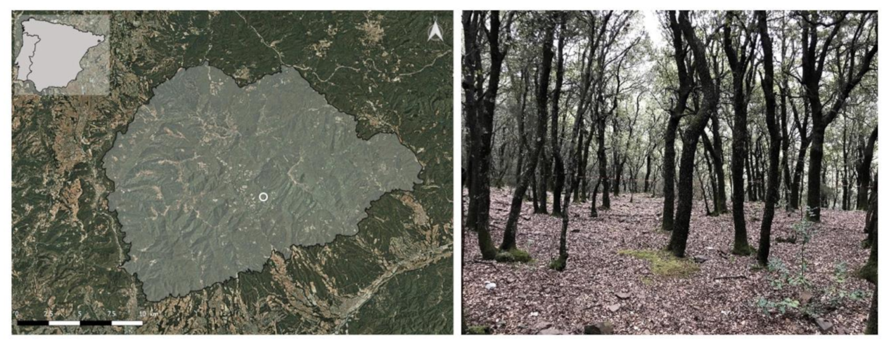
2.2. Study Site
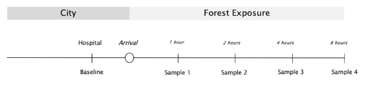
2.3. Study Design
2.4. Saliva Sampling Procedure
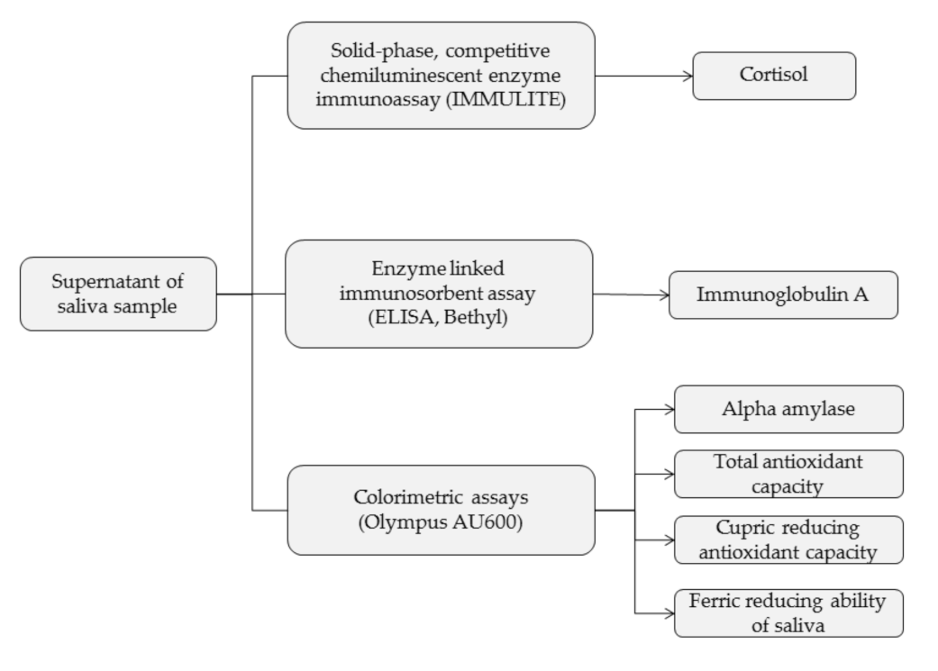
2.5. Laboratory Salivary Methods
2.6. Data Analysis
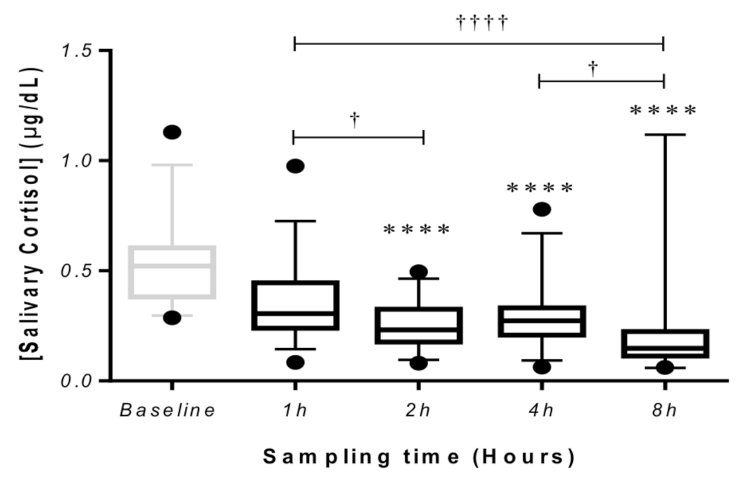
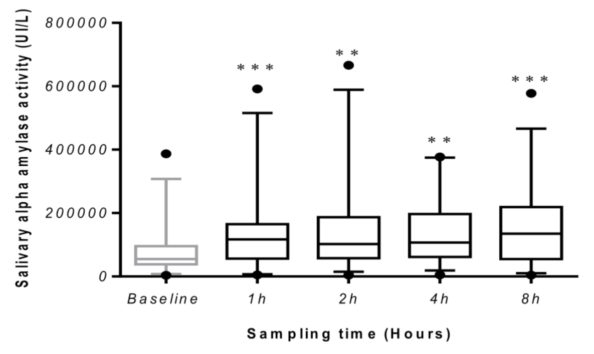
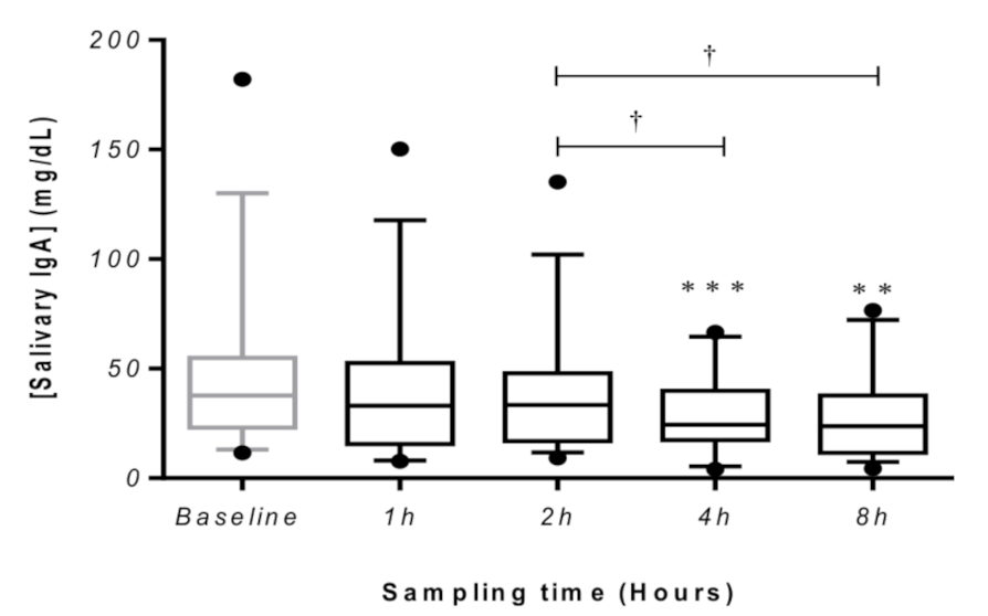
3. Results
4. Discussion
Limitations
5. Conclusions
Author Contributions
Funding
Acknowledgments
Conflicts of Interest
References
- Jordan, J.; Gurr, E.; Tinline, G.; Giga, S.; Faragher, B.; Cooper, C. HSE Health & Safety Executive Beacons of Excellence in Stress Prevention. 2003; ISBN 0717627098. Available online: https://www.hse.gov.uk/research/rrpdf/rr133.pdf (accessed on 5 November 2021).
- Goodwin, R.D.; Davidson, K.W.; Keyes, K. Mental disorders and cardiovascular disease among adults in the United States. J. Psychiatr. Res. 2009, 43, 239–246. [Google Scholar] [CrossRef] [PubMed] [Green Version]
- Todaro, J.F.; Shen, B.J.; Raffa, S.D.; Tilkemeier, P.L.; Niaura, R. Prevalence of anxiety disorders in men and women with established coronary heart disease. J. Cardiopulm. Rehabil. Prev. 2007, 27, 86–91. [Google Scholar] [CrossRef] [PubMed]
- Player, M.S.; Peterson, E.L. Anxiety disorders, hypertension, and cardiovascular risk: A review. Int. J. Psychiatry Med. 2011, 41, 365–377. [Google Scholar] [CrossRef] [PubMed]
- Aldwin, C.M. Stress, Coping and Development: An Intergrative Perspective; The Guilford Press: New York, NY, USA, 2007; ISBN 9781572308404. [Google Scholar]
- Tsunetsugu, Y.; Park, B.J.; Miyazaki, Y. Trends in research related to “shinrin-yoku” (taking in the forest atmosphere or forest bathing) in Japan. Environ. Health Prev. Med. 2010, 15, 27–37. [Google Scholar] [CrossRef] [Green Version]
- Park, B.J.; Tsunetsugu, Y.; Kasetani, T.; Kagawa, T.; Miyazaki, Y. The physiological effects of Shinrin-yoku (taking in the forest atmosphere or forest bathing): Evidence from field experiments in 24 forests across Japan. Environ. Health Prev. Med. 2010, 15, 18–26. [Google Scholar] [CrossRef] [Green Version]
- Song, C.; Ikei, H.; Miyazaki, Y. Physiological effects of nature therapy: A review of the research in Japan. Int. J. Environ. Res. Public Health 2016, 13, 781. [Google Scholar] [CrossRef] [PubMed]
- Hansen, M.M.; Jones, R.; Tocchini, K. Shinrin-yoku (Forest bathing) and nature therapy: A state-of-the-art review. Int. J. Environ. Res. Public Health 2017, 14, 851. [Google Scholar] [CrossRef] [Green Version]
- Morita, E.; Fukuda, S.; Nagano, J.; Hamajima, N.; Yamamoto, H.; Iwai, Y.; Nakashima, T.; Ohira, H.; Shirakawa, T. Psychological effects of forest environments on healthy adults: Shinrin-yoku (forest-air bathing, walking) as a possible method of stress reduction. Public Health 2007, 121, 54–63. [Google Scholar] [CrossRef] [PubMed] [Green Version]
- Park, B.J.; Tsunetsugu, Y.; Kasetani, T.; Hirano, H.; Kagawa, T.; Sato, M.; Miyazaki, Y. Physiological effects of Shinrin-yoku (taking in the atmosphere of the forest) using salivary cortisol and cerebral activity as indicators. J. Physiol. Anthropol. 2007, 26, 123–128. [Google Scholar] [CrossRef] [PubMed] [Green Version]
- Tsunetsugu, Y.; Park, B.-J.; Ishii, H.; Hirano, H.; Kagawa, T.; Miyazaki, Y. Physiological effects of shinrin-yoku (taking in the atmosphere of the forest) in an old-growth broadleaf forest in Yamagata Prefecture, Japan. J. Physiol. Anthropol. 2007, 26, 135–142. [Google Scholar] [CrossRef] [PubMed] [Green Version]
- Jia, B.B.; Yang, Z.X.; Mao, G.X.; Lyu, Y.D.; Wen, X.L.; Xu, W.H.; Lyu, X.L.; Cao, Y.B.; Wang, G.F. Health effect of forest bathing trip on elderly patients with chronic obstructive pulmonary disease. Biomed. Environ. Sci. 2016, 29, 212–218. [Google Scholar]
- Kobayashi, H.; Song, C.; Ikei, H.; Park, B.J.; Lee, J.; Kagawa, T.; Miyazaki, Y. Population-based study on the effect of a forest environment on salivary cortisol concentration. Int. J. Environ. Res. Public Health 2017, 14, 931. [Google Scholar] [CrossRef] [PubMed]
- Mao, G.X.; Lan, X.G.; Cao, Y.B.; Chen, Z.M.; He, Z.H.; LV, Y.D.; Wang, Y.Z.; Hu, X.L.; Wang, G.F.; Yan, J. Effects of short-term forest bathing on human health in a broad-leaved evergreen forest in Zhejiang Province, China. Biomed. Environ. Sci. 2012, 25, 317–324. [Google Scholar] [PubMed]
- Ochiai, H.; Ikei, H.; Song, C.; Kobayashi, M.; Miura, T.; Kagawa, T.; Li, Q.; Kumeda, S.; Imai, M.; Miyazaki, Y. Physiological and psychological effects of a forest therapy program on middle-aged females. Int. J. Environ. Res. Public Health 2015, 12, 15222–15232. [Google Scholar] [CrossRef] [Green Version]
- Komori, T.; Mitsui, M.; Togashi, K.; Matsui, J.; Kato, T.; Uei, D.; Shibayama, A.; Yamato, K.; Okumura, H.; Kinoshita, F. Relaxation effect of a 2-hour walk in Kumano-Kodo Forest. J. Neurol. Neurosci. 2017, 8. [Google Scholar] [CrossRef]
- Horiuchi, M.; Endo, J.; Akatsuka, S.; Uno, T.; Hasegawa, T. Influence of forest walking on blood pressure, profile of mood states and stress markers from the viewpoint of aging. J. Aging Gerontol. 2013, 1, 9–17. [Google Scholar] [CrossRef]
- Lee, J.; Park, B.J.; Tsunetsugu, Y.; Ohira, T.; Kagawa, T.; Miyazaki, Y. Effect of forest bathing on physiological and psychological responses in young Japanese male subjects. Public Health 2011, 125, 93–100. [Google Scholar] [CrossRef]
- Toda, M.; Den, R.; Hasegawa-Ohira, M.; Morimoto, K. Effects of woodland walking on salivary stress markers cortisol and chromogranin A. Complement. Ther. Med. 2013, 21, 29–34. [Google Scholar] [CrossRef]
- Antonelli, M.; Barbieri, G.; Donelli, D. Effects of forest bathing (shinrin-yoku) on levels of cortisol as a stress biomarker: A systematic review and meta-analysis. Int. J. Biometeorol. 2019, 63, 1117–1134. [Google Scholar] [CrossRef]
- Chrousos, G.P.; Kino, T.; Charmandari, E. Evaluation of the hypothalamic-pituitary-adrenal axis function in childhood and adolescence. Neuroimmunomodulation 2009, 16, 272–283. [Google Scholar] [CrossRef]
- Antonelli, M.; Donelli, D. Effects of balneotherapy and spa therapy on levels of cortisol as a stress biomarker: A systematic review. Int. J. Biometeorol. 2018, 62, 913–924. [Google Scholar] [CrossRef] [PubMed]
- Grassi, G.; Esler, M. How to assess sympathetic activity in humans. J. Hypertens. 1999, 17, 719–734. [Google Scholar] [CrossRef]
- Petrakova, L.; Doering, B.K.; Vits, S.; Engler, H.; Rief, W.; Schedlowski, M.; Grigoleit, J.S. Psychosocial stress increases salivary alpha-Amylase activity independently from plasma noradrenaline levels. PLoS ONE 2015, 10, e0134561. [Google Scholar] [CrossRef] [Green Version]
- Nater, U.M.; Rohleder, N. Salivary alpha-amylase as a non-invasive biomarker for the sympathetic nervous system: Current state of research. Psychoneuroendocrinology 2009, 34, 486–496. [Google Scholar] [CrossRef] [PubMed]
- DeCaro, J.A. Methodological considerations in the use of salivary α-amylase as a stress marker in field research. Am. J. Hum. Biol. 2008, 20, 617–619. [Google Scholar] [CrossRef]
- Granger, D.A.; Kivlighan, K.T.; El-Sheikh, M.; Gordis, E.B.; Stroud, L.R. Salivy α-amylase in biobehavioral research: Recent developments and applications. Ann. N. Y. Acad. Sci. 2007, 1098, 122–144. [Google Scholar] [CrossRef]
- Yamaguchi, M.; Deguchi, M.; Miyazaki, Y. The effects of exercise in forest and urban environments on sympathetic nervous activity of normal young adults. J. Int. Med. Res. 2006, 34, 152–159. [Google Scholar] [CrossRef] [PubMed]
- Carpenter, G.H.; Proctor, G.B.; Anderson, L.C.; Zhang, X.S.; Garrett, J.R. Immunoglobulin A secretion into saliva during dual sympathetic and parasympathetic nerve stimulation of rat submandibular glands. Exp. Physiol. 2000, 85, 281–286. [Google Scholar] [CrossRef]
- Allgrove, J.E.; Gomes, E.; Hough, J.; Gleeson, M. Effects of exercise intensity on salivary antimicrobial proteins and markers of stress in active men. J. Sports Sci. 2008, 26, 653–661. [Google Scholar] [CrossRef]
- Obayashi, K. Salivary mental stress proteins. Clin. Chim. Acta 2013, 425, 196–201. [Google Scholar] [CrossRef]
- Peluso, I.; Raguzzini, A. Salivary and urinary total antioxidant capacity as biomarkers of oxidative stress in humans. Pathol. Res. Int. 2016, 2016, 5480267. [Google Scholar] [CrossRef] [Green Version]
- Bach, A.; Penuelas, J.; Clarà, J.; Llusià, J.; Campillo, I.; López, F.; Maneja, R. How should forests be characterized in regard to human health? Evidence from existing literature. Int. J. Environ. Res. Public Health 2020, 17, 1027. [Google Scholar] [CrossRef] [PubMed] [Green Version]
- Park, B.J.; Tsunetsugu, Y.; Ishii, H.; Furuhashi, S.; Hirano, H.; Kagawa, T.; Miyazaki, Y. Physiological effects of Shinrin-yoku (taking in the atmosphere of the forest) in a mixed forest in Shinano Town, Japan. Scand. J. For. Res. 2008, 23, 278–283. [Google Scholar] [CrossRef]
- Lee, J.; Park, B.J.; Tsunetsugu, Y.; Kagawa, T.; Miyazaki, Y. Restorative effects of viewing real forest landscapes, based on a comparison with urban landscapes. Scand. J. For. Res. 2009, 24, 227–234. [Google Scholar] [CrossRef]
- Nater, U.M.; Rohleder, N.; Schlotz, W.; Ehlert, U.; Kirschbaum, C. Determinants of the diurnal course of salivary alpha-amylase. Psychoneuroendocrinology 2007, 32, 392–401. [Google Scholar] [CrossRef]
- Borisenkov, M.F.; Erunova, L.A.; Lyuseva, E.M.; Pozdeeva, N.V. Diurnal changes in the total antioxidant activity of human saliva. Hum. Physiol. 2007, 33, 375–376. [Google Scholar] [CrossRef]
- Fries, E.; Dettenborn, L.; Kirschbaum, C. The cortisol awakening response (CAR): Facts and future directions. Int. J. Psychophysiol. 2009, 72, 67–73. [Google Scholar] [CrossRef] [PubMed]
- Contreras-Aguilar, M.D.; Escribano, D.; Martínez-Subiela, S.; Martínez-Miró, S.; Rubio, M.; Tvarijonaviciute, A.; Tecles, F.; Cerón, J.J. Influence of the way of reporting alpha-amylase values in saliva in different naturalistic situations: A pilot study. PLoS ONE 2017, 12, e0180100. [Google Scholar] [CrossRef] [PubMed] [Green Version]
- Tecles, F.; Fuentes-Rubio, M.; Tvarijonaviciute, A.; Martínez-Subiela, S.; Fatjó, J.; Cerón, J.J. Assessment of stress associated with an oral public speech in veterinary students by salivary biomarkers. J. Vet. Med. Educ. 2014, 41, 37–43. [Google Scholar] [CrossRef] [PubMed]
- Rohleder, N.; Nater, U.M. Determinants of salivary α-amylase in humans and methodological considerations. Psychoneuroendocrinology 2009, 34, 469–485. [Google Scholar] [CrossRef]
- Van der Heiden, C.; Bais, R.; Gerhardt, W.; Lorentz, K.; Rosalki, S. IFCC methods for measurement of catalytic concentration of enzymes. Clin. Chim. Acta 1999, 281, S5–S39. [Google Scholar]
- Lopez-Jornet, P.; Cayuela, C.A.; Tvarijonaviciute, A.; Parra-Perez, F.; Escribano, D.; Ceron, J. Oral lichen planus: Salival biomarkers cortisol, immunoglobulin A., adiponectin. J. Oral Pathol. Med. 2016, 45, 211–217. [Google Scholar] [CrossRef] [PubMed]
- Campos, C.; Guzmán, R.; López-Fernández, E.; Casado, Á. Evaluation of the copper(II) reduction assay using bathocuproinedisulfonic acid disodium salt for the total antioxidant capacity assessment: The CUPRAC-BCS assay. Anal. Biochem. 2009, 392, 37–44. [Google Scholar] [CrossRef] [PubMed]
- Rhee, S.G.; Chang, T.S.; Jeong, W.; Kang, D. Methods for detection and measurement of hydrogen peroxide inside and outside of cells. Mol. Cells 2010, 29, 539–549. [Google Scholar] [CrossRef] [PubMed]
- Ochiai, H.; Ikei, H.; Song, C.; Kobayashi, M.; Takamatsu, A.; Miura, T.; Kagawa, T.; Li, Q.; Kumeda, S.; Imai, M.; et al. Physiological and psychological effects of forest therapy on middle-aged males with high-normal blood pressure. Int. J. Environ. Res. Public Health 2015, 12, 2532–2542. [Google Scholar] [CrossRef] [PubMed] [Green Version]
- Hill, E.E.; Zack, E.; Battaglini, C.; Viru, M.; Viru, A.; Hackney, A.C. Exercise and circulating cortisol levels: The intensity threshold effect. J. Endocrinol. Invest. 2008, 31, 587–591. [Google Scholar] [CrossRef] [PubMed]
- Mezzacappa, E.S.; Kelsey, R.M.; Katkin, E.S.; Sloan, R.P. Vagal rebound and recovery from psychological stress. Psychosom. Med. 2001, 63, 650–657. [Google Scholar] [CrossRef] [PubMed]
- Koibuchi, E.; Suzuki, Y. Exercise upregulates salivary amylase in humans. Exp. Ther. Med. 2014, 7, 773–777. [Google Scholar] [CrossRef] [PubMed] [Green Version]
- Hunter, M.C.R.; Gillespie, B.W.; Chen, S.Y.P. Urban nature experiences reduce stress in the context of daily life based on salivary biomarkers. Front. Psychol. 2019, 10, 722. [Google Scholar] [CrossRef] [PubMed]
- Horiuchi, M.; Endo, J.; Takayama, N.; Murase, K.; Nishiyama, N.; Saito, H.; Fujiwara, A. Impact of viewing vs. Not viewing a real forest on physiological and psychological responses in the same setting. Int. J. Environ. Res. Public Health 2014, 11, 10883–10901. [Google Scholar] [CrossRef] [PubMed] [Green Version]
- Hayase, M.; Shimada, M. Effects of maternity yoga on the autonomic nervous system during pregnancy. J. Obstet. Gynaecol. Res. 2018, 44, 1887–1895. [Google Scholar] [CrossRef] [PubMed]
- Tsujita, S.; Morimoto, K. Secretory IgA in saliva can be a useful stress marker. Environ. Health Prev. Med. 1999, 4, 1–8. [Google Scholar] [CrossRef]
- Klentrou, P.; Cieslak, T.; MacNeil, M.; Vintinner, A.; Plyley, M. Effect of moderate exercise on salivary immunoglobulin A and infection risk in humans. Eur. J. Appl. Physiol. 2002, 87, 153–158. [Google Scholar] [CrossRef] [PubMed]
- Doimo, I.; Masiero, M.; Gatto, P. Forest and wellbeing: Bridging medical and forest research for effective forest-based initiatives. Forests 2020, 11, 791. [Google Scholar] [CrossRef]
- Bach, A.; Yáñez-Serrano, A.M.; Llusi, J.; Filella, I.; Maneja, R.; Penuelas, J. Human breathable air in a Mediterranean forest: Characterization of monoterpene concentrations under the canopy. Int. J. Environ. Res. Public Health 2020, 17, 4391. [Google Scholar] [CrossRef] [PubMed]
- Antonelli, M.; Donelli, D.; Barbieri, G.; Valussi, M.; Maggini, V.; Firenzuoli, F. Forest volatile organic compounds and their effects on human health: A state-of-the-art review. Int. J. Environ. Res. Public Health 2020, 17, 6506. [Google Scholar] [CrossRef]


| Cortisol | sAA | IgA | CUPRAC | FRAS | |||
|---|---|---|---|---|---|---|---|
| F | 0.4275 | 0.0867 | 2.135 | 0.6170 | 0.7348 | ||
| p-value | 0.5316 | 0.7758 | 0.1821 | 0.4548 | 0.4163 | ||
| Median ± SD (µL/dL or UI/L) | Baseline | Women | 0.53 ± 0.11 | 62,940 ± 70,704 | 9.36 ± 22.37 | 0.19 ± 0.08 | 0.29 ± 0.22 |
| Men | 0.50 ± 0.11 | 47,300 ± 111,139 | 1.34 ± 50.50 | 0.21 ± 0.16 | 0.44 ± 0.28 | ||
| 1 h | Women | 0.27 ± 0.24 | 138,820 ± 112,519 | 3, 26.40 ± 24.65 | 0.15 ± 0.12 | 0.30 ± 0.29 | |
| Men | 0.34 ± 0.24 | 76,790 ± 171,402 | 3, 39.44 ± 39.89 | 0.21 ± 0.11 | 0.42 ± 0.25 | ||
| 2 h | Women | 0.23 ± 0.12 | 99,040 ± 141,090 | 8.14 ± 23.23 | 0.20 ± 0.10 | 0.41 ± 0.24 | |
| Men | 0.24 ± 0.12 | 103,510 ± 159,407 | 34.80 ± 35.74 | 0.17 ± 0.09 | 0.32 ± 0.19 | ||
| 4 h | Women | 0.26 ± 0.10 | 107,160 ± 108,249 | 2, 26.34 ± 17.06 | 0.19 ± 0.12 | 0.39 ± 0.28 | |
| Men | 0.30 ± 0.10 | 103,509 ± 114,329 | 23.82 ± 18.88 | 0.19 ± 0.08 | 0.36 ± 0.20 | ||
| 8 h | Women | 0.15 ± 0.08 | 147,880 ± 98,885 | 34.09 ± 18.50 | 0.20 ± 0.19 | 0.42 ± 0.37 | |
| Men | 0.21 ± 0.08 | 79,830 ± 169,277 | 13.89 ± 22.43 | 0.18 ± 0.06 | 0.31 ± 0.16 | ||
Publisher’s Note: MDPI stays neutral with regard to jurisdictional claims in published maps and institutional affiliations. |
© 2021 by the authors. Licensee MDPI, Basel, Switzerland. This article is an open access article distributed under the terms and conditions of the Creative Commons Attribution (CC BY) license (https://creativecommons.org/licenses/by/4.0/).
Share and Cite
Bach, A.; Ceron, J.J.; Maneja, R.; Llusià, J.; Penuelas, J.; Escribano, D. Evolution of Human Salivary Stress Markers during an Eight-Hour Exposure to a Mediterranean Holm Oak Forest. A Pilot Study. Forests 2021, 12, 1600. https://0-doi-org.brum.beds.ac.uk/10.3390/f12111600
Bach A, Ceron JJ, Maneja R, Llusià J, Penuelas J, Escribano D. Evolution of Human Salivary Stress Markers during an Eight-Hour Exposure to a Mediterranean Holm Oak Forest. A Pilot Study. Forests. 2021; 12(11):1600. https://0-doi-org.brum.beds.ac.uk/10.3390/f12111600
Chicago/Turabian StyleBach, Albert, Jose Joaquin Ceron, Roser Maneja, Joan Llusià, Josep Penuelas, and Damián Escribano. 2021. "Evolution of Human Salivary Stress Markers during an Eight-Hour Exposure to a Mediterranean Holm Oak Forest. A Pilot Study" Forests 12, no. 11: 1600. https://0-doi-org.brum.beds.ac.uk/10.3390/f12111600