Article

Molecular-Based Reappraisal of a Historical Record of Dothistroma Needle Blight in the Centre of the Mediterranean Region

1
Department of Agricultural, Food, Environmental and Forest Sciences and Technologies, University of Florence, Piazzale delle Cascine 18, 50144 Firenze, Italy
2
National Research Council (CNR), Institute for Sustainable Plant Protection, Via Madonna del Piano 10, 50019 Sesto Fiorentino, Italy
*
Author to whom correspondence should be addressed.
Submission received: 9 July 2021 / Revised: 20 July 2021 / Accepted: 20 July 2021 / Published: 24 July 2021
(This article belongs to the Special Issue Forest Pathology and Entomology)

Abstract

:
In this work, we rechecked, using species-specific Loop mediated isothermal AMPlification (LAMP) diagnostic assays followed by sequencing of fungal isolates at the beta-2-tubulin (tub2) gene region, a historical and never confirmed report of Dothistroma needle blight (DNB) in the introduced Monterey pine (Pinus radiata D. Don) in the mountains in the extreme tip of southern Italy. The report dates back to the mid-1970s, and predates the molecular-based taxonomic revision of the genus Dothistroma that defined the species accepted today. In the fall of 2019, symptomatic needles of Monterey pine and Corsican pine (Pinus nigra subsp. laricio (Poir.) Palib. ex Maire) were sampled in the area of the first finding. The applied diagnostic methods revealed the presence of Dothistroma septosporum (Dorogin) M. Morelet on both pine species. In this way, we: (i) confirmed the presence of the disease; (ii) clarified the taxonomic identity of the causal agent now occurring at that site; (iii) validated the species-specific LAMP diagnostic protocol we recently developed for Dothistroma for use on a portable field instrument, and (iv) showed that the pathogen now also attacks the native P. nigra subsp. laricio, a species particularly susceptible to the disease, indigenous to the mountains of Calabria, which is one of the very few areas where the species’ genetic resources are conserved. Comparative genetic analysis of the rare populations of D. septosporum found in the central Mediterranean region and in the native range of P. nigra subsp. laricio could help to clarify the history of the spread of the pathogen in southern Europe and better evaluate the risk it poses to the conservation of native pine species.

1. Introduction

Dothistroma needle blight (DNB) is a serious foliar disease of pines with a worldwide distribution [1,2]. Causal agents of the disease are two closely related ascomycete fungi, Dothistroma septosporum (Dorog.) Morelet (syn. Scirrhia pini Funk & Parker, teleomorph: Mycosphaerella pini Rostr.), and Dothistroma pini Hullbary (teleomorph:unknown) [3]. These pathogens induce quite similar symptoms consisting initially of water-soaked lesions that later develop into small brown-to-reddish spots. Within these spots, black conidiomata differentiate under favorable conditions. Spots later expand to the whole needle circumference, resulting in transverse bands, hence the alternative name “red band needle blight”. Infected needles gradually necrotize and fall [3,4].
Moisture, rainfall and temperature are primary factors in DNB incidence and severity, both in plantations and in nurseries [5]. Stagnation of air and humidity are the most significant factors contributing to the disease at the population scale. The impact of the disease is higher on lower pine branches, especially on those that are shaded and closer to the ground. Pines growing in dense stands are more susceptible than pines growing in more open stands; young pines in open stands, where canopy conditions promote the development of thick understory vegetation, are particularly susceptible; and pines growing inside the stands are always more susceptible than those growing on the margin [6]. On a single tree, infection spreads from the lower branches to the upper parts of the canopy. Under favorable climatic conditions (the optimum is high relative humidity and temperatures around 16–18 °C), the disease can be severe and cause a reduction in photosynthesis, extensive defoliation, stunted growth and mortality. The wide distribution of the disease in large areas of the northern and the southern hemispheres, under varied climatic and ecological conditions, proves the high adaptability of DNB pathogens. This is confirmed by climate models forecasting for the agents of DNB the ability to persist under a range of climates, from sub-arctic to temperate, continental, subtropical and dry tropical regions [7].
Until the 1970s, the disease was known primarily for the severe damage it caused to Monterey pine (Pinus radiata D. Don), a fast-growing species that was widely used in southern hemisphere plantations, especially in Tanzania and Kenya, and the causal agent was described as D. pini Hulbary [4]. The continued increase in demand for woody material that has driven the growth of P. radiata plantations worldwide led to a rapid epidemic and sometimes devastating spread of DNB, which appeared in Chile, New Zealand, and central and southern Africa. At the time, the disease, although present in the northern hemisphere, for instance in British Columbia, California and Oregon, had not had particularly significant epidemic manifestations [4]. However, DNB also appeared in Central France and Southern Europe (Spain, ex-Yugoslavia, Romania), where Dothistroma was reported in P. radiata and black pine (Pinus nigra J.F.Arnold) plantations [8,9,10].
Additionally, in Italy, in the post-war period, there was an increase in the number of plantations of fast-growing species for wood production, and in particular of P. radiata plantations in the central-southern regions. Magnani [11], supported by the opinion of the mycologists de Ana Magan in Spain and Morelet in France, reported the presence of D. pini Hulbary in the south of the peninsula. The report was again from an artificial plantation of the non-native P. radiata.
This first record of DNB in Italy was in the center of the Mediterranean region, in the southern end of the Apennine mountains (Calabria, southern Italy). The outbreak site, San Pietro di Caridà, is close to the mountain ridge, in an area with a microclimate characterized by high moisture and high annual rainfall [12]. Here, the temperate and humid air masses rising from the Ionian sea and the Tyrrhenian sea, the two seas separated by the small strip of land that is southern Calabria, meet on the ridge, giving rise to turbulence and frequent precipitation.
Because the taxonomy of the genus Dothistroma has been decisively revised since the disease was first reported in southern Italy more than forty years ago, and because DNB is now an important foliar disease of pines considered to be emerging globally, in the present study, a survey was carried out in this historical outbreak site in order to: (1) ascertain if DNB was still occurring in the area; and, if this was the case, (2) identify unequivocally and conclusively the causal agent; and finally (3) assess the current host species of the pathogen. A proper identification of the causal agent had both taxonomic and epidemiological relevance, since the disease was originally ascribed to D. pini on the basis of micromorphological examination alone [11]. However, later research demonstrated an accurate identification of the agent(s) of DNB to be possible only by means of molecular methods [13,14]. Recently, the differential diagnosis of the pine pathogens D. pini, D. septosporum, and Lecanosticta acicola can be performed more quickly and yet sensitively using specific assays based on loop-mediated isothermal amplification (LAMP) [15]. Considering the widespread presence of D. septosporum in Calabria on Pinus nigra subsp. laricio in the Sila National Park, located approximately 150 km to the north [16,17], the last objective of the study was related to the concern that, also in this area, the pathogen might have made a host jump from the introduced P. radiata to other pine species. In fact, the area falls within the native range of Corsican pine (Pinus nigra subsp. laricio (Poir.) Maire), which is represented by a few populations distributed between Corsica and Southern Italy [18], in which the genetic resources of the species are conserved [19,20]. P. nigra subsp. laricio has been found to be particularly susceptible to DNB both in the Sila National Park [17] and in various other localities outside its native range of distribution, for instance in continental France, in the United Kingdom, and in New Zealand, where the species is grown in artificial plantations [21,22,23,24,25,26].

2. Materials and Methods

2.1. Study Area and Plant Material

Sampling was performed in the fall of 2019 in the Prateria district, in the Municipality of San Pietro di Caridà, Province of Reggio Calabria (Calabria, southern Italy), in which the samples observed by Magnani [11] had also been collected in 1977. The Prateria district, in the Calabrian Apennine, extends between 800 and 1100 m above sea level, in a mountainous area of transition between the southern side of the Serre mountains and the northern side of the Aspromonte massif. Monitoring in search of the old DNB outbreak site focused on the south-facing slopes that drain into the Metramo lake (38°28′ N, 16°13′ E, about 900 m above sea level), an artificial water reserve built at the beginning of this century for irrigation purposes (Figure 1). The vegetation, compared to the descriptions from the 1970s, when there were in the area extensive plantations of P. radiata and P. nigra subsp. laricio cultivated since the middle of the twentieth century for the production of wood, is now very different. Broad-leaved trees prevail, mainly beech (Fagus sylvatica L.), which grows in extensive pure high forests and to a lesser extent in coppice forests, covering all the mountains around. There are, however, scattered in the beech forest, nuclei of P. nigra subsp. laricio and still some remnants of the old plantations of P. radiata that have been cut down over time.
Needle samples were collected from adult plants of P. radiata and P. nigra subsp. laricio that showed typical DNB symptoms such as defoliation, reduced growth, and characteristic red bands on the needles, with or without the presence of fruiting bodies attributable to Dothistroma on the basis of simple visual inspection with the naked eye. Samples were taken from six plant groups (4 groups of P. nigra subsp. laricio and 2 groups of P. radiata) at least 250 m apart in an area of approximately 4 square kilometers (Figure 1c). Needle samples, bagged in sealed envelopes (two separate envelopes for each pine group and each containing several dozen needles from different pine plants of the same species), were transported to the laboratory and kept refrigerated until further handling.

2.2. Isolation of Fungi

In the laboratory, symptomatic needles that clearly showed deep red bands, orange-red-brown needle apices, and green bases were examined under the stereomicroscope (Optica Lab, Geass, Torino, Italy). In order for fungal fruiting bodies to mature, needles were incubated at room temperature for 5 days in Petri dishes containing moist blotting paper. Conidia were mounted in lactophenol cotton blue and examined under a light microscope (Axiophot, Carl Zeiss) at up to ×40 magnification.
In a laminar flow cabinet, the needles were surface-sterilized by gently wiping with 70% ethanol, and conidiomata were excised from tissues with a fine-pointed scalpel under the stereomicroscope (×10 magnification) and crushed into a drop of sterile distilled water. The dispersion (10 µL) was taken with a pipette and streaked onto a 90 mm Petri dish containing 1.5% Malt Extract Agar (MEA) plus streptomycin (0.050 gL−1). Three streaks (30 µL) were made on each dish. Dishes were incubated in the dark at 20 °C according to species requirements [30].

2.3. DNA Extraction from Mycelia and Pine Needles

The mycelium of axenic cultures was grown on 1.5% MEA plus cellophane into 90 mm Petri dishes and maintained in the dark at 20 °C. After 7–15 days, ca. 70 mg (fresh weight) of mycelium was scraped from the cellophane surface and put in a 2 mL Eppendorf tube. Similarly, fragments (2–5 mm long) of symptomatic P. radiata and P. nigra subsp. laricio pine needles (ca. 70 mg fresh weight obtained by pooling together pieces of different symptomatic needles from the envelope corresponding to a sample) were transferred into 2 mL microcentrifuge tubes (12 samples for DNA extraction in total). Needle samples were frozen at −20 °C overnight and ground for 1 min at 30 Hz with two 3 mm sterile steel beads in a MM400 mixer mill (Retsch GmbH, Haan, Germany).
DNA was extracted from fungal mycelia following the CTAB (cetyl trimethylammonium bromide) protocol described in [31], while DNA was extracted from pine needles using the Nucleospin Plant II kit (Macherey-Nagel, Düren, Germany) following the manufacturer’s instructions. Total DNA concentrations were estimated using a NanoDrop® ND-1000 spectrophotometer (Thermo Fisher Scientific, Waltham, MA, USA). Eluted DNA samples were kept at −20 °C until further processing.

2.4. LAMP on DNA Extracts from Needles

Each DNA extract from pine needles was tested by LAMP reaction using the species-specific assays previously optimized for D. septosporum, D. pini and L. acicola [15]. Each run was performed separately on a portable Genie III instrument (OptiGene Limited, Horsham, UK) according to conditions described in [15]. Each sample was tested in duplicate, including in each test, positive controls (containing external target DNA extracts from D. septosporum, D. pini or L. acicola cultures) and two no-template controls (NTC, in which 5 μL of pure water were used instead of DNA), to ensure effective LAMP reaction conditions and verify the absence of contamination.

2.5. Conventional PCR and Sequencing on Mycelial DNA Extracts

DNA from mycelia of four isolates was amplified at the beta-2-tubulin (tub2) gene region using the Btub2Fd/Btub4Rd primer pairs [32]. The expected PCR product size was ca. 431 bp. Each PCR reaction was performed in a final volume of 25 µL using a MB series thermocycler (Cheimika, Italy). Each sample was amplified in duplicate, including in each run no template controls (NTC), in which DNA was substituted by dd-water and positive controls composed by D. septosporum DNA. PCR reaction cycle was as follows: a first step of 3 min at 95 °C, 40 cycles composed of (i) 30 s at 95 °C, (ii) 30 s at 54 °C, (iii) 30 s at 72 °C, a final step of 7 min at 72 °C. Each reaction mixture contained 19.25 µL of dd-water, 2.5 µL of 1× DreamTaq Green Buffer (Thermo Fisher Scientific, Waltham, USA), 0.5 µL PCR primer mixture (each at a final concentration of 50 µM), 0.5 µL of 10 mM dNTPs, 0.25 µL of 5 U* µL−1 DreamTaq polymerase (Thermo Fisher Scientific, Waltham, USA) and 2 µL of template DNA. For each sample, 5 μL of post-amplification products were visualized by electrophoresis runs in 1% agarose gel (Sigma-Aldrich, St. Louis, MO, USA) using 1× Tris-acetate-EDTA (TAE) as a buffer and ethidium bromide (0.5 μg mL−1) for staining. DNA fragments were purified from the PCR products by using NucleoSpin® Gel and PCR Clean-up (Macherey-Nagel, Düren, Germany), and were sent for Sanger sequencing to StarSEQ® GmbH (Mainz, Germany). Chromatograms were checked and nucleotide sequences trimmed using Unipro EUGENE [33]. Sequences were aligned using T-coffee [34] and alignments visualized with Mview [35] through the EMBL-EBI Job Dispatcher Web Services [36]. Identity searches of consensus sequences were performed with the Blastn tool in the NCBI GenBank database (http://www.ncbi.nlm.nih.gov, accessed on 7 may 2021).
Phylogenetic analyses were performed using a qiime2 version 2021.4 pipeline [37]. De novo multiple sequence alignment was performed with MAFFT version 7 [38] with default settings. Sequences for the outgroup were downloaded from the National Center for Biotechnology Information (NCBI: https://www.ncbi.nlm.nih.gov, accessed on 3 July 2021). Three strains of Lecanosticta were included in the analyses (L. variabilis, L. longispora and L. acicola). In order to reduce alignment errors that may introduce noise and confound phylogenetic inference, alignments were masked (i.e., filtered) before phylogenetic analysis, eliminating alignment columns that were phylogenetically uninformative or misleading [39]. Max gap frequency value was set equal to 1, retaining all columns regardless of gap character frequency. The minimum conservation value was set equal to 0.4, retaining a column only if it contains at least one character that is present in at least 40% of the sequences. Phylogenetic analyses were conducted using RAxLM (randomized accelerated maximum likelihood) version 8 [40] with the addition of rapid bootstrapping support values under the gamma model [41]. The tree with maximum likelihood score was selected, setting a seed for the parsimony starting tree equal to 1993, and a seed for rapid bootstrapping equal to 2756. Alignment had 164 distinct alignment patterns, and the proportion of gaps and completely undetermined characters in this alignment corresponded to 20.00%. RAxML rapid bootstrapping and a subsequent maximum likelihood (ML) search were performed using 1 distinct models/data partitions with joint branch length optimization, executing 1000 rapid bootstrap inferences and thereafter a thorough ML search.

3. Results

3.1. Symptoms, Signs and Conidial Micromorphology
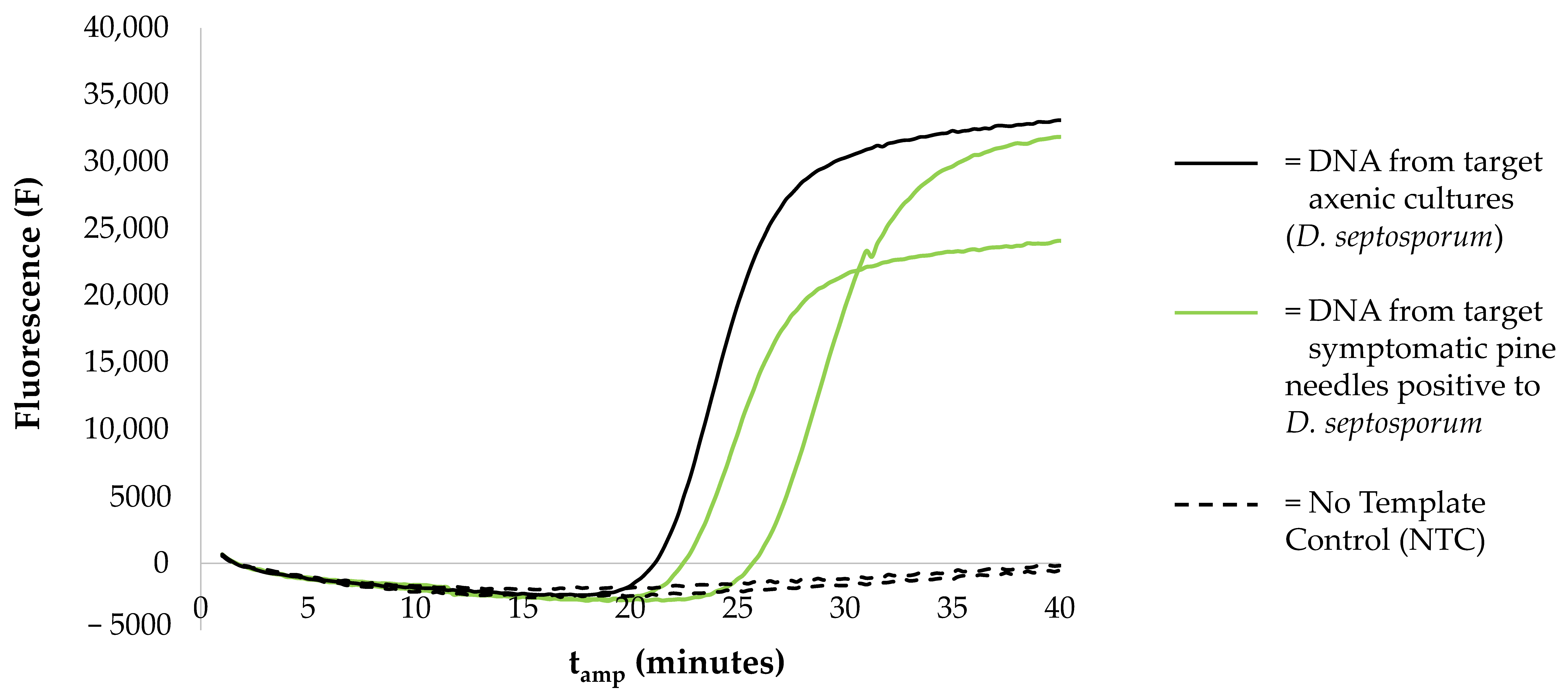
Typical DNB symptoms were present on all needle samples taken in six different groups of pines, two of P. radiata and four of P. nigra subsp. laricio, on the south-facing slopes towards Lake Metramo at elevations ranging from about 900 to 1100 m a.s.l. Trees of both species were defoliated, with transparent crowns and typical lion-tailed branches. These sampling points represent, in the first case, remains of old plantations of the introduced P. radiata, and in the second case, naturally regenerated trees of the native P. nigra subsp. laricio.
Under the stereoscope, necrotic brown spots and black stroma were visible beneath the needle epidermis and typical black conidiomata in the red bands and discolored areas appeared after a few days of incubation in high humidity conditions. Conidia had morphological characteristics consistent with attribution of the species to D. septosporum or D. pini [13], i.e., they were thin-walled, hyaline, smooth, fusiform to short-clavate, straight or more frequently curved, 2–3(5) septate, and measured 12–40 × 2–3 μm.

3.2. LAMP Assays

DNA extracts from pine needles with typical DNB symptoms, with or without fruiting bodies, all tested positive for D. septosporum and negative for both D. pini and L. acicola by applying LAMP optimized assays. Each sample that tested positive for D. septosporum on the portable instrument was amplified in about 20 min with amplification times (tamp), quality of amplification curves and fluorescence (F) (tamp range: 22–23 min, F range: 15,000–30,000) comparable to those obtained on the real time PCR instrument used during the optimization of the assay [15]. No amplification curves were obtained from negative samples. All positive controls were amplified (D. septosporum tamp 20–22 min, F 10,000–30,000; D pini tamp 15–22 min, F 20,000–25,000; L. acicola tamp 21–23 min, F 15,000–25,000), confirming the effectiveness of each run. An example of the reaction kinetics is given in Figure 2.

3.3. Isolation of Fungi and Identification by Sequencing

Numerous dark brown or blackish colonies resembling those of Dothistroma species were obtained from conidia from all the groups of sampled pines. Colonies grew slowly, as is typical of Dothistroma, and stained the substrate red for dothistromin production after 1–2 weeks of growth. Mycelial plugs from 15 axenic colonies were cut from the agar and placed into microeppendorf tubes (1.5 mL), covered with sterile distilled water and stored at 4 °C.
Partial sequences of the tub2 gene region were obtained from four isolates from Pinus nigra subsp. laricio. Amplicon size was ca. 431 bp as expected. After quality check, trimming and alignment, sequences comprised 297–372 base pairs due to poor quality sequences requiring more trimming in some cases. Blast alignment in the GeneBank database showed that all four fungal isolates could be classified as D. septosporum (99.35%–100% sequence identity, best hit D. septosporum strain CMW44656). Sequences were deposited in GenBank under accession numbers (MZ594908-MZ594911).
Dothistroma spp. included in phylogenetic analyses formed a cluster independent from Lecanosticta spp., which were selected as an outgroup (Figure 3). Sequences from the study site (RCDS001, RCDS003, RCDS007, RCDS009) were placed inside the Dothistroma septosporum clade, supporting the conclusion that these isolates belong to this species.

4. Discussion

Since the first report of DNB in the mountains of southern Calabria [11], the cultivation of P. radiata has been almost abandoned in southern Italy [42], and today, forty-four years later, in the area of that first report, only rather small nuclei of pine, especially the native P. nigra subsp. laricio, are found scattered in vast pure beech forests. Now that molecular techniques enable us to discriminate among morphologically indistinguishable species and also to recognize cryptic pathogens, we were able to test the authenticity of the record of Magnani [11], conducting our investigations at exactly the same site, and thus confirm that his observations, never verified later, were correct. In fact, the major revision of the genus Dothistroma, the name of the species of which changed [13], came long after the publication of Magnani’s report. In this study, we can now ascertain that, about half a century after the establishment of P. radiata plantations, Dothistroma is still present in the area, although trees with clear symptoms of the disease are scattered or in small, isolated groups physically separated from each other by non-host species.
In confirming the persistence of the pathogen in the area, we demonstrated that the recently published LAMP diagnostic assays discriminating the infections by DNB agents and L. acicola in pine needles [15], are efficiently applicable on the portable instrument (Genie III Optigene, UK). This result encourages their prospective use directly in the field, after optimization of a suitable crude extraction protocol that is under development. Application of LAMP diagnostics and sequencing in the tub2 gene region confirmed the pathogen found today in the study area to be the species currently identified as D. septosporum. Given the peculiar climatic characteristics of the area due to the orography, the position between two seas and also the additional presence of the artificial lake Metramo, which create conditions of higher atmospheric humidity than in the surrounding areas and favor the survival and spread of the pathogen, it is reasonable to assume that the population detected today likely descends from the one found in 1977, although the data do not allow us to confirm this assertion with certainty.
Due to changes in the politics of afforestation as well as in forest management strategies over the past 50 years, the silvicultural importance of P. radiata in the area seems today to be greatly reduced compared to the past, but the danger posed by D. septosporum to native pines remains serious. In fact, in the area included in this work, as in the Sila Massif [15] (which is also located in Calabria about 150 km north of the present finding), the pathogen was found on the native species P. nigra subsp. laricio. The few natural populations that still exist of this native species of pine are preserved today in Calabria and Sicily, in addition to the few present on the French island of Corsica. The presence of the pathogen therefore represents a threat to the genetic resources of the species.
Outside the Italian territory, reports based on molecular diagnosis of D. septosporum in the central southern Mediterranean area (i.e., Spain, Greece, Turkey) are isolated and sporadic and generally from pine plantations [2,43], with the exception of findings of the fungus in natural forests in Montenegro and southwestern Turkey [44,45,46,47,48]. The scientific debate on the geographic origin of D. septosporum is long and not without controversy [13,49,50,51,52], partly due to the lack of sufficient sampling of the fungus in some regions of its current range. Recently, a large body of work using microsatellite markers for genotyping an unprecedented number of samples collected from across the worldwide range of D. septosporum has indicated Eastern Europe (i.e., the Baltics and Western Russia) and Western Asia as the possible area of origin of the fungus, and hypothesized a possible derivation of Turkish populations from ancestral populations through an ancient separation dating prior to the colonization of Central Europe [53]. In the study by Mullet and colleagues [53], not enough isolates of the fungus from the outbreak in the Sila Massif were included to attempt reconstructing the evolutionary history of that population, while the finding of D. septosporum in the areas of the present study was yet to be confirmed. Whether or not D. septosporum is indigenous to these areas is currently unknown.
The high susceptibility of P. nigra subsp. laricio at all sites where it was planted outside its native range and came into contact with D. septosporum suggests a lack of coevolution with the pathogen. Similarly, the severe damage observed on this pine in its native range in the Sila Massif [17] also points to a recent introduction, although other possible explanations, e.g., an environment made more conducive to disease by climate change, cannot be excluded [54]. However, it is reasonable to assume that the populations of D. septosporum recently found on this pine species in its native range in Southern Italy might be recent introductions. In Corsica, to the best of our knowledge, there is only one published record of DNB, i.e., a single isolate of D. septosporum from P. nigra subsp. laricio included in [53], which has not been transposed yet into the EPPO database (https://gd.eppo.int/taxon/SCIRPI/distribution, accessed on 19 July 2021). According to EPPO, the pathogen was absent from Corsica at least until 2011, when a monitoring campaign was conducted, and no subsequent updates have been reported. Therefore, the finding in Corsica could also follow a recent introduction. No records exist on the occurrence of the pathogen in Sicily, where natural populations of P. nigra subsp. laricio are also present. As a prospect for a future study, a comparative genetic analysis of the Calabrian populations of the fungus could help to define in more detail the history of the pathogen’s spread in Southern Europe.

5. Conclusions

DNA-based approaches are increasingly used to support and extend more classical taxonomic relationships, as well as to unravel complicated taxonomic histories and circumscript species. Molecular identification as D. septosporum of the fungus initially identified as D. pini on the basis of micromorphological characterization alone in the first reported Italian outbreak of the disease is a vivid example. The development of a rapid and highly discriminating diagnostic tool such as the LAMP technique will allow us to identify the agents of DNB in the Mediterranean area more accurately and more specifically. Further genetic analysis could also provide a more complete picture of the current distribution of the causal agents of red band needle blight in the region and insights into the origin of the disease.

Author Contributions

Conceptualization, L.G., S.M., P.C.; Data curation, L.G., C.A., A.B., E.S.; Data analysis, L.G., E.S.; Methodology, L.G., C.A., E.S.; Supervision, L.G., S.M., P.C.; Writing—original draft, L.G., S.M., P.C.; Writing—review and editing, all authors. All authors have read and agreed to the published version of the manuscript.

Funding

Part of this work was funded by the project “Holistic management of emerging forest pests and diseases” (HOMED) a European Union’s Horizon 2020 Programme for Research & Innovation under grant agreement No 771271 to L.G. and P.C.

Data Availability Statement

The sequence data presented in this study were deposited in the NCBI GenBank data repository.

Acknowledgments

The authors wish to thank Sebastiano Monaco, from the Consorzio di Bonifica Tirreno Vibonese and Maurizio Angotti from the A.R.S.A.C., Azienda Regionale per lo Sviluppo dell’Agricoltura in Calabria for their availability and support during field trials. The colleagues who kindly provided DNA of fungal cultures used in this study are also acknowledged: Rein Drenkhan (Estonian University of Life sciences, Estonia), Barbara Piškur (Slovenian Forestry Institute, Slovenia), Josef Janoušek (Phytophthora Research Center, Mendel University, Brno, Czech Republic), Renaud Ioos (Plant Health Laboratory, French Agency for Food, Environmental and Occupational Health & Safety). We warmly thank Maria Teresa Ceccherini and Guido Marchi for providing part of the laboratory supplies and equipment. We would like to thank two anonymous reviewers who helped us improve the manuscript.

Conflicts of Interest

The authors declare no conflict of interest. The funders had no role in the design of the study; in the collection, analyses, or interpretation of data; in the writing of the manuscript, or in the decision to publish the results.

References

  1. Drenkhan, R.; Tomešová-Haataja, V.; Fraser, S.; Bradshaw, R.E.; Vahalik, P.; Mullett, M.S.; Martín-García, J.; Bulman, L.S.; Wingfield, M.J.; Kirisits, T.; et al. Global geographic distribution and host range of Dothistroma species: A comprehensive review. For. Pathol. 2016, 46, 408–442. [Google Scholar] [CrossRef]
  2. Mullett, M.S.; Adamson, K.; Bragança, H.; Bulgakov, T.S.; Georgieva, M.; Henriques, J.; Jürisoo, L.; Laas, M.; Drenkhan, R. New country and regional records of the pine needle blight pathogens Lecanosticta acicola, Dothistroma septosporum and Dothistroma pini. For. Pathol. 2018, 48, e12440. [Google Scholar] [CrossRef]
  3. Barnes, I.; van der Nest, A.; Mullett, M.S.; Crous, P.W.; Drenkhan, R.; Musolin, D.L.; Wingfield, M.J. Neotypification of Dothistroma septosporum and epitypification of D. pini, causal agents of Dothistroma needle blight of pine. For. Pathol. 2016, 46, 388–407. [Google Scholar] [CrossRef]
  4. Gibson, I.A.S. Dothistroma blight of Pinus radiata. Annu. Rev Phytopathol. 1972, 10, 51–72. [Google Scholar] [CrossRef]
  5. Woods, A.; Coates, K.D.; Hamann, A. Is an unprecedented Dothistroma needle blight epidemic related to climate change? BioScience 2005, 55, 761–769. [Google Scholar] [CrossRef] [Green Version]
  6. Barnes, I.; Kirisits, T.; Akulov, A.; Chhetri, D.B.; Wingfield, B.D.; Bulgakov, T.S.; Wingfield, M.J. New host and country records of the Dothistroma needle blight pathogens from Europe and Asia. For. Pathol 2008, 38, 178–195. [Google Scholar] [CrossRef]
  7. Watt, M.S.; Kriticos, D.J.; Alcaraz, S.; Brown, A.V.; Leriche, A. The hosts and potential geographic range of Dothistroma needle blight. For. Ecol Manag. 2009, 257, 1505–1519. [Google Scholar] [CrossRef]
  8. Morelet, M. Une maladie des pins, nouvelle pour la France, due à Scirrhia pini Funk et Parker et à son stade conidien Dothistroma pini Hulbary. Bulletin Mens. Soc. Linn. Lyon 1967, 36, 361–367. [Google Scholar] [CrossRef]
  9. Arthaud, J. Scirrhia pini (Ascomycetes, Dothideaceae) present in the southwest [of France]. Bull. Soc. Linn. Bordx. 1972, 2, 123–124. [Google Scholar]
  10. Fernandez de Ana Magan, F.J. Red Band Disease of Pinus Radiata; Comunicaciones I.N.I.A. Serie Proteccion Vegetal; Ministerio De Agricultura, Instituto Nacional de Investigaciones Agrarias: Madrid, Spain, 1975; Volume 3, p. 16. Available online: http://libros.inia.es/libros/product_info.php?products_id=1113 (accessed on 22 July 2021).
  11. Magnani, G. Presence of Dothistroma pini on Pinus radiata. Cellul. Carta 1977, 28, 37–42. [Google Scholar]
  12. Fratianni, S.; Acquaotta, F. The climate of Italy. In Landscapes and Landforms of Italy; Soldati, M., Marchetti, M., Eds.; World Geomorphological Landscapes; Springer International Publishing: Basel, Switzerland, 2017; pp. 29–38. [Google Scholar] [CrossRef]
  13. Barnes, I.; Crous, P.W.; Wingfield, B.D.; Wingfield, M.J. Multigene phylogenies reveal that red band needle blight of Pinus is caused by two distinct species of Dothistroma, D. septosporum and D. pini. Stud. Mycol. 2004, 50, 551–565. [Google Scholar]
  14. Ioos, R.; Fabre, B.; Saurat, C.; Fourrier, C.; Frey, P.; Marcais, B. Development, comparison, and validation of real-time and conventional PCR tools for the detection of the fungal pathogens causing brown spot and red band needle blights of pine. Phytopathology 2010, 100, 105–114. [Google Scholar] [CrossRef] [Green Version]
  15. Aglietti, C.; Meinecke, C.D.; Ghelardini, L.; Barnes, I.; van der Nest, A.; Villari, C. Rapid detection of pine pathogens Lecanosticta acicola, Dothistroma pini and D. septosporum on needles by probe-based LAMP assays. Forests 2021, 12, 479. [Google Scholar] [CrossRef]
  16. Marchi, G.; Ghelardini, L. Outbreak of Dothistroma septosporum on Corsican Pine in Southern Italy. J. Plant Pathol. 2017, 99, S53. Available online: https://iris.unito.it/retrieve/handle/2318/1651431/368356/Masino%20A.%20et%20al.%20-%20JPP%2099%20%28supp.%20XXIII%20convegno%29.pdf (accessed on 7 July 2021).
  17. Ghelardini, L.; Aglietti, C.; Loria, F.; Cerboneschi, M.; Gionni, A.; Goti, E.; Maresi, G.; Moricca, S.; Marchi, G. Dothistroma needle blight in protected pine forests in Italy. Manag. Biol. Invasions 2020, 11, 689–702. [Google Scholar] [CrossRef]
  18. Farjon, A. Pinus nigra subsp. laricio. IUCN Red List Threat. Species 2013, e.T20453493A20453502. [Google Scholar] [CrossRef]
  19. Bonavita, S.; Vendramin, G.G.; Bernardini, V.; Avolio, S.; Regina, T.M.R. The first SSR-based assessment of genetic variation and structure among Pinus laricio Poiret populations within their native area. Plant Biosyst. 2016, 150, 1271–1281. [Google Scholar] [CrossRef]
  20. Plutino, M.; Pollastrini, M.; Avolio, S.; Bernardini, V. Tree inventory data of Pinus nigra J.F.Arnold subsp. laricio (Poir.) Maire in southern Italy. Ann. Silv. Res. 2018, 42, 43–45. [Google Scholar] [CrossRef]
  21. Villebonne, D.; Maugard, F. Rapid development of Dothistroma needle blight (Scirrhia pini) on Corsican pine (Pinus nigra subsp. laricio) in France. In La Sante des Forets, Annual Report 1998; Les Cahiers du Département de la santé des forêts (DSF); DERF: Paris, France, 1999; Volume 1. [Google Scholar]
  22. Bulman, L.S.; Gadgil, P.D.; Kershaw, D.J.; Ray, J.W. Assessment and control of Dothistroma needle-blight. Res. Bull. 2004, 229, 1176–2632. [Google Scholar]
  23. Sturrock, R.N.; Frankel, S.J.; Brown, A.V.; Hennon, P.E.; Kliejunas, J.T.; Lewis, K.J.; Worrall, J.J.; Woods, A.J. Climate change and forest diseases: Climate change and forest diseases. Plant Pathol. 2011, 60, 133–149. [Google Scholar] [CrossRef]
  24. Fraser, S.; Brown, A.V.; Woodward, S. Intraspecific variation in susceptibility to Dothistroma needle blight within native Scottish Pinus sylvestris. Plant Pathol. 2015, 64, 864–870. [Google Scholar] [CrossRef]
  25. Fraser, S.; Woodward, S.; Brown, A.V. Inter- and intraspecific variation in susceptibility to Dothistroma needle blight in Britain. How susceptible are Pinus sylvestris and Pinus contorta? For. Pathol. 2016, 46, 534–546. [Google Scholar] [CrossRef]
  26. Mullett, M.; Peace, A.; Brown, A. Persistence of Dothistroma septosporum on abscised pine needles and its implications for disease management. Plant Dis. 2016, 100, 1271–1277. [Google Scholar] [CrossRef] [PubMed] [Green Version]
  27. Pebesma, E. Simple Features for R: Standardized Support for Spatial Vector Data. R J. 2018, 10, 439–446. [Google Scholar] [CrossRef] [Green Version]
  28. Wickham, H. ggplot2: Elegant Graphics for Data Analysis, 2nd ed.; Springer: New York, NY, USA, 2016; p. 260. [Google Scholar] [CrossRef]
  29. R Core Team. R: A Language and Environment for Statistical Computing; R Foundation for Statistical Computing: Vienna, Austria, 2020; Available online: https://www.R-project.org/ (accessed on 8 July 2021).
  30. Mullett, C.M.; Barnes, I. Dothistroma Isolation and Molecular Identification Methods. In Detection and Diagnostics of Dothistroma; COST ACTION FP1102 Determining Invasiveness and Risk of Dothistroma, Training School May 2012, Brno, Czech Republic. Available online: https://www.forestresearch.gov.uk/documents/305/DIAROD_052012_Isolation_and_indentification_97fNCCI.pdf (accessed on 7 July 2021).
  31. Carter-House, D.; Stajich, J.E.; Unruh, S.; Kurbessoian, T. Fungal CTAB DNA Extraction. Protocols.io 2020. [Google Scholar] [CrossRef]
  32. Woudenberg, J.H.C.; Aveskamp, M.M.; de Gruyter, J.; Spiers, A.G.; Crous, P.W. Multiple Didymella teleomorphs are linked to the Phoma clematidina morphotype. Persoonia 2009, 22, 56–62. [Google Scholar] [CrossRef] [Green Version]
  33. Okonechnikov, K.; Golosova, O.; Fursov, M.; the UGENE Team. Unipro UGENE: A unified bioinformatics toolkit. Bioinformatics 2012, 28, 1166–1167. [Google Scholar] [CrossRef] [Green Version]
  34. Notredame, C.; Higgins, D.G.; Heringa, J. T-coffee: A novel method for fast and accurate multiple sequence alignment. J. Mol. Biol. 2000, 302, 205–217. [Google Scholar] [CrossRef] [Green Version]
  35. Brown, N.P.; Leroy, C.; Sander, C. MView: A web-compatible database search or multiple alignment viewer. Bioinformatics 1998, 14, 380–381. [Google Scholar] [CrossRef]
  36. Madeira, F.; Park, Y.M.; Lee, J.; Buso, N.; Gur, T.; Madhusoodanan, N.; Basutkar, P.; Tivey, A.R.N.; Potter, S.C.; Finn, R.D.; et al. The EMBL-EBI search and sequence analysis tools APIs in 2019. Nucleic Acids Res. 2019, 47, W636–W641. [Google Scholar] [CrossRef] [Green Version]
  37. Bolyen, E.; Rideout, J.R.; Dillon, M.R.; Bokulich, N.A.; Abnet, C.C.; Al-Ghalith, G.A.; Alexander, H.; Alm, E.J.; Arumugam, M.; Asnicar, F.; et al. Reproducible, interactive, scalable and extensible microbiome data science using QIIME 2. Nat. Biotechnol. 2019, 37, 852–857. [Google Scholar] [CrossRef] [PubMed]
  38. Katoh, K.; Standley, D.M. MAFFT Multiple Sequence Alignment Software Version 7: Improvements in performance and usability. Mol. Biol. Evol. 2013, 30, 772–780. [Google Scholar] [CrossRef] [PubMed] [Green Version]
  39. Lane, D. 16S/23S rRNA Sequencing. In Nucleic Acid Techniques in Bacterial Systematics; Stackebrandt, E., Goodfellow, M., Eds.; John Wiley and Sons: New York, NY, USA, 1991; pp. 115–175. [Google Scholar]
  40. Stamatakis, A. RAxML version 8: A tool for phylogenetic analysis and post-analysis of large phylogenies. Bioinformatics 2014, 30, 1312–1313. [Google Scholar] [CrossRef] [PubMed]
  41. Stamatakis, A.; Hoover, P.; Rougemont, J. A Rapid Bootstrap Algorithm for the RAxML Web Servers. Syst. Biol. 2008, 57, 758–771. [Google Scholar] [CrossRef] [PubMed]
  42. Ciancio, O.; Garfi, V.; Iovino, F.; Menguzzato, G. Il pino insigne nell’arboricoltura da legno: Analisi di interventi realizzati nell’Italia meridionale. Ital. For. Mont. 2006, 61, 217–231. Available online: http://ojs.aisf.it/index.php/ifm/article/view/36 (accessed on 7 July 2021). [CrossRef] [Green Version]
  43. Tsopelas, P.; Barnes, I.; Soulioti, N.; Wingfield, M.J. Dothistroma septosporum identified in Greece on Pinus brutia and Pinus nigra plantations. Plant Dis. 2013, 97, 1247. [Google Scholar] [CrossRef]
  44. Doğmuş-Lehtijärvi, H.T.; Lehtijärvi, A.; Oskay, F.; Aday Kaya, A.G.; Örtel, E.; Datumani, A. Dothistroma needle blight in Turkey. In Book of Abstract, IUFRO WP 7.02.02 Foliage Shoot and Stems Diseases: Biosecurity in Natural Forests and Plantations, Genomics and Biotechnology for Biosecurity and Forestry; Cerno Hora and Brno,: Czech Republic, 20–25 May 2013; Mendel University: Brno, Czech Republic, 2013; pp. 68–69. [Google Scholar]
  45. Tunalı, Z.; Doğmuș-Lehtİjärvİ, H.T.; Oskay, F. Detection of Fungal Needle Disease Agents of Turkish Pine (Pinus brutia Ten.) forests in Burdur Province Using Molecular Techniques. Süleyman Demirel Üniversitesi Fen Bilimleri Enstitüsü Dergisi; 2018; Volume 22, pp. 628–636, (In Turkish with English Abstract); Available online: https://www.cabdirect.org/cabdirect/abstract/20203105032 (accessed on 7 July 2021).
  46. Oskay, F.; Tunalı, Z.; Lehtijärvi, A.T.; Doğmuş-Lehtijärvi, H.T.; Woodward, S.; Mullett, M. Distribution and genetic diversity of Dothistroma septosporum in Pinus brutia forests of south-western Turkey. Plant Pathol. 2020, 69, 1551–1564. [Google Scholar] [CrossRef]
  47. Lazarević, J.; Davidenko, K.; Millberg, H. Incidence of Dothistroma septosporum in different pine forests in Montenegro. Mycol. Montenegrina 2014, 17, 119–131. [Google Scholar]
  48. Lazarević, J.; Davidenko, K.; Millberg, H. Dothistroma needle blight on high altitude pine forests in Montenegr. Balt. For. 2017, 23, 294–302. [Google Scholar]
  49. Adamson, K.; Mullett, M.S.; Solheim, H.; Barnes, I.; Müller, M.M.; Hantula, J.; Vuorinen, M.; Kačergius, A.; Markovskaja, S.; Musolin, D.L.; et al. Looking for relationships between the populations of Dothistroma septosporum in northern Europe and Asia. Fungal Genet. Biol. 2018, 110, 15–25. [Google Scholar] [CrossRef]
  50. Dale, A.L.; Lewis, K.J.; Murray, B.W. Sexual reproduction and gene flow in the pine pathogen Dothistroma septosporum in British Columbia. Phytopathology 2011, 101, 68–76. [Google Scholar] [CrossRef] [PubMed] [Green Version]
  51. Mullett, M.S.; Brown, A.V.; Fraser, S.; Baden, R.; Tubby, K.V. Insights into the pathways of spread and potential origins of Dothistroma septosporum in Britain. Fungal Ecol. 2017, 26, 85–98. [Google Scholar] [CrossRef]
  52. Capron, A.; Feau, N.; Heinzelmann, R.; Barnes, I.; Benowicz, A.; Bradshaw, R.E.; Dale, A.; Lewis, K.J.; Owen, T.J.; Reich, R.; et al. Signatures of post-glacial genetic isolation and human-driven migration in the Dothistroma needle blight pathogen in Western Canada. Phytopathology 2021, 111, 116–127. [Google Scholar] [CrossRef] [PubMed]
  53. Mullett, M.S.; Drenkhan, R.; Adamson, K.; Boroń, P.; Lenart-Boroń, A.; Barnes, I.; Tomšovský, M.; Jánošíková, Z.; Adamčíková, K.; Ondrušková, E.; et al. Worldwide genetic structure elucidates the Eurasian origin and invasion pathways of Dothistroma septosporum, causal agent of Dothistroma needle blight. J. Fungi 2021, 7, 111. [Google Scholar] [CrossRef] [PubMed]
  54. Woods, A.J.; Martín-García, J.; Bulman, L.; Vasconcelos, M.W.; Boberg, J.; La Porta, N.; Peredo, H.; Vergara, G.; Ahumada, R.; Brown, A.; et al. Dothistroma needle blight, weather and possible climatic triggers for the disease’s recent emergence. For. Pathol. 2016, 46, 443–452. [Google Scholar] [CrossRef] [Green Version]
Figure 1. Map position of the study area of Prateria (red symbols) in the center of the Mediterranean region (a,b) on the south-facing slope of the Serre mountains towards the Metramo Lake (San Pietro di Caridà, Reggio Calabria, Italy) (c). The approximate local distribution range of Pinus nigra subsp. laricio (adapted from the European Forest Genetic Resources Programme, EUFORGEN http://www.euforgen.org/species/pinus-nigra/ (accessed on 3 July 2021)) is shown in magenta coloring (b). Maps were drawn using the sf [27] and ggplot2 [28] packages in R [29].
Forests 12 00983 g001
Figure 2. Selection of kinetics showing amplification results of the LAMP assay for the detection of D. septosporum [15] on a portable instrument (Genie III OptiGene, Limited, Horsham, UK).
Forests 12 00983 g002
Figure 3. Phylogenetic tree with maximum likelihood score (branch support computed out of 1000 rapid bootstrapped trees) based on partial tub2 gene sequences of isolates RCDSS001, RCDS003, RCDS007, RCDS009 from Pinus nigra subsp. laricio in Prateria (San Pietro di Caridà, Reggio Calabria, Italy) and of Dothistroma species isolates retrieved from GenBank.
Forests 12 00983 g003
Publisher’s Note: MDPI stays neutral with regard to jurisdictional claims in published maps and institutional affiliations.

