Article

Antiviral RNA Interference Activity in Cells of the Predatory Mosquito, Toxorhynchites amboinensis

by
Claire L. Donald
1,*,
Margus Varjak
1,
Eric Roberto Guimarães Rocha Aguiar
2,†,
João T. Marques
2,
Vattipally B. Sreenu
1,
Esther Schnettler
1,‡,§ and
Alain Kohl
1,*
1
MRC-University of Glasgow Centre for Virus Research, Glasgow, Scotland G61 1QH, UK
2
Departamento de Bioquímica e Imunologia, Instituto de Ciências Biológicas, Universidade Federal de Minas Gerais, 6627-Pampulha-Belo Horizonte-MG, CEP 31270-901, Brazil
*
Authors to whom correspondence should be addressed.
Current address: Instituto de Ciências da Saúde, Universidade Federal da Bahia, Salvador, BA 40110-100, Brazil.
Current address: Bernhard-Nocht-Institute for Tropical Medicine, Bernhard-Nocht-Strasse 74, 20359 Hamburg, Germany.
§
Current address: German Centre for Infection Research (DZIF), Partner Site Hamburg-Lübeck-Borstel-Riems, 20359 Hamburg, Germany.
Submission received: 10 October 2018 / Revised: 19 November 2018 / Accepted: 4 December 2018 / Published: 6 December 2018
(This article belongs to the Special Issue Breakthroughs in Viral Replication)

Abstract

:
Arthropod vectors control the replication of arboviruses through their innate antiviral immune responses. In particular, the RNA interference (RNAi) pathways are of notable significance for the control of viral infections. Although much has been done to understand the role of RNAi in vector populations, little is known about its importance in non-vector mosquito species. In this study, we investigated the presence of an RNAi response in Toxorhynchites amboinensis, which is a non-blood feeding species proposed as a biological control agent against pest mosquitoes. Using a derived cell line (TRA-171), we demonstrate that these mosquitoes possess a functional RNAi response that is active against a mosquito-borne alphavirus, Semliki Forest virus. As observed in vector mosquito species, small RNAs are produced that target viral sequences. The size and characteristics of these small RNAs indicate that both the siRNA and piRNA pathways are induced in response to infection. Taken together, this data suggests that Tx. amboinensis are able to control viral infections in a similar way to natural arbovirus vector mosquito species. Understanding their ability to manage arboviral infections will be advantageous when assessing these and similar species as biological control agents.

1. Introduction

Toxorhynchites (Diptera: Culicidae) mosquitoes or “elephant mosquitoes” are the largest mosquitoes on the planet, with a wingspan surpassing 12 mm for some species [1]. Unlike most mosquito species, they are autogenous and do not require a blood meal for egg production. Instead, all instars of larvae are predatory against other mosquito larvae, including those of medical relevance, such as Aedes aegypti, which is a key vector for many important human arboviruses. As a result, various species, such as Tx. splendens, Tx. rutilus, and Tx. amboinensis, have been proposed as biological control agents against pest species [2,3,4].
As a consequence of adult Toxorhynchites being exclusively nectarivorous, they are not considered to be natural vectors for arboviruses. However, previous work has demonstrated that several species of Toxorhynchites are susceptible to important arboviruses and, as such, have a role as artificial hosts for their detection and propagation. Tx. amboinensis, Tx. brevipalpis, Tx. rutilus rutilus, Tx. theobaldi, and Tx. splendens have previously been shown to be susceptible to a number of flaviviruses including dengue virus (DENV) serotypes 1–4, Japanese encephalitis virus (JEV), yellow fever virus (YFV), and Zika virus (ZIKV) [5,6,7]. Furthermore, certain species have also demonstrated the capacity for generating greater viral titers, especially each of the four DENV serotypes, compared to vector species or mammalian cells commonly used to produce virus. For instance, Tx. amboinensis and Tx. brevipalpis generate greater titers of DENV compared to Ae. albopictus or their derived cell line, C6/36 [5,8]. Tx. amboinensis were also shown to be susceptible to JEV and allowed it to replicate to high titres [5]. In addition to DENV and other flaviviruses, Tx. amboinensis have been shown to efficiently propagate alphaviruses (chikungunya (CHIKV), Ross River (RRV), and Venezuelan equine encephalitis (VEEV) viruses) and bunyaviruses (La Crosse (LACV), San Angelo (SAV), and Keystone (KEYV)) viruses [5,9,10].
Several continuous cell lines have been derived from Toxorhynchites to facilitate virus propagation and isolation in vitro. Cell cultures derived from Tx. amboinensis have been established which show comparative levels of sensitivity as the adults and commonly used vector cell lines to DENV and other arboviruses [11,12,13,14]. These cultures provide a useful in vitro system for the study of interactions between arboviruses and Toxorhynchites mosquitoes.
Despite their usability for the propagation of arboviruses, nothing is known about the antiviral responses in this mosquito genus. In nature, Toxorhynchites spp. may become exposed to arboviruses by predating on vertically infected larvae [15], and it is therefore valuable to understand their antiviral capabilities when considering their use as an alternative to chemical pesticides against vector species. Historically, much of our understanding of mosquito immunity came from extensive research carried out in the Drosophila melanogaster model, although an increasingly detailed picture of mosquito immunity in vector species is now emerging which highlights a number of key differences [16,17,18,19,20,21]. The major antiviral mechanism for the control of arboviral infections in mosquitoes is RNA interference (RNAi), which is divided into several pathways differentiated by their effector proteins, small RNA molecules, and their method of induction. The exogenous small interfering RNA (exo-siRNA), and to a lesser extent, the PIWI-interacting RNA (piRNA) pathways are highly important in the context of a viral infection [22,23,24,25,26,27,28,29,30,31,32,33,34,35,36,37,38,39]. The exo-siRNA pathway detects the production of virus-derived long double-stranded RNA (dsRNA). These dsRNAs are cleaved into 21 nucleotide (nt) long virus-specific siRNAs (vsiRNAs) by the exoribonuclease, Dicer 2 (Dcr2). The vsiRNAs are transferred to the RNA-induced silencing complex (RISC) and loaded into the effector protein, Argonaute 2 (Ago2). While one strand of the vsiRNA duplex is degraded, Ago2 uses the other strand to recognize complementary viral RNA, which leads to the cleavage and degradation of the target sequence. The piRNA pathway is not as well-characterized and it’s antiviral role(s) are less clear [40]. It also differs considerably in mosquitoes compared to D. melanogaster [41]. In D. melanogaster, the pathway involves PIWI proteins Piwi, Aub, and Ago3. However, Ae. aegypti lack orthologues of Aub and Piwi, but express Ago3 and an additional 7 PIWI family proteins, Piwi1-7 [41]. The pathway involves piRNA molecules, which are between 24–29 nt in length and are generated through a “ping-pong” amplification system. Intermediate piRNAs are initially produced against genomic transposons and display a characteristic uridine as the first nucleotide (U1). These are loaded into the Piwi complex and are further processed to produce mature piRNAs with an adenine at the 10th nucleotide position (A10). The mature piRNAs are bound by Ago3 and target complementary antisense RNA transcripts to produce more piRNAs. Therefore, a typical characteristic of ping-pong derived piRNAs is not only the A10 and U1 bias but also a high frequency of 10 nt complementarity to opposing small RNAs.
In this study, we describe an active antiviral immune response in Tx. amboinensis-derived TRA-171 cells. Our observations indicate that these cells possess a functional RNAi response that is effective against Semliki Forest virus (SFV, Togaviridae, Alphavirus) infection. We used deep sequencing analysis to show the production of both vsiRNAs and virus-specific piRNAs derived from SFV. In addition, silencing assays showed that RNAi responses are induced by the presence of sequence-specific dsRNA against both viral RNAs and mRNAs transcribed from transfected plasmid DNA. This evidence suggests that Tx. amboinensis is able to mount a classical RNAi immune response against viral infections in a similar manner to what is known for mosquito vector species.

2. Materials and Methods

2.1. Cell Lines

Tx. amboinensis-derived TRA-171 cells (European Collection of Authenticated Cell Cultures (ECACC), 90120514) were grown in media prepared in house consisting of L-15 (Leibovitz) growth culture medium (Life Technologies, Carlsbad, CA, USA) mixed 1:1 with Mitsuhashi and Maramorosch basal media prepared in house (CaCl22H2O (250 mg/lt), MgCl26H2O (125 mg/lt), KCl (250 mg/lt), NaHCO3 (150 mg/lt), NaCl (8750 mg/lt), NaH2PO4H2O (250 mg/lt), d-glucose (500 mg/lt), lactalbumin hydrolysate (8125 mg/lt), and yeastolate (0.75 mL/lt)) supplemented with 10% tryptose phosphate broth (TPB, Life Technologies), 10% fetal bovine serum (FBS, Life Technologies), 0.05% bovine serum albumin (BSA) (Sigma-Aldrich, St. Louis, MO, USA), 1% non-essential amino acids (Sigma-Aldrich), and penicillin-streptomycin (final concentration 100 units/mL, 100 µg/mL respectively, Life Technologies). D. melanogaster-derived S2 cells [38,42] were cultured in Schneider’s growth media supplemented with 10% FBS and penicillin-streptomycin (final concentration 100 units/mL, 100 µg/mL, respectively). TRA-171 and S2 cells were maintained at 28 °C with no additional CO2. Baby hamster kidney (BHK-21) [43] cells were grown in Glasgow’s minimal essential medium (GMEM, Life Technologies) supplemented with 10% TPB, 10% newborn calf serum (NBCS, Life Technologies), and penicillin-streptomycin (final concentration 100 units/mL, 100 µg/mL, respectively) at 37 °C with 5% CO2.

2.2. Viruses

The prototype molecular clone of SFV, SFV4, and two derived reporter viruses either expressing Firefly luciferase (FFLuc) (SFV4(3H)-FFLuc) or Renilla luciferase (RLuc) (SFV4(3H)-RLuc) inserted between duplicated nsP2 cleavage sites at the nsP3/4 junction, were grown and titered by plaque assay in BHK-21 cells, as described previously [24]. Infections were performed for 1 h at 28 °C by diluting virus stocks in the appropriate volume of PBSA (phosphate buffered saline with 0.75% BSA) before removing the inoculum and applying fresh growth media.

2.3. Plasmids

The FFLuc and RLuc luciferase expression plasmids, pIZ-Fluc and pAcIE1-Rluc, have been previously described [37,38,44,45].

2.4. In Vitro Transcription of dsRNA

dsRNA molecules against either FFLuc or RLuc were produced using a T7 RNA polymerase in vitro transcription kit (Megascript RNAi kit, Ambion, Foster City, CA, USA) using a PCR product template flanked by T7 RNA polymerase promoter sequences. pIZ-Fluc [45] and pRL-CMV (Promega, Madison, WI, USA) were used as a template for the amplification of dsRNAs targeting FFLuc and RLuc, respectively. An eGFP-derived dsRNA was taken as a control. This sequence was obtained from a gel-purified PCR product using peGFP-C1 (Clontech, Mountain View, CA, USA) as a template. Primer sequences can be found in Table S1.
Internally radio-labelled dsRNAs were prepared by combining 5 µL 114 nt eGFP PCR product with T7 polymerase sites, 4 µL 5× Transcription buffer (Ambion), 2 µL DTT (0.1 M, Invitrogen, Carlsbad, CA, USA), 1 µL rNTPs (10 mM each ATP, GTP and UTP with 0.1 mM CTP) (Promega), 3 µL α-32P rCTP (Perkin Elmer, Waltham, MA, USA), 1 µL T7 RNA polymerase (Ambion), 1 µL RNase inhibitor (Invitrogen), and 3 µL RNase/DNase free H2O. The reaction was incubated for 1 to 3 h at 37 °C before heating to 65 °C for 5 min and allowing it to gradually cool to room temperature. Following this, 2 µL of DNase I and 1 µL of RNase A were added and the reaction was incubated for a further 30 min at 37 °C. The dsRNA could then be purified by running on an 8% native acrylamide gel.

2.5. Nucleic Acid Transfection

Cells were seeded at a density of 2.2 × 10⁵ cells per well of a 24-well plate 24 h prior to transfection with Dharmafect 2 (GE Healthcare, Chicago, IL, USA) following the manufacturer’s instructions. For plasmid transfection experiments, each well was co-transfected with 100 ng pIZ-Fluc, 100 ng pAcIE1-Rluc (as an internal control) with either 1 ng of dsRNA (FFLuc-specific or eGFP control dsRNA), or 2 ng siRNAs (FFLuc-specific or Hygromycin B resistance gene control siRNA [39]). Cells were lysed 24 h post transfection (p.t.) and luciferase activity determined.
For infection experiments, viral reporter gene transcripts were silenced by transfecting 50 ng RLuc or eGFP-specific control dsRNA. After 24 h, cells were infected with SFV4(3H)-RLuc at a multiplicity of infection (MOI) of 0.005. Cells were lysed 24 h post infection (p.i.) and luciferase activities were determined.

2.6. Luciferase Assay

The cells were lysed in Passive Lysis Buffer (Promega) and luciferase expression was determined with either the Renilla-Glo Luciferase assay system (Promega) or the Dual Luciferase assay system (Promega) and a GloMax luminometer.

2.7. Small RNA Sequencing and Analysis

The cells were grown at a density of 9 × 10⁵ per well of a six-well plate. RNA extraction was performed using 1 mL TRIzol (Life Technologies) as per the manufacturer’s instructions, with the addition of glycogen as a carrier. DNA libraries consisting of small RNAs between 15–40 nt were gel purified and sequenced using the Illumina Hiseq 4000 platform at BGI Tech (Shenzhen, China). Data analysis was carried out as described previously by aligning sequence reads to the SFV4 reference genome (Genbank accession number: KP699763) [24,37]. A maximum of one mismatch or indel was allowed in the alignments. Alignment lengths between 18–36 nt were selected for further analysis. These were separated into two groups according to their orientation, i.e., if they mapped to the genome (positive) or antigenome (negative). Coverage plots of mapped reads were generated using R programming language. Sequence logos were generated using a Bioconductor package, motifStack [46]. The mapping positions of each small RNA that aligned to the positive strand of the SFV4 genome was compared to the positions of small RNAs that aligned to the negative strand (antigenome). Any overlaps between these were recorded. Similarly, small RNA pairs of 25–29 nt were compared and their overlapping nucleotide frequencies were aggregated [47]. Using python program coding and the R statistical package, standard scores (z-scores) of the overlapping nucleotides and their frequencies were calculated and plotted. Small RNA sequencing data is available at Sequence Read Archive (https://0-www-ncbi-nlm-nih-gov.brum.beds.ac.uk/sra) under the accession number: PRJNA486770.

2.8. In Vitro Dicer Cleavage Assay

TRA-171 and S2 cells were seeded at 9 × 10⁵ and 1 × 10⁶ cells per well of a six-well plate respectively. Following a 24 h incubation, the media was removed. The cells were re-suspended in sterile PBS and centrifuged for 5 min at 1500 rpm. The supernatant was removed and the procedure repeated a second time as described. Following this, the pellet was re-suspended in 200 µL 1× lysis buffer (10 mM MgAc (Sigma-Aldrich) and 150 mM Hepes-KOH (pH 7.5)) and homogenized using a micro-pestle. A further centrifugation step (14,000 rpm for 20 min at 4 °C) was performed to remove cell debris, after which 5 µL of the supernatant was transferred to a fresh tube. To this was added, 3 µL 32P labelled dsRNA, 1 µL H2O, and a 3 µL creatine mix (consisting of 1 µL DTT (1 M), 10 µL creatine phosphate (12 mg/100 µL) (Calbiochem, San Diego, CA, USA), 20 µL 5× lysis buffer (10 mM MgAc and 150 mM Hepes-KOH (pH 7.5)), 20 µL glycerol, 2 µL RNase Inhibitor (Promega), 2 µL ATP (100 mM) (Thermo Fisher Scientific, Waltham, MA, USA), 0.3 µL 20 mg/mL creatine phosphate kinase in 1× storage buffer (40 mg/mL lyophilized creatine kinase (Calbiochem) in 2× storage buffer (ice-cold 40 mM tris-acetate (pH 6.8), 200 mM KAc, 0.2 mM EDTA, 20 mM β-mercaptoethanol) diluted 1:1 in an equal volume of 100% ice-cold glycerol), and 4.7 µL RNase/DNase free H2O). The reactions were incubated at 28 °C overnight. Subsequently, 200 µL 2× PK buffer (200 mM Tris (pH 7.5), 300 mM NaCl, 5 mM EDTA, 2% SDS), 1 µL glycogen (10 mg/mL) (Roche, Basel, Switzerland), and 0.3 µL proteinase K (10 mg/mL) (Sigma-Aldrich) were added and each reaction was incubated for a further 10 min at 65 °C. Following this, 200 µL phenol/chloroform/isoamylalcohol (25:24:1) (Ambion) was added. Reactions were vortexed for 15 sec and centrifuged for 10 min at 10,000 rpm. The aqueous phase was transferred to a new tube containing 450 µL ice-cold 96% EtOH and centrifuged at 13,000 rpm for 10 min. The supernatant was removed and the pellet washed with 400 µL ice cold 70% EtOH. The samples were further centrifuged as described and the supernatant was removed. The resulting pellets were air dried for 5–10 min prior to resuspension in 15 µL 2× RNA gel loading buffer (Thermo Fisher Scientific). The samples were boiled for 5 min at 65 °C before placing on ice for 2 min. On completion, the samples were loaded onto a 0.75 mm 12% denaturing acrylamide gel with 0.96% urea chilled by submerging the tank in ice-cold water. Electrophoresis was carried out at 200 V. The gel was then transferred to a gel dryer and allowed to dry at 80 °C for 2 h. The resulting bands were detected by exposing the gel to a phosphor imaging screen for ≥16 h and viewed using a personal molecular imager (Bio-Rad, Hercules, CA, USA).

2.9. Statistical Analysis

Statistical analysis was performed using GraphPad Prism. Data was analyzed using an unpaired, two-tailed t-test.

2.10. Data Availability

Source data for the figures can be found at http://0-dx-doi-org.brum.beds.ac.uk/10.5525/gla.researchdata.703. Small RNA sequencing data can be found under the accession number described above.

3. Results

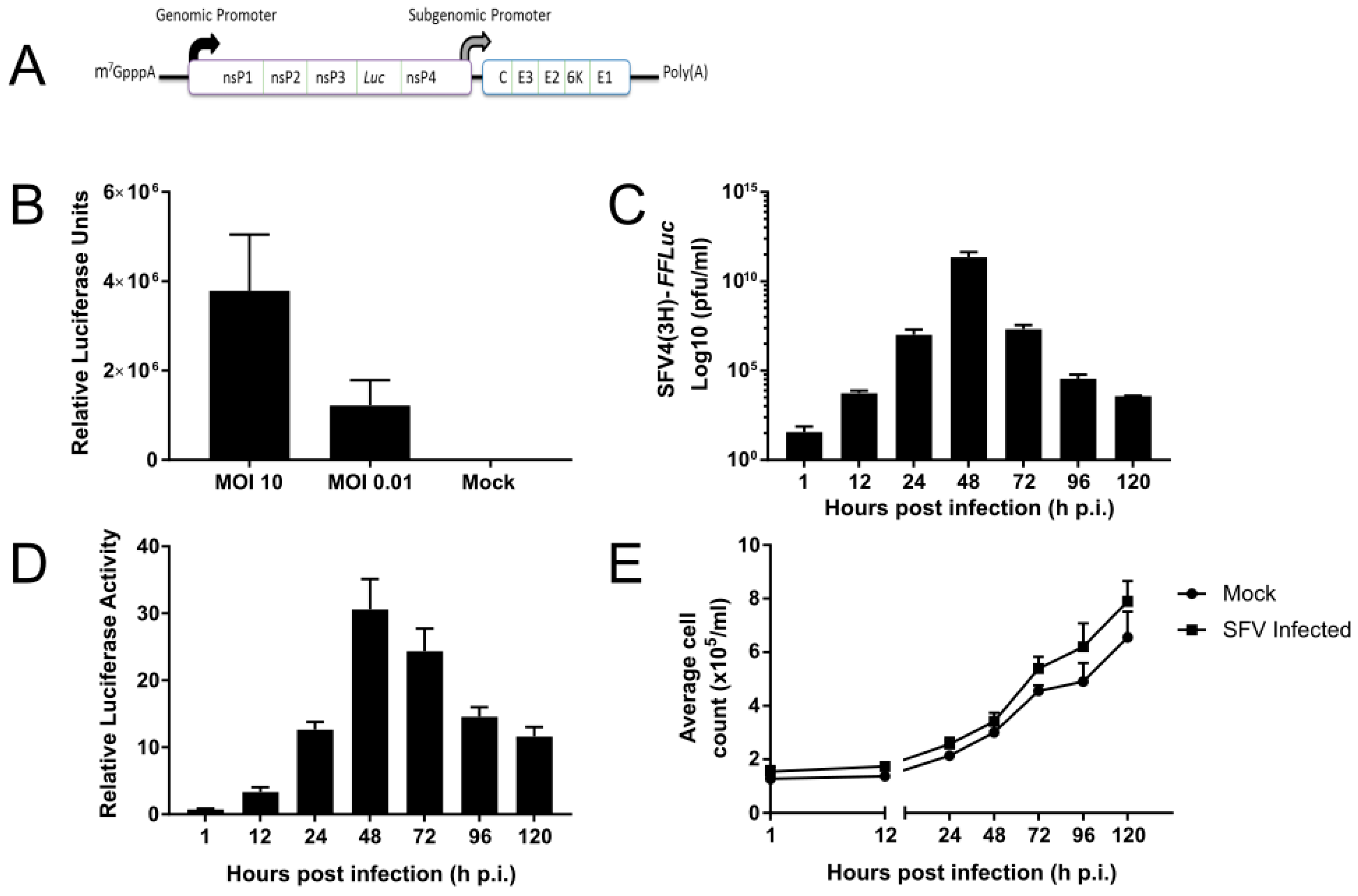
3.1. SFV Infects Tx. amboinensis-Derived TRA-171 Cells

Tx. amboinensis-derived TRA-171 cells are known to be permissive to infection by CHIKV [14], but it has not been shown that they can be infected by SFV, a related alphavirus. To answer this question, TRA-171 cells were infected with SFV expressing luciferase (either RLuc or FFLuc) (Figure 1A), which allows replication to be monitored directly. Cells were infected with SFV4(3H)-RLuc at a high (10) or low (0.01) MOI and incubated for 24 h prior to lysing. As anticipated, proportional SFV replication was detected at each MOI used (Figure 1B). To understand virus kinetics during infection, the cells were infected with a second SFV reporter strain (SFV4(3H)-FFLuc) at MOI 10 and its replication efficiency was monitored at regular intervals over 120 h. Both viral titres (Figure 1C) and luciferase expression (Figure 1D) peaked after 48 h p.i. before decreasing. It was also determined that cell numbers between infected and uninfected cultures were similar and increased at a comparable rate over the 120 h observation period (Figure 1E). These observations are consistent with previous studies that show a similar pattern of SFV infection in Ae. albopictus-derived cell lines [48,49]. This data therefore suggests that SFV infection in the TRA-171 cell line displays similar kinetics to an arboviral infection in vector cells.

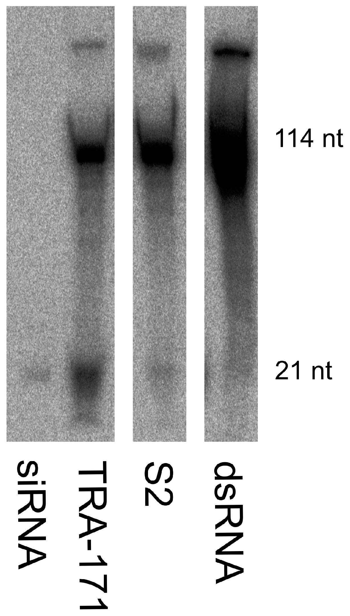
3.2. Functional RNAi Pathways are Present in TRA-171 Cells

A distinguishing feature of the exo-siRNA pathway in vector mosquitoes is that, through Dcr2 cleavage of dsRNA, it produces 21 nt siRNAs that are complementary to the target sequence. To investigate if TRA-171 cells express a functional Dicer enzyme and are thereby capable of generating siRNAs, we used an in vitro Dicer cleavage assay. The cell extracts were incubated with 32P internally radio-labeled dsRNA and incubated overnight before isolating the small RNAs. Extracts were run on an acrylamide gel alongside size markers; input dsRNA (114 nt) and siRNAs (21 nt), as well as an extract from S2 cells as a positive control. Both samples showed the detection of input dsRNA in addition to discernible bands at the size expected for 21 nt siRNAs (Figure 2, Figure S1). This data suggests that TRA-171 cells possess an active dicing enzyme that is effectively able to cleave long dsRNA molecules into small RNAs of approximately the expected size for siRNAs.
The siRNA pathway is induced in a sequence specific manner through the detection of dsRNAs by Dcr2. To determine the ability of TRA-171 cells to silence a FFLuc reporter gene via the RNAi pathway, we performed a previously described reporter RNAi assay [50]. Cells were co-transfected with both pIZ-Fluc and pAcIE1-Rluc (as an internal control), as well as either FFLuc-specific or negative control dsRNA/siRNAs. Each condition was lysed 24 h p.t. and luciferase expression assessed. Our data demonstrates that the cells that received either FFLuc-specific dsRNAs (Figure 3A) or siRNAs (Figure 3B) showed a significant decrease in relative luciferase activity compared to the cells treated with control dsRNA/siRNAs. This suggests that TRA-171 cells are able to induce a gene silencing response, which is mediated by the presence of both sequence-specific dsRNA and siRNAs.

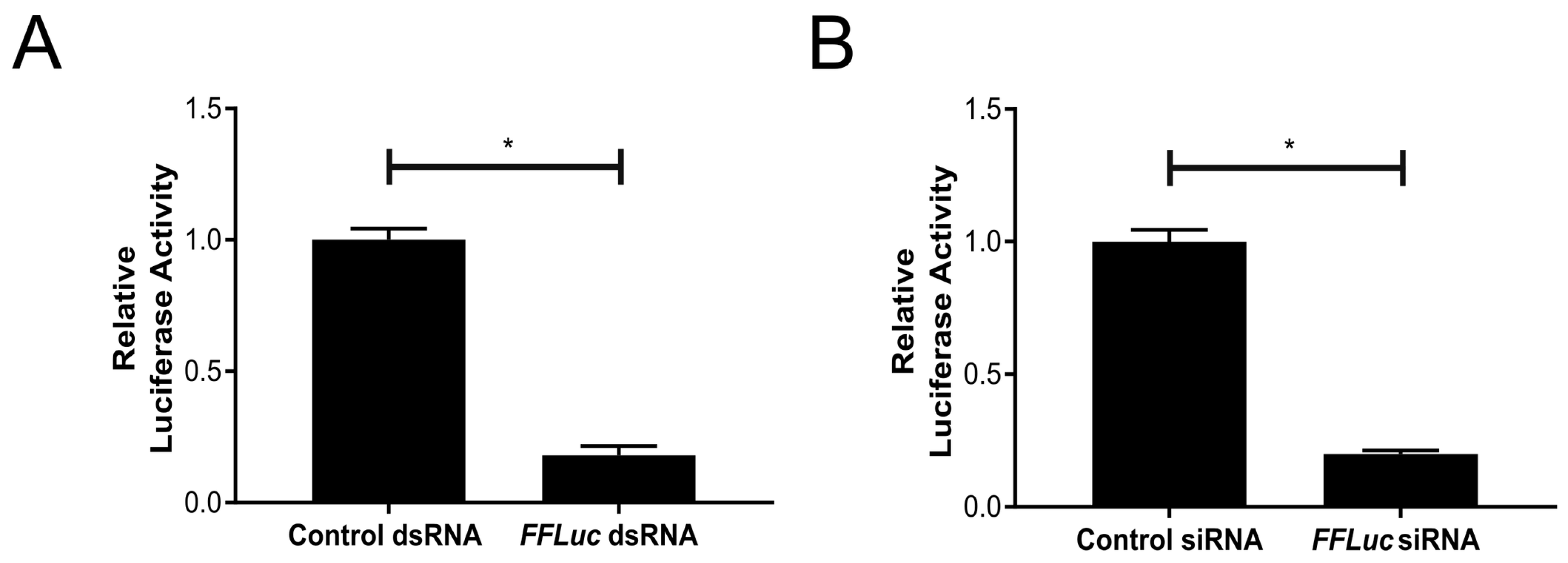
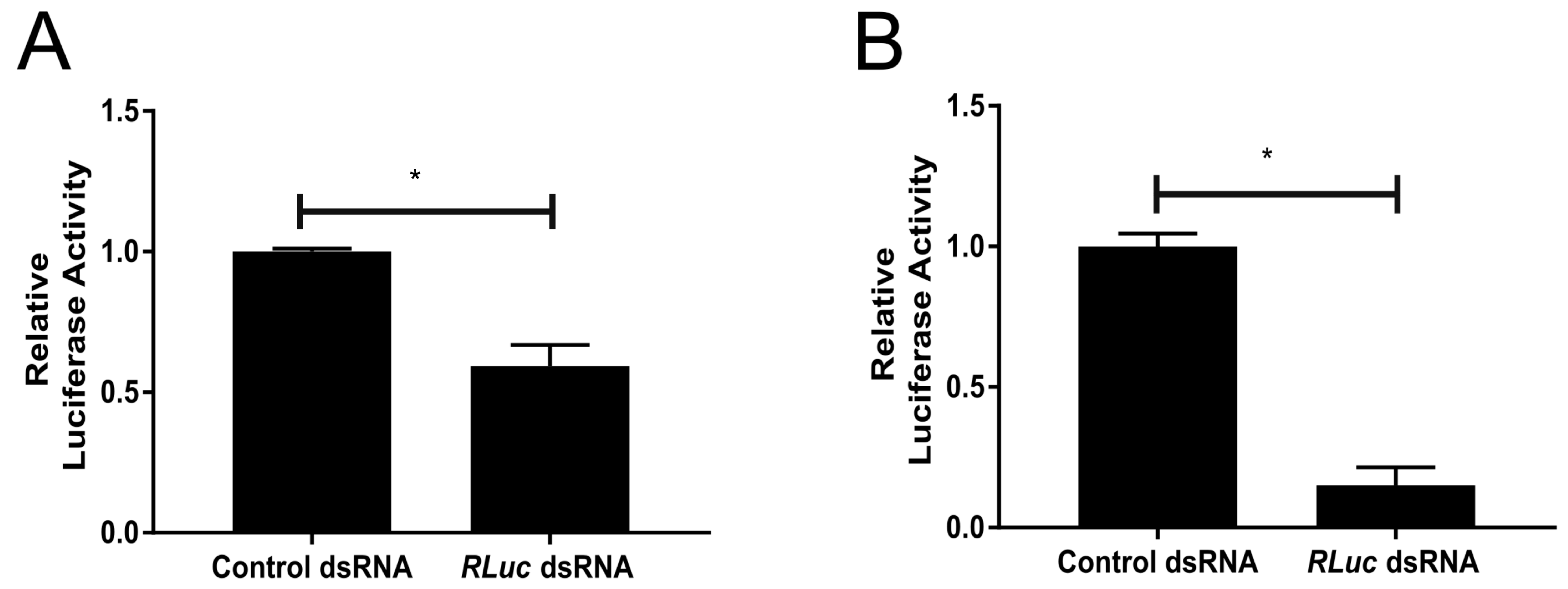
3.3. An Active dsRNA-Inducible RNAi Response has Antiviral Activity against SFV Infection in TRA-171 Cells

Next, we assessed if this silencing pathway has an inducible antiviral function against SFV. The cells were first treated with either RLuc-specific or eGFP-specific control dsRNA prior to infection with SFV4(3H)-RLuc. The infections were performed at either a high (10) or low (0.005) MOI. The cells were lysed 24 h p.i. and luciferase expression was assessed. A decrease in the relative luciferase activity was observed in cells treated with RLuc-specific dsRNA when compared to those that received control eGFP-specific dsRNA at both MOIs, which directly indicates a decrease in viral replication and, therefore, a reduction in virus production (Figure 4A,B). These findings suggest that TRA-171 cells possess a sequence specific antiviral response that can be externally induced by dsRNA.

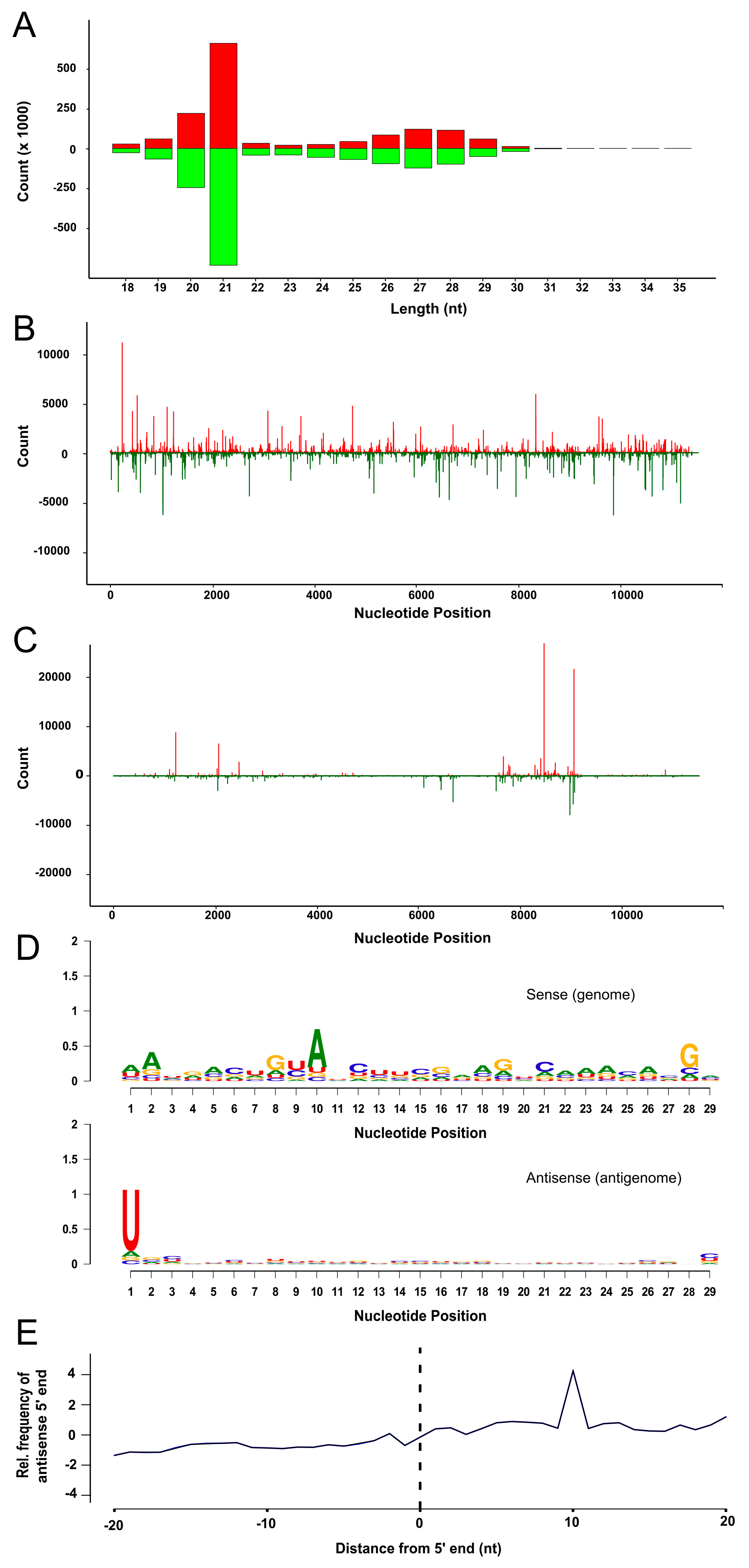
3.4. SFV Infection Induces Small RNA Production in TRA-171 Cells

It has previously been shown that, following infection, non-vector mosquito cells are capable of generating small RNAs of the expected size and properties expected of vsiRNAs and vpiRNAs [22,50]. Having shown that TRA-171 cells have an inducible RNAi response that is capable of controlling SFV replication, we next wanted to establish if TRA-171 cells have the capacity to generate vsiRNAs and/or vpiRNAs which specifically target SFV. TRA-171 cells were infected with SFV4 at MOI 10 and RNA isolated 24 h p.i. for sequencing. Small RNAs were sequenced and analyzed by mapping to both the SFV genome and the antigenome (Figure 5, Supplementary Table S2). SFV-specific vsiRNAs predominantly 21 nt in length were found to be produced within infected cells (Figure 5A). These reads were found to map to both the viral genome and the antigenome in approximately equal quantities. The pattern of vsiRNA distribution observed (Figure 5B) indicates regions of many reads (hot spots) and regions with few reads (cold spots). A second class of SFV-specific small RNAs were also identified, which ranged from 24–29 nt and again mapped to both the genome and antigenome of SFV (Figure 5A). These preferentially targeted specific regions of the coding strand (Figure 5C) and presented with the characteristic signature of piRNAs; a bias for A at position 10 and a U at position 1 (Figure 5D). The 5′ ends of these complementary small RNAs overlapped most frequently by 10 nt, which suggests that these are vpiRNAs produced via the ‘ping-pong’ mechanism (Figure 5E). Taken together, this data suggests that the antiviral RNAi response in TRA-171 cells is induced following SFV infection and produces both vsiRNAs and vpiRNAs which target viral sequences.
Other analysis of the sequencing data revealed the presence of putative novel insect specific viruses (ISVs), although further studies are needed to confirm their presence (Supplemental Materials and Methods, Supplemental Table S3 and Figure S2). ISVs lack the ability to replicate in vertebrate cells but have been naturally shown to infect a variety of arthropods in nature including mosquitoes and are present in several mosquito derived cell lines [51,52,53,54,55,56,57,58,59]. It will be beneficial to assess their impact on the host and their involvement in pathogen transmission in order to understand how this may affect vector competence and arbovirus transmission within specific populations. How these ISVs, as well as other sequences such as transposable elements (TEs) and endogenous viral elements (EVEs), interact with the RNAi response will be important to develop a more complete awareness of the global role of RNAi out with arboviral infections. For instance, given the known correlation between EVEs and the piRNA pathway [60], this information may provide relevant data for understanding how viruses establish a persistent infection and the potential for mosquitoes to pass on heritable immune indicators.

4. Discussion

Studies in mosquito vector species have shown that RNAi is the predominant antiviral response against infections [17,19,20]. Specifically, the exo-siRNA has been demonstrated to be important in regulating this antiviral activity, although the antiviral role of piRNAs is unclear. Previous research has used D. melanogaster as a model for RNAi studies [61,62,63,64,65,66,67,68,69], but very few studies have investigated the role of the RNAi pathways in non-vector mosquito species. Toxorhynchites is one of three mosquito genera, along with Malaya and Topomyia, which do not require a blood meal during their adult life stages to initiate egg development and, as such, do not exhibit host-seeking behavior [70,71]. Their lack of importance as a medically relevant pest species has meant that their general biology has been largely neglected. However, in their role as a biological control agent against pest species, Toxorhynchites may be at risk of an arbovirus infection due to ingesting vertically infected larvae [15]. Therefore, it is important to understand their antiviral capabilities.
This study identifies the presence of a functional RNAi response within Tx. amboinensis-derived TRA-171 cells. SFV infection induced the production of 21 nt vsiRNAs derived from both the viral genome and antigenome, which is indicative of Dcr2 cleavage of dsRNA [20]. A similar enrichment of 21 nt vsiRNAs has previously been reported for SFV, as well as other alphaviruses [24,26,28,31,32,37,39,72] and members of the Bunyaviridae and Flaviviridae [18,19,20,21]. Deep sequencing of SFV-infected TRA-171 cells indicates that these vsiRNAs are distributed across the genome and antigenome with hot spot and cold spot areas. This is consistent with previous SFV data obtained from aedine cell lines [24,28,37,39]. Further work would be required to determine whether vsiRNAs derived from cold spot regions are able to inhibit SFV replication significantly more effectively than hot spot derived vsiRNAs, as has been shown previously [28].
In addition, larger classes of SFV-derived small RNAs between 24–29 nt were detected which presented with the hallmark characteristic ping-pong motif of piRNAs; an A10/U1 bias and a 10 nt overlap between the 5′ ends of piRNAs from different strands, which has been previously described in aedine cells [24,31,32,37]. Consistent with previous findings for SFV infection of Aag2 and U4.4 cells, these were less widely distributed and preferentially targeted specific regions of the coding strand [24,31,32]. However, unlike data from aedine cells, these vpiRNAs are present in approximately equal quantities against both sense and antisense sequences, rather than with a bias towards the sense strand. The identification of 24–29 nt small RNAs displaying a A10 and U1 bias and a 10 nt overlap between the 5′ ends of different strands suggests that TRA-171 cells encode PIWI clade proteins.
Our findings show that the exo-siRNA pathway can be artificially induced in TRA-171 cells by the transfection of long dsRNA molecules, which leads to sequence-specific silencing. Similarly, our results also show that sequence-specific siRNAs are capable of achieving efficient gene silencing, which supports the presence of a natural antiviral RNAi pathway within TRA-171 cells. Key mediator proteins of the exo-siRNA pathway, such as Dcr2 and Ago2, are highly conserved between mosquito and drosophila and the effector mechanisms are considered to be similar [73]. Previous studies have confirmed the importance of these proteins as mediators of infection as viral replication increases following their knockdown [23,24,27,33,37,74]. Similarly, recent studies in Ae. aegypti show that Ago3, Piwi5, and, to a lesser extent, Piwi6 participate in the production of viral-derived piRNAs [35,36]. Given the lack of genomic information available for Tx. amboinensis, it was not possible to identify RNAi effector-encoding sequences and, therefore, we can only speculate if these proteins are present in the Toxorhychites genome. However, the results presented here are a strong indicator for the presence of RNAi machinery comparable to that of aedine species. Previous research has shown that arboviruses are able to replicate to high levels within Toxorhynchites mosquitoes and their derived cell lines. The data shown here for SFV replication and production is congruous with these findings. Further work will be required to determine the exact temporal and spatial mechanisms, as well as the proteins involved in the Toxorhynchites immune response, which permit enhanced arbovirus replication.
In conclusion, we have demonstrated the presence of an active antiviral RNAi response in Toxorhynchites cells which successfully modulates SFV infection in a manner similar to that of natural arboviral vector species. Only once we fully understand their ability to manage infections will we be able to make an informed decision regarding the suitability of certain species as biological control agents against vector mosquito species. Our data also expands on the current knowledge of ISVs, TEs, and EVEs in mosquitoes that may influence pathogen transmission in vector populations and highlights their interactions with the exo-siRNA and piRNA pathways.

Supplementary Materials

The following are available online at https://0-www-mdpi-com.brum.beds.ac.uk/1999-4915/10/12/694/s1, Supplemental Materials and Methods: Small RNA analysis for the identification of novel putative insect specific viruses, Figure S1: Production of small RNAs from long dsRNA, Figure S2: Characteristics of contigs derived from an EVE, TE, and a potential novel virus, Table S1: Primer sequences, Table S2: Sequencing reads results, Table S3: Overview of assembled contigs showing similarity to viruses or TEs.

Author Contributions

C.L.D., E.S., and A.K. conceived and designed the experiments. C.L.D. and M.V. performed the experiments. E.R.G.R.A., J.T.M., and V.B.S. conducted bioinformatics analysis of small RNA data. C.L.D. and A.K. wrote the paper. All authors contributed to the data analysis and manuscript revision.

Funding

This study was supported by the UK Medical Research Council (MC_UU_12014/8).

Acknowledgments

The authors wish to thank A. Merits (University of Tartu) for providing the SFV constructs.

Conflicts of Interest

The authors declare no conflict of interest.

References

  1. Zuharah, W.F.; Fadzly, N.; Yusof, N.A.; Dieng, H. Risky behaviors: Effects of toxorhynchites splendens (diptera: Culicidae) predator on the behavior of three mosquito species. J. Insect Sci. 2015, 15. [Google Scholar] [CrossRef] [PubMed]
  2. Huang, Y.S.; Higgs, S.; Vanlandingham, D.L. Biological control strategies for mosquito vectors of arboviruses. Insects 2017, 8, 21. [Google Scholar] [CrossRef] [PubMed]
  3. Schreiber, E.T. Toxorhynchites. J. Am. Mosq. Control. Assoc. 2007, 23, 129–132. [Google Scholar] [CrossRef]
  4. Focks, D.A. Toxorhynchites as biocontrol agents. J. Am. Mosq. Control. Assoc. 2007, 23, 118–127. [Google Scholar] [CrossRef]
  5. Rosen, L. The use of toxorhynchites mosquitoes to detect and propagate dengue and other arboviruses. Am. J. Trop. Med. Hyg. 1981, 30, 177–183. [Google Scholar] [CrossRef] [PubMed]
  6. Rosen, L.; Shroyer, D.A. Comparative susceptibility of five species of toxorhynchites mosquitoes to parenteral infection with dengue and other flaviviruses. Am. J. Trop. Med. Hyg. 1985, 34, 805–809. [Google Scholar] [CrossRef] [PubMed]
  7. Rosen, L.; Tesh, R.B.; Lien, J.C.; Cross, J.H. Transovarial transmission of Japanese encephalitis virus by mosquitoes. Science 1978, 199, 909–911. [Google Scholar] [CrossRef] [PubMed]
  8. Tesh, R.B. A method for the isolation and identification of dengue viruses, using mosquito cell cultures. Am. J. Trop. Med. Hyg. 1979, 28, 1053–1059. [Google Scholar] [CrossRef]
  9. Tesh, R.B.; McLean, R.G.; Shroyer, D.A.; Calisher, C.H.; Rosen, L. Ross river virus (togaviridae: Alphavirus) infection (epidemic polyarthritis) in American Samoa. Trans. R. Soc. Trop. Med. Hyg. 1981, 75, 426–431. [Google Scholar] [CrossRef]
  10. Scherer, W.F.; Chin, J. Sensitivity of toxorhynchites amboinensis mosquitoes versus chicken embryonic cell cultures for assays of Venezuelan encephalitis virus. J. Clin. Microbiol. 1981, 13, 947–950. [Google Scholar]
  11. Kuno, G.; Gubler, D.J.; Velez, M.; Oliver, A. Comparative sensitivity of three mosquito cell lines for isolation of dengue viruses. Bull. World Health Organ. 1985, 63, 279–286. [Google Scholar] [PubMed]
  12. Kuno, G. Persistent infection of a nonvector mosquito cell line (TRA-171) with dengue viruses. Intervirology 1982, 18, 45–55. [Google Scholar] [CrossRef] [PubMed]
  13. Kuno, G. Replication of dengue, yellow fever, St. Louis encephalitis and vesicular stomatitis viruses in a cell line (TRA-171) derived from Toxorhynchites amboinensis. In Vitro 1981, 17, 1011–1015. [Google Scholar] [CrossRef] [PubMed]
  14. Sanchez Legrand, F.; Hotta, S. Susceptibility of cloned toxorhynchites amboinensis cells to dengue and chikungunya viruses. Microbiol. Immunol. 1983, 27, 101–105. [Google Scholar] [CrossRef] [PubMed]
  15. Lequime, S.; Lambrechts, L. Vertical transmission of arboviruses in mosquitoes: A historical perspective. Infect. Genet. Evol. 2014, 28, 681–690. [Google Scholar] [CrossRef] [PubMed]
  16. Merkling, S.H.; van Rij, R.P. Beyond RNAi: Antiviral defense strategies in drosophila and mosquito. J. Insect Physiol. 2013, 59, 159–170. [Google Scholar] [CrossRef]
  17. Blair, C.D. Mosquito RNAi is the major innate immune pathway controlling arbovirus infection and transmission. Future Microbiol. 2011, 6, 265–277. [Google Scholar] [CrossRef] [Green Version]
  18. Blair, C.D.; Olson, K.E. The role of RNA interference (RNAi) in arbovirus-vector interactions. Viruses 2015, 7, 820–843. [Google Scholar] [CrossRef]
  19. Olson, K.E.; Blair, C.D. Arbovirus-mosquito interactions: RNAi pathway. Curr. Opin. Virol. 2015, 15, 119–126. [Google Scholar] [CrossRef]
  20. Donald, C.L.; Kohl, A.; Schnettler, E. New insights into control of arbovirus replication and spread by insect RNA interference pathways. Insects 2012, 3, 511–531. [Google Scholar] [CrossRef]
  21. Samuel, G.H.; Adelman, Z.N.; Myles, K.M. Antiviral immunity and virus-mediated antagonism in disease vector mosquitoes. Trends Microbiol. 2018, 26, 447–461. [Google Scholar] [CrossRef] [PubMed]
  22. Dietrich, I.; Shi, X.; McFarlane, M.; Watson, M.; Blomstrom, A.L.; Skelton, J.K.; Kohl, A.; Elliott, R.M.; Schnettler, E. The antiviral RNAi response in vector and non-vector cells against orthobunyaviruses. PLoS Negl. Trop. Dis. 2017, 11, e0005272. [Google Scholar] [CrossRef] [PubMed]
  23. McFarlane, M.; Arias-Goeta, C.; Martin, E.; O’Hara, Z.; Lulla, A.; Mousson, L.; Rainey, S.M.; Misbah, S.; Schnettler, E.; Donald, C.L.; et al. Characterization of Aedes aegypti innate-immune pathways that limit chikungunya virus replication. PLoS Negl. Trop. Dis. 2014, 8, e2994. [Google Scholar] [CrossRef] [PubMed]
  24. Schnettler, E.; Donald, C.L.; Human, S.; Watson, M.; Siu, R.W.; McFarlane, M.; Fazakerley, J.K.; Kohl, A.; Fragkoudis, R. Knockdown of piRNA pathway proteins results in enhanced semliki forest virus production in mosquito cells. J. Gen. Virol. 2013, 94, 1680–1689. [Google Scholar] [CrossRef] [PubMed] [Green Version]
  25. Varjak, M.; Donald, C.L.; Mottram, T.J.; Sreenu, V.B.; Merits, A.; Maringer, K.; Schnettler, E.; Kohl, A. Characterization of the zika virus induced small RNA response in Aedes aegypti cells. PLoS Negl. Trop. Dis. 2017, 11, e0006010. [Google Scholar] [CrossRef] [PubMed]
  26. Myles, K.M.; Wiley, M.R.; Morazzani, E.M.; Adelman, Z.N. Alphavirus-derived small RNAs modulate pathogenesis in disease vector mosquitoes. Proc. Natl. Acad. Sci. USA 2008, 105, 19938–19943. [Google Scholar] [CrossRef] [PubMed] [Green Version]
  27. Sanchez-Vargas, I.; Scott, J.C.; Poole-Smith, B.K.; Franz, A.W.; Barbosa-Solomieu, V.; Wilusz, J.; Olson, K.E.; Blair, C.D. Dengue virus type 2 infections of Aedes aegypti are modulated by the mosquito’s RNA interference pathway. PLoS Pathog. 2009, 5, e1000299. [Google Scholar] [CrossRef] [PubMed]
  28. Siu, R.W.; Fragkoudis, R.; Simmonds, P.; Donald, C.L.; Chase-Topping, M.E.; Barry, G.; Attarzadeh-Yazdi, G.; Rodriguez-Andres, J.; Nash, A.A.; Merits, A.; et al. Antiviral RNA interference responses induced by semliki forest virus infection of mosquito cells: Characterization, origin, and frequency-dependent functions of virus-derived small interfering RNAs. J. Virol. 2011, 85, 2907–2917. [Google Scholar] [CrossRef]
  29. Scott, J.C.; Brackney, D.E.; Campbell, C.L.; Bondu-Hawkins, V.; Hjelle, B.; Ebel, G.D.; Olson, K.E.; Blair, C.D. Comparison of dengue virus type 2-specific small RNAs from RNA interference-competent and -incompetent mosquito cells. PLoS Negl. Trop. Dis. 2010, 4, e848. [Google Scholar] [CrossRef]
  30. Leger, P.; Lara, E.; Jagla, B.; Sismeiro, O.; Mansuroglu, Z.; Coppee, J.Y.; Bonnefoy, E.; Bouloy, M. Dicer-2- and piwi-mediated RNA interference in rift valley fever virus-infected mosquito cells. J. Virol. 2013, 87, 1631–1648. [Google Scholar] [CrossRef]
  31. Vodovar, N.; Bronkhorst, A.W.; van Cleef, K.W.; Miesen, P.; Blanc, H.; van Rij, R.P.; Saleh, M.C. Arbovirus-derived piRNAs exhibit a ping-pong signature in mosquito cells. PLoS ONE 2012, 7, e30861. [Google Scholar] [CrossRef] [PubMed]
  32. Morazzani, E.M.; Wiley, M.R.; Murreddu, M.G.; Adelman, Z.N.; Myles, K.M. Production of virus-derived ping-pong-dependent piRNA-like small RNAs in the mosquito soma. PLoS Pathog. 2012, 8, e1002470. [Google Scholar] [CrossRef] [PubMed]
  33. Campbell, C.L.; Keene, K.M.; Brackney, D.E.; Olson, K.E.; Blair, C.D.; Wilusz, J.; Foy, B.D. Aedes aegypti uses RNA interference in defense against sindbis virus infection. BMC Microbiol 2008, 8, 47. [Google Scholar] [CrossRef] [PubMed]
  34. Brackney, D.E.; Beane, J.E.; Ebel, G.D. RNAi targeting of West Nile virus in mosquito midguts promotes virus diversification. PLoS Pathog. 2009, 5, e1000502. [Google Scholar] [CrossRef] [PubMed]
  35. Miesen, P.; Girardi, E.; van Rij, R.P. Distinct sets of piwi proteins produce arbovirus and transposon-derived piRNAs in Aedes aegypti mosquito cells. Nucleic Acids Res. 2015, 43, 6545–6556. [Google Scholar] [CrossRef] [PubMed]
  36. Miesen, P.; Ivens, A.; Buck, A.H.; van Rij, R.P. Small RNA profiling in dengue virus 2-infected Aedes mosquito cells reveals viral piRNAs and novel host mirnas. PLoS Negl. Trop. Dis. 2016, 10, e0004452. [Google Scholar] [CrossRef] [PubMed]
  37. Varjak, M.; Maringer, K.; Watson, M.; Sreenu, V.B.; Fredericks, A.C.; Pondeville, E.; Donald, C.L.; Sterk, J.; Kean, J.; Vazeille, M.; et al. Aedes aegypti piwi4 is a noncanonical piwi protein involved in antiviral responses. mSphere 2017, 2. [Google Scholar] [CrossRef] [PubMed]
  38. Dietrich, I.; Jansen, S.; Fall, G.; Lorenzen, S.; Rudolf, M.; Huber, K.; Heitmann, A.; Schicht, S.; Ndiaye, E.H.; Watson, M.; et al. RNA interference restricts rift valley fever virus in multiple insect systems. mSphere 2017, 2. [Google Scholar] [CrossRef]
  39. Varjak, M.; Dietrich, I.; Sreenu, V.B.; Till, B.E.; Merits, A.; Kohl, A.; Schnettler, E. Spindle-e acts antivirally against alphaviruses in mosquito cells. Viruses 2018, 10, 88. [Google Scholar] [CrossRef]
  40. Varjak, M.; Leggewie, M.; Schnettler, E. The antiviral piRNA response in mosquitoes? J. Gen. Virol. 2018. [Google Scholar] [CrossRef]
  41. Miesen, P.; Joosten, J.; van Rij, R.P. Piwis go viral: Arbovirus-derived piRNAs in vector mosquitoes. PLoS Pathog. 2016, 12, e1006017. [Google Scholar] [CrossRef] [PubMed]
  42. Schneider, I. Cell lines derived from late embryonic stages of drosophila melanogaster. J. Embryol. Exp. Morphol. 1972, 27, 353–365. [Google Scholar]
  43. Rainey, S.M.; Martinez, J.; McFarlane, M.; Juneja, P.; Sarkies, P.; Lulla, A.; Schnettler, E.; Varjak, M.; Merits, A.; Miska, E.A.; et al. Wolbachia blocks viral genome replication early in infection without a transcriptional response by the endosymbiont or host small RNA pathways. PLoS Pathog. 2016, 12, e1005536. [Google Scholar] [CrossRef] [PubMed]
  44. Schnettler, E.; Hemmes, H.; Goldbach, R.; Prins, M. The NS3 protein of rice hoja blanca virus suppresses RNA silencing in mammalian cells. J. Gen. Virol. 2008, 89, 336–340. [Google Scholar] [CrossRef] [PubMed] [Green Version]
  45. Ongus, J.R.; Roode, E.C.; Pleij, C.W.; Vlak, J.M.; van Oers, M.M. The 5’ non-translated region of varroa destructor virus 1 (genus iflavirus): Structure prediction and IRES activity in lymantria dispar cells. J. Gen. Virol. 2006, 87, 3397–3407. [Google Scholar] [CrossRef]
  46. Ou, J.; Wolfe, S.A.; Brodsky, M.H.; Zhu, L.J. Motifstack for the analysis of transcription factor binding site evolution. Nat. Methods 2018, 15, 8–9. [Google Scholar] [CrossRef]
  47. Antoniewski, C. Computing siRNA and piRNA overlap signatures. Methods Mol. Biol. 2014, 1173, 135–146. [Google Scholar]
  48. Fragkoudis, R.; Chi, Y.; Siu, R.W.; Barry, G.; Attarzadeh-Yazdi, G.; Merits, A.; Nash, A.A.; Fazakerley, J.K.; Kohl, A. Semliki forest virus strongly reduces mosquito host defence signaling. Insect Mol. Biol. 2008, 17, 647–656. [Google Scholar] [CrossRef] [PubMed]
  49. Davey, M.W.; Dalgarno, L. Semliki forest virus replication in cultured Aedes albopictus cells: Studies on the establishment of persistence. J. Gen. Virol. 1974, 24, 453–463. [Google Scholar] [CrossRef]
  50. Schnettler, E.; Ratinier, M.; Watson, M.; Shaw, A.E.; McFarlane, M.; Varela, M.; Elliott, R.M.; Palmarini, M.; Kohl, A. RNA interference targets arbovirus replication in culicoides cells. J. Virol. 2013, 87, 2441–2454. [Google Scholar] [CrossRef] [Green Version]
  51. Bolling, B.G.; Weaver, S.C.; Tesh, R.B.; Vasilakis, N. Insect-specific virus discovery: Significance for the arbovirus community. Viruses 2015, 7, 4911–4928. [Google Scholar] [CrossRef] [PubMed]
  52. Schnettler, E.; Sreenu, V.B.; Mottram, T.; McFarlane, M. Wolbachia restricts insect-specific flavivirus infection in Aedes aegypti cells. J. Gen. Virol. 2016, 97, 3024–3029. [Google Scholar] [CrossRef] [PubMed]
  53. Stollar, V.; Thomas, V.L. An agent in the Aedes aegypti cell line (peleg) which causes fusion of Aedes albopictus cells. Virology 1975, 64, 367–377. [Google Scholar] [CrossRef]
  54. Crabtree, M.B.; Sang, R.C.; Stollar, V.; Dunster, L.M.; Miller, B.R. Genetic and phenotypic characterization of the newly described insect flavivirus, kamiti river virus. Arch. Virol. 2003, 148, 1095–1118. [Google Scholar] [CrossRef] [PubMed]
  55. Nouri, S.; Matsumura, E.E.; Kuo, Y.W.; Falk, B.W. Insect-specific viruses: From discovery to potential translational applications. Curr. Opin. Virol. 2018, 33, 33–41. [Google Scholar] [CrossRef]
  56. Halbach, R.; Junglen, S.; van Rij, R.P. Mosquito-specific and mosquito-borne viruses: Evolution, infection, and host defense. Curr. Opin. Insect Sci. 2017, 22, 16–27. [Google Scholar] [CrossRef] [PubMed]
  57. Hall, R.A.; Bielefeldt-Ohmann, H.; McLean, B.J.; O’Brien, C.A.; Colmant, A.M.; Piyasena, T.B.; Harrison, J.J.; Newton, N.D.; Barnard, R.T.; Prow, N.A.; et al. Commensal viruses of mosquitoes: Host restriction, transmission, and interaction with arboviral pathogens. Evol. Bioinform. Online 2016, 12, 35–44. [Google Scholar] [CrossRef]
  58. Roundy, C.M.; Azar, S.R.; Rossi, S.L.; Weaver, S.C.; Vasilakis, N. Insect-specific viruses: A historical overview and recent developments. Adv. Virus Res. 2017, 98, 119–146. [Google Scholar]
  59. Bolling, B.G.; Vasilakis, N.; Guzman, H.; Widen, S.G.; Wood, T.G.; Popov, V.L.; Thangamani, S.; Tesh, R.B. Insect-specific viruses detected in laboratory mosquito colonies and their potential implications for experiments evaluating arbovirus vector competence. Am. J. Trop. Med. Hyg. 2015, 92, 422–428. [Google Scholar] [CrossRef]
  60. Arensburger, P.; Hice, R.H.; Wright, J.A.; Craig, N.L.; Atkinson, P.W. The mosquito Aedes aegypti has a large genome size and high transposable element load but contains a low proportion of transposon-specific pirnas. BMC Genom. 2011, 12, 606. [Google Scholar] [CrossRef]
  61. Aliyari, R.; Wu, Q.; Li, H.W.; Wang, X.H.; Li, F.; Green, L.D.; Han, C.S.; Li, W.X.; Ding, S.W. Mechanism of induction and suppression of antiviral immunity directed by virus-derived small RNAs in drosophila. Cell Host Microbe 2008, 4, 387–397. [Google Scholar] [CrossRef] [PubMed]
  62. Chotkowski, H.L.; Ciota, A.T.; Jia, Y.; Puig-Basagoiti, F.; Kramer, L.D.; Shi, P.Y.; Glaser, R.L. West Nile virus infection of drosophila melanogaster induces a protective RNAi response. Virology 2008, 377, 197–206. [Google Scholar] [CrossRef] [PubMed]
  63. Galiana-Arnoux, D.; Dostert, C.; Schneemann, A.; Hoffmann, J.A.; Imler, J.L. Essential function in vivo for dicer-2 in host defense against RNA viruses in drosophila. Nat. Immunol. 2006, 7, 590–597. [Google Scholar] [CrossRef] [PubMed]
  64. Saleh, M.C.; Tassetto, M.; van Rij, R.P.; Goic, B.; Gausson, V.; Berry, B.; Jacquier, C.; Antoniewski, C.; Andino, R. Antiviral immunity in drosophila requires systemic RNA interference spread. Nature 2009, 458, 346–350. [Google Scholar] [CrossRef] [PubMed]
  65. Zambon, R.A.; Vakharia, V.N.; Wu, L.P. RNAi is an antiviral immune response against a dsrna virus in drosophila melanogaster. Cell. Microbiol. 2006, 8, 880–889. [Google Scholar] [CrossRef] [PubMed]
  66. Marques, J.T.; Wang, J.P.; Wang, X.; de Oliveira, K.P.; Gao, C.; Aguiar, E.R.; Jafari, N.; Carthew, R.W. Functional specialization of the small interfering RNA pathway in response to virus infection. PLoS Pathog. 2013, 9, e1003579. [Google Scholar] [CrossRef]
  67. van Rij, R.P.; Saleh, M.C.; Berry, B.; Foo, C.; Houk, A.; Antoniewski, C.; Andino, R. The RNA silencing endonuclease argonaute 2 mediates specific antiviral immunity in drosophila melanogaster. Genes Dev. 2006, 20, 2985–2995. [Google Scholar] [CrossRef]
  68. Han, Y.H.; Luo, Y.J.; Wu, Q.; Jovel, J.; Wang, X.H.; Aliyari, R.; Han, C.; Li, W.X.; Ding, S.W. RNA-based immunity terminates viral infection in adult drosophila in the absence of viral suppression of RNA interference: Characterization of viral small interfering RNA populations in wild-type and mutant flies. J. Virol 2011, 85, 13153–13163. [Google Scholar] [CrossRef]
  69. Wang, X.H.; Aliyari, R.; Li, W.X.; Li, H.W.; Kim, K.; Carthew, R.; Atkinson, P.; Ding, S.W. RNA interference directs innate immunity against viruses in adult drosophila. Science 2006, 312, 452–454. [Google Scholar] [CrossRef]
  70. Steffan, W.A.; Neal, L.E. Biology of toxorhynchites. Annu. Rev. Entomol. 1981, 26, 159–181. [Google Scholar] [CrossRef]
  71. Zhou, X.; Rinker, D.C.; Pitts, R.J.; Rokas, A.; Zwiebel, L.J. Divergent and conserved elements comprise the chemoreceptive repertoire of the nonblood-feeding mosquito toxorhynchites amboinensis. Genome Biol. Evol. 2014, 6, 2883–2896. [Google Scholar] [CrossRef] [PubMed]
  72. Myles, K.M.; Morazzani, E.M.; Adelman, Z.N. Origins of alphavirus-derived small RNAs in mosquitoes. RNA Biol. 2009, 6, 387–391. [Google Scholar] [CrossRef] [PubMed] [Green Version]
  73. Lewis, S.H.; Salmela, H.; Obbard, D.J. Duplication and diversification of dipteran argonaute genes, and the evolutionary divergence of piwi and aubergine. Genome Biol. Evol. 2016, 8, 507–518. [Google Scholar] [CrossRef] [PubMed]
  74. Keene, K.M.; Foy, B.D.; Sanchez-Vargas, I.; Beaty, B.J.; Blair, C.D.; Olson, K.E. RNA interference acts as a natural antiviral response to o’nyong-nyong virus (alphavirus; togaviridae) infection of anopheles gambiae. Proc. Natl. Acad. Sci. USA 2004, 101, 17240–17245. [Google Scholar] [CrossRef] [PubMed]
Figure 1. Infection of TRA-171 cells with SFV. (A) Schematic representation of the design of the reporter strains of SFV expressing luciferase (Luc: either RLuc, SFV4(3H)-RLuc, or FFLuc, SFV4(3H)-FFLuc) inserted between duplicated nsP2-protease cleavage sites at the nsP3/4 junction. (B) TRA-171 cells were either mock infected or infected with SFV4(3H)-RLuc at MOI 10 or 0.01. Luciferase expression was determined at 24 h p.i. by luciferase assay and relative luciferase activity was normalized against background, shown on the Y-axis. (C) TRA-171 cells were infected with SFV4(3H)-FFLuc at MOI 10. Cell growth media was collected and replaced with fresh media at the given time points. Virus production was determined by plaque assay and the titre was measured in plaque forming units (PFU/mL). (D) The cells were infected as in (C) and lysed at the given time points to monitor viral replication. Luciferase expression was determined by luciferase assay and relative luciferase units are shown on the Y-axis. (E) The cells were either infected as in (C) (■) or mock infected (●) and cell numbers were counted at the given time points. Mean values with standard error are shown for three (B) or two (CE) independent experiments conducted in triplicate. The analysis used the average of each triplicate per experiment.
Viruses 10 00694 g001
Figure 2. Production of small RNAs from long dsRNA. The cellular extracts were prepared from TRA-171 and S2 cells. The extracts were incubated with 32P internally-labeled dsRNA (114 nt). Size markers of long dsRNA (114 nt) and siRNAs (21 nt) are indicated to show approximate sizes. The image shown is representative of three independent experiments and shows relevant individual lanes. The complete image is shown in Supplementary Figure S1.
Viruses 10 00694 g002
Figure 3. dsRNA or siRNA mediated gene silencing in TRA-171 cells. The cells were co-transfected with pIZ-Fluc and pAcIE1-Rluc (as an internal control) alongside either dsRNA (A) or siRNAs (B) targeting FFLuc or a control. Luciferase expression was determined by luciferase assay 24 h p.t. and relative luciferase activity (FFLuc/RLuc) is shown on the Y-axis. Mean values with standard error are shown for four independent experiments performed in triplicate. The analysis used the average of each triplicate per experiment. * indicates significance, p < 0.05 by Student t-test.
Viruses 10 00694 g003
Figure 4. TRA-171 cells possess a dsRNA-inducible antiviral RNAi pathway. The cells were transfected with dsRNA against either RLuc or eGFP (as a control) 24 h prior to being either mock infected or infected with SFV(3H)-RLuc at MOI 10 (A) or 0.005 (B). Luciferase expression was determined by luciferase assay 24 h p.i. and luciferase activity is shown on the Y-axis. Mean values with standard error are shown for three independent experiments performed in triplicate. The analysis used the average of each triplicate per experiment. * indicates significance, p < 0.05 by Student t-test.
Viruses 10 00694 g004
Figure 5. Characteristics of SFV-derived small RNAs in TRA-171 cells. RNA was isolated from TRA-171 cells 24 h p.i. with SFV4 at MOI 10 followed by small RNA sequencing. (A) The size distribution of small RNAs from SFV infected cells mapping to the SFV4 genome (red, positive numbers) or antigenome (green, negative numbers). The distribution of 21 nt (B) or 28 nt (C) small RNAs across the length of the SFV genome (red, positive numbers) or antigenome (green, negative numbers). The Y-axis shows the frequency of small RNAs mapping to the corresponding nucleotide location on the X-axis. (D) The conserved relative nucleotide frequency at each position of 29 nt long small RNAs mapping to the SFV4 genome or antigenome represented on a web logo diagram. The height of each nucleotide represents the degree of sequence conservation. The level of conservation is indicated by the Y-axis. (E) Frequency map showing the distance between the 5′ ends of 25–29 nt small RNAs mapping to the opposite strand of the SFV4 reference sequence. Position 0 represents the first nucleotide. The results shown are representative of two independent experiments.
Viruses 10 00694 g005

