Next Issue
Volume 10, April
Previous Issue
Volume 10, February


Viruses, Volume 10, Issue 3 (March 2018) – 41 articles

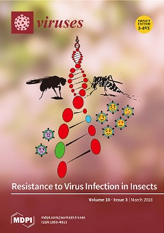
Cover Story (view full-size image): Studies of dipteran insects, such as fruit flies and virus-transmitting mosquitoes, have greatly aided our understanding of virus–insect interactions. Comparative analyses, genetic mapping, and experimental control of the microbiota and environment have highlighted variation in these interactions across hosts and viruses. In this review, we describe the diversity of immune responses to dipteran viruses and the sources of variation in viral resistance across host individuals. View this paper
  • Issues are regarded as officially published after their release is announced to the table of contents alert mailing list.
  • You may sign up for e-mail alerts to receive table of contents of newly released issues.
  • PDF is the official format for papers published in both, html and pdf forms. To view the papers in pdf format, click on the "PDF Full-text" link, and use the free Adobe Reader to open them.
Order results
Result details
Section
Select all
Export citation of selected articles as:
8 pages, 762 KiB  
Review
Enterovirus Transmission by Secretory Autophagy
by Yael Mutsafi and Nihal Altan-Bonnet
Viruses 2018, 10(3), 139; https://0-doi-org.brum.beds.ac.uk/10.3390/v10030139 - 20 Mar 2018
Cited by 46 | Viewed by 7140
Abstract
Present in many cell types, non-degradative secretory autophagy is a newly discovered pathway in which autophagosomes fuse with the plasma membrane instead of lysosomes. Surprisingly, some viruses exploit secretory autophagy to exit cells non-lytically, shedding into the extracellular environment as particle populations contained [...] Read more.
Present in many cell types, non-degradative secretory autophagy is a newly discovered pathway in which autophagosomes fuse with the plasma membrane instead of lysosomes. Surprisingly, some viruses exploit secretory autophagy to exit cells non-lytically, shedding into the extracellular environment as particle populations contained within vesicles. As a result, this significantly enhances the infectivity of these viruses. In this paper, this novel cellular exit pathway is highlighted and its advantages for viral transmission discussed. Full article
(This article belongs to the Special Issue Viruses and Autophagy)
Show Figures

Figure 1

15 pages, 2054 KiB  
Review
Alphavirus Nucleocapsid Packaging and Assembly
by Adriano Mendes and Richard J. Kuhn
Viruses 2018, 10(3), 138; https://0-doi-org.brum.beds.ac.uk/10.3390/v10030138 - 20 Mar 2018
Cited by 36 | Viewed by 6157
Abstract
Alphavirus nucleocapsids are assembled in the cytoplasm of infected cells from 240 copies of the capsid protein and the approximately 11 kb positive strand genomic RNA. However, the challenge of how the capsid specifically selects its RNA package and assembles around it has [...] Read more.
Alphavirus nucleocapsids are assembled in the cytoplasm of infected cells from 240 copies of the capsid protein and the approximately 11 kb positive strand genomic RNA. However, the challenge of how the capsid specifically selects its RNA package and assembles around it has remained an elusive one to solve. In this review, we will summarize what is known about the alphavirus capsid protein, the packaging signal, and their roles in the mechanism of packaging and assembly. We will review the discovery of the packaging signal and how there is as much evidence for, as well as against, its requirement to specify packaging of the genomic RNA. Finally, we will compare this model with those of other viral systems including particular reference to a relatively new idea of RNA packaging based on the presence of multiple minimal packaging signals throughout the genome known as the two stage mechanism. This review will provide a basis for further investigating the fundamental ways of how RNA viruses are able to select their own cargo from the relative chaos that is the cytoplasm. Full article
(This article belongs to the Special Issue Advances in Alphavirus Research)
Show Figures

Figure 1

20 pages, 9735 KiB  
Article
Beet Necrotic Yellow Vein Virus Noncoding RNA Production Depends on a 5′→3′ Xrn Exoribonuclease Activity
by Alyssa Flobinus, Nicolas Chevigny, Phillida A. Charley, Tanja Seissler, Elodie Klein, Claudine Bleykasten-Grosshans, Claudio Ratti, Salah Bouzoubaa, Jeffrey Wilusz and David Gilmer
Viruses 2018, 10(3), 137; https://0-doi-org.brum.beds.ac.uk/10.3390/v10030137 - 19 Mar 2018
Cited by 26 | Viewed by 5026
Abstract
The RNA3 species of the beet necrotic yellow vein virus (BNYVV), a multipartite positive-stranded RNA phytovirus, contains the ‘core’ nucleotide sequence required for its systemic movement in Beta macrocarpa. Within this ‘core’ sequence resides a conserved “coremin” motif of 20 nucleotides that [...] Read more.
The RNA3 species of the beet necrotic yellow vein virus (BNYVV), a multipartite positive-stranded RNA phytovirus, contains the ‘core’ nucleotide sequence required for its systemic movement in Beta macrocarpa. Within this ‘core’ sequence resides a conserved “coremin” motif of 20 nucleotides that is absolutely essential for long-distance movement. RNA3 undergoes processing steps to yield a noncoding RNA3 (ncRNA3) possessing “coremin” at its 5′ end, a mandatory element for ncRNA3 accumulation. Expression of wild-type (wt) or mutated RNA3 in Saccharomyces cerevisiae allows for the accumulation of ncRNA3 species. Screening of S. cerevisiae ribonuclease mutants identified the 5′-to-3′ exoribonuclease Xrn1 as a key enzyme in RNA3 processing that was recapitulated both in vitro and in insect cell extracts. Xrn1 stalled on ncRNA3-containing RNA substrates in these decay assays in a similar fashion as the flavivirus Xrn1-resistant structure (sfRNA). Substitution of the BNYVV-RNA3 ‘core’ sequence by the sfRNA sequence led to the accumulation of an ncRNA species in yeast in vitro but not in planta and no viral long distance occurred. Interestingly, XRN4 knockdown reduced BNYVV RNA accumulation suggesting a dual role for the ribonuclease in the viral cycle. Full article
(This article belongs to the Section Viral Immunology, Vaccines, and Antivirals)
Show Figures

Figure 1

15 pages, 1090 KiB  
Article
The Incidence and Genetic Diversity of Apple Mosaic Virus (ApMV) and Prune Dwarf Virus (PDV) in Prunus Species in Australia
by Wycliff M. Kinoti, Fiona E. Constable, Narelle Nancarrow, Kim M. Plummer and Brendan Rodoni
Viruses 2018, 10(3), 136; https://0-doi-org.brum.beds.ac.uk/10.3390/v10030136 - 19 Mar 2018
Cited by 10 | Viewed by 4840
Abstract
Apple mosaic virus (ApMV) and prune dwarf virus (PDV) are amongst the most common viruses infecting Prunus species worldwide but their incidence and genetic diversity in Australia is not known. In a survey of 127 Prunus tree samples collected from five states in [...] Read more.
Apple mosaic virus (ApMV) and prune dwarf virus (PDV) are amongst the most common viruses infecting Prunus species worldwide but their incidence and genetic diversity in Australia is not known. In a survey of 127 Prunus tree samples collected from five states in Australia, ApMV and PDV occurred in 4 (3%) and 13 (10%) of the trees respectively. High-throughput sequencing (HTS) of amplicons from partial conserved regions of RNA1, RNA2, and RNA3, encoding the methyltransferase (MT), RNA-dependent RNA polymerase (RdRp), and the coat protein (CP) genes respectively, of ApMV and PDV was used to determine the genetic diversity of the Australian isolates of each virus. Phylogenetic comparison of Australian ApMV and PDV amplicon HTS variants and full length genomes of both viruses with isolates occurring in other countries identified genetic strains of each virus occurring in Australia. A single Australian Prunus infecting ApMV genetic strain was identified as all ApMV isolates sequence variants formed a single phylogenetic group in each of RNA1, RNA2, and RNA3. Two Australian PDV genetic strains were identified based on the combination of observed phylogenetic groups in each of RNA1, RNA2, and RNA3 and one Prunus tree had both strains. The accuracy of amplicon sequence variants phylogenetic analysis based on segments of each virus RNA were confirmed by phylogenetic analysis of full length genome sequences of Australian ApMV and PDV isolates and all published ApMV and PDV genomes from other countries. Full article
(This article belongs to the Special Issue Fruit Tree Viruses and Viroids)
Show Figures

Figure 1

19 pages, 5286 KiB  
Article
Transcriptome Analysis of Epithelioma Papulosum Cyprini Cells Infected by Reovirus Isolated from Allogynogenetic Silver Crucian Carp
by Cui-Yu Ba, Xiao-Yan Du, Pei-Jun Zhang, Ping Chen, Ya-Nan Cai and Yue-Hong Li
Viruses 2018, 10(3), 135; https://0-doi-org.brum.beds.ac.uk/10.3390/v10030135 - 18 Mar 2018
Cited by 3 | Viewed by 3619
Abstract
The present study aimed to identify differentially expressed genes (DEGs) and major signal transduction pathways that were related to the immune response of epithelioma papulosum cyprinid (EPC) cells to reoviruses isolated from allogynogenetic silver crucian carp. The study also lays a theoretical foundation [...] Read more.
The present study aimed to identify differentially expressed genes (DEGs) and major signal transduction pathways that were related to the immune response of epithelioma papulosum cyprinid (EPC) cells to reoviruses isolated from allogynogenetic silver crucian carp. The study also lays a theoretical foundation for the pathogenesis and immunity of the reovirus, which is helpful to the breeding of cyprinids fish. Reovirus infected and uninfected EPC cells were analyzed by using a new-generation high-throughput sequencing technology. DEGs were identified, annotated, and classified, and the signal pathways involved in the response to reovirus infection were identified by using bioinformatics tool. The data were assembled into 92,101 contigs with an average length of 835.24 bp and an N50 value of 1432 nt. Differential expression analysis of all the genes identified 3316 DEGs at a false discovery rate (FDR) of <0.01 and a fold-change of ≥3, of which 1691 were upregulated genes, 1625 were downregulated, and about 305 were immune-related genes. Gene Ontology (GO) enrichment analysis resulted in the annotation of 3941 GO terms, including 2719 biological processes (37,810 unigenes), 376 cell components (7943 unigenes), and 846 molecular functions (11,750 unigenes). KEGG metabolic pathway analysis matched the DEGs from pre-and post-infection EPC cells to 193 pathways, of which 35 were immune-related, including the Toll-like receptor, cytokine-cytokine receptor interaction, and the JAK-STAT signaling pathways. Full article
Show Figures

Figure 1

15 pages, 1904 KiB  
Article
A Novel Betabaculovirus Isolated from the Monocot Pest Mocis latipes (Lepidoptera: Noctuidae) and the Evolution of Multiple-Copy Genes
by Daniel M. P. Ardisson-Araújo, Ana Maria Rodrigues Da Silva, Fernando L. Melo, Ethiane Rozo Dos Santos, Daniel R. Sosa-Gómez and Bergmann M. Ribeiro
Viruses 2018, 10(3), 134; https://0-doi-org.brum.beds.ac.uk/10.3390/v10030134 - 16 Mar 2018
Cited by 8 | Viewed by 4535
Abstract
In this report, we described the genome of a novel baculovirus isolated from the monocot insect pest Mocis latipes, the striped grass looper. The genome has 134,272 bp in length with a G + C content of 38.3%. Based on the concatenated [...] Read more.
In this report, we described the genome of a novel baculovirus isolated from the monocot insect pest Mocis latipes, the striped grass looper. The genome has 134,272 bp in length with a G + C content of 38.3%. Based on the concatenated sequence of the 38 baculovirus core genes, we found that the virus is a betabaculovirus closely related to the noctuid-infecting betabaculoviruses including Pseudaletia unipuncta granulovirus (PsunGV), Trichoplusia ni granulovirus (TnGV), Helicoverpa armigera granulovirus (HearGV), and Xestia c-nigrum granulovirus (XecnGV). The virus may constitute a new Betabaculovirus species tentatively named Mocis latipes granulovirus (MolaGV). After gene content analysis, five open reading frames (ORFs) were found to be unique to MolaGV and several auxiliary genes were found including iap-3, iap-5, bro-a, bro-b, and three enhancins. The virus genome lacked both chitinase and cathepsin. We then looked at the evolutionary history of the enhancin gene and found that betabaculovirus acquired this gene from an alphabaculovirus followed by several duplication events. Gene duplication also happened to an endonuclease-like gene. Genomic and gene content analyses revealed both a strict collinearity and gene expansion into the genome of the MolaGV-related species. We also characterized the granulin gene using a recombinant Autographa californica multiple nucleopolyhedrovirus (AcMNPV) and found that occlusion bodies were produced into the nucleus of infected cells and presented a polyhedral shape and no occluded virions within. Overall, betabaculovirus genome sequencing is of importance to the field as few genomes are publicly accessible. Mocis latipes is a secondary pest of maize, rice, and wheat crops in Brazil. Certainly, both the discovery and description of novel baculoviruses may lead to development of greener and safer pesticides in order to counteract and effectively control crop damage-causing insect populations Full article
(This article belongs to the Section Insect Viruses)
Show Figures

Graphical abstract

11 pages, 1771 KiB  
Review
Antiviral Defense and Innate Immune Memory in the Oyster
by Timothy J. Green and Peter Speck
Viruses 2018, 10(3), 133; https://0-doi-org.brum.beds.ac.uk/10.3390/v10030133 - 16 Mar 2018
Cited by 45 | Viewed by 7835
Abstract
The Pacific oyster, Crassostrea gigas, is becoming a valuable model for investigating antiviral defense in the Lophotrochozoa superphylum. In the past five years, improvements to laboratory-based experimental infection protocols using Ostreid herpesvirus I (OsHV-1) from naturally infected C. gigas combined with next-generation [...] Read more.
The Pacific oyster, Crassostrea gigas, is becoming a valuable model for investigating antiviral defense in the Lophotrochozoa superphylum. In the past five years, improvements to laboratory-based experimental infection protocols using Ostreid herpesvirus I (OsHV-1) from naturally infected C. gigas combined with next-generation sequencing techniques has revealed that oysters have a complex antiviral response involving the activation of all major innate immune pathways. Experimental evidence indicates C. gigas utilizes an interferon-like response to limit OsHV-1 replication and spread. Oysters injected with a viral mimic (polyI:C) develop resistance to OsHV-1. Improved survival following polyI:C injection was found later in life (within-generational immune priming) and in the next generation (multi-generational immune priming). These studies indicate that the oyster’s antiviral defense system exhibits a form of innate immune-memory. An important priority is to identify the molecular mechanisms responsible for this phenomenon. This knowledge will motivate the development of practical and cost-effective treatments for improving oyster health in aquaculture. Full article
(This article belongs to the Special Issue Antiviral Defense in Invertebrates)
Show Figures

Figure 1

16 pages, 3821 KiB  
Article
High Mobility Group Box 1 Influences HSV1716 Spread and Acts as an Adjuvant to Chemotherapy
by Leslee Sprague, Joel M. Lee, Brian J. Hutzen, Pin-Yi Wang, Chun-Yu Chen, Joe Conner, Lynne Braidwood, Kevin A. Cassady and Timothy P. Cripe
Viruses 2018, 10(3), 132; https://0-doi-org.brum.beds.ac.uk/10.3390/v10030132 - 15 Mar 2018
Cited by 12 | Viewed by 3959
Abstract
High Mobility Group Box 1 (HMGB1) is a multifunctional protein that plays various roles in the processes of inflammation, cancer, and other diseases. Many reports document abundant HMGB1 release following infection with oncolytic viruses (OVs). Further, other groups including previous reports from our [...] Read more.
High Mobility Group Box 1 (HMGB1) is a multifunctional protein that plays various roles in the processes of inflammation, cancer, and other diseases. Many reports document abundant HMGB1 release following infection with oncolytic viruses (OVs). Further, other groups including previous reports from our laboratory highlight the synergistic effects of OVs with chemotherapy drugs. Here, we show that virus-free supernatants have varying cytotoxic potential, and HMGB1 is actively secreted by two established fibroblast cell lines (NIH 3T3 and 3T6-Swiss albino) following HSV1716 infection in vitro. Further, pharmacologic inhibition or genetic knock-down of HMGB1 reveals a role for HMGB1 in viral restriction, the ability to modulate bystander cell proliferation, and drug sensitivity in 3T6 cells. These data further support the multifactorial role of HMGB1, and suggest it could be a target for modulating the efficacy of oncolytic virus therapies alone or in combination with other frontline cancer treatments. Full article
Show Figures

Graphical abstract

22 pages, 3959 KiB  
Article
Intermolecular RNA Recombination Occurs at Different Frequencies in Alternate Forms of Brome Mosaic Virus RNA Replication Compartments
by Hernan Garcia-Ruiz, Arturo Diaz and Paul Ahlquist
Viruses 2018, 10(3), 131; https://0-doi-org.brum.beds.ac.uk/10.3390/v10030131 - 15 Mar 2018
Cited by 15 | Viewed by 4179
Abstract
Positive-strand RNA viruses replicate their genomes in membrane-bound replication compartments. Brome mosaic virus (BMV) replicates in vesicular invaginations of the endoplasmic reticulum membrane. BMV has served as a productive model system to study processes like virus-host interactions, RNA replication and recombination. Here we [...] Read more.
Positive-strand RNA viruses replicate their genomes in membrane-bound replication compartments. Brome mosaic virus (BMV) replicates in vesicular invaginations of the endoplasmic reticulum membrane. BMV has served as a productive model system to study processes like virus-host interactions, RNA replication and recombination. Here we present multiple lines of evidence showing that the structure of the viral RNA replication compartments plays a fundamental role and that recruitment of parental RNAs to a common replication compartment is a limiting step in intermolecular RNA recombination. We show that a previously defined requirement for an RNA recruitment element on both parental RNAs is not to function as a preferred crossover site, but in order for individual RNAs to be recruited into the replication compartments. Moreover, modulating the form of the replication compartments from spherular vesicles (spherules) to more expansive membrane layers increased intermolecular RNA recombination frequency by 200- to 1000-fold. We propose that intermolecular RNA recombination requires parental RNAs to be recruited into replication compartments as monomers, and that recruitment of multiple RNAs into a contiguous space is much more common for layers than for spherules. These results could explain differences in recombination frequencies between viruses that replicate in association with smaller spherules versus larger double-membrane vesicles and convoluted membranes. Full article
(This article belongs to the Special Issue Viral Recombination: Ecology, Evolution and Pathogenesis)
Show Figures

Graphical abstract

11 pages, 1146 KiB  
Article
Utilisation of Chimeric Lyssaviruses to Assess Vaccine Protection against Highly Divergent Lyssaviruses
by Jennifer S. Evans, Guanghui Wu, David Selden, Hubert Buczkowski, Leigh Thorne, Anthony R. Fooks and Ashley C. Banyard
Viruses 2018, 10(3), 130; https://0-doi-org.brum.beds.ac.uk/10.3390/v10030130 - 15 Mar 2018
Cited by 11 | Viewed by 4058
Abstract
Lyssaviruses constitute a diverse range of viruses with the ability to cause fatal encephalitis known as rabies. Existing human rabies vaccines and post exposure prophylaxes (PEP) are based on inactivated preparations of, and neutralising antibody preparations directed against, classical rabies viruses, respectively. Whilst [...] Read more.
Lyssaviruses constitute a diverse range of viruses with the ability to cause fatal encephalitis known as rabies. Existing human rabies vaccines and post exposure prophylaxes (PEP) are based on inactivated preparations of, and neutralising antibody preparations directed against, classical rabies viruses, respectively. Whilst these prophylaxes are highly efficient at neutralising and preventing a productive infection with rabies virus, their ability to neutralise other lyssaviruses is thought to be limited. The remaining 15 virus species within the lyssavirus genus have been divided into at least three phylogroups that generally predict vaccine protection. Existing rabies vaccines afford protection against phylogroup I viruses but offer little to no protection against phylogroup II and III viruses. As such, work involving sharps with phylogroup II and III must be considered of high risk as no PEP is thought to have any effect on the prevention of a productive infection with these lyssaviruses. Whilst rabies virus itself has been characterised in a number of different animal models, data on the remaining lyssaviruses are scarce. As the lyssavirus glycoprotein is considered to be the sole target of neutralising antibodies we generated a vaccine strain of rabies using reverse genetics expressing highly divergent glycoproteins of West Caucasian Bat lyssavirus and Ikoma lyssavirus. Using these recombinants, we propose that recombinant vaccine strain derived lyssaviruses containing heterologous glycoproteins may be a suitable surrogate for wildtype viruses when assessing vaccine protection for the lyssaviruses. Full article
(This article belongs to the Section Animal Viruses)
Show Figures

Figure 1

21 pages, 3959 KiB  
Article
Tomato Spotted Wilt Virus NSs Protein Supports Infection and Systemic Movement of a Potyvirus and Is a Symptom Determinant
by Hernan Garcia-Ruiz, Sergio M. Gabriel Peralta and Patricia A. Harte-Maxwell
Viruses 2018, 10(3), 129; https://0-doi-org.brum.beds.ac.uk/10.3390/v10030129 - 14 Mar 2018
Cited by 14 | Viewed by 5499
Abstract
Plant viruses are inducers and targets of antiviral RNA silencing. To condition susceptibility, most plant viruses encode silencing suppressor proteins that interfere with antiviral RNA silencing. The NSs protein is an RNA silencing suppressor in orthotospoviruses, such as the tomato spotted wilt virus [...] Read more.
Plant viruses are inducers and targets of antiviral RNA silencing. To condition susceptibility, most plant viruses encode silencing suppressor proteins that interfere with antiviral RNA silencing. The NSs protein is an RNA silencing suppressor in orthotospoviruses, such as the tomato spotted wilt virus (TSWV). The mechanism of RNA silencing suppression by NSs and its role in virus infection and movement are poorly understood. Here, we cloned and tagged TSWV NSs and expressed it from a GFP-tagged turnip mosaic virus (TuMV-GFP) carrying either a wild-type or suppressor-deficient (AS9) helper component proteinase (HC-Pro). When expressed in cis, NSs restored pathogenicity and promoted systemic infection of suppressor-deficient TuMV-AS9-GFP in Nicotiana benthamiana and Arabidopsis thaliana. Inactivating mutations were introduced in NSs RNA-binding domain one. A genetic analysis with active and suppressor-deficient NSs, in combination with wild-type and mutant plants lacking essential components of the RNA silencing machinery, showed that the NSs insert is stable when expressed from a potyvirus. NSs can functionally replace potyviral HC-Pro, condition virus susceptibility, and promote systemic infection and symptom development by suppressing antiviral RNA silencing through a mechanism that partially overlaps that of potyviral HC-Pro. The results presented provide new insight into the mechanism of silencing suppression by NSs and its effect on virus infection. Full article
(This article belongs to the Section Viruses of Plants, Fungi and Protozoa)
Show Figures

Graphical abstract

14 pages, 2373 KiB  
Article
Molecular Diagnosis of Felis catus Gammaherpesvirus 1 (FcaGHV1) Infection in Cats of Known Retrovirus Status with and without Lymphoma
by Alicia J. McLuckie, Vanessa R. Barrs, Scott Lindsay, Mahdis Aghazadeh, Cheryl Sangster and Julia A. Beatty
Viruses 2018, 10(3), 128; https://0-doi-org.brum.beds.ac.uk/10.3390/v10030128 - 14 Mar 2018
Cited by 23 | Viewed by 4911
Abstract
The pathogenicity of Felis catus gammaherpesvirus 1 (FcaGHV1), a common infection of domestic cats, is unknown. To explore an association between FcaGHV1 detection and feline lymphoma, a retrospective, cross-sectional, disease-association study was conducted. The infection status of all cats for feline immunodeficiency virus [...] Read more.
The pathogenicity of Felis catus gammaherpesvirus 1 (FcaGHV1), a common infection of domestic cats, is unknown. To explore an association between FcaGHV1 detection and feline lymphoma, a retrospective, cross-sectional, disease-association study was conducted. The infection status of all cats for feline immunodeficiency virus and feline leukaemia virus was determined. Neither a molecular diagnosis of FcaGHV1 nor whole-blood FcaGHV1 load was related to outcome in 122 lymphoma cases compared with 71 controls matched for age and sex. Molecular analysis of lymphoma-derived DNA paired with autologous uninvolved tissue did not suggest restriction of FcaGHV1 DNA to tumour tissue. FcaGHV1 DNA detection was associated with significantly shorter survival in lymphoma cases, an observation that could not be adequately explained by treatment differences. In addition, regressive feline leukaemia virus infection was identified as a risk factor for lymphoma. A history of fighting or roaming was identified as a novel epidemiological risk factor for FcaGHV1 detection, lending support to intercat aggression as a potential route of transmission. Studies investigating the cellular location and expression of FcaGHV1 are indicated to assist in ruling out a lymphomagenic role for this virus. Prospective investigation of FcaGHV1 DNA detection as a prognostic marker in feline lymphoma is warranted. Full article
(This article belongs to the Section Animal Viruses)
Show Figures

Figure 1

10 pages, 6008 KiB  
Article
Cellular hnRNP A1 Interacts with Nucleocapsid Protein of Porcine Epidemic Diarrhea Virus and Impairs Viral Replication
by Zhonghua Li, Wei Zeng, Shiyi Ye, Jian Lv, Axiu Nie, Bingzhou Zhang, Yumei Sun, Heyou Han and Qigai He
Viruses 2018, 10(3), 127; https://0-doi-org.brum.beds.ac.uk/10.3390/v10030127 - 13 Mar 2018
Cited by 22 | Viewed by 4137
Abstract
The nucleocapsid (N) protein is a major structural component of porcine epidemic diarrhea virus (PEDV), which is predicted to be a multifunctional protein in viral replication. Heterogeneous nuclear ribonucleoprotein A1 (hnRNP A1) is a cellular protein participating in the splicing of pre-mRNA in [...] Read more.
The nucleocapsid (N) protein is a major structural component of porcine epidemic diarrhea virus (PEDV), which is predicted to be a multifunctional protein in viral replication. Heterogeneous nuclear ribonucleoprotein A1 (hnRNP A1) is a cellular protein participating in the splicing of pre-mRNA in the nucleus and translation regulation in the cytoplasm. According to our previous proteomic study about PEDV infection in vivo, hnRNP A1 was thought to be a cellular factor influencing PEDV replication. In this report, PEDV N protein was discovered to colocalize with cellular hnRNP A1 in perinuclear region of PEDV infected cells. Co-immunoprecipitation (CO-IP) results clearly demonstrated that PEDV N protein could bind to human hnRNP A1. Replication of PEDV was inhibited by silencing the expression of hnRNP A1 in CCL-81 cells, suggesting the positive effect of hnRNP A1 on PEDV infection. Full article
(This article belongs to the Section Animal Viruses)
Show Figures

Figure 1

10 pages, 193 KiB  
Article
Development and Testing of a Method for Validating Chemical Inactivation of Ebola Virus
by Kendra J. Alfson and Anthony Griffiths
Viruses 2018, 10(3), 126; https://0-doi-org.brum.beds.ac.uk/10.3390/v10030126 - 13 Mar 2018
Cited by 15 | Viewed by 5599
Abstract
Complete inactivation of infectious Ebola virus (EBOV) is required before a sample may be removed from a Biosafety Level 4 laboratory. The United States Federal Select Agent Program regulations require that procedures used to demonstrate chemical inactivation must be validated in-house to confirm [...] Read more.
Complete inactivation of infectious Ebola virus (EBOV) is required before a sample may be removed from a Biosafety Level 4 laboratory. The United States Federal Select Agent Program regulations require that procedures used to demonstrate chemical inactivation must be validated in-house to confirm complete inactivation. The objective of this study was to develop a method for validating chemical inactivation of EBOV and then demonstrate the effectiveness of several commonly-used inactivation methods. Samples containing infectious EBOV (Zaire ebolavirus) in different matrices were treated, and the sample was diluted to limit the cytopathic effect of the inactivant. The presence of infectious virus was determined by assessing the cytopathic effect in Vero E6 cells. Crucially, this method did not result in a loss of infectivity in control samples, and we were able to detect less than five infectious units of EBOV (Zaire ebolavirus). We found that TRIzol LS reagent and RNA-Bee inactivated EBOV in serum; TRIzol LS reagent inactivated EBOV in clarified cell culture media; TRIzol reagent inactivated EBOV in tissue and infected Vero E6 cells; 10% neutral buffered formalin inactivated EBOV in tissue; and osmium tetroxide vapors inactivated EBOV on transmission electron microscopy grids. The methods described herein are easily performed and can be adapted to validate inactivation of viruses in various matrices and by various chemical methods. Full article
(This article belongs to the Collection Advances in Ebolavirus, Marburgvirus, and Cuevavirus Research)
Show Figures

Graphical abstract

14 pages, 3129 KiB  
Article
Human Parainfluenza Virus Type 3 Matrix Protein Reduces Viral RNA Synthesis of HPIV3 by Regulating Inclusion Body Formation
by Shengwei Zhang, Qi Cheng, Chenxi Luo, Yali Qin and Mingzhou Chen
Viruses 2018, 10(3), 125; https://0-doi-org.brum.beds.ac.uk/10.3390/v10030125 - 11 Mar 2018
Cited by 8 | Viewed by 4977
Abstract
Human parainfluenza virus type 3 is one of the main causes of lower respiratory illness in newborns and infants. The role of the matrix protein (M) in viral budding is extensively studied, but the effect of M on viral replication remains to be [...] Read more.
Human parainfluenza virus type 3 is one of the main causes of lower respiratory illness in newborns and infants. The role of the matrix protein (M) in viral budding is extensively studied, but the effect of M on viral replication remains to be determined. Using an HPIV3 minigenome assay, we found that M reduced HPIV3 mingenome-encoded reporter activity even though it had an unspecific effect on the expression of cellular genes. Furthermore, the inhibition effect of M on viral RNA synthesis was proven to be independent of its virus-like particles (VLPs)’ release ability. A VLP’s defective mutant (ML302A) decreased the expression of minigenome reporter as wild type M did. Using an immunofluorescence assay, we found that M weakened the formation of inclusion bodies (IBs), although it did not co-localize with the IBs. Moreover, using another mutant, ML305A , which is defective in M-nucleoprotein (N) interaction, we found that ML305A had no effect on reporter activity and IB formation as the wild type of M did. Taken together, we conclude that M reduces the replication of HPIV3 and IB formation by M–N interaction. Full article
Show Figures

Figure 1

13 pages, 620 KiB  
Review
Stem Cell-Derived Models of Viral Infections in the Gastrointestinal Tract
by Wyatt E. Lanik, Madison A. Mara, Belgacem Mihi, Carolyn B. Coyne and Misty Good
Viruses 2018, 10(3), 124; https://0-doi-org.brum.beds.ac.uk/10.3390/v10030124 - 10 Mar 2018
Cited by 17 | Viewed by 4978
Abstract
Studies on the intestinal epithelial response to viral infection have previously been limited by the absence of in vitro human intestinal models that recapitulate the multicellular complexity of the gastrointestinal tract. Recent technological advances have led to the development of “mini-intestine” models, which [...] Read more.
Studies on the intestinal epithelial response to viral infection have previously been limited by the absence of in vitro human intestinal models that recapitulate the multicellular complexity of the gastrointestinal tract. Recent technological advances have led to the development of “mini-intestine” models, which mimic the diverse cellular nature and physiological activity of the small intestine. Utilizing adult or embryonic intestinal tissue, enteroid and organoid systems, respectively, represent an opportunity to effectively model cellular differentiation, proliferation, and interactions that are specific to the specialized environment of the intestine. Enteroid and organoid systems represent a significant advantage over traditional in vitro methods because they model the structure and function of the small intestine while also maintaining the genetic identity of the host. These more physiologic models also allow for novel approaches to investigate the interaction of enteric viruses with the gastrointestinal tract, making them ideal to study the complexities of host-pathogen interactions in this unique cellular environment. This review aims to provide a summary on the use of human enteroid and organoid systems as models to study virus pathogenesis. Full article
(This article belongs to the Special Issue Viruses–Bacteria Interactions in the Gut)
Show Figures

Graphical abstract

7 pages, 8133 KiB  
Conference Report
“French Phage Network”—Third Meeting Report
by Mireille Ansaldi, Laurent Debarbieux, Sylvain Gandon, Marie-Agnès Petit, Paulo Tavares and Pascale Boulanger
Viruses 2018, 10(3), 123; https://0-doi-org.brum.beds.ac.uk/10.3390/v10030123 - 10 Mar 2018
Cited by 2 | Viewed by 4959
Abstract
In its third year of existence, the French Phage Network (Phages.fr) is pursuing its expansion. With more than 25 groups, mostly based in France, working on the various aspects of phage research, the network has increased its visibility, interactivity, and activity. The third [...] Read more.
In its third year of existence, the French Phage Network (Phages.fr) is pursuing its expansion. With more than 25 groups, mostly based in France, working on the various aspects of phage research, the network has increased its visibility, interactivity, and activity. The third meeting of the Phages.fr network, held on November 2017 at the Gif-sur-Yvette Centre National de la Recherche Scientifique (CNRS) campus, was a great opportunity for many young scientists to present their work and interact with more senior scientists, amongst which several were invited from abroad. Here we provide a summary of the work presented at this occasion during the oral presentations and poster sessions. Full article
(This article belongs to the Special Issue Bacteriophage Genomes and Genomics: News from the Wild)
Show Figures

Figure 1

9 pages, 424 KiB  
Review
Together Forever: Bacterial–Viral Interactions in Infection and Immunity
by Zhenda Shi and Andrew T. Gewirtz
Viruses 2018, 10(3), 122; https://0-doi-org.brum.beds.ac.uk/10.3390/v10030122 - 10 Mar 2018
Cited by 30 | Viewed by 6130
Abstract
Most viruses first encounter host cells at mucosal surfaces, which are typically colonized by a complex ecosystem of microbes collectively referred to as the microbiota. Recent studies demonstrate the microbiota plays an important role in mediating host–viral interactions and determining the outcomes of [...] Read more.
Most viruses first encounter host cells at mucosal surfaces, which are typically colonized by a complex ecosystem of microbes collectively referred to as the microbiota. Recent studies demonstrate the microbiota plays an important role in mediating host–viral interactions and determining the outcomes of these encounters. This review outlines recently described examples of how bacteria and viruses impact each other particularly during infectious processes. Mechanistically, these effects can be broadly categorized as reflecting direct bacterial–viral interactions and/or involving microbial impacts upon innate and/or adaptive immunity. Full article
(This article belongs to the Section Bacterial Viruses)
Show Figures

Figure 1

16 pages, 250 KiB  
Review
Challenge for One Health: Co-Circulation of Zoonotic H5N1 and H9N2 Avian Influenza Viruses in Egypt
by Shin-Hee Kim
Viruses 2018, 10(3), 121; https://0-doi-org.brum.beds.ac.uk/10.3390/v10030121 - 09 Mar 2018
Cited by 43 | Viewed by 6851
Abstract
Highly pathogenic avian influenza (HPAI) H5N1 viruses are currently endemic in poultry in Egypt. Eradication of the viruses has been unsuccessful due to improper application of vaccine-based control strategies among other preventive measures. The viruses have evolved rapidly with increased bird-to-human transmission efficacy, [...] Read more.
Highly pathogenic avian influenza (HPAI) H5N1 viruses are currently endemic in poultry in Egypt. Eradication of the viruses has been unsuccessful due to improper application of vaccine-based control strategies among other preventive measures. The viruses have evolved rapidly with increased bird-to-human transmission efficacy, thus affecting both animal and public health. Subsequent spread of potentially zoonotic low pathogenic avian influenza (LPAI) H9N2 in poultry has also hindered efficient control of avian influenza. The H5N1 viruses acquired enhanced bird-to-human transmissibility by (1) altering amino acids in hemagglutinin (HA) that enable binding affinity to human-type receptors, (2) loss of the glycosylation site and 130 loop in the HA protein and (3) mutation of E627K in the PB2 protein to enhance viral replication in mammalian hosts. The receptor binding site of HA of Egyptian H9N2 viruses has been shown to contain the Q234L substitution along with a H191 mutation, which can increase human-like receptor specificity. Therefore, co-circulation of H5N1 and H9N2 viruses in poultry farming and live bird markets has increased the risk of human exposure, resulting in complication of the epidemiological situation and raising a concern for potential emergence of a new influenza A virus pandemic. For efficient control of infection and transmission, the efficacy of vaccine and vaccination needs to be improved with a comprehensive control strategy, including enhanced biosecurity, education, surveillance, rapid diagnosis and culling of infected poultry. Full article
13 pages, 2611 KiB  
Review
Bovine Vaccinia: Insights into the Disease in Cattle
by Ana Carolina Diniz Matos, Izabelle Silva Rehfeld, Maria Isabel Maldonado Coelho Guedes and Zélia Inês Portela Lobato
Viruses 2018, 10(3), 120; https://0-doi-org.brum.beds.ac.uk/10.3390/v10030120 - 09 Mar 2018
Cited by 14 | Viewed by 6574
Abstract
Bovine vaccinia (BV), caused by Vaccinia virus (VACV), is a zoonosis characterized by exanthematous lesions in the teats of dairy cows and the hands of milkers and is an important public health issue. Severe VACV-induced lesions in the teats and udder of cows [...] Read more.
Bovine vaccinia (BV), caused by Vaccinia virus (VACV), is a zoonosis characterized by exanthematous lesions in the teats of dairy cows and the hands of milkers and is an important public health issue. Severe VACV-induced lesions in the teats and udder of cows and buffaloes could lead to mastitis and other secondary infections, thereby reducing productivity and resulting in economic losses to the dairy industry. In Brazil, BV re-emerged in the late 1990s and is now endemic in most of the Brazilian territory. In the last 15 years, much effort has been made to know more about this disease and its epidemiology, etiologic agents, and interactions with the host and the environment. In this review, we describe the known dynamics of VACV infection in cattle and the viral shedding routes, as well as the relevance of BV for animal and public health. Full article
(This article belongs to the Special Issue Smallpox and Emerging Zoonotic Orthopoxviruses: What Is Coming Next?)
Show Figures

Figure 1

15 pages, 5501 KiB  
Article
Antiviral Effects of ABMA against Herpes Simplex Virus Type 2 In Vitro and In Vivo
by Wenwen Dai, Yu Wu, Jinpeng Bi, Shuai Wang, Fang Li, Wei Kong, Julien Barbier, Jean-Christophe Cintrat, Feng Gao, Daniel Gillet, Weiheng Su and Chunlai Jiang
Viruses 2018, 10(3), 119; https://0-doi-org.brum.beds.ac.uk/10.3390/v10030119 - 09 Mar 2018
Cited by 23 | Viewed by 6429
Abstract
Herpes simplex virus type 2 (HSV-2) is the causative pathogen of genital herpes and is closely associated with the occurrence of cervical cancer and human immunodeficiency virus (HIV) infection. The absence of an effective vaccine and the emergence of drug resistance to commonly [...] Read more.
Herpes simplex virus type 2 (HSV-2) is the causative pathogen of genital herpes and is closely associated with the occurrence of cervical cancer and human immunodeficiency virus (HIV) infection. The absence of an effective vaccine and the emergence of drug resistance to commonly used nucleoside analogs emphasize the urgent need for alternative antivirals against HSV-2. Recently, ABMA [1-adamantyl (5-bromo-2-methoxybenzyl) amine] has been demonstrated to be an inhibitor of several pathogens exploiting host-vesicle transport, which also participates in the HSV-2 lifecycle. Here, we showed that ABMA inhibited HSV-2-induced cytopathic effects and plaque formation with 50% effective concentrations of 1.66 and 1.08 μM, respectively. We also preliminarily demonstrated in a time of compound addition assay that ABMA exerted a dual antiviral mechanism by impairing virus entry, as well as the late stages of the HSV-2 lifecycle. Furthermore, in vivo studies showed that ABMA protected BALB/c mice from intravaginal HSV-2 challenge with an improved survival rate of 50% at 5 mg/kg (8.33% for the untreated virus infected control). Consequently, our study has identified ABMA as an effective inhibitor of HSV-2, both in vitro and in vivo, for the first time and presents an alternative to nucleoside analogs for HSV-2 infection treatment. Full article
(This article belongs to the Section Viral Immunology, Vaccines, and Antivirals)
Show Figures

Figure 1

37 pages, 1585 KiB  
Review
Natural Variation in Resistance to Virus Infection in Dipteran Insects
by William H. Palmer, Finny S. Varghese and Ronald P. Van Rij
Viruses 2018, 10(3), 118; https://0-doi-org.brum.beds.ac.uk/10.3390/v10030118 - 09 Mar 2018
Cited by 52 | Viewed by 11075
Abstract
The power and ease of Drosophila genetics and the medical relevance of mosquito-transmitted viruses have made dipterans important model organisms in antiviral immunology. Studies of virus–host interactions at the molecular and population levels have illuminated determinants of resistance to virus infection. Here, we [...] Read more.
The power and ease of Drosophila genetics and the medical relevance of mosquito-transmitted viruses have made dipterans important model organisms in antiviral immunology. Studies of virus–host interactions at the molecular and population levels have illuminated determinants of resistance to virus infection. Here, we review the sources and nature of variation in antiviral immunity and virus susceptibility in model dipteran insects, specifically the fruit fly Drosophila melanogaster and vector mosquitoes of the genera Aedes and Culex. We first discuss antiviral immune mechanisms and describe the virus-specificity of these responses. In the following sections, we review genetic and microbiota-dependent variation in antiviral immunity. In the final sections, we explore less well-studied sources of variation, including abiotic factors, sexual dimorphism, infection history, and endogenous viral elements. We borrow from work on other pathogen types and non-dipteran species when it parallels or complements studies in dipterans. Understanding natural variation in virus–host interactions may lead to the identification of novel restriction factors and immune mechanisms and shed light on the molecular determinants of vector competence. Full article
(This article belongs to the Special Issue Antiviral Defense in Invertebrates)
Show Figures

Figure 1

18 pages, 2901 KiB  
Article
San Wu Huangqin Decoction, a Chinese Herbal Formula, Inhibits Influenza a/PR/8/34 (H1N1) Virus Infection In Vitro and In Vivo
by Qinhai Ma, Qingtian Yu, Xuefeng Xing, Sinian Liu, Chunyu Shi and Jiabo Luo
Viruses 2018, 10(3), 117; https://0-doi-org.brum.beds.ac.uk/10.3390/v10030117 - 09 Mar 2018
Cited by 39 | Viewed by 6990
Abstract
The San Wu Huangqin Decoction (SWHD), a traditional Chinese medicine formula, is used to treat colds caused by exposure to wind-pathogen, hyperpyrexia, infectious diseases and cancer; moreover, it is used for detoxification. The individual herbs of SWHD, such as Sophora flavescens and Scutellaria [...] Read more.
The San Wu Huangqin Decoction (SWHD), a traditional Chinese medicine formula, is used to treat colds caused by exposure to wind-pathogen, hyperpyrexia, infectious diseases and cancer; moreover, it is used for detoxification. The individual herbs of SWHD, such as Sophora flavescens and Scutellaria baicalensis, exhibit a wide spectrum of antiviral, anti-inflammatory, antibacterial, anticancer and other properties. The Chinese compound formula of SWHD is composed of S. flavescens, S. baicalensis and Rehmannia glutinosa. However, the effect of SWHD on the influenza virus (IFV) and its mechanism remain unknown. The aim of this study was to evaluate, for the first time, whether SWHD could be used to treat influenza. Results showed that SWHD could effectively inhibit influenza A/PR/8/34 (H1N1) virus at different stages of viral replication (confirmed through antiviral effect assay, penetration assay, attachment assay and internalization assay) in vitro. It could reduce the infection of the virus in a dose- and time-dependent manner, as confirmed by observing the cell cytopathic effect and calculating the cell viability (p < 0.05). SWHD demonstrated better antiviral activity than oseltamivir in the evaluation of antiviral prophylaxis on influenza (p < 0.05). The antiviral activity of SWHD may be related to its regulation ability on the immune system. Western blot, real-time polymerase chain reaction and indirect immunofluorescence assay showed that the expression of the four target viral proteins of the IFV (namely, haemagglutinin (HA), neuraminidase (NA), nucleoprotein (NP) and matrix-2 (M2)) reduced significantly (p < 0.05). Moreover, SWHD (23.40 and 11.70 g/kg) significantly alleviated the clinical signs, reduced the mortality and increased the survival time of infected mice (p < 0.05). The lung index, virus titres, pathological changes in lung tissues and the expression of key proteins of the IFV in mice also decreased (p < 0.05). In conclusion, SWHD possessed anti-influenza activity. This work provided a new view of complementary therapy and drug discovery for clinical treatment. Full article
Show Figures

Graphical abstract

17 pages, 1671 KiB  
Article
Proviruses with Long-Term Stable Expression Accumulate in Transcriptionally Active Chromatin Close to the Gene Regulatory Elements: Comparison of ASLV-, HIV- and MLV-Derived Vectors
by Dalibor Miklík, Filip Šenigl and Jiří Hejnar
Viruses 2018, 10(3), 116; https://0-doi-org.brum.beds.ac.uk/10.3390/v10030116 - 08 Mar 2018
Cited by 10 | Viewed by 4790
Abstract
Individual groups of retroviruses and retroviral vectors differ in their integration site preference and interaction with the host genome. Hence, immediately after infection genome-wide distribution of integrated proviruses is non-random. During long-term in vitro or persistent in vivo infection, the genomic position and [...] Read more.
Individual groups of retroviruses and retroviral vectors differ in their integration site preference and interaction with the host genome. Hence, immediately after infection genome-wide distribution of integrated proviruses is non-random. During long-term in vitro or persistent in vivo infection, the genomic position and chromatin environment of the provirus affects its transcriptional activity. Thus, a selection of long-term stably expressed proviruses and elimination of proviruses, which have been gradually silenced by epigenetic mechanisms, helps in the identification of genomic compartments permissive for proviral transcription. We compare here the extent and time course of provirus silencing in single cell clones of the K562 human myeloid lymphoblastoma cell line that have been infected with retroviral reporter vectors derived from avian sarcoma/leukosis virus (ASLV), human immunodeficiency virus type 1 (HIV) and murine leukaemia virus (MLV). While MLV proviruses remain transcriptionally active, ASLV proviruses are prone to rapid silencing. The HIV provirus displays gradual silencing only after an extended time period in culture. The analysis of integration sites of long-term stably expressed proviruses shows a strong bias for some genomic features—especially integration close to the transcription start sites of active transcription units. Furthermore, complex analysis of histone modifications enriched at the site of integration points to the accumulation of proviruses of all three groups in gene regulatory segments, particularly close to the enhancer loci. We conclude that the proximity to active regulatory chromatin segments correlates with stable provirus expression in various retroviral species. Full article
Show Figures

Graphical abstract

18 pages, 1756 KiB  
Article
The Bacteriophage Lambda CII Phenotypes for Complementation, Cellular Toxicity and Replication Inhibition Are Suppressed in cII-oop Constructs Expressing the Small RNA OOP
by Karthic Rajamanickam and Sidney Hayes
Viruses 2018, 10(3), 115; https://0-doi-org.brum.beds.ac.uk/10.3390/v10030115 - 07 Mar 2018
Cited by 6 | Viewed by 5227
Abstract
The temperate bacteriophage lambda (λ) CII protein is a positive regulator of transcription from promoter pE, a component of the lysogenic response. The expression of cII was examined in vectors devoid of phage transcription-modulating elements. Their removal enabled evaluating if the expression [...] Read more.
The temperate bacteriophage lambda (λ) CII protein is a positive regulator of transcription from promoter pE, a component of the lysogenic response. The expression of cII was examined in vectors devoid of phage transcription-modulating elements. Their removal enabled evaluating if the expression of the small RNA OOP, on its own, could suppress CII activities, including complementing for a lysogenic response, cell toxicity and causing rapid cellular loss of ColE1 plasmids. The results confirm that OOP RNA expression from the genetic element pO-oop-to can prevent the ability of plasmid-encoded CII to complement for a lysogenic response, suggesting that it serves as a powerful regulatory pivot in λ development. Plasmids with a pO promoter sequence of 45 nucleotides (pO45), containing the −10 and −35 regions for oop, were non-functional; whereas, plasmids with pO94 prevented CII complementation, CII-dependent plasmid loss and suppressed CII toxicity, suggesting the pO promoter has an extended DNA sequence. All three CII activities were eliminated by the deletion of the COOH-terminal 20 amino acids of CII. Host mutations in the hflA locus, in pcnB and in rpoB influenced CII activities. These studies suggest that the COOH-terminal end of CII likely interacts with the β-subunit of RNA polymerase. Full article
(This article belongs to the Section Bacterial Viruses)
Show Figures

Figure 1

23 pages, 9171 KiB  
Article
Upregulation of Glucose Uptake and Hexokinase Activity of Primary Human CD4+ T Cells in Response to Infection with HIV-1
by Maia Kavanagh Williamson, Naomi Coombes, Florian Juszczak, Marios Athanasopoulos, Mariam B. Khan, Thomas R. Eykyn, Ushani Srenathan, Leonie S. Taams, Julianna Dias Zeidler, Andrea T. Da Poian and Hendrik Huthoff
Viruses 2018, 10(3), 114; https://0-doi-org.brum.beds.ac.uk/10.3390/v10030114 - 07 Mar 2018
Cited by 59 | Viewed by 8274
Abstract
Infection of primary CD4+ T cells with HIV-1 coincides with an increase in glycolysis. We investigated the expression of glucose transporters (GLUT) and glycolytic enzymes in human CD4+ T cells in response to infection with HIV-1. We demonstrate the co-expression of GLUT1, GLUT3, [...] Read more.
Infection of primary CD4+ T cells with HIV-1 coincides with an increase in glycolysis. We investigated the expression of glucose transporters (GLUT) and glycolytic enzymes in human CD4+ T cells in response to infection with HIV-1. We demonstrate the co-expression of GLUT1, GLUT3, GLUT4, and GLUT6 in human CD4+ T cells after activation, and their concerted overexpression in HIV-1 infected cells. The investigation of glycolytic enzymes demonstrated activation-dependent expression of hexokinases HK1 and HK2 in human CD4+ T cells, and a highly significant increase in cellular hexokinase enzyme activity in response to infection with HIV-1. HIV-1 infected CD4+ T cells showed a marked increase in expression of HK1, as well as the functionally related voltage-dependent anion channel (VDAC) protein, but not HK2. The elevation of GLUT, HK1, and VDAC expression in HIV-1 infected cells mirrored replication kinetics and was dependent on virus replication, as evidenced by the use of reverse transcription inhibitors. Finally, we demonstrated that the upregulation of HK1 in HIV-1 infected CD4+ T cells is independent of the viral accessory proteins Vpu, Vif, Nef, and Vpr. Though these data are consistent with HIV-1 dependency on CD4+ T cell glucose metabolism, a cellular response mechanism to infection cannot be ruled out. Full article
(This article belongs to the Section Animal Viruses)
Show Figures

Figure 1

14 pages, 3008 KiB  
Article
Deciphering the Human Virome with Single-Virus Genomics and Metagenomics
by Maria José De la Cruz Peña, Francisco Martinez-Hernandez, Inmaculada Garcia-Heredia, Mónica Lluesma Gomez, Òscar Fornas and Manuel Martinez-Garcia
Viruses 2018, 10(3), 113; https://0-doi-org.brum.beds.ac.uk/10.3390/v10030113 - 06 Mar 2018
Cited by 30 | Viewed by 12658
Abstract
Single-cell genomics has unveiled the metabolic potential of dominant microbes inhabiting different environments, including the human body. The lack of genomic information for predominant microbes of the human body, such as bacteriophages, hinders our ability to answer fundamental questions about our viral communities. [...] Read more.
Single-cell genomics has unveiled the metabolic potential of dominant microbes inhabiting different environments, including the human body. The lack of genomic information for predominant microbes of the human body, such as bacteriophages, hinders our ability to answer fundamental questions about our viral communities. Here, we applied single-virus genomics (SVGs) to natural human salivary samples in combination with viral metagenomics to gain some insights into the viral community structure of the oral cavity. Saliva samples were processed for viral metagenomics (n = 15) and SVGs (n = 3). A total of 1328 uncultured single viruses were sorted by fluorescence-activated virus sorting followed by whole genome amplification. Sequencing of 24 viral single amplified genomes (vSAGs) showed that half of the vSAGs contained viral hallmark genes. Among those bona fide viruses, the uncultured single virus 92-C13 putatively infecting oral Streptococcus-like species was within the top ≈10 most abundant viruses in the oral virome. Viral gene network and viral metagenomics analyses of 439 oral viruses from cultures, metagenomics, and SVGs revealed that salivary viruses were tentatively structured into ≈200 major viral clusters, corresponding to approximately genus-level groupings. Data showed that none of the publicly available viral isolates, excepting an Actinomyces phage, were significantly abundant in the oral viromes. In addition, none of the obtained viral contigs and vSAGs from this study were present in all viromes. Overall, the data demonstrates that most viral isolates are not naturally abundant in saliva, and furthermore, the predominant viruses in the oral cavity are yet uncharacterized. Results suggest a variable, complex, and interpersonal viral profile. Finally, we demonstrated the power of SVGs in combination with viral metagenomics to unveil the genetic information of the uncultured viruses of the human virome. Full article
(This article belongs to the Special Issue Bacteriophage Genomes and Genomics: News from the Wild)
Show Figures

Figure 1

11 pages, 10609 KiB  
Article
Overexpression of MAP2 and NF-H Associated with Dendritic Pathology in the Spinal Cord of Mice Infected with Rabies Virus
by Jeison Monroy-Gómez, Gerardo Santamaría and Orlando Torres-Fernández
Viruses 2018, 10(3), 112; https://0-doi-org.brum.beds.ac.uk/10.3390/v10030112 - 06 Mar 2018
Cited by 9 | Viewed by 5008
Abstract
Rabies is a viral infection that targets the nervous system, specifically neurons. The clinical manifestations of the disease are dramatic and their outcome fatal; paradoxically, conventional histopathological descriptions reveal only subtle changes in the affected nervous tissue. Some researchers have considered that the [...] Read more.
Rabies is a viral infection that targets the nervous system, specifically neurons. The clinical manifestations of the disease are dramatic and their outcome fatal; paradoxically, conventional histopathological descriptions reveal only subtle changes in the affected nervous tissue. Some researchers have considered that the pathophysiology of rabies is based more on biochemical changes than on structural alterations, as is the case with some psychiatric diseases. However, we believe that it has been necessary to resort to other methods that allow us to analyze the effect of the infection on neurons. The Golgi technique is the gold standard for studying the morphology of all the components of a neuron and the cytoskeletal proteins are the structural support of dendrites and axons. We have previously shown, in the mouse cerebral cortex and now with this work in spinal cord, that rabies virus generates remarkable alterations in the morphological pattern of the neurons and that this effect is associated with the increase in the expression of two cytoskeletal proteins (MAP2 and NF-H). It is necessary to deepen the investigation of the pathogenesis of rabies in order to find therapeutic alternatives to a disease to which the World Health Organization classifies as a neglected disease. Full article
(This article belongs to the Special Issue Cytoskeleton in Virus Infections)
Show Figures

Figure 1

14 pages, 5231 KiB  
Article
Loss of Actin-Based Motility Impairs Ectromelia Virus Release In Vitro but Is Not Critical to Spread In Vivo
by Melanie Laura Duncan, Jacquelyn Horsington, Preethi Eldi, Zahrah Al Rumaih, Gunasegaran Karupiah and Timothy P. Newsome
Viruses 2018, 10(3), 111; https://0-doi-org.brum.beds.ac.uk/10.3390/v10030111 - 05 Mar 2018
Cited by 4 | Viewed by 4458
Abstract
Ectromelia virus (ECTV) is an orthopoxvirus and the causative agent of mousepox. Like other poxviruses such as variola virus (agent of smallpox), monkeypox virus and vaccinia virus (the live vaccine for smallpox), ECTV promotes actin-nucleation at the surface of infected cells during virus [...] Read more.
Ectromelia virus (ECTV) is an orthopoxvirus and the causative agent of mousepox. Like other poxviruses such as variola virus (agent of smallpox), monkeypox virus and vaccinia virus (the live vaccine for smallpox), ECTV promotes actin-nucleation at the surface of infected cells during virus release. Homologs of the viral protein A36 mediate this function through phosphorylation of one or two tyrosine residues that ultimately recruit the cellular Arp2/3 actin-nucleating complex. A36 also functions in the intracellular trafficking of virus mediated by kinesin-1. Here, we describe the generation of a recombinant ECTV that is specifically disrupted in actin-based motility allowing us to examine the role of this transport step in vivo for the first time. We show that actin-based motility has a critical role in promoting the release of virus from infected cells in vitro but plays a minor role in virus spread in vivo. It is likely that loss of microtubule-dependent transport is a major factor for the attenuation observed when A36R is deleted. Full article
(This article belongs to the Special Issue Cytoskeleton in Virus Infections)
Show Figures

Figure 1

35 pages, 1540 KiB  
Review
Are microRNAs Important Players in HIV-1 Infection? An Update
by Muthukumar Balasubramaniam, Jui Pandhare and Chandravanu Dash
Viruses 2018, 10(3), 110; https://0-doi-org.brum.beds.ac.uk/10.3390/v10030110 - 03 Mar 2018
Cited by 58 | Viewed by 8195
Abstract
HIV-1 has already claimed over 35 million human lives globally. No curative treatments are currently available, and the only treatment option for over 36 million people currently living with HIV/AIDS are antiretroviral drugs that disrupt the function of virus-encoded proteins. However, such virus-targeted [...] Read more.
HIV-1 has already claimed over 35 million human lives globally. No curative treatments are currently available, and the only treatment option for over 36 million people currently living with HIV/AIDS are antiretroviral drugs that disrupt the function of virus-encoded proteins. However, such virus-targeted therapeutic strategies are constrained by the ability of the virus to develop drug-resistance. Despite major advances in HIV/AIDS research over the years, substantial knowledge gaps exist in many aspects of HIV-1 replication, especially its interaction with the host. Hence, understanding the mechanistic details of virus–host interactions may lead to novel therapeutic strategies for the prevention and/or management of HIV/AIDS. Notably, unprecedented progress in deciphering host gene silencing processes mediated by several classes of cellular small non-coding RNAs (sncRNA) presents a promising and timely opportunity for developing non-traditional antiviral therapeutic strategies. Cellular microRNAs (miRNA) belong to one such important class of sncRNAs that regulate protein synthesis. Evidence is mounting that cellular miRNAs play important roles in viral replication, either usurped by the virus to promote its replication or employed by the host to control viral infection by directly targeting the viral genome or by targeting cellular proteins required for productive virus replication. In this review, we summarize the findings to date on the role of miRNAs in HIV-1 biology. Full article
Show Figures

Figure 1

Previous Issue
Next Issue
Back to TopTop