Next Issue
Volume 10, September
Previous Issue
Volume 10, July


Viruses, Volume 10, Issue 8 (August 2018) – 57 articles

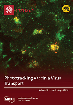
Cover Story (view full-size image): We have constructed recombinant vaccinia viruses that express the photoconvertible protein Dendra2. This novel approach enabled us to accurately track the dispersal of subsets of virus particles over extended periods of time, a challenge with classical live-cell microscopy. Here, we use this system to demonstrate the importance of kinesin-1 engagement in mediating virus exit from the trans-Golgi network. View Paper here.
  • Issues are regarded as officially published after their release is announced to the table of contents alert mailing list.
  • You may sign up for e-mail alerts to receive table of contents of newly released issues.
  • PDF is the official format for papers published in both, html and pdf forms. To view the papers in pdf format, click on the "PDF Full-text" link, and use the free Adobe Reader to open them.
Order results
Result details
Section
Select all
Export citation of selected articles as:
22 pages, 12635 KiB  
Article
The Morphology and Assembly of Respiratory Syncytial Virus Revealed by Cryo-Electron Tomography
by Zunlong Ke, Rebecca S. Dillard, Tatiana Chirkova, Fredrick Leon, Christopher C. Stobart, Cheri M. Hampton, Joshua D. Strauss, Devi Rajan, Christina A. Rostad, Jeannette V. Taylor, Hong Yi, Raven Shah, Mengtian Jin, Tina V. Hartert, R. Stokes Peebles, Jr., Barney S. Graham, Martin L. Moore, Larry J. Anderson and Elizabeth R. Wright
Viruses 2018, 10(8), 446; https://0-doi-org.brum.beds.ac.uk/10.3390/v10080446 - 20 Aug 2018
Cited by 51 | Viewed by 10232
Abstract
Human respiratory syncytial virus (RSV) is the leading cause of lower respiratory tract disease in young children. With repeat infections throughout life, it can also cause substantial disease in the elderly and in adults with compromised cardiac, pulmonary and immune systems. RSV is [...] Read more.
Human respiratory syncytial virus (RSV) is the leading cause of lower respiratory tract disease in young children. With repeat infections throughout life, it can also cause substantial disease in the elderly and in adults with compromised cardiac, pulmonary and immune systems. RSV is a pleomorphic enveloped RNA virus in the Pneumoviridae family. Recently, the three-dimensional (3D) structure of purified RSV particles has been elucidated, revealing three distinct morphological categories: spherical, asymmetric, and filamentous. However, the native 3D structure of RSV particles associated with or released from infected cells has yet to be investigated. In this study, we have established an optimized system for studying RSV structure by imaging RSV-infected cells on transmission electron microscopy (TEM) grids by cryo-electron tomography (cryo-ET). Our results demonstrate that RSV is filamentous across several virus strains and cell lines by cryo-ET, cryo-immuno EM, and thin section TEM techniques. The viral filament length varies from 0.5 to 12 μm and the average filament diameter is approximately 130 nm. Taking advantage of the whole cell tomography technique, we have resolved various stages of RSV assembly. Collectively, our results can facilitate the understanding of viral morphogenesis in RSV and other pleomorphic enveloped viruses. Full article
(This article belongs to the Special Issue Advances in Structural Virology via Cryo-EM)
Show Figures

Figure 1

25 pages, 12707 KiB  
Review
US28: HCMV’s Swiss Army Knife
by Benjamin A. Krishna, William E. Miller and Christine M. O’Connor
Viruses 2018, 10(8), 445; https://0-doi-org.brum.beds.ac.uk/10.3390/v10080445 - 20 Aug 2018
Cited by 43 | Viewed by 6602
Abstract
US28 is one of four G protein coupled receptors (GPCRs) encoded by human cytomegalovirus (HCMV). The US28 protein (pUS28) is a potent signaling molecule that alters a variety of cellular pathways that ultimately alter the host cell environment. This viral GPCR is expressed [...] Read more.
US28 is one of four G protein coupled receptors (GPCRs) encoded by human cytomegalovirus (HCMV). The US28 protein (pUS28) is a potent signaling molecule that alters a variety of cellular pathways that ultimately alter the host cell environment. This viral GPCR is expressed not only in the context of lytic replication but also during viral latency, highlighting its multifunctional properties. pUS28 is a functional GPCR, and its manipulation of multiple signaling pathways likely impacts HCMV pathogenesis. Herein, we will discuss the impact of pUS28 on both lytic and latent infection, pUS28-mediated signaling and its downstream consequences, and the influence this viral GPCR may have on disease states, including cardiovascular disease and cancer. We will also discuss the potential for and progress towards exploiting pUS28 as a novel therapeutic to combat HCMV. Full article
(This article belongs to the Special Issue Recent Advances in Cytomegalovirus Research)
Show Figures

Figure 1

27 pages, 1947 KiB  
Review
Molecular Determinants and the Regulation of Human Cytomegalovirus Latency and Reactivation
by Donna Collins-McMillen, Jason Buehler, Megan Peppenelli and Felicia Goodrum
Viruses 2018, 10(8), 444; https://0-doi-org.brum.beds.ac.uk/10.3390/v10080444 - 20 Aug 2018
Cited by 84 | Viewed by 9382
Abstract
Human cytomegalovirus (HCMV) is a beta herpesvirus that establishes a life-long persistence in the host, like all herpesviruses, by way of a latent infection. During latency, viral genomes are maintained in a quieted state. Virus replication can be reactivated from latency in response [...] Read more.
Human cytomegalovirus (HCMV) is a beta herpesvirus that establishes a life-long persistence in the host, like all herpesviruses, by way of a latent infection. During latency, viral genomes are maintained in a quieted state. Virus replication can be reactivated from latency in response to changes in cellular signaling caused by stress or differentiation. The past decade has brought great insights into the molecular basis of HCMV latency. Here, we review the complex persistence of HCMV with consideration of latent reservoirs, viral determinants and their host interactions, and host signaling and the control of cellular and viral gene expression that contributes to the establishment of and reactivation from latency. Full article
(This article belongs to the Special Issue Recent Advances in Cytomegalovirus Research)
Show Figures

Graphical abstract

14 pages, 1573 KiB  
Article
New Adenovirus Groups in Western Palaearctic Bats
by Maria Iglesias-Caballero, Javier Juste, Sonia Vázquez-Morón, Ana Falcon, Carolina Aznar-Lopez, Carlos Ibáñez, Francisco Pozo, Guillermo Ruiz, Jose M. Berciano, Inazio Garin, Joxerra Aihartza, Juan E. Echevarría and Inmaculada Casas
Viruses 2018, 10(8), 443; https://0-doi-org.brum.beds.ac.uk/10.3390/v10080443 - 20 Aug 2018
Cited by 13 | Viewed by 4641
Abstract
In the context of long-term screening for viruses on Western Palaearctic bats, we tested for the presence of adenovirus 1392 oropharyngeal swabs and 325 stool samples taken from 27 bat species. Adenoviruses were detected in 12 species of the Vespertilionidae and the Rhinolophidae [...] Read more.
In the context of long-term screening for viruses on Western Palaearctic bats, we tested for the presence of adenovirus 1392 oropharyngeal swabs and 325 stool samples taken from 27 bat species. Adenoviruses were detected in 12 species of the Vespertilionidae and the Rhinolophidae families. Fifty positive respiratory and 26 positive stool samples were studied. Phylogenetic analyses of partial hexon protein and partial DNA-dependent DNA polymerase genes indicate that all these bat adenoviruses belong to the genus Mastadenovirus but without constituting a monophyletic cluster. According to genetic identities, the new groups are distinct to the previously described Bat mastadenovirus A and B species and contribute with potentially new members. Our data support that diversity of bat mastadenovirus is host-dependent and increase the knowledge of potentially pathogenic virus from bats. Due to the active role of bats as viral reservoirs, the characterization of these viruses is relevant for Public Health. Full article
(This article belongs to the Special Issue Emerging Viruses)
Show Figures

Figure 1

4 pages, 187 KiB  
Opinion
Is the Virus Important? And Some Other Questions
by Ruth-Anne Sandaa and Gunnar Bratbak
Viruses 2018, 10(8), 442; https://0-doi-org.brum.beds.ac.uk/10.3390/v10080442 - 19 Aug 2018
Cited by 3 | Viewed by 3581
Abstract
The motivation for focusing on a specific virus is often its importance in terms of impact on human interests. The chlorella viruses are a notable exception and 40 years of research has made them the undisputed model system for large icosahedral dsDNA viruses [...] Read more.
The motivation for focusing on a specific virus is often its importance in terms of impact on human interests. The chlorella viruses are a notable exception and 40 years of research has made them the undisputed model system for large icosahedral dsDNA viruses infecting eukaryotes. Their status has changed from inconspicuous and rather odd with no ecological relevance to being the Phycodnaviridae type strain possibly affecting humans and human cognitive functioning in ways that remain to be understood. The Van Etten legacy is the backbone for research on Phycodnaviridae. After highlighting some of the peculiarities of chlorella viruses, we point to some issues and questions related to the viruses we choose for our research, our prejudices, what we are still missing, and what we should be looking for. Full article
(This article belongs to the Special Issue Algae Virus)
11 pages, 3430 KiB  
Article
Rapidity of Genomic Adaptations to Prasinovirus Infection in a Marine Microalga
by Sheree Yau, Gaëtan Caravello, Nadège Fonvieille, Élodie Desgranges, Hervé Moreau and Nigel Grimsley
Viruses 2018, 10(8), 441; https://0-doi-org.brum.beds.ac.uk/10.3390/v10080441 - 19 Aug 2018
Cited by 4 | Viewed by 4176
Abstract
Prasinoviruses are large dsDNA viruses commonly found in aquatic systems worldwide, where they can infect and lyse unicellular prasinophyte algae such as Ostreococcus. Host susceptibility is virus strain-specific, but resistance of susceptible Ostreococcus tauri strains to a virulent virus arises frequently. In [...] Read more.
Prasinoviruses are large dsDNA viruses commonly found in aquatic systems worldwide, where they can infect and lyse unicellular prasinophyte algae such as Ostreococcus. Host susceptibility is virus strain-specific, but resistance of susceptible Ostreococcus tauri strains to a virulent virus arises frequently. In clonal resistant lines that re-grow, viruses are usually present for many generations, and genes clustered on chromosome 19 show physical rearrangements and differential expression. Here, we investigated changes occurring during the first two weeks after inoculation of the prasinovirus OtV5. By serial dilutions of cultures at the time of inoculation, we estimated the frequency of resistant cells arising in virus-challenged O. tauri cultures to be 10−3–10−4 of the inoculated population. Re-growing resistant cells were detectable by flow cytometry 3 days post-inoculation (dpi), visible re-greening of cultures occurred by 6 dpi, and karyotypic changes were visually detectable at 8 dpi. Resistant cell lines showed a modified spectrum of host-virus specificities and much lower levels of OtV5 adsorption. Full article
(This article belongs to the Special Issue Algae Virus)
Show Figures

Graphical abstract

26 pages, 1329 KiB  
Review
The Diverse Roles of microRNAs at the Host–Virus Interface
by Annie Bernier and Selena M. Sagan
Viruses 2018, 10(8), 440; https://0-doi-org.brum.beds.ac.uk/10.3390/v10080440 - 19 Aug 2018
Cited by 73 | Viewed by 9014
Abstract
MicroRNAs (miRNAs) are small, non-coding RNAs that regulate gene expression at the post-transcriptional level. Through this activity, they are implicated in almost every cellular process investigated to date. Hence, it is not surprising that miRNAs play diverse roles in regulation of viral infections [...] Read more.
MicroRNAs (miRNAs) are small, non-coding RNAs that regulate gene expression at the post-transcriptional level. Through this activity, they are implicated in almost every cellular process investigated to date. Hence, it is not surprising that miRNAs play diverse roles in regulation of viral infections and antiviral responses. Diverse families of DNA and RNA viruses have been shown to take advantage of cellular miRNAs or produce virally encoded miRNAs that alter host or viral gene expression. MiRNA-mediated changes in gene expression have been demonstrated to modulate viral replication, antiviral immune responses, viral latency, and pathogenesis. Interestingly, viruses mediate both canonical and non-canonical interactions with miRNAs to downregulate specific targets or to promote viral genome stability, translation, and/or RNA accumulation. In this review, we focus on recent findings elucidating several key mechanisms employed by diverse virus families, with a focus on miRNAs at the host–virus interface during herpesvirus, polyomavirus, retroviruses, pestivirus, and hepacivirus infections. Full article
(This article belongs to the Special Issue CSV2018: The 2nd symposium of the Canadian Society for Virology (CSV))
Show Figures

Graphical abstract

9 pages, 210 KiB  
Review
Catching Chances: The Movement to Be on the Ground and Research Ready before an Outbreak
by David Brett-Major and James Lawler
Viruses 2018, 10(8), 439; https://0-doi-org.brum.beds.ac.uk/10.3390/v10080439 - 19 Aug 2018
Cited by 6 | Viewed by 4851
Abstract
After more than 28,000 Ebola virus disease cases and at least 11,000 deaths in West Africa during the 2014–2016 epidemic, the world remains without a licensed vaccine or therapeutic broadly available and demonstrated to alleviate suffering. This deficiency has been felt acutely in [...] Read more.
After more than 28,000 Ebola virus disease cases and at least 11,000 deaths in West Africa during the 2014–2016 epidemic, the world remains without a licensed vaccine or therapeutic broadly available and demonstrated to alleviate suffering. This deficiency has been felt acutely in the two, short, following years with two Ebola virus outbreaks in the Democratic Republic of Congo (DRC), and a Marburg virus outbreak in Uganda. Despite billions of U.S. dollars invested in developing medical countermeasures for filoviruses in the antecedent decades, resulting in an array of preventative, diagnostic, and therapeutic products, none are available on commercial shelves. This paper explores why just-in-time research efforts in the field during the West Africa epidemic failed, as well as some recent initiatives to prevent similarly lost opportunities. Full article
(This article belongs to the Collection Advances in Ebolavirus, Marburgvirus, and Cuevavirus Research)
17 pages, 2379 KiB  
Article
Synergistic Removal of Static and Dynamic Staphylococcus aureus Biofilms by Combined Treatment with a Bacteriophage Endolysin and a Polysaccharide Depolymerase
by Nanna M. C. Olsen, Elowine Thiran, Tobias Hasler, Thomas Vanzieleghem, Georgios N. Belibasakis, Jacques Mahillon, Martin J. Loessner and Mathias Schmelcher
Viruses 2018, 10(8), 438; https://0-doi-org.brum.beds.ac.uk/10.3390/v10080438 - 18 Aug 2018
Cited by 58 | Viewed by 7174
Abstract
Staphylococcus aureus is an important pathogen and biofilm former. Biofilms cause problems in clinics and food production and are highly recalcitrant to antibiotics and sanitizers. Bacteriophage endolysins kill bacteria by degrading their cell wall and are therefore deemed promising antimicrobials and anti-biofilm agents. [...] Read more.
Staphylococcus aureus is an important pathogen and biofilm former. Biofilms cause problems in clinics and food production and are highly recalcitrant to antibiotics and sanitizers. Bacteriophage endolysins kill bacteria by degrading their cell wall and are therefore deemed promising antimicrobials and anti-biofilm agents. Depolymerases targeting polysaccharides in the extracellular matrix have been suggested as parts of a multi-enzyme approach to eradicate biofilms. The efficacy of endolysins and depolymerases against S. aureus biofilms in static models has been demonstrated. However, there is a lack of studies evaluating their activity against biofilms grown under more realistic conditions. Here, we investigated the efficacy of the endolysin LysK and the poly-N-acetylglucosamine depolymerase DA7 against staphylococcal biofilms in static and dynamic (flow cell-based) models. LysK showed activity against multiple S. aureus strains, and both LysK and DA7 removed static and dynamic biofilms from polystyrene and glass surfaces at low micromolar and nanomolar concentrations, respectively. When combined, the enzymes acted synergistically, as demonstrated by crystal violet staining of static biofilms, significantly reducing viable cell counts compared to individual enzyme treatment in the dynamic model, and confocal laser scanning microscopy. Overall, our results suggest that LysK and DA7 are potent anti-biofilm agents, alone and in combination. Full article
(This article belongs to the Special Issue Phage Lytic Enzymes and Their Applications)
Show Figures

Figure 1

15 pages, 3505 KiB  
Article
Shell-Less Egg Syndrome (SES) Widespread in Western Canadian Layer Operations Is Linked to a Massachusetts (Mass) Type Infectious Bronchitis Virus (IBV) Isolate
by Aruna Amarasinghe, Shelly Popowich, Upasama De Silva Senapathi, Mohamed Sarjoon Abdul-Cader, Frank Marshall, Frank Van der Meer, Susan C. Cork, Susantha Gomis and Mohamed Faizal Abdul-Careem
Viruses 2018, 10(8), 437; https://0-doi-org.brum.beds.ac.uk/10.3390/v10080437 - 18 Aug 2018
Cited by 17 | Viewed by 4671
Abstract
A disease with a sudden drop in egg production and shell-less eggs called, shell-less egg syndrome (SES) has been observed in Western Canada egg layer flocks since 2010. The etiology of this disease is not known. We hypothesize that SES is caused by [...] Read more.
A disease with a sudden drop in egg production and shell-less eggs called, shell-less egg syndrome (SES) has been observed in Western Canada egg layer flocks since 2010. The etiology of this disease is not known. We hypothesize that SES is caused by an infectious bronchitis virus (IBV) strain since it is known that IBV replicates in the shell gland causing various eggshell abnormalities. In this study, we screened egg layer flocks, in the provinces of Alberta (AB) and Saskatchewan (SK), with and without a history of SES for the presence of IBV infection. During 2015–2016, a total of 27 egg layer flocks were screened in AB (n = 7) and SK (n = 20). Eighty-one percent of the screened flocks (n = 22) were positive for IBV infection. Thirty of these isolates were successfully characterized using molecular tools targeting the most variable spike (S) 1 gene. IBV isolates from this study clustered into three genotypes based on partial S1 gene variability. The majority of the IBV isolates (70%) were Massachusetts (Mass) type, and the rest were either Connecticut (Conn) type or an uncharacterized genotype with genetic characteristics of Mass and Conn types. Since the majority of the IBV isolates included within the Mass type, we used a Mass type IBV isolate to reproduce SES in specific pathogen free (SPF) white leghorn chickens in lay. Further studies are warranted to investigate whether other IBV isolates can cause SES, to clarify the pathogenesis of SES and to develop a vaccine in order to prevent SES as observed in Western Canadian layer flocks. Full article
(This article belongs to the Section Animal Viruses)
Show Figures

Figure 1

23 pages, 318 KiB  
Review
Recent Advances on Detection and Characterization of Fruit Tree Viruses Using High-Throughput Sequencing Technologies
by Varvara I. Maliogka, Angelantonio Minafra, Pasquale Saldarelli, Ana B. Ruiz-García, Miroslav Glasa, Nikolaos Katis and Antonio Olmos
Viruses 2018, 10(8), 436; https://0-doi-org.brum.beds.ac.uk/10.3390/v10080436 - 17 Aug 2018
Cited by 87 | Viewed by 7098
Abstract
Perennial crops, such as fruit trees, are infected by many viruses, which are transmitted through vegetative propagation and grafting of infected plant material. Some of these pathogens cause severe crop losses and often reduce the productive life of the orchards. Detection and characterization [...] Read more.
Perennial crops, such as fruit trees, are infected by many viruses, which are transmitted through vegetative propagation and grafting of infected plant material. Some of these pathogens cause severe crop losses and often reduce the productive life of the orchards. Detection and characterization of these agents in fruit trees is challenging, however, during the last years, the wide application of high-throughput sequencing (HTS) technologies has significantly facilitated this task. In this review, we present recent advances in the discovery, detection, and characterization of fruit tree viruses and virus-like agents accomplished by HTS approaches. A high number of new viruses have been described in the last 5 years, some of them exhibiting novel genomic features that have led to the proposal of the creation of new genera, and the revision of the current virus taxonomy status. Interestingly, several of the newly identified viruses belong to virus genera previously unknown to infect fruit tree species (e.g., Fabavirus, Luteovirus) a fact that challenges our perspective of plant viruses in general. Finally, applied methodologies, including the use of different molecules as templates, as well as advantages and disadvantages and future directions of HTS in fruit tree virology are discussed. Full article
(This article belongs to the Special Issue Fruit Tree Viruses and Viroids)
33 pages, 3352 KiB  
Review
Prospects in Innate Immune Responses as Potential Control Strategies against Non-Primate Lentiviruses
by Lorena De Pablo-Maiso, Ana Doménech, Irache Echeverría, Carmen Gómez-Arrebola, Damián De Andrés, Sergio Rosati, Esperanza Gómez-Lucia and Ramsés Reina
Viruses 2018, 10(8), 435; https://0-doi-org.brum.beds.ac.uk/10.3390/v10080435 - 17 Aug 2018
Cited by 17 | Viewed by 5900
Abstract
Lentiviruses are infectious agents of a number of animal species, including sheep, goats, horses, monkeys, cows, and cats, in addition to humans. As in the human case, the host immune response fails to control the establishment of chronic persistent infection that finally leads [...] Read more.
Lentiviruses are infectious agents of a number of animal species, including sheep, goats, horses, monkeys, cows, and cats, in addition to humans. As in the human case, the host immune response fails to control the establishment of chronic persistent infection that finally leads to a specific disease development. Despite intensive research on the development of lentivirus vaccines, it is still not clear which immune responses can protect against infection. Viral mutations resulting in escape from T-cell or antibody-mediated responses are the basis of the immune failure to control the infection. The innate immune response provides the first line of defense against viral infections in an antigen-independent manner. Antiviral innate responses are conducted by dendritic cells, macrophages, and natural killer cells, often targeted by lentiviruses, and intrinsic antiviral mechanisms exerted by all cells. Intrinsic responses depend on the recognition of the viral pathogen-associated molecular patterns (PAMPs) by pathogen recognition receptors (PRRs), and the signaling cascades leading to an antiviral state by inducing the expression of antiviral proteins, including restriction factors. This review describes the latest advances on innate immunity related to the infection by animal lentiviruses, centered on small ruminant lentiviruses (SRLV), equine infectious anemia virus (EIAV), and feline (FIV) and bovine immunodeficiency viruses (BIV), specifically focusing on the antiviral role of the major restriction factors described thus far. Full article
(This article belongs to the Special Issue Nonprimate Lentivirus)
Show Figures

Figure 1

9 pages, 280 KiB  
Article
Colonized Sabethes cyaneus, a Sylvatic New World Mosquito Species, Shows a Low Vector Competence for Zika Virus Relative to Aedes aegypti
by Ajit K. Karna, Sasha R. Azar, Jessica A. Plante, Rumei Yun, Nikos Vasilakis, Scott C. Weaver, Immo A. Hansen and Kathryn A. Hanley
Viruses 2018, 10(8), 434; https://0-doi-org.brum.beds.ac.uk/10.3390/v10080434 - 16 Aug 2018
Cited by 24 | Viewed by 5558
Abstract
The introduction of Zika virus (ZIKV) to the Americas raised concern that the virus would spill back from human transmission, perpetuated by Aedes aegypti, into a sylvatic cycle maintained in wildlife and forest-living mosquitoes. In the Americas, Sabethes species are vectors of [...] Read more.
The introduction of Zika virus (ZIKV) to the Americas raised concern that the virus would spill back from human transmission, perpetuated by Aedes aegypti, into a sylvatic cycle maintained in wildlife and forest-living mosquitoes. In the Americas, Sabethes species are vectors of sylvatic yellow fever virus (YFV) and are therefore candidate vectors of a sylvatic ZIKV cycle. To test the potential of Sabethes cyaneus to transmit ZIKV, Sa. cyaneus and Ae. aegypti were fed on A129 mice one or two days post-infection (dpi) with a ZIKV isolate from Mexico. Sa. cyaneus were sampled at 3, 4, 5, 7, 14, and 21 days post-feeding (dpf) and Ae. aegypti were sampled at 14 and 21 dpf. ZIKV was quantified in mosquito bodies, legs, and saliva to measure infection, dissemination, and potential transmission, respectively. Of 69 Sa. cyaneus that fed, ZIKV was detected in only one, in all body compartments, at 21 dpf. In contrast, at 14 dpf 100% of 20 Ae. aegypti that fed on mice at 2 dpi were infected and 70% had virus in saliva. These data demonstrate that Sa. cyaneus is a competent vector for ZIKV, albeit much less competent than Ae. aegypti. Full article
(This article belongs to the Special Issue 6th Pan-American Dengue Research Network Meeting)
16 pages, 8341 KiB  
Article
Potential Therapeutic Agents for Feline Calicivirus Infection
by Tulio M. Fumian, Daniel Enosi Tuipulotu, Natalie E. Netzler, Jennifer H. Lun, Alice G. Russo, Grace J. H. Yan and Peter A. White
Viruses 2018, 10(8), 433; https://0-doi-org.brum.beds.ac.uk/10.3390/v10080433 - 16 Aug 2018
Cited by 17 | Viewed by 16029
Abstract
Feline calicivirus (FCV) is a major cause of upper respiratory tract disease in cats, with widespread distribution in the feline population. Recently, virulent systemic diseases caused by FCV infection has been associated with mortality rates up to 50%. Currently, there are no direct-acting [...] Read more.
Feline calicivirus (FCV) is a major cause of upper respiratory tract disease in cats, with widespread distribution in the feline population. Recently, virulent systemic diseases caused by FCV infection has been associated with mortality rates up to 50%. Currently, there are no direct-acting antivirals approved for the treatment of FCV infection. Here, we tested 15 compounds from different antiviral classes against FCV using in vitro protein and cell culture assays. After the expression of FCV protease-polymerase protein, we established two in vitro assays to assess the inhibitory activity of compounds directly against the FCV protease or polymerase. Using this recombinant enzyme, we identified quercetagetin and PPNDS as inhibitors of FCV polymerase activity (IC50 values of 2.8 μM and 2.7 μM, respectively). We also demonstrate the inhibition of FCV protease activity by GC376 (IC50 of 18 µM). Using cell culture assays, PPNDS, quercetagetin and GC376 did not display antivirals effects, however, we identified nitazoxanide and 2′-C-methylcytidine (2CMC) as potent inhibitors of FCV replication, with EC50 values in the low micromolar range (0.6 μM and 2.5 μM, respectively). In conclusion, we established two in vitro assays that will accelerate the research for FCV antivirals and can be used for the high-throughput screening of direct-acting antivirals. Full article
(This article belongs to the Section Viral Immunology, Vaccines, and Antivirals)
Show Figures

Graphical abstract

11 pages, 2655 KiB  
Communication
Molecular Characterization and Geographic Distribution of a Mymonavirus in the Population of Botrytis cinerea
by Fangmin Hao, Mingde Wu and Guoqing Li
Viruses 2018, 10(8), 432; https://0-doi-org.brum.beds.ac.uk/10.3390/v10080432 - 15 Aug 2018
Cited by 34 | Viewed by 4741
Abstract
Here, we characterized a negative single-stranded (−ss)RNA mycovirus, Botrytis cinerea mymonavirus 1 (BcMyV1), isolated from the phytopathogenic fungus Botrytis cinerea. The genome of BcMyV1 is 7863 nt in length, possessing three open reading frames (ORF1–3). The ORF1 encodes a large polypeptide containing [...] Read more.
Here, we characterized a negative single-stranded (−ss)RNA mycovirus, Botrytis cinerea mymonavirus 1 (BcMyV1), isolated from the phytopathogenic fungus Botrytis cinerea. The genome of BcMyV1 is 7863 nt in length, possessing three open reading frames (ORF1–3). The ORF1 encodes a large polypeptide containing a conserved mononegaviral RNA-dependent RNA polymerase (RdRp) domain showing homology to the protein L of mymonaviruses, whereas the possible functions of the remaining two ORFs are still unknown. The internal cDNA sequence (10-7829) of BcMyV1 was 97.9% identical to the full-length cDNA sequence of Sclerotinia sclerotiorum negative stranded RNA virus 7 (SsNSRV7), a virus-like contig obtained from Sclerotinia sclerotiorum metatranscriptomes, indicating BcMyV1 should be a strain of SsNSRV7. Phylogenetic analysis based on RdRp domains showed that BcMyV1 was clustered with the viruses in the family Mymonaviridae, suggesting it is a member of Mymonaviridae. BcMyV1 may be widely distributed in regions where B. cinerea occurs in China and even over the world, although it infected only 0.8% of tested B. cinerea strains. Full article
(This article belongs to the Special Issue Mycoviruses)
Show Figures

Figure 1

18 pages, 2361 KiB  
Communication
Bacteriophage Sf6 Tailspike Protein for Detection of Shigella flexneri Pathogens
by Sonja Kunstmann, Tom Scheidt, Saskia Buchwald, Alexandra Helm, Laurence A. Mulard, Angelika Fruth and Stefanie Barbirz
Viruses 2018, 10(8), 431; https://0-doi-org.brum.beds.ac.uk/10.3390/v10080431 - 15 Aug 2018
Cited by 23 | Viewed by 5417
Abstract
Bacteriophage research is gaining more importance due to increasing antibiotic resistance. However, for treatment with bacteriophages, diagnostics have to be improved. Bacteriophages carry adhesion proteins, which bind to the bacterial cell surface, for example tailspike proteins (TSP) for specific recognition of bacterial O-antigen [...] Read more.
Bacteriophage research is gaining more importance due to increasing antibiotic resistance. However, for treatment with bacteriophages, diagnostics have to be improved. Bacteriophages carry adhesion proteins, which bind to the bacterial cell surface, for example tailspike proteins (TSP) for specific recognition of bacterial O-antigen polysaccharide. TSP are highly stable proteins and thus might be suitable components for the integration into diagnostic tools. We used the TSP of bacteriophage Sf6 to establish two applications for detecting Shigella flexneri (S. flexneri), a highly contagious pathogen causing dysentery. We found that Sf6TSP not only bound O-antigen of S. flexneri serotype Y, but also the glucosylated O-antigen of serotype 2a. Moreover, mass spectrometry glycan analyses showed that Sf6TSP tolerated various O-acetyl modifications on these O-antigens. We established a microtiter plate-based ELISA like tailspike adsorption assay (ELITA) using a Strep-tag®II modified Sf6TSP. As sensitive screening alternative we produced a fluorescently labeled Sf6TSP via coupling to an environment sensitive dye. Binding of this probe to the S. flexneri O-antigen Y elicited a fluorescence intensity increase of 80% with an emission maximum in the visible light range. The Sf6TSP probes thus offer a promising route to a highly specific and sensitive bacteriophage TSP-based Shigella detection system. Full article
(This article belongs to the Special Issue Biotechnological Applications of Phage and Phage-Derived Proteins)
Show Figures

Figure 1

11 pages, 2659 KiB  
Brief Report
Molecular and Biological Characterisation of Turnip mosaic virus Isolates Infecting Poppy (Papaver somniferum and P. rhoeas) in Slovakia
by Miroslav Glasa, Katarína Šoltys, Lukáš Predajňa, Nina Sihelská, Slavomíra Nováková, Zdeno Šubr, Ján Kraic and Daniel Mihálik
Viruses 2018, 10(8), 430; https://0-doi-org.brum.beds.ac.uk/10.3390/v10080430 - 14 Aug 2018
Cited by 6 | Viewed by 4249
Abstract
In recent years, the accumulated molecular data of Turnip mosaic virus (TuMV) isolates from various hosts originating from different parts of the world considerably helped to understand the genetic complexity and evolutionary history of the virus. In this work, four complete TuMV genomes [...] Read more.
In recent years, the accumulated molecular data of Turnip mosaic virus (TuMV) isolates from various hosts originating from different parts of the world considerably helped to understand the genetic complexity and evolutionary history of the virus. In this work, four complete TuMV genomes (HC9, PK1, MS04, MS15) were characterised from naturally infected cultivated and wild-growing Papaver spp., hosts from which only very scarce data were available previously. Phylogenetic analyses showed the affiliation of Slovak Papaver isolates to the world-B and basal-B groups. The PK1 isolate showed a novel intra-lineage recombination pattern, further confirming the important role of recombination in the shaping of TuMV genetic diversity. Biological assays indicated that the intensity of symptoms in experimentally inoculated oilseed poppy are correlated to TuMV accumulation level in leaves. This is the first report of TuMV in poppy plants in Slovakia. Full article
(This article belongs to the Special Issue Plant Virus Ecology and Biodiversity)
Show Figures

Figure 1

25 pages, 2377 KiB  
Review
Overview of Trends in the Application of Metagenomic Techniques in the Analysis of Human Enteric Viral Diversity in Africa’s Environmental Regimes
by Cecilia Oluseyi Osunmakinde, Ramganesh Selvarajan, Timothy Sibanda, Bhekie B Mamba and Titus A.M Msagati
Viruses 2018, 10(8), 429; https://0-doi-org.brum.beds.ac.uk/10.3390/v10080429 - 14 Aug 2018
Cited by 8 | Viewed by 5080
Abstract
There has been an increase in the quest for metagenomics as an approach for the identification and study of the diversity of human viruses found in aquatic systems, both for their role as waterborne pathogens and as water quality indicators. In the last [...] Read more.
There has been an increase in the quest for metagenomics as an approach for the identification and study of the diversity of human viruses found in aquatic systems, both for their role as waterborne pathogens and as water quality indicators. In the last few years, environmental viral metagenomics has grown significantly and has enabled the identification, diversity and entire genome sequencing of viruses in environmental and clinical samples extensively. Prior to the arrival of metagenomics, traditional molecular procedures such as the polymerase chain reaction (PCR) and sequencing, were mostly used to identify and classify enteric viral species in different environmental milieu. After the advent of metagenomics, more detailed reports have emerged about the important waterborne viruses identified in wastewater treatment plant effluents and surface water. This paper provides a review of methods that have been used for the concentration, detection and identification of viral species from different environmental matrices. The review also takes into consideration where metagenomics has been explored in different African countries, as well as the limitations and challenges facing the approach. Procedures including sample processing, experimental design, sequencing technology, and bioinformatics analysis are discussed. The review concludes by summarising the current thinking and practices in the field and lays bare key issues that those venturing into this field need to consider and address. Full article
(This article belongs to the Section Animal Viruses)
Show Figures

Figure 1

18 pages, 750 KiB  
Review
Mycobacteriophage Lysis Enzymes: Targeting the Mycobacterial Cell Envelope
by Maria João Catalão and Madalena Pimentel
Viruses 2018, 10(8), 428; https://0-doi-org.brum.beds.ac.uk/10.3390/v10080428 - 14 Aug 2018
Cited by 35 | Viewed by 7071
Abstract
Mycobacteriophages are viruses that specifically infect mycobacteria, which ultimately culminate in host cell death. Dedicated enzymes targeting the complex mycobacterial cell envelope arrangement have been identified in mycobacteriophage genomes, thus being potential candidates as antibacterial agents. These comprise lipolytic enzymes that target the [...] Read more.
Mycobacteriophages are viruses that specifically infect mycobacteria, which ultimately culminate in host cell death. Dedicated enzymes targeting the complex mycobacterial cell envelope arrangement have been identified in mycobacteriophage genomes, thus being potential candidates as antibacterial agents. These comprise lipolytic enzymes that target the mycolic acid-containing outer membrane and peptidoglycan hydrolases responsive to the atypical mycobacterial peptidoglycan layer. In the recent years, a remarkable progress has been made, particularly on the comprehension of the mechanisms of bacteriophage lysis proteins activity and regulation. Notwithstanding, information about mycobacteriophages lysis strategies is limited and is mainly represented by the studies performed with mycobacteriophage Ms6. Since mycobacteriophages target a specific group of bacteria, which include Mycobacterium tuberculosis responsible for one of the leading causes of death worldwide, exploitation of the use of these lytic enzymes demands a special attention, as they may be an alternative to tackle multidrug resistant tuberculosis. This review focuses on the current knowledge of the function of lysis proteins encoded by mycobacteriophages and their potential applications, which may contribute to increasing the effectiveness of antimycobacterial therapy. Full article
(This article belongs to the Special Issue Phage Lytic Enzymes and Their Applications)
Show Figures

Figure 1

22 pages, 5112 KiB  
Article
Structure and Analysis of R1 and R2 Pyocin Receptor-Binding Fibers
by Sergey A. Buth, Mikhail M. Shneider, Dean Scholl and Petr G. Leiman
Viruses 2018, 10(8), 427; https://0-doi-org.brum.beds.ac.uk/10.3390/v10080427 - 14 Aug 2018
Cited by 30 | Viewed by 5855
Abstract
The R-type pyocins are high-molecular weight bacteriocins produced by some strains of Pseudomonas aeruginosa to specifically kill other strains of the same species. Structurally, the R-type pyocins are similar to “simple” contractile tails, such as those of phage P2 and Mu. The pyocin [...] Read more.
The R-type pyocins are high-molecular weight bacteriocins produced by some strains of Pseudomonas aeruginosa to specifically kill other strains of the same species. Structurally, the R-type pyocins are similar to “simple” contractile tails, such as those of phage P2 and Mu. The pyocin recognizes and binds to its target with the help of fibers that emanate from the baseplate structure at one end of the particle. Subsequently, the pyocin contracts its sheath and drives the rigid tube through the host cell envelope. This causes depolarization of the cytoplasmic membrane and cell death. The host cell surface-binding fiber is ~340 Å-long and is attached to the baseplate with its N-terminal domain. Here, we report the crystal structures of C-terminal fragments of the R1 and R2 pyocin fibers that comprise the distal, receptor-binding part of the protein. Both proteins are ~240 Å-long homotrimers in which slender rod-like domains are interspersed with more globular domains—two tandem knob domains in the N-terminal part of the fragment and a lectin-like domain at its C-terminus. The putative substrate binding sites are separated by about 100 Å, suggesting that binding of the fiber to the cell surface causes the fiber to adopt a certain orientation relative to the baseplate and this then triggers sheath contraction. Full article
(This article belongs to the Special Issue Biotechnological Applications of Phage and Phage-Derived Proteins)
Show Figures

Graphical abstract

20 pages, 3980 KiB  
Article
Removal of the N-Glycosylation Sequon at Position N116 Located in p27 of the Respiratory Syncytial Virus Fusion Protein Elicits Enhanced Antibody Responses after DNA Immunization
by Annelies Leemans, Marlies Boeren, Winke Van der Gucht, Isabel Pintelon, Kenny Roose, Bert Schepens, Xavier Saelens, Dalan Bailey, Wim Martinet, Guy Caljon, Louis Maes, Paul Cos and Peter Delputte
Viruses 2018, 10(8), 426; https://0-doi-org.brum.beds.ac.uk/10.3390/v10080426 - 14 Aug 2018
Cited by 12 | Viewed by 5112
Abstract
Prevention of severe lower respiratory tract infections in infants caused by the human respiratory syncytial virus (hRSV) remains a major public health priority. Currently, the major focus of vaccine development relies on the RSV fusion (F) protein since it is the main target [...] Read more.
Prevention of severe lower respiratory tract infections in infants caused by the human respiratory syncytial virus (hRSV) remains a major public health priority. Currently, the major focus of vaccine development relies on the RSV fusion (F) protein since it is the main target protein for neutralizing antibodies induced by natural infection. The protein conserves 5 N-glycosylation sites, two of which are located in the F2 subunit (N27 and N70), one in the F1 subunit (N500) and two in the p27 peptide (N116 and N126). To study the influence of the loss of one or more N-glycosylation sites on RSV F immunogenicity, BALB/c mice were immunized with plasmids encoding RSV F glycomutants. In comparison with F WT DNA immunized mice, higher neutralizing titres were observed following immunization with F N116Q. Moreover, RSV A2-K-line19F challenge of mice that had been immunized with mutant F N116Q DNA was associated with lower RSV RNA levels compared with those in challenged WT F DNA immunized animals. Since p27 is assumed to be post-translationally released after cleavage and thus not present on the mature RSV F protein, it remains to be elucidated how deletion of this glycan can contribute to enhanced antibody responses and protection upon challenge. These findings provide new insights to improve the immunogenicity of RSV F in potential vaccine candidates. Full article
(This article belongs to the Section Viral Immunology, Vaccines, and Antivirals)
Show Figures

Figure 1

10 pages, 247 KiB  
Article
Reported Direct and Indirect Contact with Dromedary Camels among Laboratory-Confirmed MERS-CoV Cases
by Romy Conzade, Rebecca Grant, Mamunur Rahman Malik, Amgad Elkholy, Mohamed Elhakim, Dalia Samhouri, Peter K. Ben Embarek and Maria D. Van Kerkhove
Viruses 2018, 10(8), 425; https://0-doi-org.brum.beds.ac.uk/10.3390/v10080425 - 13 Aug 2018
Cited by 69 | Viewed by 7903
Abstract
Dromedary camels (Camelus dromedarius) are now known to be the vertebrate animal reservoir that intermittently transmits the Middle East respiratory syndrome coronavirus (MERS-CoV) to humans. Yet, details as to the specific mechanism(s) of zoonotic transmission from dromedaries to humans remain unclear. [...] Read more.
Dromedary camels (Camelus dromedarius) are now known to be the vertebrate animal reservoir that intermittently transmits the Middle East respiratory syndrome coronavirus (MERS-CoV) to humans. Yet, details as to the specific mechanism(s) of zoonotic transmission from dromedaries to humans remain unclear. The aim of this study was to describe direct and indirect contact with dromedaries among all cases, and then separately for primary, non-primary, and unclassified cases of laboratory-confirmed MERS-CoV reported to the World Health Organization (WHO) between 1 January 2015 and 13 April 2018. We present any reported dromedary contact: direct, indirect, and type of indirect contact. Of all 1125 laboratory-confirmed MERS-CoV cases reported to WHO during the time period, there were 348 (30.9%) primary cases, 455 (40.4%) non-primary cases, and 322 (28.6%) unclassified cases. Among primary cases, 191 (54.9%) reported contact with dromedaries: 164 (47.1%) reported direct contact, 155 (44.5%) reported indirect contact. Five (1.1%) non-primary cases also reported contact with dromedaries. Overall, unpasteurized milk was the most frequent type of dromedary product consumed. Among cases for whom exposure was systematically collected and reported to WHO, contact with dromedaries or dromedary products has played an important role in zoonotic transmission. Full article
23 pages, 3542 KiB  
Article
Potent HIV-1-Specific CD8 T Cell Responses Induced in Mice after Priming with a Multiepitopic DNA-TMEP and Boosting with the HIV Vaccine MVA-B
by Beatriz Perdiguero, Suresh C. Raman, Cristina Sánchez-Corzo, Carlos Oscar S. Sorzano, José Ramón Valverde, Mariano Esteban and Carmen Elena Gómez
Viruses 2018, 10(8), 424; https://0-doi-org.brum.beds.ac.uk/10.3390/v10080424 - 13 Aug 2018
Cited by 9 | Viewed by 3651
Abstract
An effective vaccine against Human Immunodeficiency Virus (HIV) still remains the best solution to provide a sustainable control and/or eradication of the virus. We have previously generated the HIV-1 vaccine modified vaccinia virus Ankara (MVA)-B, which exhibited good immunogenicity profile in phase I [...] Read more.
An effective vaccine against Human Immunodeficiency Virus (HIV) still remains the best solution to provide a sustainable control and/or eradication of the virus. We have previously generated the HIV-1 vaccine modified vaccinia virus Ankara (MVA)-B, which exhibited good immunogenicity profile in phase I prophylactic and therapeutic clinical trials, but was unable to prevent viral rebound after antiretroviral (ART) removal. To potentiate the immunogenicity of MVA-B, here we described the design and immune responses elicited in mice by a new T cell multi-epitopic B (TMEP-B) immunogen, vectored by DNA, when administered in homologous or heterologous prime/boost regimens in combination with MVA-B. The TMEP-B protein contained conserved regions from Gag, Pol, and Nef proteins including multiple CD4 and CD8 T cell epitopes functionally associated with HIV control. Heterologous DNA-TMEP/MVA-B regimen induced higher HIV-1-specific CD8 T cell responses with broader epitope recognition and higher polyfunctional profile than the homologous DNA-TMEP/DNA-TMEP or the heterologous DNA-GPN/MVA-B combinations. Moreover, higher HIV-1-specific CD4 and Tfh immune responses were also detected using this regimen. After MVA-B boost, the magnitude of the anti-VACV CD8 T cell response was significantly compromised in DNA-TMEP-primed animals. Our results revealed the immunological potential of DNA-TMEP prime/MVA-B boost regimen and supported the application of these combined vectors in HIV-1 prevention and/or therapy. Full article
(This article belongs to the Special Issue HIV Vaccines)
Show Figures

Figure 1

13 pages, 4832 KiB  
Review
A Systems Approach to Study Immuno- and Neuro-Modulatory Properties of Antiviral Agents
by Eva Zusinaite, Aleksandr Ianevski, Diana Niukkanen, Minna M. Poranen, Magnar Bjørås, Jan Egil Afset, Tanel Tenson, Vidya Velagapudi, Andres Merits and Denis E. Kainov
Viruses 2018, 10(8), 423; https://0-doi-org.brum.beds.ac.uk/10.3390/v10080423 - 12 Aug 2018
Cited by 7 | Viewed by 4996
Abstract
There are dozens of approved, investigational and experimental antiviral agents. Many of these agents cause serious side effects, which can only be revealed after drug administration. Identification of the side effects prior to drug administration is challenging. Here we describe an ex vivo [...] Read more.
There are dozens of approved, investigational and experimental antiviral agents. Many of these agents cause serious side effects, which can only be revealed after drug administration. Identification of the side effects prior to drug administration is challenging. Here we describe an ex vivo approach for studying immuno- and neuro-modulatory properties of antiviral agents, which may be associated with potential side effects of these therapeutics. The current approach combines drug toxicity/efficacy tests and transcriptomics, which is followed by mRNA, cytokine and metabolite profiling. We demonstrated the utility of this approach with several examples of antiviral agents. We also showed that the approach can utilize different immune stimuli and cell types. It can also include other omics techniques, such as genomics and epigenomics, to allow identification of individual markers associated with adverse reactions to antivirals with immuno- and neuro-modulatory properties. Full article
(This article belongs to the Special Issue Viruses and Cellular Metabolism)
Show Figures

Figure 1

28 pages, 7627 KiB  
Article
Functional Genomics and Immunologic Tools: The Impact of Viral and Host Genetic Variations on the Outcome of Zika Virus Infection
by Sang-Im Yun, Byung-Hak Song, Jordan C. Frank, Justin G. Julander, Aaron L. Olsen, Irina A. Polejaeva, Christopher J. Davies, Kenneth L. White and Young-Min Lee
Viruses 2018, 10(8), 422; https://0-doi-org.brum.beds.ac.uk/10.3390/v10080422 - 11 Aug 2018
Cited by 12 | Viewed by 4908
Abstract
Zika virus (ZIKV) causes no-to-mild symptoms or severe neurological disorders. To investigate the importance of viral and host genetic variations in determining ZIKV infection outcomes, we created three full-length infectious cDNA clones as bacterial artificial chromosomes for each of three spatiotemporally distinct and [...] Read more.
Zika virus (ZIKV) causes no-to-mild symptoms or severe neurological disorders. To investigate the importance of viral and host genetic variations in determining ZIKV infection outcomes, we created three full-length infectious cDNA clones as bacterial artificial chromosomes for each of three spatiotemporally distinct and genetically divergent ZIKVs: MR-766 (Uganda, 1947), P6-740 (Malaysia, 1966), and PRVABC-59 (Puerto Rico, 2015). Using the three molecularly cloned ZIKVs, together with 13 ZIKV region-specific polyclonal antibodies covering nearly the entire viral protein-coding region, we made three conceptual advances: (i) We created a comprehensive genome-wide portrait of ZIKV gene products and their related species, with several previously undescribed gene products identified in the case of all three molecularly cloned ZIKVs. (ii) We found that ZIKV has a broad cell tropism in vitro, being capable of establishing productive infection in 16 of 17 animal cell lines from 12 different species, although its growth kinetics varied depending on both the specific virus strain and host cell line. More importantly, we identified one ZIKV-non-susceptible bovine cell line that has a block in viral entry but fully supports the subsequent post-entry steps. (iii) We showed that in mice, the three molecularly cloned ZIKVs differ in their neuropathogenicity, depending on the particular combination of viral and host genetic backgrounds, as well as in the presence or absence of type I/II interferon signaling. Overall, our findings demonstrate the impact of viral and host genetic variations on the replication kinetics and neuropathogenicity of ZIKV and provide multiple avenues for developing and testing medical countermeasures against ZIKV. Full article
(This article belongs to the Special Issue New Advances on Zika Virus Research)
Show Figures

Figure 1

26 pages, 1233 KiB  
Review
Going (Reo)Viral: Factors Promoting Successful Reoviral Oncolytic Infection
by Tarryn Bourhill, Yoshinori Mori, Derrick E. Rancourt, Maya Shmulevitz and Randal N. Johnston
Viruses 2018, 10(8), 421; https://0-doi-org.brum.beds.ac.uk/10.3390/v10080421 - 11 Aug 2018
Cited by 18 | Viewed by 4184
Abstract
Oncolytic viruses show intriguing potential as cancer therapeutic agents. These viruses are capable of selectively targeting and killing cancerous cells while leaving healthy cells largely unaffected. The use of oncolytic viruses for cancer treatments in selected circumstances has recently been approved by the [...] Read more.
Oncolytic viruses show intriguing potential as cancer therapeutic agents. These viruses are capable of selectively targeting and killing cancerous cells while leaving healthy cells largely unaffected. The use of oncolytic viruses for cancer treatments in selected circumstances has recently been approved by the Food and Drug Administration (FDA) of the US and work is progressing on engineering viral vectors for enhanced selectivity, efficacy and safety. However, a better fundamental understanding of tumour and viral biology is essential for the continued advancement of the oncolytic field. This knowledge will not only help to engineer more potent and effective viruses but may also contribute to the identification of biomarkers that can determine which patients will benefit most from this treatment. A mechanistic understanding of the overlapping activity of viral and standard chemotherapeutics will enable the development of better combinational approaches to improve patient outcomes. In this review, we will examine each of the factors that contribute to productive viral infections in cancerous cells versus healthy cells. Special attention will be paid to reovirus as it is a well-studied virus and the only wild-type virus to have received orphan drug designation by the FDA. Although considerable insight into reoviral biology exists, there remain numerous deficiencies in our understanding of the factors regulating its successful oncolytic infection. Here we will discuss what is known to regulate infection as well as speculate about potential new mechanisms that may enhance successful replication. A joint appreciation of both tumour and viral biology will drive innovation for the next generation of reoviral mediated oncolytic therapy. Full article
(This article belongs to the Section Animal Viruses)
Show Figures

Figure 1

21 pages, 4142 KiB  
Article
Influenza Virus Infection of Human Lymphocytes Occurs in the Immune Cell Cluster of the Developing Antiviral Response
by David J. Mock, Mark W. Frampton, Joan E. Nichols, Frank M. Domurat, Denise J. Signs and Norbert J. Roberts, Jr.
Viruses 2018, 10(8), 420; https://0-doi-org.brum.beds.ac.uk/10.3390/v10080420 - 10 Aug 2018
Cited by 7 | Viewed by 4185
Abstract
Monocytes-macrophages and lymphocytes are recruited to the respiratory tract in response to influenza virus challenge and are exposed to the virus during the establishment of immune defenses. The susceptibility of human lymphocytes to infection was assessed. The presence of monocytes-macrophages was required to [...] Read more.
Monocytes-macrophages and lymphocytes are recruited to the respiratory tract in response to influenza virus challenge and are exposed to the virus during the establishment of immune defenses. The susceptibility of human lymphocytes to infection was assessed. The presence of monocytes-macrophages was required to attain infection of both resting and proliferating lymphocytes. Lymphocyte infection occurred in the context of immune cell clusters and was blocked by the addition of anti-intercellular adhesion molecule-1 (ICAM-1) antibody to prevent cell clustering. Both peripheral blood-derived and bronchoalveolar lymphocytes were susceptible to infection. Both CD4+ and CD8+ T lymphocytes were susceptible to influenza virus infection, and the infected CD4+ and CD8+ lymphocytes served as infectious foci for other nonpermissive or even virus-permissive cells. These data show that monocytes-macrophages and both CD4+ and CD8+ lymphocytes can become infected during the course of an immune response to influenza virus challenge. The described leukocyte interactions during infection may play an important role in the development of effective anti-influenza responses. Full article
(This article belongs to the Special Issue What’s New with Flu?)
Show Figures

Figure 1

18 pages, 11780 KiB  
Article
Swine Influenza Virus Induces RIPK1/DRP1-Mediated Interleukin-1 Beta Production
by Hong-Su Park, Guanqun Liu, Qiang Liu and Yan Zhou
Viruses 2018, 10(8), 419; https://0-doi-org.brum.beds.ac.uk/10.3390/v10080419 - 09 Aug 2018
Cited by 20 | Viewed by 4594
Abstract
Nucleotide-binding domain and leucine-rich repeat-containing protein 3 (NLRP3) inflammasome plays a pivotal role in modulating lung inflammation in response to the influenza A virus infection. We previously showed that the swine influenza virus (SIV) infection induced NLRP3 inflammasome-mediated IL-1β production in primary porcine [...] Read more.
Nucleotide-binding domain and leucine-rich repeat-containing protein 3 (NLRP3) inflammasome plays a pivotal role in modulating lung inflammation in response to the influenza A virus infection. We previously showed that the swine influenza virus (SIV) infection induced NLRP3 inflammasome-mediated IL-1β production in primary porcine alveolar macrophages (PAMs), and we were interested in examining the upstream signaling events that are involved in this process. Here, we report that the SIV-infection led to dynamin-related protein 1 (DRP1) phosphorylation at serine 579 and mitochondrial fission in PAMs. IL-1β production was dependent on the reactive oxygen species (ROS) production, and DRP1 phosphorylation resulted in the upregulation of the NLRP3 inflammasome. Furthermore, the requirement of the kinase activity of receptor-interacting protein kinase 1 (RIPK1) for the IL-1β production and RIPK1-DRP1 association suggested that RIPK1 is an upstream kinase for DRP1 phosphorylation. Our results reveal a critical role of the RIPK1/DRP1 signaling axis, whose activation leads to mitochondrial fission and ROS release, in modulating porcine NLRP3 inflammasome-mediated IL-1β production in SIV-infected PAMs. Full article
(This article belongs to the Section Animal Viruses)
Show Figures

Figure 1

12 pages, 3677 KiB  
Article
Xenotropic Mouse Gammaretroviruses Isolated from Pre-Leukemic Tissues Include a Recombinant
by Devinka Bamunusinghe, Matthew Skorski, Alicia Buckler-White and Christine A. Kozak
Viruses 2018, 10(8), 418; https://0-doi-org.brum.beds.ac.uk/10.3390/v10080418 - 09 Aug 2018
Cited by 1 | Viewed by 2728
Abstract
Naturally-occurring lymphomagenesis is induced by mouse leukemia viruses (MLVs) carried as endogenous retroviruses (ERVs). Replicating the ecotropic MLVs recombines with polytropic (P-ERVs) and xenotropic ERVs (X-ERVs) to generate pathogenic viruses with an altered host range. While most recovered nonecotropic recombinants have a polytropic [...] Read more.
Naturally-occurring lymphomagenesis is induced by mouse leukemia viruses (MLVs) carried as endogenous retroviruses (ERVs). Replicating the ecotropic MLVs recombines with polytropic (P-ERVs) and xenotropic ERVs (X-ERVs) to generate pathogenic viruses with an altered host range. While most recovered nonecotropic recombinants have a polytropic host range, the X-MLVs are also present in the pre-leukemic tissues. We analyzed two such isolates from the AKR mice to identify their ERV progenitors and to look for evidence of recombination. AKR40 resembles the active X-ERV Bxv1, while AKR6 has a Bxv1-like backbone with substitutions that alter the long terminal repeat (LTR) enhancer and the envelope (env). AKR6 has a modified xenotropic host range, and its Env residue changes all lie outside of the domain that governs the receptor choice. The AKR6 segment spanning the two substitutions, but not the entire AKR6 env-LTR, exists as an ERV, termed Xmv67, in AKR, but not in the C57BL/6 mice. This suggests that AKR6 is the product of one, not two, recombination events. Xmv67 originated in the Asian mice. These data indicate that the recombinant X-MLVs that can be generated during lymphomagenesis, describe a novel X-ERV subtype found in the AKR genome, but not in the C57BL/6 reference genome, and identify residues in the envelope C-terminus that may influence the host range. Full article
(This article belongs to the Section Animal Viruses)
Show Figures

Figure 1

18 pages, 2179 KiB  
Article
Neutralizing Epitopes and Residues Mediating the Potential Antigenic Drift of the Hemagglutinin-Esterase Protein of Influenza C Virus
by Yoko Matsuzaki, Kanetsu Sugawara, Yuki Furuse, Yoshitaka Shimotai, Seiji Hongo, Katsumi Mizuta and Hidekazu Nishimura
Viruses 2018, 10(8), 417; https://0-doi-org.brum.beds.ac.uk/10.3390/v10080417 - 09 Aug 2018
Cited by 12 | Viewed by 3483
Abstract
We mapped the hemagglutinin-esterase (HE) antigenic epitopes of the influenza C virus on the three-dimensional (3D) structure of the HE glycoprotein using 246 escape mutants that were selected by a panel of nine anti-HE monoclonal antibodies (MAbs), including seven of the C/Ann Arbor/1/50 [...] Read more.
We mapped the hemagglutinin-esterase (HE) antigenic epitopes of the influenza C virus on the three-dimensional (3D) structure of the HE glycoprotein using 246 escape mutants that were selected by a panel of nine anti-HE monoclonal antibodies (MAbs), including seven of the C/Ann Arbor/1/50 virus and two of the C/Yamagata/15/2004 virus. The frequency of variant selection in the presence of anti-HE MAbs was very low, with frequencies ranging from 10−4.62 to 10−7.58 for the C/Ann Arbor/1/50 virus and from 10−7.11 to 10−9.25 for the C/Yamagata/15/2004 virus. Sequencing of mutant HE genes revealed 25 amino acid substitutions at 16 positions in three antigenic sites: A-1, A-2, and A-3, and a newly designated Y-1 site. In the 3D structure, the A-1 site was widely located around the receptor-binding site, the A-2 site was near the receptor-destroying enzyme site, and the Y-1 site was located in the loop on the topside of HE. The hemagglutination inhibition reactions of the MAbs with influenza C viruses, circulating between 1947 and 2016, were consistent with the antigenic-site amino acid changes. We also found some amino acid variations in the antigenic site of recently circulating strains with antigenic changes, suggesting that viruses that have the potential to alter antigenicity continue to circulate in humans. Full article
(This article belongs to the Section Animal Viruses)
Show Figures

Figure 1

Previous Issue
Back to TopTop