Article

Detection and Characterization of Distinct Alphacoronaviruses in Five Different Bat Species in Denmark

1
DTU National Veterinary Institute, Technical University of Denmark, Lindholm, DK-4771 Kalvehave, Denmark
2
The Natural History Museum of Denmark, University of Copenhagen, 2100 Copenhagen, Denmark
3
School of Animal Science, Xichang College, Xichang 615013, China
4
Wageningen Bioveterinary Research, 8221 RA Lelystad, The Netherlands
*
Author to whom correspondence should be addressed.
Submission received: 3 August 2018 / Revised: 3 September 2018 / Accepted: 10 September 2018 / Published: 11 September 2018
(This article belongs to the Special Issue Viruses and Bats 2019)

Abstract

:
Bat populations harbour a multitude of viruses; some of these are pathogenic or potentially pathogenic in other animals or humans. Therefore, it is important to monitor the populations and characterize these viruses. In this study, the presence of coronaviruses (CoVs) in different species of Danish bats was investigated using active surveillance at different geographical locations in Denmark. Faecal samples were screened for the presence of CoVs using pan-CoV real-time RT-PCR assays. The amplicons, obtained from five different species of bats, were sequenced. Phylogenetic analysis revealed a species-specific clustering with the samples from Myotis daubentonii, showing a close resemblance to coronavirus sequences obtained from the same species of bat in Germany and the United Kingdom. Our results show, for the first time, that multiple, distinct alphacoronaviruses are present in the Danish bat populations.

1. Introduction

Coronaviruses (CoVs) are a diverse group of large, single-stranded, positive-sense RNA viruses in the family Coronaviridae, order Nidovirales. Certain CoVs can cause severe diseases in animals and humans. Most recently, the SARS (severe acute respiratory syndrome) and MERS (Middle East respiratory syndrome) epidemics in humans have sparked a renewed interest in coronaviruses. Nucleotide sequencing and phylogenetic analyses have enabled the discovery of spill-over events and back-tracing of the origin of these viruses from their animal hosts.
Bat populations worldwide are now generally considered to be the natural reservoirs for alpha- and betacoronaviruses, with delta- and gammacoronaviruses having avian reservoirs [1]. However, incomplete information exists about which bat species host specific CoVs and which genera and strains of bat CoVs are circulating in Europe [2].
Seventeen species of bats have been registered as occurring in Denmark [3]. They all belong to the family Vespertilionidae, with Myotis daubentonii (M. daubentonii), Nyctalus noctula, Pipistrellus pygmaeus (P. pygmaeus), Eptesicus serotinus (E. serotinus), and Vespertilio murinus being among the most common and widespread. Important Danish hibernacula for some bat species, of mainly the Myotis genus, are the limestone mines of Daugbjerg and Mønsted located in the Western peninsula of Jutland, where each autumn, thousands of bats from a large area of Jutland come to spend the cold season in hibernation [3,4,5,6]. Five species of bats are known to occur in Daugbjerg and Mønsted, with estimated winter populations for 2009 of: M. daubentonii (16,200 (±1500) and 10,000 (±1000) individuals, respectively); Myotis dasycneme (M. dasycneme) (approximately 3400 and 4600 individuals, respectively); and low numbers of Myotis brandtii (M. brandtii), Myotis nattereri (M. nattereri), and Plecotus auritus (P. auritus) [5].
The purpose of this study was to investigate the presence of CoVs in healthy Danish bat populations by: (a) active targeted surveillance of bats associated with the hibernacula at Mønsted; and (b) general surveillance by opportunistic sampling around the country from bats in breeding colonies, day roosts, or caught when flying in the field. In addition, we wanted to relate the different viruses found to the various bat species and to make comparisons to the CoV strains reported from neighbouring countries.

2. Materials and Methods

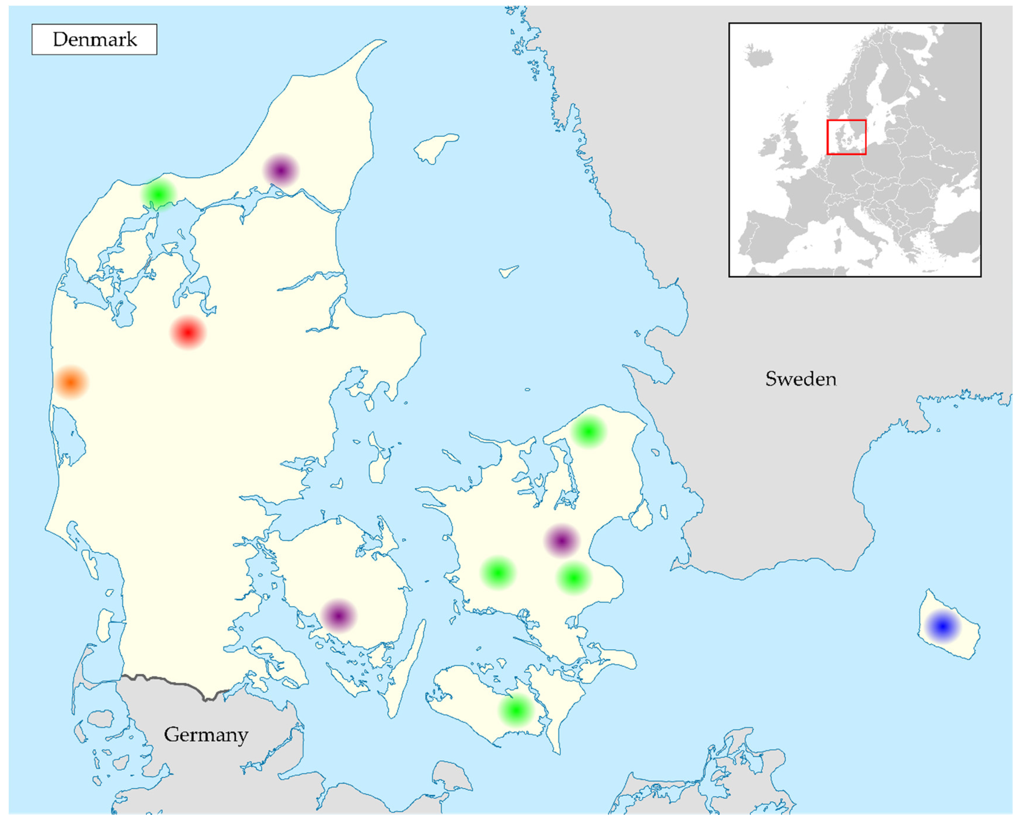
2.1. Collection of Samples

Samples were collected from bats in Denmark between 2013 and 2017 (see Table 1). The limestone mines of Mønsted were chosen as sampling locations due to their important status as hibernacula. In this study, samples were actively collected at least once a year when the bats departed from the mines during spring or entered during autumn.
At Mønsted limestone mines, bats were captured using a modified version of a harp trap placed in front of the entrance to the mines [7,8,9]. Faecal pellets were collected from individual bats using sterile tweezers. Bats were handled carefully and for the shortest possible time. Bat species were identified based on morphological features. Capturing was performed with permission from the Nature Agency of the Ministry of Environment and Food in Denmark.
Additionally, faecal samples were collected from bats during the non-hibernating seasons, from spring to autumn, in connection with other projects where bats had to be handled or roosts/colonies had to be visited. Samples were taken directly from individual bats when handled or from the floor under roosting bats (see Figure 1).

2.2. Screening of Faecal Samples Using pan-CoV RT-PCR Assays

The faecal samples were homogenized for 1 min in 500 µL Eagle’s medium (in-house production) using the TissueLyser II (Qiagen, Hilden, Germany) followed by centrifugation at 3000 rpm for 5 min. The RNA was extracted from 200 µL of the supernatant using the MagNaPure 96 robot with the MagNa Pure 96 DNA and Viral Small Volume kit (Roche, Basel, Switzerland) and the Pathogen Universal Protocol. Extracted RNA samples were screened for the presence of CoV RNA using two independent real-time RT-PCRs (screening assays B and C, see Supplementary File 1), each based on previously described pan-CoV primers [10,11] targeting conserved regions of the ORF1b which encodes part of the viral replicase complex.

2.3. Sequencing of Samples

Amplicons, generated using screening assays B and C, were sequenced to identify the CoVs. The resulting sequences without primer binding regions were 130 bp for the B assay (corresponding to nucleotide position 14911–15040 of the reference sequence NC_009657) and 208 bp for the C assay (nucleotide position 14251–14458). Selected positive samples from the different species of bats were also tested with a nested pan-CoV RT-PCR [12] and amplicons generated, using this assay A, were sequenced for further phylogenetic comparisons. Sequences obtained from this assay were 381 bp in length (corresponding to reference nucleotide position 14493 to 14873). Sequencing was performed using the BigDye Terminator v. 3.1 Cycle Sequencing Kit on an ABI PRISM 3500 DNA analyser (Applied Biosystems, Foster City, CA, USA).

2.4. Sequence Analysis

The Danish bat CoV sequences were assembled and analysed using the CLC Main Workbench and subjected to a BLASTn search to identify the CoV genus. The sequences were aligned with selected reference coronavirus strains approved by ICTV [13], as well as relevant and/or more recent European bat coronavirus sequences found by BLAST searching in the NCBI database. The phylogenies were constructed using the Jukes-Cantor Neighbor-Joining method with 1000 bootstrap replicates and a minimum bootstrap value of 50 for phylogenies based on the A and C assays and a minimum of 20 for the B assay-based phylogeny. Nucleotide (nt) sequences were translated to the predicted amino acid (aa) sequences and compared for synonymity using an alignment. All alignments and phylogenies were made in CLC Main Workbench.

2.5. Statistical Analysis

Coronavirus prevalence data was analysed using R [14], investigating the effects of season (spring or autumn), year (2013–2016), and bat gender. Odds-ratio [15] and Fisher’s exact test were performed for each variable of season, year, and gender. A significance level of 0.05 was used in the statistical analysis.

2.6. Confirmation of Sampled Host Species

A single M. dasycneme faecal sample containing a coronavirus very similar to the coronaviruses from M. daubentonii was selected for confirmatory species testing, together with an M. daubentonii sample as the control, using a bat species determination protocol. Mouth swabs collected from the same animals were submerged in 500 µL phosphate buffered saline and the nucleic acids extracted from 200 µL on the MagnaPure 96 (Roche, Basel, Switzerland) robot using the same kit and protocol as for the faecal samples. The extracted DNA from the mouth swab samples, as well as from the faecal samples, was tested with two different real-time PCR assays targeting the bat mitochondrial DNA; one assay was specific for the cytochrome B gene and the other for the 16S rRNA gene using previously designed primers ([16] and Supplementary File 1). PCR products were visualized on agarose gels, purified using the GeneJet purification kit, and sequenced as above. Sequencing reads were assembled in CLC Main Workbench and consensus sequences were subjected to BLASTn-search to identify the sampled bat species.

3. Results

3.1. Samples Positive for Coronavirus RNA

In total, 271 faecal samples were obtained; of these, 187 were from M. daubentonii, while P. pygmaeus was the second most sampled bat with 28 samples and M. dasycneme was third with 19 faecal samples. Faecal samples from five of the 10 sampled bat species tested positive for the presence of CoV RNA (as an indicator of the presence of the virus) in screening assays B and/or C. The positive samples were obtained from M. daubentonii, P. pygmaeus, M. dasycneme, M. nattereri, and E. serotinus (see Table 1 and Table 2). The three most sampled bat species had an observed prevalence of coronavirus RNA in their faeces of around 20–25% (Table 2). Only one out of eleven samples from M. nattereri was CoV positive. Three samples from a maternity colony of E. serotinus proved positive in the CoV screening, while a fourth sample, collected at a different location, tested negative.
The bats at Mønsted were sampled twice a year, but only a few bats provided a faecal sample when captured departing from the mines after winter hibernation (11 from M. daubentonii). Therefore, the majority of samples were collected during autumn (172 from M. daubentonii). Due to the seasonally skewed sample collection, it was not possible to infer a seasonal pattern for the presence of virus from the available data. Odds-ratio testing of the different years revealed significantly higher odds (5:1, 95% CI [2; 17]) for the presence of coronaviruses in the year 2015 compared to 2013, but not in the years 2014 or 2016. Odds-ratio testing of bat gender indicated slightly higher odds of coronavirus presence in females than in males (1.4:1, 95% CI [0.7; 2.9]), but this was not significant.
The samples not originating from Mønsted were collected from individual bats caught in the field, from maternity colonies, or bat roosts with single bats from June through October between the years 2013 and 2017. It is not possible to infer a seasonal, year, or gender specific pattern from these sparse samples, as they were collected from different species and from different geographical locations.

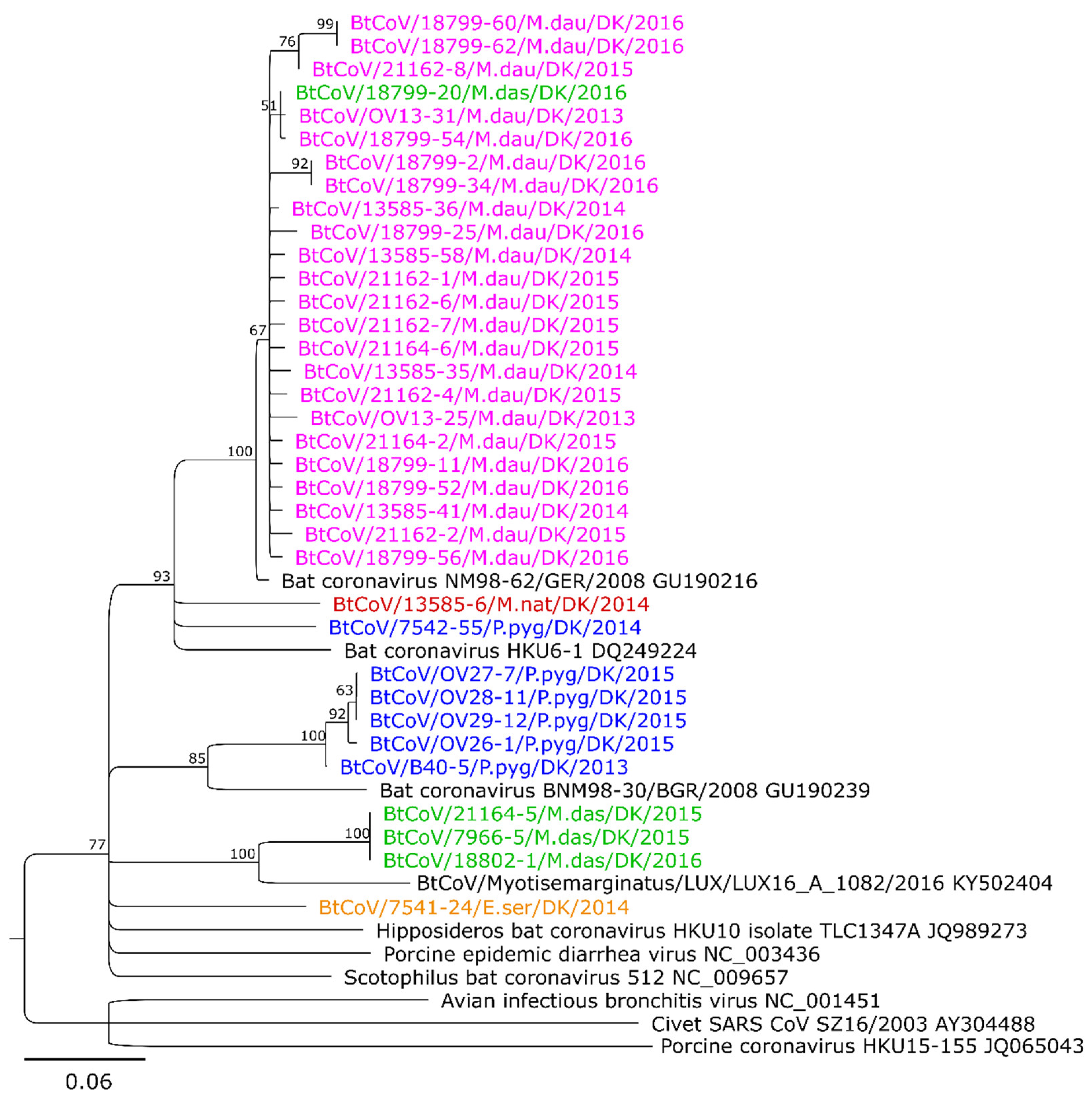
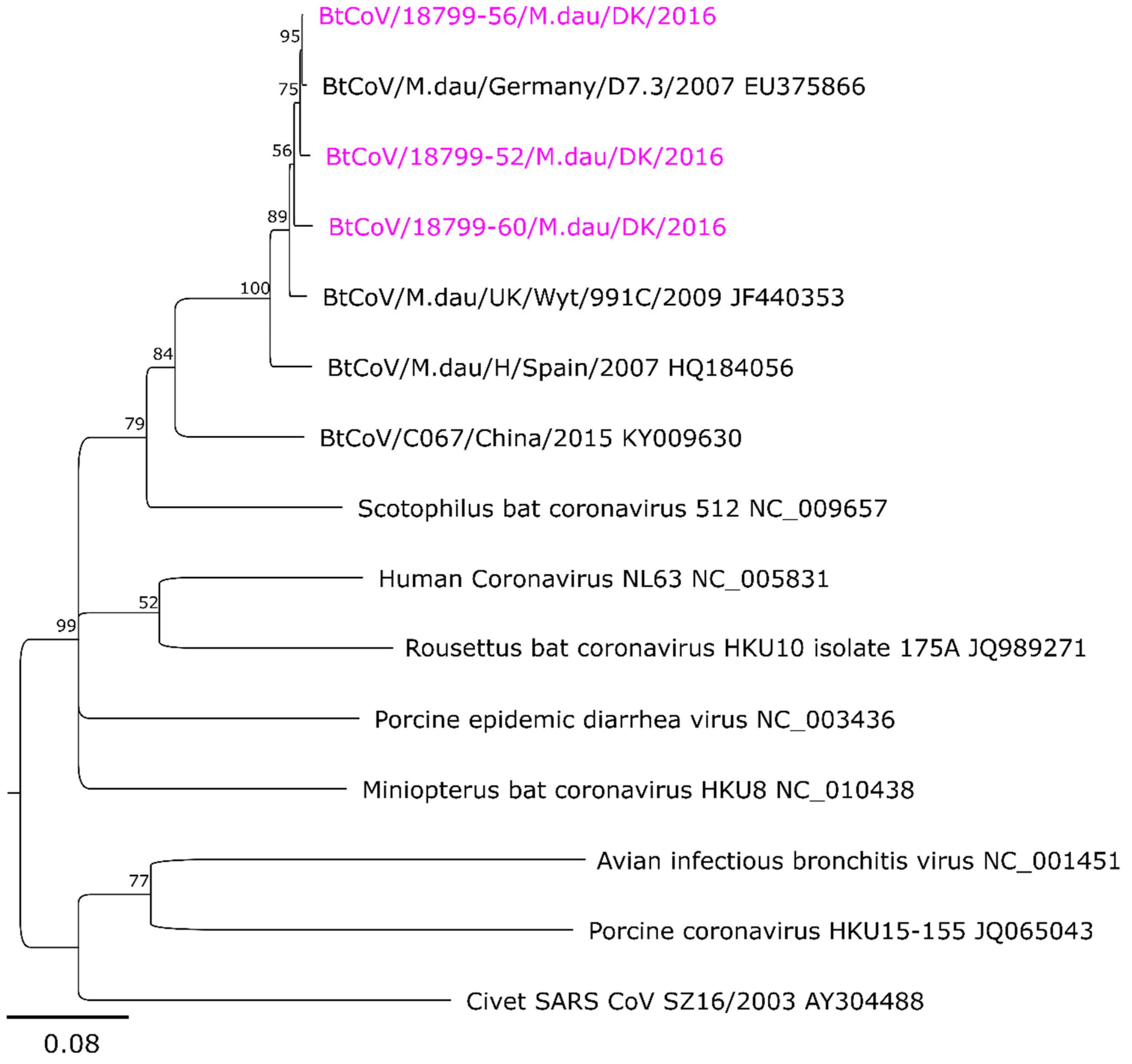
3.2. Sequence Analysis and Phylogeny

All generated coronavirus sequences belong to the genus Alphacoronavirus, as determined by BLASTn searches and phylogenetic analysis, and they are deposited in the European Nucleic Archive (ENA) database under study accession no. PRJEB28001, PRJEB28437, and PRJEB28438.
Separate phylogenetic trees were generated for sequences obtained from the two screening assays C and B (Figure 2 and Supplementary Figure S3). The phylogenetic tree for the 208 bp sequences obtained from screening assay C included 35 Danish CoV sequences (Figure 2). The samples from M. daubentonii, M. dasycneme, and M. nattereri were all collected at Mønsted, while one of the P. pygmaeus samples was from an area near Mønsted (Vadum). All the Danish M. daubentonii CoV sequences are closely related (>95% nt identity, maximum 9 nt differences), or identical, and form a single cluster within the tree. This cluster also includes one of the Danish M. dasycneme CoV sequences and a German CoV sequence from M. daubentonii (Accession number GU190216). The latter sequence has only 3 nt differences from the Danish M. dasycneme derived sequence and a maximum of 9 nt differences compared to any of the Danish CoV sequences in the cluster. Outside of this cluster, branching from an unresolved node, are two other Danish CoV sequences from P. pygmaeus and M. nattereri, along with the closest BLAST search result that is the HKU6-1 bat coronavirus from Hong Kong in 2006. This HKU6-1 sequence has at least 24 nt differences compared to the M. daubentonii cluster, 25 nt differences to the P. pygmaeus sequence, and 29 nt differences to the M. nattereri sequence. The five remaining Danish CoV sequences from P. pygmaeus form a separate cluster with 0–3 nt differences between them, and with a minimum of 27 nt differences compared to the closest reference virus sequence that was from a sample collected from a Nyctalus leisleri specimen in Bulgaria in 2008 (Accession number GU190239). This particular reference sequence was described as forming its own RNA-dependent RNA polymerase-based grouping unit (RGU) in a previous study [17]. The three remaining Danish M. dasycneme virus sequences are identical. Their closest reference sequence, with 25 nt differences (out of 208 nt), is from a Myotis emarginatus in Luxembourg from 2016. The single Danish CoV sequence from E. serotinus does not cluster with any of the other Danish virus sequences, and it branches from an unresolved node.
In the phylogenetic tree based on sequences (120 bp) from the screening assay B, 34 Danish virus sequences were included (Supplementary Figure S3). The M. daubentonii sample sequences form a single cluster, which also includes the single M. dasycneme sequence. There is a maximum of 5 nt differences within the cluster, but none of the bat CoV reference sequences fall within this cluster.
The single P. pygmaeus and M. nattereri virus sequences are similar to, but separate from, this cluster. There are 16 nt differences between these two sequences and from the P. pygmaeus to the M. daubentonii cluster. The closest reference sequence (KF294382) is from China, with 16 nt differences to the P. pygmaeus sequence, 20 nt differences to the M. nattereri sequence, and 17 nt differences to the closest M. daubentonii viruses.
The other available Danish P. pygmaeus CoV sequence is most closely related to a sequence from a Pipistrellus kuhlii specimen in Italy from 2010. As in Figure 2, the Danish E. serotinus CoV sequence does not cluster with any of the other reference viruses or other Danish CoV sequences, and it branches from an unresolved node. The closest related sequence in the NCBI database is from Kenya (HQ728480), with 20 nt differences.
Sequences (381 bp in length) were also obtained from selected M. daubentonii bat samples using assay A to further characterize the relationships with sequences reported from the same bat species in Germany, Spain, and the United Kingdom [18,19,20]. Samples from other species of bats tested negative in assay A, so no products were available for sequencing. In this tree (Figure 3), the CoV sequences obtained from M. daubentonii in Denmark form a cluster with the sequences reported from Germany and from the United Kingdom (UK). There are only 1 to 8 nt differences compared to the German sequence, but 6 to 11 nt differences compared to the U.K. sequences and 17 to 22 nt differences from the Spanish sequence. Between the three Danish samples, there are 4 to 9 nt differences.

3.3. Confirmation of Sampled Host Species

The sequenced mouth swab samples yielded a higher quality sequence than the faecal samples in the species identification PCR assays. The sample no. 18799-20 was confirmed to be from the species M. dasycneme on the basis of BLASTn searches of the sequenced PCR products from both assays. The PCR products generated with the mitochondrial 16S-gene specific assay were most similar to reference sequences from M. fimbriatus and M. pilosus, with E-values of 3.55 × 10−110 (no 16S reference sequences were found for M. dasycneme in GenBank). For the cytochrome B gene-specific assay, the lowest E-value was found for M. dasycneme (2.52 × 10−168).
The sample 18799-52 was included as the control and was confirmed to be from M. daubentonii, with the sequenced PCR products from both assays yielding the lowest BLASTn E-values for M. daubentonii references (E-values: 4.14 × 10−141 for the 16S PCR product and indistinguishable from 0 for the cytochrome B PCR product).

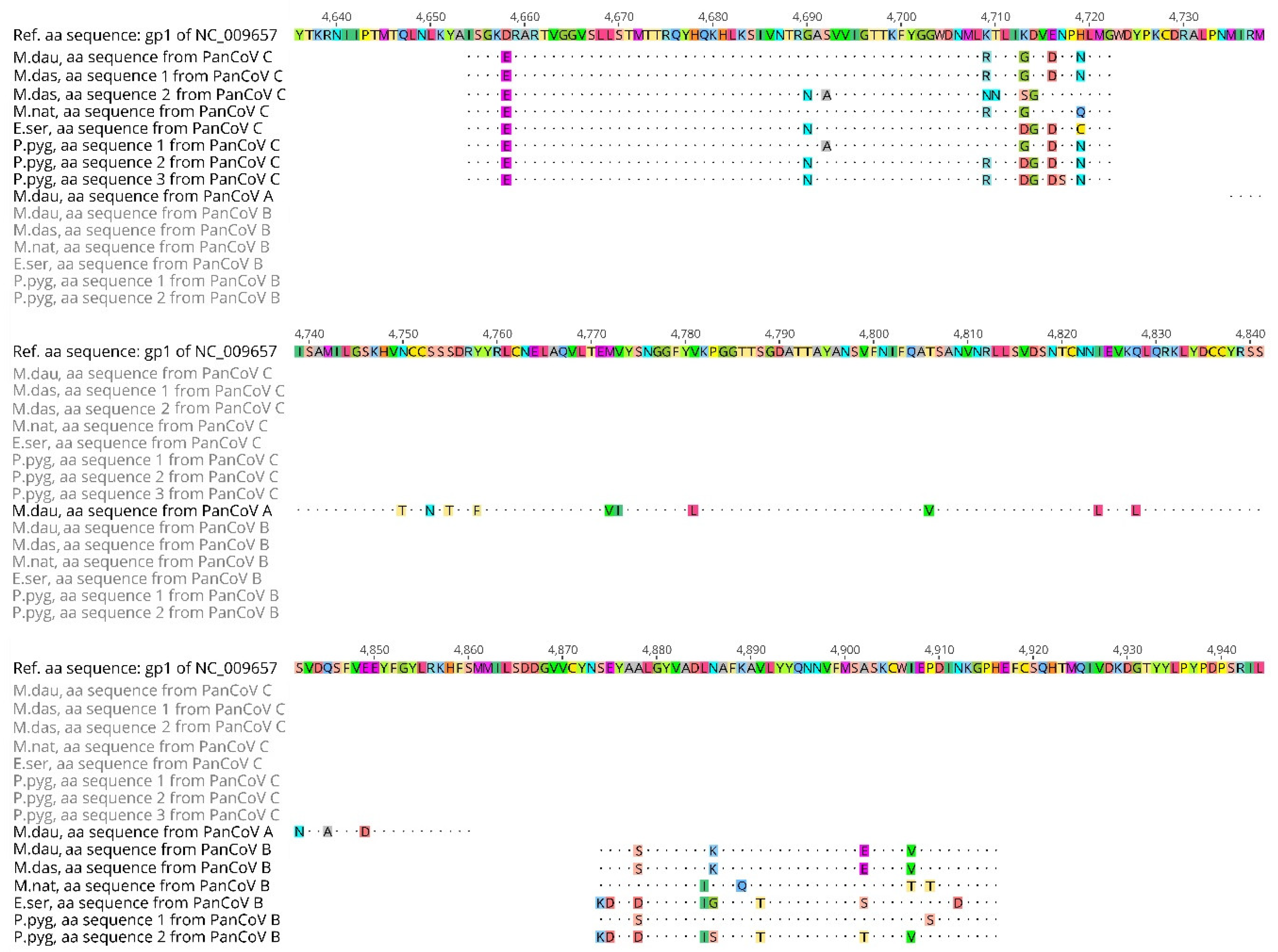
3.4. Predicted Amino Acid Sequences and Host Restriction

All the new nt sequences were translated to their predicted amino acid (aa) sequences and aligned, and identical aa sequences from the same species of bat were removed (see Figure 4).
The predicted aa sequences from all of the M. daubentonii samples were identical as the nt sequences within each of the amplicons from each assay only had synonymous nt differences.
For the short B assay amplicons, encoding 43 aa residues between the primer regions, five different aa sequences were found. The sequences had 4–10 aa differences between each other, with the CoVs from E. serotinus, M. nattereri and M. daubentonii each having one distinct aa sequence and the P. pygmaeus CoVs having two. The aa sequence predicted from the single M. dasycneme CoV nt sequence was identical to the aa sequence from the M. daubentonii samples.
Seven different aa sequences, with 2–8 aa differences between them, were predicted for the C assay amplicon encoding 69 aa between the primer regions. Again, the predicted aa sequence from one of the M. dasycneme CoV sequences was identical to the predicted aa sequence from the M. daubentonii samples. The other three nt sequences from M. dasycneme samples were predicted to encode a single distinct aa sequence. Three different aa sequences were predicted for the six CoV positive samples from P. pygmaeus, with four of the samples having identical predicted aa sequences and two nt sequences encoding two distinct aa sequences. The last two different aa sequences were predicted from the two M. nattereri and E. serotinus coronavirus samples.

4. Discussion

In this study, we wanted to investigate the presence of CoVs in healthy Danish bat populations in different geographical locations, including the major colonies at Mønsted, to relate the findings to the bat species and to make comparisons to the bat CoV strains reported from neighbouring countries.
The average prevalence, at around 20%, observed in this study for coronavirus RNA positive faecal samples from the different bat species, is high in comparison with other European studies reporting prevalences of, e.g., 9.8% [18], 4.2% [21], and 1.8% [22]. This could possibly be due to the testing strategy employed, with two different, sensitive screening assays used in this study. Of the 58 CoV positive samples, 48 of these samples provided a nucleotide sequence from at least one of the assays. The remaining 10 samples only yielded partial sequences or were judged positive based on the real-time RT-PCR analysis. Twenty-one of the 48 sequenced samples were positive in both screening assays B and C, 14 samples were only positive in PanCoV C, and 13 only in PanCoV B.
The samples collected from M. daubentonii at Mønsted were analysed for seasonal, annual, and gender variability to obtain information about the dynamics of bat coronavirus infection within the bat populations. Previously, reports have been made of a higher coronavirus prevalence in young bats and lactating bats in maternity colonies [18,23,24], suggesting this could be due to the young animals being a susceptible population with a lower immunity that could then amplify the virus and transmit the infection to the adults [18]. Seasonal fluctuations in coronavirus prevalence could be indications of re-introductions of virus and/or the presence of persistently infected animals with periods of virus shedding [25]. Unfortunately, we did not obtain enough samples collected in the springtime to assess seasonal variability in the CoV prevalence in the Danish bats. Our study indicates a higher, but not significant, odds-ratio for females to carry coronaviruses than for males. We observed a significantly higher odds-ratio for coronavirus presence in the M. daubentonii samples from Mønsted from the year 2015 compared to years 2013, 2014, and 2016. The reason behind this is unknown.
The co-hibernation of several bat species in one location might facilitate the mixing of viruses between the species. However, the bats at Mønsted are rarely observed to have physical contact with other species, except for occasional aggregations of lethargic hibernating bats. Previously, a high degree of host restriction for the different bat coronavirus strains has been described [2,18,26,27,28]. Therefore, it is relevant to group the detected coronaviruses by bat species and indeed the nt sequencing in this study revealed distinct CoV sequences to be present within each of the five bat species, indicating that specific CoVs are, at least predominantly, restricted to individual host species.
When comparing strains in a phylogeny, it is important to look at variable areas in the genome. In the study by Xu et al., (2003), conserved areas or aa motifs in the ORF1ab encoded RNA-dependent RNA polymerase gene of the SARS coronavirus were inferred by comparison to other coronavirus strains and to other RNA viruses [29]. The Danish bat coronavirus RT-PCR amplicons were generated with primers spanning the nt sequences of some of these motifs. The predicted aa sequence (126 aa) encoded by the region targeted by RT-PCR assay A contains two aa from a conserved motif near its C-terminus, as well as a large motif of 33 aa in the middle of the region. The RT-PCR assay B targets a region encoding 43 aa. This region contains conserved motifs at both ends, as well as an internal motif of 26 aa, meaning that this region of the genome does not represent a variable area for coronaviruses and thus is not optimal for determining phylogeny. The region targeted by RT-PCR assay C encodes 69 aa. The ends of this region likewise contain conserved motifs, but there are no internal conserved motifs. With this in mind, the three different phylogenetic trees should only be regarded as an initial assessment of the geographical connections between the Danish bat coronaviruses and the bat coronaviruses of neighbouring countries. The nt sequences also function as a tool for establishing the genus of the coronaviruses detected in the samples. Furthermore, they provide evidence for species specificity; however, it is not possible to establish firm evolutionary relationships without analysing larger regions of the coronavirus genome that will allow the use of more reference data.
The Danish samples positive for CoV from M. daubentonii, M. dasycneme, and M. nattereri were all collected at Mønsted, but in the phylogenetic trees, there was a clear species-specific clustering, even within the Myotis genus. However, a single M. dasycneme sample had a coronavirus similar to that found in the M. daubentonii bats, and the M. nattereri had a distinct, but related, coronavirus to the M. daubentonii sequences. The CoV positive samples from P. pygmaeus collected from different geographical regions in Denmark, were to some extent, reflected in the genetic relatedness shown in the phylogenetic trees. For instance, one P. pygmaeus sequence sampled close to Mønsted grouped close to the Mønsted M. daubentonii-cluster. The two other P. pygmaeus collection sites, at Borup and Sollerup, are more than 100 km apart and separated by a large stretch of water (Storebælt), but these P. pygmaeus coronavirus sequences were still very closely related, with only three synonymous nt differences between the 208 nt sequences in the C assay amplicons from Borup and Sollerup. Unfortunately, the coronavirus present in the positive sample from E. serotinus could only be detected using the screening assays and no clear phylogenetic relationship could be established based on the available reference sequences.
The host species differentiation described above became more apparent when the predicted aa sequences derived from the different nt sequences were compared (Figure 4). Here, it was obvious that each species of bat harboured CoVs with distinct predicted amino acid sequences.
From the phylogenetic analysis, we see a close resemblance between the Danish M. daubentonii bat CoVs and CoVs from the same bat species in Germany and the U.K., especially in relation to the CoVs from M. daubentonii sampled at the Bad Segeberg caves in Germany, located roughly 330 km from the Danish limestone mines [18].
Some bat species in Europe, such as P. nathusii, N. noctula, and V. murinus, are known to be capable of long distance flights. Others, e.g., M. nattereri, Plecotus auritus, and E. serotinus, are rated as much more sedentary, with flights between summer and winter roosts of generally max. 50–100 km. Species like M. daubentonii and M. dasycneme are normally rated as intermediate, i.e., generally moving or migrating short to medium long distances of up to 300–400 km [30], probably depending on distances to available, suitable, and well-known hibernation or swarming sites [31,32]. However, more recent research indicates that the distinction between these categories is not so sharp [33]. Of the several hundred bats ringed in the Danish limestone mines in the 1950’s and 60’s [34], none have been recaptured in northern Germany and likewise, bats ringed in northern Germany have, to the knowledge of the authors, hardly ever been found in the Danish limestone mines. However, during the intensive population research in Mønsted in 2009 [5], a single M. dasycneme female was found that had been ringed as a juvenile the year before in a maternity colony near Kiel in northern Germany. The following year, this individual was captured in northern Germany hibernating in a bunker near Itzehoe, and the following summer, it was back in the colony where it was born (P. Borkenhagen pers. com. cited in [6]).
Furthermore, it has been shown that in late summer and autumn, both M. daubentonii and M. dasycneme can be frequently observed hunting insects and in more directional flight far at sea in the Baltic [33]. M. daubentonii is one of the species that each year gathers at certain points on the coasts of southern Sweden and southern Denmark (Lolland-Falster) and can be seen flying out from these points directly over the sea on what seems to be regular migration. Such M. daubentonii specimens in unidirectional flight have been observed in the middle of the Baltic Sea and also migrating out from the south-western point of Bornholm [6,33,35].
Finally, M. daubentonii is a very common bat distributed widely and continuously, e.g., in Denmark and northern Germany [4,36], presumably with breeding colonies, intermediate day roosts, etc. all over the region. In the active part of the year, many bats often roost together, in confined spaces in hollow trees, enabling faecal-oral transmission of viruses and it is more than likely that there is frequent contact between animals from neighbouring roosting sites.
In summary, several indications point to a more dynamic situation than previously described, involving actual migration and more flux between M. daubentonii and M. dasycneme populations in Denmark and northern Germany. This might explain the phylogenetically closely related CoV sequences described in this study. Sequencing of larger and less conserved regions of the coronavirus genome is needed to confirm whether there is, indeed, this close relationship with the bat coronavirus populations of the neighbouring countries.
In this study, only coronaviruses belonging to the genus Alphacoronavirus were detected in the Danish bats. These CoVs were obtained from five different species of bats. Four of these species (M. dasycneme [18,37], M. daubentonii [18,19,20,37], P. pygmaeus [18], and M. nattereri [20,24]) have previously been reported to harbour coronaviruses of this genus in other countries. To our knowledge, this is the first report of an alphacoronavirus detected from E. serotinus. Betacoronaviruses have previously been isolated from both E. serotinus [38] and P. pygmaeus [39] in other European countries. Therefore, it is likely that Danish bats of these species could also harbour betacoronaviruses, but these were not detected in the limited number of samples analysed in this study. In the five other sampled bat species, no coronavirus positive samples were detected. This could be due to the low number of samples, with only two to nine samples assayed per species. The majority of the samples from these species were collected on the island of Bornholm located in the eastern part of Denmark. Bornholm is the only place where M. bechsteinii and M. mystacinus were registered during a thorough survey of bat species in Denmark [3]. Of the 24 samples collected from bats in Bornholm, none tested positive for coronavirus RNA; otherwise, CoVs were detected in samples from each of the three major geographical regions of Denmark, namely Jutland, Funen, and Zealand.

Supplementary Materials

Author Contributions

Conceptualization, T.R., M.C., and H.B.; Validation, T.R.; Formal Analysis, C.L. and T.R.; Investigation, C.L., H.B., E.F., and Y.D.; Resources, E.K.; Writing—Original Draft Preparation, C.L.; Writing—Review & Editing, all authors.; Visualization, C.L.; Supervision, T.R., G.B., and A.B.; Project Administration, T.R. and M.C.; Funding Acquisition, T.R. and M.C.

Funding

Collection in the field and laboratory processing of bat faeces samples at the National Veterinary Institute of the Technical University of Denmark, Lindholm, was performed as part of a surveillance program for coronaviruses and lyssaviruses in Danish bats funded by the Danish Veterinary and Food Administration. Y.D. was supported by the China Scholarship Council (CSC201408515154).

Acknowledgments

We are grateful for the technical assistance of laboratory technician Helle Rasmussen, who received, sorted, prepared, and tested the samples for CoVs.

Conflicts of Interest

The authors declare no conflict of interest. The funders had no role in the design of the study; in the collection, analyses, or interpretation of data; in the writing of the manuscript, and in the decision to publish the results.

References

  1. Woo, P.C.Y.; Lau, S.K.P.; Lam, C.S.F.; Lau, C.C.Y.; Tsang, A.K.L.; Lau, J.H.N.; Bai, R.; Teng, J.L.L.; Tsang, C.C.C.; Wang, M.; et al. Discovery of Seven Novel Mammalian and Avian Coronaviruses in the Genus Deltacoronavirus Supports Bat Coronaviruses as the Gene Source of Alphacoronavirus and Betacoronavirus and Avian Coronaviruses as the Gene Source of Gammacoronavirus and Deltacoronavi. J. Virol. 2012, 86, 3995–4008. [Google Scholar] [CrossRef] [PubMed]
  2. Drexler, J.F.; Corman, V.M.; Drosten, C. Ecology, Evolution and Classification of Bat Coronaviruses in the Aftermath of SARS. Antivir. Res. 2014, 101, 45–56. [Google Scholar] [CrossRef] [PubMed]
  3. Baagøe, H.J. Danish Bats (Mammalia: Chiroptera): Atlas and Analysis of Distribution, Occurrence and Abundance; Steenstrupia: Copenhagen, Denmark, 2001; ISSN 0375-2909. [Google Scholar]
  4. Baagøe, H.J. Flagermus, Chiroptera. In Dansk Pattedyratlas; Baagøe, H.J., Jensen, T.S., Eds.; Gyldendal: Copenhagen, Denmark, 2007; pp. 38–98. ISBN 9788702055061. [Google Scholar]
  5. Baagøe, H.J.; Degn, H.J. Flagermusene i Daugbjerg og Mønsted Kalkgruber i Udflyvningsperioden 2009; Danish Forest and Nature Agency: Midtjylland, Denmark, 2009. [Google Scholar]
  6. Møller, J.D.; Baagøe, H.J.; Degn, H.J. Forvaltningsplan for Flagermus; Naturstyrelsen, Miljøministeriet: Copenhagen, Denmark, 2013; ISBN 9788772794075. [Google Scholar]
  7. Constantine, D.G. An Automatic Bat-Collecting Device. J. Wildl. Manag. 1958, 22, 17–22. [Google Scholar] [CrossRef]
  8. Tuttle, M.D. An Improved Trap for Bats. J. Mammal. 1974, 55, 475–477. [Google Scholar] [CrossRef] [PubMed]
  9. Baagøe, H.J.; Degn, H.J. Flagermusene i Daugbjerg og Mønsted Kalkgruber i Udflyvningsperioden 2003; National Environmental Research Institute of Denmark: Roskilde, Denmark, 2004. [Google Scholar]
  10. Escutenaire, S.; Mohamed, N.; Isaksson, M.; Thorén, P.; Klingeborn, B.; Belák, S.; Berg, M.; Blomberg, J. SYBR Green Real-Time Reverse Transcription-Polymerase Chain Reaction Assay for the Generic Detection of Coronaviruses. Arch. Virol. 2007, 152, 41–58. [Google Scholar] [CrossRef] [PubMed]
  11. Vijgen, L.; Moës, E.; Keyaerts, E.; Li, S.; Van Ranst, M. A Pancoronavirus RT-PCR Assay for Detection of All Known Coronaviruses. Methods Mol. Biol. 2008, 454, 3–12. [Google Scholar] [CrossRef] [PubMed]
  12. De Souza Luna, L.K.; Heiser, V.; Regamey, N.; Panning, M.; Drexler, J.F.; Mulangu, S.; Poon, L.; Baumgarte, S.; Haijema, B.J.; Kaiser, L.; et al. Generic Detection of Coronaviruses and Differentiation at the Prototype Strain Level by Reverse Transcription-PCR and Nonfluorescent Low-Density Microarray. J. Clin. Microbiol. 2007, 45, 1049–1052. [Google Scholar] [CrossRef] [PubMed] [Green Version]
  13. International Committee on Taxonomy of Viruses (ICTV) Virus Metadata Repository: Version 21 March 2018; MSL32. Available online: https://talk.ictvonline.org/taxonomy/vmr/ (accessed on 15 May 2018).
  14. R Core Team. R: A Language and Environment for Statistical Computing; R Foundation for Statistical Computing: Vienna, Austria, 2017. [Google Scholar]
  15. Aragon, T.J. Epitools: Epidemiology Tools. Available online: https://cran.r-project.org/package=epitools (accessed on 15 May 2018).
  16. Schlegel, M.; Ali, H.S.; Stieger, N.; Groschup, M.H.; Wolf, R.; Ulrich, R.G. Molecular Identification of Small Mammal Species Using Novel Cytochrome b Gene-Derived Degenerated Primers. Biochem. Genet. 2012, 50, 440–447. [Google Scholar] [CrossRef] [PubMed]
  17. Drexler, J.F.; Gloza-Rausch, F.; Glende, J.; Corman, V.M.; Muth, D.; Goettsche, M.; Seebens, A.; Niedrig, M.; Pfefferle, S.; Yordanov, S.; et al. Genomic Characterization of Severe Acute Respiratory Syndrome-Related Coronavirus in European Bats and Classification of Coronaviruses Based on Partial RNA-Dependent RNA Polymerase Gene Sequences. J. Virol. 2010, 84, 11336–11349. [Google Scholar] [CrossRef] [PubMed] [Green Version]
  18. Gloza-Rausch, F.; Ipsen, A.; Seebens, A.; Göttsche, M.; Panning, M.; Drexler, J.F.; Petersen, N.; Annan, A.; Grywna, K.; Müller, M.; et al. Detection and Prevalence Patterns of Group I Coronaviruses in Bats, Northern Germany. Emerg. Infect. Dis. 2008, 14, 626–631. [Google Scholar] [CrossRef] [PubMed]
  19. Falcón, A.; Vázquez-Morón, S.; Casas, I.; Aznar, C.; Ruiz, G.; Pozo, F.; Perez-Breña, P.; Juste, J.; Ibáñez, C.; Garin, I.; et al. Detection of Alpha and Betacoronaviruses in Multiple Iberian Bat Species. Arch. Virol. 2011, 156, 1883–1890. [Google Scholar] [CrossRef] [PubMed] [Green Version]
  20. August, T.A.; Mathews, F.; Nunn, M.A. Alphacoronavirus Detected in Bats in the United Kingdom. Vector-Borne Zoonotic Dis. 2012, 12, 530–533. [Google Scholar] [CrossRef] [PubMed]
  21. Goffard, A.; Demanche, C.; Arthur, L.; Pinçon, C.; Michaux, J.; Dubuisson, J. Alphacoronaviruses detected in french bats are phylogeographically linked to coronaviruses of european bats. Viruses 2015, 7, 6279–6290. [Google Scholar] [CrossRef] [PubMed] [Green Version]
  22. Kemenesi, G.; Dallos, B.; Gorfol, T.; Boldogh, S.; Estok, P.; Kurucz, K.; Kutas, A.; Foldes, F.; Oldal, M.; Nemeth, V.; et al. Molecular survey of RNA viruses in hungarian bats: Discovering novel astroviruses, coronaviruses, and caliciviruses. Vector-Borne Zoonotic Dis. 2014, 14, 846–855. [Google Scholar] [CrossRef] [PubMed]
  23. Drexler, J.F.; Corman, V.M.; Wegner, T.; Tateno, A.F.; Zerbinati, R.M.; Gloza-Rausch, F.; Seebens, A.; Müller, M.A.; Drosten, C. Amplification of Emerging Viruses in a Bat Colony. Emerg. Infect. Dis. 2011, 17, 449–456. [Google Scholar] [CrossRef] [PubMed]
  24. Rizzo, F.; Edenborough, K.M.; Toffoli, R.; Culasso, P.; Zoppi, S.; Dondo, A.; Robetto, S.; Rosati, S.; Lander, A.; Kurth, A.; et al. Coronavirus and Paramyxovirus in Bats from Northwest Italy. BMC Vet. Res. 2017, 13, 1–11. [Google Scholar] [CrossRef] [PubMed]
  25. Jeong, J.; Smith, C.S.; Peel, A.J.; Plowright, R.K.; Kerlin, D.H.; McBroom, J.; McCallum, H. Persistent Infections Support Maintenance of a Coronavirus in a Population of Australian Bats (Myotis macropus). Epidemiol. Infect. 2017, 145, 2053–2061. [Google Scholar] [CrossRef] [PubMed]
  26. Corman, V.M.; Rasche, A.; Diallo, T.D.; Cottontail, V.M.; Stöcker, A.; Souza, B.F.d.C.D.; Corrêa, J.I.; Carneiro, A.J.B.; Franke, C.R.; Nagy, M.; et al. Highly Diversified Coronaviruses in Neotropical Bats. J. Gen. Virol. 2013, 94, 1984–1994. [Google Scholar] [CrossRef] [PubMed]
  27. Cui, J.; Han, N.; Streicker, D.; Li, G.; Tang, X.; Shi, Z.; Hu, Z.; Zhao, G.; Fontanet, A.; Guan, Y.; et al. Evolutionary Relationships between Bat Coronaviruses and Their Hosts. Emerg. Infect. Dis. 2007, 13, 1526–1532. [Google Scholar] [CrossRef] [PubMed] [Green Version]
  28. Tang, X.C.; Zhang, J.X.; Zhang, S.Y.; Wang, P.; Fan, X.H.; Li, L.F.; Li, G.; Dong, B.Q.; Liu, W.; Cheung, C.L.; et al. Prevalence and Genetic Diversity of Coronaviruses in Bats from China. J. Virol. 2006, 80, 7481–7490. [Google Scholar] [CrossRef] [PubMed] [Green Version]
  29. Xu, X.; Liu, Y.; Weiss, S.; Arnold, E.; Sarafianos, S.G.; Ding, J. Molecular Model of SARS Coronavirus Polymerase: Implications for Biochemical Functions and Drug Design. Nucleic Acids Res. 2003, 31, 7117–7130. [Google Scholar] [CrossRef] [PubMed]
  30. Gaisler, J.; Hanak, V.; Hanzal, V.; Jarský, V. Results of bat banding in the Czech and Slovak Republics, 1948–2000. Vespertilio 2003, 7, 3–61. [Google Scholar]
  31. Dietz, C.; von Helversen, O.; Nill, D. Bats of Britain, Europe and Northwest Africa, 1st ed.; A & C Black Publishers Ltd.: London, UK, 2009; ISBN 9781408105313. [Google Scholar]
  32. Steffens, R.; Zöphel, U.; Brockmann, D. 40th Anniversary Bat Marking Centre Dresden—Evaluation of Methods and Overview of Results; Saxon State Office for Environment and Geology: Dresden, Germany, 2004; ISBN 3-00-016143-0. [Google Scholar]
  33. Ahlén, I.; Baagøe, H.J.; Bach, L. Behavior of Scandinavian Bats during Migration and Foraging at Sea. J. Mammal. 2009, 90, 1318–1323. [Google Scholar] [CrossRef] [Green Version]
  34. Egsbæk, W.; Jensen, B. Results of bat banding in Denmark. Vidensk. Medd. Dansk Naturh. Foren. 1963, 125, 269–296. [Google Scholar]
  35. Baagøe, H.J. Bornholms flagermus-status 2010. Nat. Bornh. 2011, 9, 22–30. [Google Scholar]
  36. Borkenhagen, P. Die Säugetiere Schleswig-Holsteins, 1st ed.; Husum Druck- und Verlagsgesellschaft: Husum, Germany, 2011; ISBN 978-3898765619. [Google Scholar]
  37. Reusken, C.C.B.E.M.; Lina, P.H.C.C.; Pielaat, A.; de Vries, A.; Dam-Deisz, C.; Adema, J.; Drexler, J.F.; Drosten, C.; Kooi, E.A. Circulation of Group 2 Coronaviruses in a Bat Species Common to Urban Areas in Western Europe. Vector-Borne Zoonotic Dis. 2010, 10, 785–791. [Google Scholar] [CrossRef] [PubMed]
  38. De Benedictis, P.; Marciano, S.; Scaravelli, D.; Priori, P.; Zecchin, B.; Capua, I.; Monne, I.; Cattoli, G. Alpha and Lineage C BetaCoV Infections in Italian Bats. Virus Genes 2014, 48, 366–371. [Google Scholar] [CrossRef] [PubMed]
  39. Annan, A.; Baldwin, H.J.; Corman, V.M.; Klose, S.M.; Owusu, M.; Nkrumah, E.E.; Badu, E.K.; Anti, P.; Agbenyega, O.; Meyer, B.; et al. Human Betacoronavirus 2c EMC/2012-Related Viruses in Bats, Ghana and Europe. Emerg. Infect. Dis. 2013, 19, 456–459. [Google Scholar] [CrossRef] [PubMed]
Figure 1. Locations of sampling in Denmark and the sampled bat species. The number of samples collected from each bat species is listed with the number of positive samples shown in parenthesis. (Red) Mønsted, sampled species: Myotis daubentonii 183 (43), Myotis dasycneme 19 (5), Myotis nattereri 3 (1), Myotis brandtii 1 (0). (Purple) Locations with positive soprano pipistrelle samples, sampled species: Pipistrellus pygmaeus 23 (6). (Orange) Location with positive serotine bat samples, sampled species: Eptesicus serotinus 3 (3). (Blue) Bornholm, only negative samples, sampled species: Myotis nattereri 7 (0), Myotis bechsteinii 11 (0), Myotis mystacinus 2 (0), Myotis brandtii 2 (0), Nyctalus noctula 4 (0), Plecotus auritus 2 (0). (Green) Other locations with only negative samples, sampled species: Myotis daubentonii 4 (0), Myotis nattereri 1 (0), Pipistrellus pygmaeus 5 (0), Eptesicus serotinus 1 (0). Graphics edited from Wikimedia user NordNordWest published under the conditions of GNU Free Documentation License Version 1.2 or later.
Viruses 10 00486 g001
Figure 2. Phylogenetic tree of Danish bat coronavirus sequences. The tree was constructed based on sequenced RT-PCR amplicons from the screening assay C (208 bp without primer sequence [11]). The minimum bootstrap value was set to 50. The Danish CoV sequences are shown in species-specific colours. The distance bar in the bottom left corner indicates the number of nucleotide differences per site.
Viruses 10 00486 g002
Figure 3. Phylogenetic tree of Danish bat coronavirus sequences. The tree was constructed based on an alignment of 381 bp length references and sequenced RT-PCR amplicons from assay A [12]. The minimum bootstrap value was set to 50. The Danish CoV sequences are shown in pink. The distance bar in the bottom left corner indicates the number of nucleotide differences per site.
Viruses 10 00486 g003
Figure 4. Predicted amino acid sequences derived from the sequenced RT-PCR products of the Danish bat coronaviruses tested with assays A, B, and C. The amino acid sequences are arbitrarily numbered and named according to the sampled bat host species and RT-PCR assay. The most closely related reference sequence Scotophilus bat coronavirus 512 NC_009657 was selected for comparison with the Danish coronavirus predicted amino acid sequences. Numbers refer to amino acid numbers of the annotated gp1 (ORF1ab) of the reference sequence.
Viruses 10 00486 g004
Table 1. Faecal sample collection details for the detection of coronaviruses in Danish bats.
Bat SpeciesNo. Sampled (No. Positive)Collection DateCollection LocationSample Types
M. daubentonii4 (0)September 2013Vesløs2 individuals and 2 from nest box
40 (8)October 2013MønstedIndividual
53 (9)October 2014MønstedIndividual
4 (1)April 2015MønstedIndividual
10 (6)October 2015MønstedIndividual
11 (6)November 2015MønstedIndividual
5 (0)May 2016MønstedIndividual
58 (13)September 2016MønstedIndividual
2 (0)April 2017MønstedIndividual
M. dasycneme2 (0)October 2013MønstedIndividual
6 (0)October 2014MønstedIndividual
2 (1)April 2015MønstedIndividual
1 (1)November 2015MønstedIndividual
6 (1)September 2016MønstedIndividual
2 (2)November 2016MønstedIndividual
M. nattereri7 (0)August–September 2013BornholmIndividual
1 (0)September 2013Lolland, Agerup forestIndividual
2 (1)October 2014MønstedIndividual
1 (0)September 2016MønstedIndividual
M. bechsteinii4 (0)August 2013BornholmIndividual
2 (0)August 2015Bornholm1 individual and 1 from nest box
3 (0)August 2016Bornholm3 nest boxes
2 (0)September 2017BornholmIndividual
M. mystacinus1 (0)August 2013BornholmIndividual
1 (0)August 2016BornholmIndividual
M. brandtii2 (0)August 2013BornholmIndividual
1 (0)April 2015MønstedIndividual
N. noctula4 (0)August 2013Bornholm2 individual samples and 2 from nest boxes
P. pygmaeus6 (1)August 2013Funen, Sollerup forestIndividual
9 (1)June 2014VadumMix of individual samples and floor samples from colony
1 (0)Summer 2015SorøSample from nest box
8 (4)September 2015Borup5 individual samples, 3 from floor
1 (0)September–October 2015HelsingeSample from abandoned colony
3 (0)August 2016TurebyFrom colony
P. auritus1 (0)August 2013BornholmIndividual
1 (0)September–October 2015BornholmSample from floor
E. serotinus3 (3)June 2014Tim1 individual sample, 2 from floor
1 (0)June 2017SakskøbingSample from floor
Table 2. Coronavirus prevalence in the samples from different bat species in Denmark.
Bat SpeciesTotal No. Sampled (No. Positive)Observed Coronavirus Prevalence in Samples95% Confidence Interval of Sample Mean
M. daubentonii187 (43)23%[0.17; 0.29]
M. dasycneme19 (5)26%[0.07; 0.46]
M. nattereri11 (1)9%[0.00; 0.26]
M. bechsteinii11 (0)0%NA 1
M. mystacinus2 (0)0%NA
M. brandtii3 (0)0%NA
N. noctula4 (0)0%NA
P. pygmaeus28 (6)21%[0.06; 0.37]
P. auritus2 (0)0%NA
E. serotinus4 (3)75%[0.33; 1.00]
Total (all species)271 (58)21%[0.17; 0.26]
1 NA: not applicable.

