Article

Antigenic Change in Human Influenza A(H2N2) Viruses Detected by Using Human Plasma from Aged and Younger Adult Individuals

1
Division of Virology, Department of Microbiology and Immunology, Institute of Medical Science, University of Tokyo, Minato-ku, Tokyo 108-8639, Japan
2
Center for Supercentenarian Medical Research, Shinjuku-ku, Tokyo 160-8582, Japan
3
Department of Viroscience, Erasmus Medical Center, Doctor Molewaterplein 40, 3015 GD Rotterdam, The Netherlands
4
Department of Special Pathogens, International Research Center for Infectious Diseases, Institute Medical Science, University of Tokyo, Minato-ku, Tokyo 108-8639, Japan
5
Influenza Research Institute, Department of Pathobiological Sciences, School of Veterinary Medicine, University of Wisconsin-Madison, Madison, WI 53706, USA
*
Author to whom correspondence should be addressed.
Submission received: 30 September 2019 / Revised: 21 October 2019 / Accepted: 21 October 2019 / Published: 23 October 2019
(This article belongs to the Section Animal Viruses)

Abstract

:
Human influenza A(H2N2) viruses emerged in 1957 and were replaced by A(H3N2) viruses in 1968. The antigenicity of human H2N2 viruses has been tested by using ferret antisera or mouse and human monoclonal antibodies. Here, we examined the antigenicity of human H2N2 viruses by using human plasma samples obtained from 50 aged individuals who were born between 1928 and 1933 and from 33 younger adult individuals who were born after 1962. The aged individuals possessed higher neutralization titers against H2N2 viruses isolated in 1957 and 1963 than those against H2N2 viruses isolated in 1968, whereas the younger adults who were born between 1962 and 1968 possessed higher neutralization titers against H2N2 viruses isolated in 1963 than those against other H2N2 viruses. Antigenic cartography revealed the antigenic changes that occurred in human H2N2 viruses during circulation in humans for 11 years, as detected by ferret antisera. These results show that even though aged individuals were likely exposed to more recent H2N2 viruses that are antigenically distinct from the earlier H2N2 viruses, they did not possess high neutralizing antibody titers to the more recent viruses, suggesting immunological imprinting of these individuals with the first H2N2 viruses they encountered and that this immunological imprinting lasts for over 50 years.

1. Introduction

In 1957, an influenza A(H2N2) virus caused the so-called Asian flu. The pandemic H2N2 virus became a seasonal influenza virus and continued to circulate until 1968, when the H3N2 pandemic virus replaced the H2N2 virus [1]. The H2N2 viruses have not circulated in the human population since then. Therefore, most humans who were born after 1968 lack immunity, including neutralizing antibodies, to the H2N2 viruses [2,3].
Between 1957 and 1968, the human H2N2 viruses accumulated amino acid mutations in their HAs to escape from neutralizing antibodies [4] and adapt to humans [5], which may have altered their antigenicity. In fact, ferret antisera against several H2N2 isolates were used to reveal differences in antigenicity among the human H2N2 isolates [6,7,8]. Mouse or human monoclonal antibodies were also used to characterize antigenic changes in the HA [9,10]; however, these experiments used H2N2 isolates that were propagated in embryonated chicken eggs with egg-adapted amino acid mutations in the HA that affect the HA antigenicity. To solve this problem, Linster et al. [11] collected H2N2 isolates that were obtained by culture in tertiary monkey kidney cells and/or MDCK cells for a maximum of five passages, without prior inoculation into embryonated chicken eggs. Using such isolates and ferret antisera against these isolates, they found antigenic changes among H2N2 viruses isolated between 1957 and 1968 [11]. The T128D and N139K substitutions together with the E126T, S154P, A184T, and A188T substitutions around the receptor-binding site of HA were important for the antigenic changes in the H2N2 viruses [11].
In this study, we performed a serological examination of human plasma samples obtained from 50 aged individuals who were born between 1928 and 1933 and 33 younger adult individuals who were born after 1962 to examine how the human immune system sees antigenic changes in human H2N2 viruses.

2. Materials and Methods

2.1. Ethical Issues

All protocols and experiments in this study were approved by the ethics committees of the Institute of Medical Science at the University of Tokyo (ID: 26-65-1119; date: 7 April 2018) and the Keio University School of Medicine (ID: 20160297; date: 2 April 2018. Written informed consent was obtained from all participants in this study. The Kawasaki Wellbeing Project is registered in the University Hospital Medical Information Network Clinical Trial Registry (ID: UMIN000026053).

2.2. Sample Collection

Plasma samples were collected from 50 aged individuals who were born between 1928 and 1933 (85–89 years old) (Table 1). These individuals had participated in the Kawasaki Wellbeing Project, which recruits healthy individuals aged 85–89 years-old, from May to July 2018. Plasma samples from 33 younger adult volunteers born after 1962 (23–55 years old), who participated in a seasonal influenza vaccine surveillance project, were collected before these individuals received the seasonal influenza vaccine from January to November 2018 (Table 1). Information on the vaccination history of most of the individuals was limited. Each blood sample was collected in a 5-mL vacuum tube containing EDTA-2Na (Terumo, Tokyo, Japan). Plasma was separated from whole blood by using a Leucosep (Greiner Bio-One, Kremsmünster, Austria) and was stored at −20 °C until use.

2.3. Biosafety Consideration

All experiments with human H2N2 viruses were conducted in a biosafety level (BSL) 3 laboratory at the University of Tokyo, which is approved for such use by the Ministry of Agriculture, Forestry, and Fisheries, Japan.

2.4. Cells and Viruses

Madin–Darby canine kidney (MDCK) cells were maintained in Eagle’s minimal essential medium (MEM) containing 5% newborn calf serum at 37 °C in 5% CO2. Influenza A(H2N2) viruses, A/Netherlands/M1/57 (M1/57), A/Netherlands/K1/63 (K1/63), A/Netherlands/B1/68 (B1/68), and A/Netherlands/B2/68 (B2/68), were kindly provided by Ron Fouchier (Eramus Medical Center, Rotterdam, Netherlands) [11]. The isolates were obtained by culture in tertiary monkey kidney cells and/or MDCK cells for a maximum of five passages, without prior inoculation into embryonated chicken eggs [11]. The 50% tissue culture infectious dose (TCID50) was determined with MDCK cells and the Reed and Muench formula.

2.5. Virus Neutralization Assay

Virus neutralization assays were conducted by using the methodology described in the WHO manual on Animal Influenza Diagnosis and Surveillance (http://www.wpro.who.int/emerging_diseases/documents/docs/manualonanimalaidiagnosisandsurveillance.pdf) with some modifications. Briefly, plasma samples were initially treated with a receptor-destroying enzyme (RDE II, DENKA SEIKEN Co., LTD, Tokyo, Japan) to remove non-specific inhibitors. Two-fold serially diluted plasma samples were mixed with 100 TCID50 of each isolate. The mixtures were incubated for 30 min at 37 °C and were then inoculated into an MDCK monolayer in a 96-well microplate. Cytopathic effect was observed to detect the presence of viral propagation 3 days after inoculation. The reciprocal number of the minimum dilution of plasma needed to suppress the appearance of CPE was used as the neutralization titer.

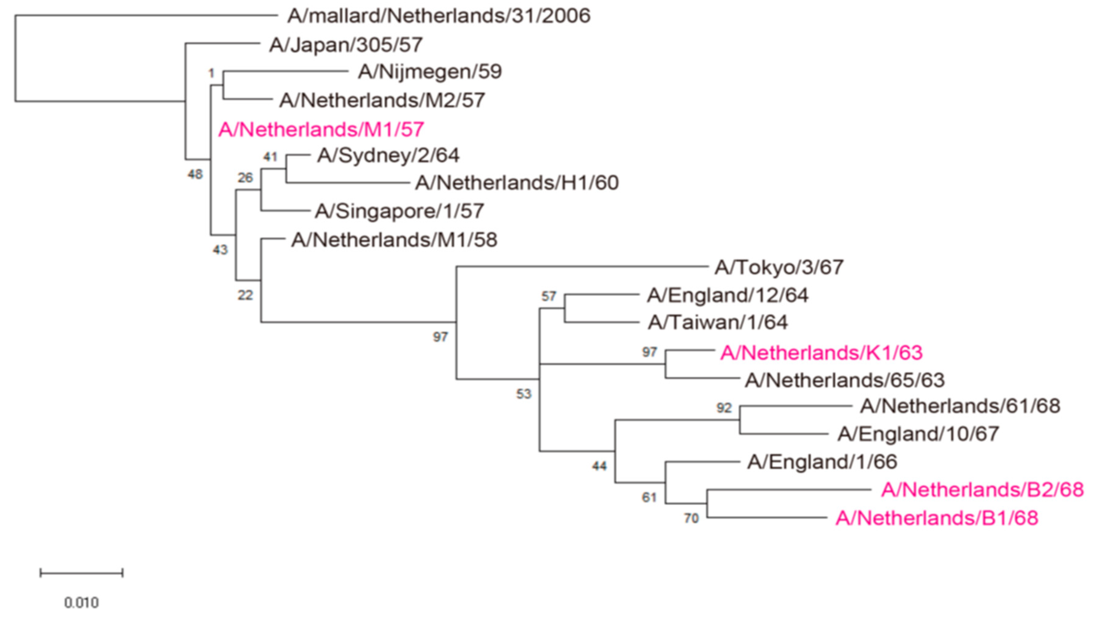
2.6. Phylogenetic Analysis

The HA sequences of 17 viruses described previously [11] were aligned using the software of ClustalW. The phylogenetic tree was inferred by using the Maximum Likelihood method and Jones-Taylor-Thornton (JTT) matrix-based model [12]. The tree with the highest log likelihood (−1787.57) is shown. The percentage of trees in which the associated taxa clustered together is shown next to the branches. Initial tree(s) for the heuristic search were obtained automatically by applying Neighbor-Join and BioNJ algorithms to a matrix of pairwise distances estimated using a JTT model, and then selecting the topology with the superior log likelihood value. The tree is drawn to scale, with branch lengths measured in the number of substitutions per site. There were 329 positions in the final dataset. Evolutionary analyses were conducted by using the software MEGA X [4]. A/mallard/Netherlands/31/2006 virus is defined as an outgroup.

2.7. Statistical Analysis

A non-parametric, repeated measures, one-way ANOVA Friedman’s test followed by a two-stage linear step-up procedure of Benjamini, Krieger, and Yekutieli was performed by using GraphPad Prism 7.04 software. p values <0.05 were considered significantly different. No samples were excluded from the analysis.

2.8. Antigenic Cartography

The neutralization data were analyzed by using antigenic cartography (https://acmacs-web.antigenic-cartography.org/), which is a method to visualize and increase the resolution of neutralization results, as detailed previously [13]. Plasma samples with no or only one numerical antibody titer were not included because they cannot be placed properly in an antigenic map.

3. Results and Discussions

To examine the antigenicity of the H2N2 viruses that circulated in humans, we used 50 plasma samples that were obtained from aged individuals (Table 2) because individuals who were born between 1928 and 1933 were likely exposed to human H2N2 viruses between 1957 and 1968. We also used 33 plasma samples from younger adults who were born after 1962 (Table 2). We chose 4 human H2N2 isolates—A/Netherlands/M1/57 (H2N2; M1/57), A/Netherlands/K1/63 (H2N2; K1/63), A/Netherlands/B1/68 (H2N2; B1/68), and A/Netherlands/B2/68 (H2N2; B2/68)—based on phylogenic analysis (Figure 1) and a previous report [11]. These isolates were obtained by culture in tertiary monkey kidney cells and MDCK cells for a maximum of five passages, without prior inoculation into embryonated chicken eggs [11].
Using these 83 plasma samples and four human H2N2 isolates, we performed a virus neutralization assay to compare the antigenicity of the human H2N2 isolates (Table 2). The aged individuals (ID #1–50) possessed higher neutralization titers against M1/57 and K1/63 than those against B1/68 and B2/68 (Figure 2A,C). The neutralizing titers against B1/68 of the aged individuals were lower than those against B2/68, whereas those against M1/57 were similar to those against K1/63 (Figure 2A,C). The neutralizing titers against K1/63 and B2/68 of the younger adult individuals were higher than those against M1/57 and B1/68 (Figure 2B,C). Younger adults who were born after 1972 (ID #75–83) had minimal neutralization titers against the human H2N2 viruses tested (Figure 2C). To visualize the antigenicity of the human H2N2 viruses, we constructed an antigenic map using the neutralizing titers of Table 2. This map revealed the antigenic differences among the H2N2 viruses tested and the gradual drift in their antigenicity during circulation in humans (Figure 3). The results obtained with these human sera were similar to those previously obtained using ferret antisera [11].
Human H2N2 viruses circulated in the human population between 1957 and 1968. Mouse and human monoclonal antibodies [9,10] and ferret antisera [6,7,8,11] revealed the antigenic change in H2N2 viruses. Here, we elucidated the antigenic change in H2N2 viruses by using 83 human plasma samples that were obtained from the aged and younger adult individuals. Overall, the antigenicity of H2N2 viruses revealed by human plasma was similar to that revealed by ferret antisera. Ferret antisera showed that the antigenic change between M1/57 and B1/68 was caused by the T128D and R139K substitutions together with other five other changes (Table 3). These substitutions may play a central role in the antigenic change revealed by human plasma since some human neutralizing monoclonal antibodies against H2-HA recognize the region surrounding these seven amino acids [10]. Furthermore, human plasma showed that the antigenicity of B1/68 differed from that of B2/68. At the seven amino acid positions that were important for the antigenic change between M1/57 and B1/68, E126K, P154Q, and A184E substitutions are present between B1/68 and B2/68 (Table 3). These 3 amino acid substitutions may, therefore, contribute to the antigenic difference between the viruses.
Although some of the aged individuals likely have been infected with or exposed to later H2N2 viruses, almost of all of the aged individuals maintained high neutralization titers against the H2N2 viruses that caused the pandemic or its antigenically closely related viruses, but possessed low neutralization titers against later H2N2 viruses. These data suggest that immunological imprinting toward the pandemic H2N2 virus occurred. This would also mean that the immunological imprinting was maintained for 50 years.
Antigenic cartography is usually performed by using antisera obtained from ferrets infected with an influenza virus [13]. In this study, we used human plasma samples obtained from aged and younger adults for the antigenic cartography of human H2N2 viruses. Previous studies showed that human infant sera and ferret antisera see the antigenicity of H1N1 and H3N2 viruses differently [14,15]. However, the positions of four H2N2 viruses in the antigenic map we made with the human plasma samples were similar to those obtained with ferret antisera (Figure 3 and ref [11]). This variation in findings with respect to the antigenic differences recognized by human sera/plasma and ferret antisera between H2N2 and H1N1 or H3N2 is interesting and deserves further study.
Human H2N2 viruses were replaced by human H3N2 viruses in 1968. Therefore, we had thought that people who were born after 1968 were unlikely to possess neutralizing antibodies against human H2N2 viruses. However, they did possess some neutralization titers against the H2N2 viruses. This finding could be explained by antibodies against the HA stem and/or NA because anti-HA stem antibodies are usually cross-reactive with members of group 1 HA [16,17,18] and antibodies against the NA of the seasonal H3N2 virus could suppress H2N2 virus replication, given that the NA of H3N2 virus was inherited from human H2N2 virus [1].
In conclusion, the plasma of individuals who were born between 1928 and 1933 revealed the antigenic change in human H2N2 viruses and the possibility of immunological imprinting of infected individuals against pandemic H2N2 virus, which lasts for half a century.

Author Contributions

Y.M., K.I.-H., T.W., M.I., and Y.K. designed this study. Y.M. and K.I-H. performed experiments. S.Y. and Y.K. analyzed the data and wrote the manuscript. Y.N., Y.A. (Yukiko Abe), and Y.A. (Yasumichi Arai) collected blood samples from volunteers. S.F. coordinated blood sample collection. T.H., M.O., and Y.G. contributed to the preparation of the plasma samples. R.A.M.F. provided human H2N2 isolates. All of the authors reviewed the manuscript and approved the final version.

Funding

This research was supported by Leading Advanced Projects for medical innovation (LEAP) from the Japan Agency for Medical Research and Development (AMED) (JP18am001007), by Grants-in-Aid for Scientific Research on Innovative Areas from the Ministry of Education, Culture, Science, Sports, and Technology (MEXT) of Japan (Nos. 16H06429, 16K21723, and 16H06434), by Grant-in-Aid for Scientific Research (B) from Japan Society for the Promotion of Science (JSPS) (18H03055), by the Japan Initiative for Global Research Network on Infectious Diseases (J-GRID) from AMED (JP19fm0108006), by a Research Program on Emerging and Re-emerging Infectious Diseases from AMED (JP19fk0108031, JP19fk0108056 and JP19fk0108066), and by JST Research Complex Program (JP15667051). R.F. is supported by National Institute of Allergy and Infectious Diseases/NIH contract no. HHSN272201400008C.

Acknowledgments

We thank Susan Watson for scientific editing, and Hiroshi Ueki and Daisuke Jubishi for blood sample collection.

Conflicts of Interest

Y.K. has received speaker’s honoraria from Toyama Chemical and Astellas Inc.; has received grant support from Chugai Pharmaceuticals, Daiichi Sankyo Pharmaceutical, Toyama Chemical, Tauns Laboratories, Inc., Otsuka Pharmaceutical Co., Ltd., and Denka Seiken Co., Ltd.; and is a co-founder of FluGen. Y.Arai. has received a research grant from Daiichi Sankyo Co, Ltd. and Novartis Pharma K K. The other authors have no conflicts of interest.

References

  1. Neumann, G.; Noda, T.; Kawaoka, Y. Emergence and pandemic potential of swine-origin H1N1 influenza virus. Nature 2009, 459, 931–939. [Google Scholar] [CrossRef] [PubMed] [Green Version]
  2. Nabel, G.J.; Wei, C.J.; Ledgerwood, J.E. Vaccinate for the next H2N2 pandemic now. Nature 2011, 471, 157–158. [Google Scholar] [CrossRef]
  3. Babu, T.M.; Perera, R.; Wu, J.T.; Fitzgerald, T.; Nolan, C.; Cowling, B.J.; Krauss, S.; Treanor, J.J.; Peiris, M. Population Serologic Immunity to Human and Avian H2N2 Viruses in the United States and Hong Kong for Pandemic Risk Assessment. J. Infect. Dis. 2018, 218, 1054–1060. [Google Scholar] [CrossRef] [Green Version]
  4. Kumar, S.; Stecher, G.; Li, M.; Knyaz, C.; Tamura, K. MEGA X: Molecular Evolutionary Genetics Analysis across Computing Platforms. Mol. Biol. Evol. 2018, 35, 1547–1549. [Google Scholar] [CrossRef] [PubMed]
  5. Joseph, U.; Linster, M.; Suzuki, Y.; Krauss, S.; Halpin, R.A.; Vijaykrishna, D.; Fabrizio, T.P.; Bestebroer, T.M.; Maurer-Stroh, S.; Webby, R.J.; et al. Adaptation of pandemic H2N2 influenza A viruses in humans. J. Virol. 2015, 89, 2442–2447. [Google Scholar] [CrossRef] [PubMed]
  6. Nerome, K.; Yoshioka, Y.; Torres, C.A.; Oya, A.; Bachmann, P.; Ottis, K.; Webster, R.G. Persistence of Q strain of H2N2 influenza virus in avian species: Antigenic, biological and genetic analysis of avian and human H2N2 viruses. Arch. Virol. 1984, 81, 239–250. [Google Scholar] [CrossRef] [PubMed]
  7. Yamada, A.; Brown, L.E.; Webster, R.G. Characterization of H2 influenza virus hemagglutinin with monoclonal antibodies: Influence of receptor specificity. Virology 1984, 138, 276–286. [Google Scholar] [CrossRef]
  8. Chen, G.L.; Lamirande, E.W.; Yang, C.F.; Jin, H.; Kemble, G.; Subbarao, K. Evaluation of replication and cross-reactive antibody responses of H2 subtype influenza viruses in mice and ferrets. J. Virol. 2010, 84, 7695–7702. [Google Scholar] [CrossRef] [PubMed]
  9. Tsuchiya, E.; Sugawara, K.; Hongo, S.; Matsuzaki, Y.; Muraki, Y.; Li, Z.N.; Nakamura, K. Antigenic structure of the haemagglutinin of human influenza A/H2N2 virus. J. Gen. Virol. 2001, 82, 2475–2484. [Google Scholar] [CrossRef] [PubMed]
  10. Krause, J.C.; Tsibane, T.; Tumpey, T.M.; Huffman, C.J.; Albrecht, R.; Blum, D.L.; Ramos, I.; Fernandez-Sesma, A.; Edwards, K.M.; Garcia-Sastre, A.; et al. Human monoclonal antibodies to pandemic 1957 H2N2 and pandemic 1968 H3N2 influenza viruses. J. Virol. 2012, 86, 6334–6340. [Google Scholar] [CrossRef] [PubMed]
  11. Linster, M.; Schrauwen, E.J.A.; van der Vliet, S.; Burke, D.F.; Lexmond, P.; Bestebroer, T.M.; Smith, D.J.; Herfst, S.; Koel, B.F.; Fouchier, R.A.M. The Molecular Basis for Antigenic Drift of Human A/H2N2 Influenza Viruses. J. Virol. 2019, 93, e01907-18. [Google Scholar] [CrossRef] [PubMed]
  12. Jones, D.T.; Taylor, W.R.; Thornton, J.M. The rapid generation of mutation data matrices from protein sequences. Comput. Appl. Biosci. 1992, 8, 275–282. [Google Scholar] [CrossRef] [PubMed]
  13. Smith, D.J.; Lapedes, A.S.; de Jong, J.C.; Bestebroer, T.M.; Rimmelzwaan, G.F.; Osterhaus, A.D.; Fouchier, R.A. Mapping the antigenic and genetic evolution of influenza virus. Science 2004, 305, 371–376. [Google Scholar] [CrossRef] [PubMed]
  14. Koel, B.F.; Mogling, R.; Chutinimitkul, S.; Fraaij, P.L.; Burke, D.F.; van der Vliet, S.; de Wit, E.; Bestebroer, T.M.; Rimmelzwaan, G.F.; Osterhaus, A.D.; et al. Identification of amino acid substitutions supporting antigenic change of influenza A(H1N1)pdm09 viruses. J. Virol. 2015, 89, 3763–3775. [Google Scholar] [CrossRef] [PubMed]
  15. Fonville, J.M.; Fraaij, P.L.; de Mutsert, G.; Wilks, S.H.; van Beek, R.; Fouchier, R.A.; Rimmelzwaan, G.F. Antigenic Maps of Influenza A(H3N2) Produced With Human Antisera Obtained After Primary Infection. J. Infect. Dis. 2016, 213, 31–38. [Google Scholar] [CrossRef] [PubMed]
  16. Yamayoshi, S.; Uraki, R.; Ito, M.; Kiso, M.; Nakatsu, S.; Yasuhara, A.; Oishi, K.; Sasaki, T.; Ikuta, K.; Kawaoka, Y. A Broadly Reactive Human Anti-hemagglutinin Stem Monoclonal Antibody That Inhibits Influenza A Virus Particle Release. EBioMedicine 2017, 17, 182–191. [Google Scholar] [CrossRef] [PubMed] [Green Version]
  17. Wu, N.C.; Yamayoshi, S.; Ito, M.; Uraki, R.; Kawaoka, Y.; Wilson, I.A. Recurring and Adaptable Binding Motifs in Broadly Neutralizing Antibodies to Influenza Virus Are Encoded on the D3-9 Segment of the Ig Gene. Cell Host Microbe 2018, 24, 569–578.e4. [Google Scholar] [Green Version]
  18. Yamayoshi, S.; Yasuhara, A.; Ito, M.; Uraki, R.; Kawaoka, Y. Differences in the ease with which mutant viruses escape from human monoclonal antibodies against the HA stem of influenza A virus. J. Clin. Virol. 2018, 108, 105–111. [Google Scholar] [CrossRef] [PubMed]
Figure 1. Phylogenetic tree based on the amino acid sequences of HA1 derived from human H2N2 viruses. This tree was constructed by using the Maximum Likelihood method and JTT matrix-based model. Virus isolates used for antigenic analysis are highlighted in magenta.
Viruses 11 00978 g001
Figure 2. Neutralization titers of aged individuals (A) and younger adult individuals (B) against human H2N2 viruses. The neutralization titers against the viruses indicated in Table 2 were plotted. A non-parametric Friedman’s test followed by a two-stage linear step-up procedure of Benjamini, Krieger, and Yekutieli was performed. * and *** mean p < 0.05 and p < 0.0001, respectively. (C) Bubble chart of the neutralization titers of all of the individuals. The bigger the circle, the higher the neutralization titer.
Viruses 11 00978 g002
Figure 3. An antigenic map of human H2N2 viruses. An antigenic map was generated from the neutralization data shown in Table 2. Viruses are represented as circles and plasma samples obtained from aged or younger adults are represented as black or grey squares, respectively. Sera with no or only 1 numerical antibody titer are not shown as they cannot be placed properly in an antigenic map. The grid indicates one unit of antigenic distance, a two-fold dilution in neutralization titer.
Viruses 11 00978 g003
Table 1. Characteristics of the participants in this study.
No. of Individuals
Birth Year
Aged IndividualsYounger Adult Individuals
1928192919301931193219331960s1970s1980s1990s
Males44444410512
Females5544449321
Total99888819833
Medical history bHeart disease3/91/93/81/81/81/8NA aNANANA
Diabetes0/90/91/82/81/81/8NANANANA
Cancer3/92/92/84/82/82/8NANANANA
a Not available. b Summary of available medical history of each individual.
Table 2. Neutralization titers of aged and younger adult individuals.
IDBirth Year (age)M1/57 aK1/63 bB1/68 cB2/68 d
11928 (89)128888
21928 (89)832416
31928 (89)6432<416
41928 (89)323248
51928 (89)256128328
61928 (89)6432<416
71928 (89)256321616
81928 (89)12864432
91928 (89)816<4<4
101929 (89)6432816
111929 (89)832<4<4
121929 (89)1286448
131929 (89)832<48
141929 (89)2566444
151929 (89)328<44
161929 (89)32324<4
171929 (88)326488
181929 (88)32128416
191930 (88)1616<48
201930 (88)16844
211930 (88)3264<48
221930 (88)2566484
231930 (87)6464416
241930 (87)12832432
251930 (87)25616416
261930 (87)2566488
271931 (87)12864432
281931 (87)1616<4<4
291931 (87)1664816
301931 (87)3216416
311931 (86)3216<4<4
321931 (86)324<44
331931 (86)3232<44
341931 (86)1283248
351932 (86)2563284
361932 (86)3232<432
371932 (86)2563284
381932 (86)168<48
391932 (86)256128<48
401932 (85)25632816
411932 (85)3232<44
421932 (85)646444
431933 (85)1616816
441933 (85)2566488
451933 (85)256324<4
461933 (85)321644
471933 (85)12864816
481933 (85)>5121286464
491933 (85)6464816
501933 (85)812844
511962 (56)4648128
521962 (55)<41644
531962 (55)8321632
541962 (55)163288
551963 (55)32>5123264
561963 (55)32>51264256
571963 (55)81286464
581963 (55)162561632
591964 (54)8128832
601965 (53)464432
611966 (52)832832
621966 (51)81281664
631967 (51)4641616
641967 (50)<43248
651968 (50)<44<4<4
661968 (49)41648
671969 (49)432832
681969 (49)<4<4<4<4
691969 (48)416416
701970 (48)84816
711970 (47)<48432
721970 (47)<48<416
731970 (47)<41684
741970 (47)<44<48
751972 (45)<4<4<4<4
761974 (44)<4<4<44
771978 (39)<4<4<4<4
781980 (37)<4<4<4<4
791982 (36)<4<4<4<4
801985 (33)<4<4<4<4
811990 (27)<4<4<4<4
821992 (26)<4<4<44
831994 (23)<4<4<44
Neutralization titers of plasma samples of aged individuals (ID 1–50) and younger adult individuals (ID 51–83) against A/Netherland/M1/57 a, A/Netherland/K1/63 b, A/Netherland/B1/68 c, or A/Netherland/B2/68 d were measured.
Table 3. Comparison of amino acids that are important for HA antigenicity a.
IsolateAmino Acid Position at
126128132139154184188
A/Netherland/M1/57TTRNSTT
A/Netherland/K1/63TTRNPAA
A/Netherland/B1/68EDKKPAA
A/Netherland/B2/68KDKKQEA
a Amino acids that are different among the HAs of the four isolates at previously identified antigenic determinants [11] are listed.

