Next Issue
Volume 11, May
Previous Issue
Volume 11, March


Viruses, Volume 11, Issue 4 (April 2019) – 86 articles

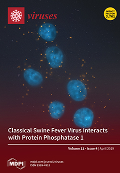
Cover Story (view full-size image): Classical swine fever virus (CSFV) structural glycoprotein E2 induces a protective immune response and is critical for virulence in swine by interacting with the Protein Phosphatase 1 (PP1) complex. In this study, we determined that CSFV E2 specifically binds to PPP1CB, a component of the PP1 complex, and activation of the PP1 pathway has a negative effect on CSFV replication. This suggests that CSFV inhibits the activation of PP1 pathway for its own survival. View this paper.
  • Issues are regarded as officially published after their release is announced to the table of contents alert mailing list.
  • You may sign up for e-mail alerts to receive table of contents of newly released issues.
  • PDF is the official format for papers published in both, html and pdf forms. To view the papers in pdf format, click on the "PDF Full-text" link, and use the free Adobe Reader to open them.
Order results
Result details
Section
Select all
Export citation of selected articles as:
18 pages, 4852 KiB  
Article
Porcine Parvovirus Infection Causes Pig Placenta Tissue Damage Involving Nonstructural Protein 1 (NS1)-Induced Intrinsic ROS/Mitochondria-Mediated Apoptosis
by Jianlou Zhang, Jinghui Fan, Yan Li, Shuang Liang, Shanshan Huo, Xing Wang, Yuzhu Zuo, Dan Cui, Wenyan Li, Zhenyu Zhong and Fei Zhong
Viruses 2019, 11(4), 389; https://0-doi-org.brum.beds.ac.uk/10.3390/v11040389 - 25 Apr 2019
Cited by 25 | Viewed by 4855
Abstract
Porcine parvovirus (PPV) is an important pathogen causing reproductive failure in pigs. PPV-induced cell apoptosis has been recently identified as being involved in PPV-induced placental tissue damages resulting in reproductive failure. However, the molecular mechanism was not fully elucidated. Here we demonstrate that [...] Read more.
Porcine parvovirus (PPV) is an important pathogen causing reproductive failure in pigs. PPV-induced cell apoptosis has been recently identified as being involved in PPV-induced placental tissue damages resulting in reproductive failure. However, the molecular mechanism was not fully elucidated. Here we demonstrate that PPV nonstructural protein 1 (NS1) can induce host cell apoptosis and death, thereby indicating the NS1 may play a crucial role in PPV-induced placental tissue damages and reproductive failure. We have found that NS1-induced apoptosis was significantly inhibited by caspase 9 inhibitor, but not caspase 8 inhibitor, and transfection of NS1 gene into PK-15 cells significantly inhibited mitochondria-associated antiapoptotic molecules Bcl-2 and Mcl-1 expressions and enhanced proapoptotic molecules Bax, P21, and P53 expressions, suggesting that NS1-induced apoptosis is mainly through the mitochondria-mediated intrinsic apoptosis pathway. We also found that both PPV infection and NS1 vector transfection could cause host DNA damage resulting in cell cycle arrest at the G1 and G2 phases, trigger mitochondrial ROS accumulation resulting in mitochondria damage, and therefore, induce the host cell apoptosis. This study provides a molecular basis for elucidating PPV-induced cell apoptosis and reproductive failure. Full article
(This article belongs to the Special Issue Porcine Viruses 2019)
Show Figures

Graphical abstract

15 pages, 3742 KiB  
Article
Prevalence and Diversity Analysis of Candidate Prophages to Provide An Understanding on Their Roles in Bacillus Thuringiensis
by Yajuan Fu, Yan Wu, Yihui Yuan and Meiying Gao
Viruses 2019, 11(4), 388; https://0-doi-org.brum.beds.ac.uk/10.3390/v11040388 - 25 Apr 2019
Cited by 15 | Viewed by 3792
Abstract
Bacillus thuringiensis (Bt) is widely used in producing biological insecticides. Phage contaminations during Bt fermentation can cause severe losses of yields. Lots of strategies have been engaged to control extrinsic phage contamination during Bt fermentation, but their effectiveness is low. In this study, [...] Read more.
Bacillus thuringiensis (Bt) is widely used in producing biological insecticides. Phage contaminations during Bt fermentation can cause severe losses of yields. Lots of strategies have been engaged to control extrinsic phage contamination during Bt fermentation, but their effectiveness is low. In this study, the candidate endogenous prophages (prophages) in 61 Bt chromosomes that had been deposited in GenBank database were analyzed. The results revealed that all chromosomes contained prophage regions, and 398 candidate prophage regions were predicted, including 135 putative complete prophages and 263 incomplete prophage regions. These putative complete prophages showed highly diverse genetic backgrounds. The inducibility of the prophages of ten Bt strains (4AJ1, 4BD1, HD-1, HD-29, HD-73, HD-521, BMB171, 4CC1, CT-43, and HD-1011) was tested, and the results showed that seven of the ten strains’ prophages were inducible. These induced phages belonged to the Siphoviridae family and exhibited a broad host spectrum against the non-original strains. The culture supernatants of the two strains (BMB171, 4CC1) could lyse Bt cells, but no virions were observed, which was speculated to be caused by lysin. The functional analysis of the putative complete prophage proteins indicated that some proteins, such as antibiotic resistance-associated proteins and restriction endonucleases, might increase the fitness of the Bt strains to different environments. The findings of this study provided understanding on the high prevalence and diversity of Bt prophages, as well as pointed out the role of prophages in the life cycle of Bt. Full article
(This article belongs to the Special Issue Diversity and Evolution of Phage Genomes)
Show Figures

Figure 1

15 pages, 3646 KiB  
Article
Susceptibility of Exopalaemon carinicauda to the Infection with Shrimp Hemocyte Iridescent Virus (SHIV 20141215), a Strain of Decapod Iridescent Virus 1 (DIV1)
by Xing Chen, Liang Qiu, Hailiang Wang, Peizhuo Zou, Xuan Dong, Fuhua Li and Jie Huang
Viruses 2019, 11(4), 387; https://0-doi-org.brum.beds.ac.uk/10.3390/v11040387 - 25 Apr 2019
Cited by 51 | Viewed by 6566
Abstract
In this study, ridgetail white prawns—Exopalaemon carinicauda—were infected per os (PO) with debris of Penaeus vannamei infected with shrimp hemocyte iridescent virus (SHIV 20141215), a strain of decapod iridescent virus 1 (DIV1), and via intramuscular injection (IM with raw extracts of [...] Read more.
In this study, ridgetail white prawns—Exopalaemon carinicauda—were infected per os (PO) with debris of Penaeus vannamei infected with shrimp hemocyte iridescent virus (SHIV 20141215), a strain of decapod iridescent virus 1 (DIV1), and via intramuscular injection (IM with raw extracts of SHIV 20141215. The infected E. carinicauda showed obvious clinical symptoms, including weakness, empty gut and stomach, pale hepatopancreas, and partial death with mean cumulative mortalities of 42.5% and 70.8% by nonlinear regression, respectively. Results of TaqMan probe-based real-time quantitative PCR showed that the moribund and surviving individuals with clinical signs of infected E. carinicauda were DIV1-positive. Histological examination showed that there were darkly eosinophilic and cytoplasmic inclusions, of which some were surrounded with or contained tiny basophilic staining, and pyknosis in hemocytes in hepatopancreatic sinus, hematopoietic cells, cuticular epithelium, etc. On the slides of in situ DIG-labeling-loop-mediated DNA amplification (ISDL), positive signals were observed in hematopoietic tissue, stomach, cuticular epithelium, and hepatopancreatic sinus of infected prawns from both PO and IM groups. Transmission electron microscopy (TEM) of ultrathin sections showed that icosahedral DIV1 particles existed in hepatopancreatic sinus and gills of the infected E. carinicauda from the PO group. The viral particles were also observed in hepatopancreatic sinus, gills, pereiopods, muscles, and uropods of the infected E. carinicauda from the IM group. The assembled virions, which mostly distributed along the edge of the cytoplasmic virogenic stromata near cellular membrane of infected cells, were enveloped and approximately 150 nm in diameter. The results of molecular tests, histopathological examination, ISDL, and TEM confirmed that E. carinicauda is a susceptible host of DIV1. This study also indicated that E. carinicauda showed some degree of tolerance to the infection with DIV1 per os challenge mimicking natural pathway. Full article
Show Figures

Figure 1

17 pages, 4034 KiB  
Article
Hepatitis B Surface Antigen Activates Unfolded Protein Response in Forming Ground Glass Hepatocytes of Chronic Hepatitis B
by Yao Li, Yuchen Xia, Xiaoming Cheng, David E. Kleiner, Stephen M. Hewitt, Julia Sproch, Tong Li, Hui Zhuang and T. Jake Liang
Viruses 2019, 11(4), 386; https://0-doi-org.brum.beds.ac.uk/10.3390/v11040386 - 25 Apr 2019
Cited by 32 | Viewed by 5886
Abstract
Ground glass hepatocytes (GGHs), a histological hallmark of chronic hepatitis B virus (HBV) infection, contain excessive hepatitis surface antigen (HBsAg) in the endoplasmic reticulum (ER), which is linked to unfolded protein response (UPR). The mechanism by which HBV activates UPR has not been [...] Read more.
Ground glass hepatocytes (GGHs), a histological hallmark of chronic hepatitis B virus (HBV) infection, contain excessive hepatitis surface antigen (HBsAg) in the endoplasmic reticulum (ER), which is linked to unfolded protein response (UPR). The mechanism by which HBV activates UPR has not been fully defined. To investigate this, HepG2-NTCP cells and primary human hepatocytes (PHHs) were either infected with HBV or transduced with adenoviral vectors expressing replication-competent HBV genome or individual HBV genes. UPR markers were evaluated by qPCR, Western blotting, and immunofluorescence. Apoptosis and cell viability were measured by Caspase-3/7 and ATPlite assay respectively. We found that UPR markers were induced by the overexpression of HBsAg in HepG2-NTCP cells and PHHs. Elevation of UPR-induced genes showed a dose-dependent correlation with HBsAg levels. In HBV-infected livers, GGHs also demonstrated excessive accumulation of HBsAg associated with increased BIP/GRP78 staining, a marker of UPR. Prolonged activation of UPR by HBsAg overexpression induced signs of apoptosis. Overexpression of HBsAg can induce ER stress through protein kinase RNA-like endoplasmic reticulum kinase (PERK) pathway in vitro, and may be linked to the appearance of GGHs. The activation of UPR by HBsAg may sensitize hepatocytes to cell death and result in possible subsequent cellular changes leading to a premalignant phenotype. Full article
(This article belongs to the Section Animal Viruses)
Show Figures

Figure 1

13 pages, 5434 KiB  
Article
Screening of an FDA-Approved Drug Library with a Two-Tier System Identifies an Entry Inhibitor of Severe Fever with Thrombocytopenia Syndrome Virus
by Shuofeng Yuan, Jasper Fuk-Woo Chan, Zi-Wei Ye, Lei Wen, Terance Gi-Wai Tsang, Jianli Cao, Jingjing Huang, Chris Chun-Yiu Chan, Kenn Ka-Heng Chik, Garnet Kwan-Yue Choi, Jian-Piao Cai, Feifei Yin, Hin Chu, Mifang Liang, Dong-Yan Jin and Kwok-Yung Yuen
Viruses 2019, 11(4), 385; https://0-doi-org.brum.beds.ac.uk/10.3390/v11040385 - 25 Apr 2019
Cited by 21 | Viewed by 6056
Abstract
Severe fever with thrombocytopenia syndrome virus (SFTSV) is an emerging tick-borne bunyavirus that causes severe disease in humans with case-fatality rates of up to 30%. There are currently very limited treatment options for SFTSV infection. We conducted a drug repurposing program by establishing [...] Read more.
Severe fever with thrombocytopenia syndrome virus (SFTSV) is an emerging tick-borne bunyavirus that causes severe disease in humans with case-fatality rates of up to 30%. There are currently very limited treatment options for SFTSV infection. We conducted a drug repurposing program by establishing a two-tier test system to rapidly screen a Food and Drug Administration- (FDA)-approved drug library for drug compounds with anti-SFTSV activity in vitro. We identified five drug compounds that inhibited SFTSV replication at low micromolar concentrations, including hexachlorophene, triclosan, regorafenib, eltrombopag, and broxyquinoline. Among them, hexachlorophene was the most potent with an IC50 of 1.3 ± 0.3 µM and a selectivity index of 18.7. Mechanistic studies suggested that hexachlorophene was a virus entry inhibitor, which impaired SFTSV entry into host cells by interfering with cell membrane fusion. Molecular docking analysis predicted that the binding of hexachlorophene with the hydrophobic pocket between domain I and domain III of the SFTSV Gc glycoprotein was highly stable. The novel antiviral activity and mechanism of hexachlorophene in this study would facilitate the use of hexachlorophene as a lead compound to develop more entry inhibitors with higher anti-SFTSV potency and lower toxicity. Full article
(This article belongs to the Special Issue Viral Entry Pathways)
Show Figures

Figure 1

13 pages, 729 KiB  
Article
Lower Broadly Neutralizing Antibody Responses in Female Versus Male HIV-1 Infected Injecting Drug Users
by Zelda EULER, Tom L. VAN DEN KERKHOF, Roger D. KOUYOS, Damien C. TULLY, Todd M. ALLEN, Alexandra TRKOLA, Rogier W. SANDERS, Hanneke SCHUITEMAKER and Marit J. VAN GILS
Viruses 2019, 11(4), 384; https://0-doi-org.brum.beds.ac.uk/10.3390/v11040384 - 25 Apr 2019
Cited by 5 | Viewed by 3273
Abstract
Understanding the factors involved in the development of broadly neutralizing antibody (bNAb) responses in natural infection can guide vaccine design aimed at eliciting protective bNAb responses. Most of the studies to identify and study the development of bNAb responses have been performed in [...] Read more.
Understanding the factors involved in the development of broadly neutralizing antibody (bNAb) responses in natural infection can guide vaccine design aimed at eliciting protective bNAb responses. Most of the studies to identify and study the development of bNAb responses have been performed in individuals who had become infected via homo- or heterosexual HIV-1 transmission; however, the prevalence and characteristics of bNAb responses in injecting drug users (IDUs) have been underrepresented. We retrospectively studied the prevalence of bNAb responses in HIV-1 infected individuals in the Amsterdam Cohort, including 50 male and 35 female participants who reported injecting drug use as the only risk factor. Our study revealed a significantly lower prevalence of bNAb responses in females compared to males. Gender, transmission route and CD4+ count at set point, but not viral load, were independently associated with the development of bNAb responses in IDUs. To further explore the influences of gender in the setting of IDU, we also looked into the Swiss 4.5k Screen. There we observed lower bNAb responses in female IDUs as well. These results reveal that the emergence of bNAbs may be dependent on multiple factors, including gender. Therefore, the effect of gender on the development of bNAb responses is a factor that should be taken into account when designing vaccine efficacy trials. Full article
(This article belongs to the Section Animal Viruses)
Show Figures

Figure 1

14 pages, 1533 KiB  
Article
Dual Transcriptomic Analysis Reveals a Delayed Antiviral Response of Haliotis diversicolor supertexta against Haliotid Herpesvirus-1
by Chang-Ming Bai, Shu-Min Zhang, Ya-Na Li, Lu-Sheng Xin, Umberto Rosani and Chong-Ming Wang
Viruses 2019, 11(4), 383; https://0-doi-org.brum.beds.ac.uk/10.3390/v11040383 - 24 Apr 2019
Cited by 10 | Viewed by 3294
Abstract
Haliotid herpesvirus-1 (HaHV-1) is the first identified gastropod herpesvirus, causing a highly lethal neurologic disease of abalone species. The genome of HaHV-1 has been sequenced, but the functions of the putative genes and their roles during infection are still poorly understood. In the [...] Read more.
Haliotid herpesvirus-1 (HaHV-1) is the first identified gastropod herpesvirus, causing a highly lethal neurologic disease of abalone species. The genome of HaHV-1 has been sequenced, but the functions of the putative genes and their roles during infection are still poorly understood. In the present study, transcriptomic profiles of Haliotis diversicolor supertexta at 0, 24 and 60 h post injection (hpi) with HaHV-1 were characterized through high-throughput RNA sequencing. A total of 448 M raw reads were obtained and assembled into 2.08 × 10⁵ unigenes with a mean length of 1486 bp and an N50 of 2455 bp. Although we detected increased HaHV-1 DNA loads and active viral expression at 24 hpi, this evidence was not linked to significant changes of host transcriptomic profiles between 0 and 24 hpi, whereas a rich immune-related gene set was over-expressed at 60 hpi. These results indicate that, at least at the beginning of HaHV-1 infection, the virus can replicate with no activation of the host immune response. We propose that HaHV-1 may evolve more effective strategies to modulate the host immune response and hide during replication, so that it could evade the immune surveillance at the early stage of infection. Full article
(This article belongs to the Section Animal Viruses)
Show Figures

Figure 1

15 pages, 21174 KiB  
Article
Porcine Epidemic Diarrhea Virus (PEDV) ORF3 Interactome Reveals Inhibition of Virus Replication by Cellular VPS36 Protein
by Challika Kaewborisuth, Yodying Yingchutrakul, Sittiruk Roytrakul and Anan Jongkaewwattana
Viruses 2019, 11(4), 382; https://0-doi-org.brum.beds.ac.uk/10.3390/v11040382 - 24 Apr 2019
Cited by 14 | Viewed by 5161
Abstract
The accessory protein ORF3 of porcine epidemic diarrhea virus (PEDV) has been proposed to play a key role in virus replication. However, our understanding of its function regarding virus and host interaction is still limited. In this study, we employed immunoprecipitation and mass [...] Read more.
The accessory protein ORF3 of porcine epidemic diarrhea virus (PEDV) has been proposed to play a key role in virus replication. However, our understanding of its function regarding virus and host interaction is still limited. In this study, we employed immunoprecipitation and mass spectrometry to screen for cellular interacting partners of ORF3. Gene ontology analysis of the host interactome highlighted the involvement of ORF3 in endosomal and immune signaling pathways. Among the identified ORF3-interacting proteins, the vacuolar protein-sorting-associated protein 36 (VPS36) was assessed for its role in PEDV replication. VPS36 was found to interact with ORF3 regardless of its GLUE domain. As a result of VPS36–ORF3 interaction, PEDV replication was substantially suppressed in cells overexpressing VPS36. Interestingly, the ORF3 protein expression was diminished in VPS36-overexpressing cells, an effect that could not be restored by treatment of lysosomal inhibitors. In addition, disruption of endogenously-expressed VPS36 by siRNA could partially augment PEDV replication. Taken together, our study provides mechanistic insights into the contribution of ORF3 in PEDV replication. Full article
(This article belongs to the Special Issue Porcine Viruses 2019)
Show Figures

Figure 1

13 pages, 3356 KiB  
Article
Lack of Middle East Respiratory Syndrome Coronavirus Transmission in Rabbits
by W. Widagdo, Nisreen M. A. Okba, Mathilde Richard, Dennis de Meulder, Theo M. Bestebroer, Pascal Lexmond, Elmoubasher A. B. A. Farag, Mohammed Al-Hajri, Koert J. Stittelaar, Leon de Waal, Geert van Amerongen, Judith M. A. van den Brand, Bart L. Haagmans and Sander Herfst
Viruses 2019, 11(4), 381; https://0-doi-org.brum.beds.ac.uk/10.3390/v11040381 - 24 Apr 2019
Cited by 10 | Viewed by 5266
Abstract
Middle East respiratory syndrome coronavirus (MERS-CoV) transmission from dromedaries to humans has resulted in major outbreaks in the Middle East. Although some other livestock animal species have been shown to be susceptible to MERS-CoV, it is not fully understood why the spread of [...] Read more.
Middle East respiratory syndrome coronavirus (MERS-CoV) transmission from dromedaries to humans has resulted in major outbreaks in the Middle East. Although some other livestock animal species have been shown to be susceptible to MERS-CoV, it is not fully understood why the spread of the virus in these animal species has not been observed in the field. In this study, we used rabbits to further characterize the transmission potential of MERS-CoV. In line with the presence of MERS-CoV receptor in the rabbit nasal epithelium, high levels of viral RNA were shed from the nose following virus inoculation. However, unlike MERS-CoV-infected dromedaries, these rabbits did not develop clinical manifestations including nasal discharge and did shed only limited amounts of infectious virus from the nose. Consistently, no transmission by contact or airborne routes was observed in rabbits. Our data indicate that despite relatively high viral RNA levels produced, low levels of infectious virus are excreted in the upper respiratory tract of rabbits as compared to dromedary camels, thus resulting in a lack of viral transmission. Full article
(This article belongs to the Special Issue MERS-CoV)
Show Figures

Figure 1

12 pages, 1547 KiB  
Article
Characterization of EIAV env Quasispecies during Long-Term Passage In Vitro: Gradual Loss of Pathogenicity
by Cong Liu, Xue-Feng Wang, Yan Wang, Jie Chen, Zhaohua Zhong, Yuezhi Lin and Xiaojun Wang
Viruses 2019, 11(4), 380; https://0-doi-org.brum.beds.ac.uk/10.3390/v11040380 - 24 Apr 2019
Cited by 4 | Viewed by 3012
Abstract
As the only widely used live lentiviral vaccine, the equine infectious anima virus (EIAV) attenuated vaccine was developed by in vitro passaging of a virulent strain for 121 generations. In our previous study, we observed that the attenuated vaccine was gradually selected under [...] Read more.
As the only widely used live lentiviral vaccine, the equine infectious anima virus (EIAV) attenuated vaccine was developed by in vitro passaging of a virulent strain for 121 generations. In our previous study, we observed that the attenuated vaccine was gradually selected under increased environmental pressure at the population level (termed a quasispecies). To further elucidate the potential correlation between viral quasispecies evolution and pathogenesis, a systematic study was performed by sequencing env using several methods. Some key mutations were identified within Env, and we observed that increased percentages of these mutations were accompanied by an increased passage number and attenuated virulence. Phylogenetic analysis revealed that env mutations related to the loss of virulence might have occurred evolutionarily. Among these mutations, deletion of amino acid 236 in the V4 region of Env resulted in the loss of one N-glycosylation site that was crucial for virulence. Notably, the 236-deleted sequence represented a “vaccine-specific” mutation that was also found in wild EIAVLN40 strains based on single genome amplification (SGA) analysis. Therefore, our results suggest that the EIAV attenuated vaccine may originate from a branch of quasispecies of EIAVLN40. Generally, the presented results may increase our understanding of the attenuation mechanism of the EIAV vaccine and provide more information about the evolution of other lentiviruses. Full article
(This article belongs to the Special Issue Nonprimate Lentivirus)
Show Figures

Figure 1

19 pages, 4673 KiB  
Article
Characterization of a New Member of Alphacoronavirus with Unique Genomic Features in Rhinolophus Bats
by Ning Wang, Chuming Luo, Haizhou Liu, Xinglou Yang, Ben Hu, Wei Zhang, Bei Li, Yan Zhu, Guangjian Zhu, Xurui Shen, Cheng Peng and Zhengli Shi
Viruses 2019, 11(4), 379; https://0-doi-org.brum.beds.ac.uk/10.3390/v11040379 - 24 Apr 2019
Cited by 23 | Viewed by 12790
Abstract
Bats have been identified as a natural reservoir of a variety of coronaviruses (CoVs). Several of them have caused diseases in humans and domestic animals by interspecies transmission. Considering the diversity of bat coronaviruses, bat species and populations, we expect to discover more [...] Read more.
Bats have been identified as a natural reservoir of a variety of coronaviruses (CoVs). Several of them have caused diseases in humans and domestic animals by interspecies transmission. Considering the diversity of bat coronaviruses, bat species and populations, we expect to discover more bat CoVs through virus surveillance. In this study, we described a new member of alphaCoV (BtCoV/Rh/YN2012) in bats with unique genome features. Unique accessory genes, ORF4a and ORF4b were found between the spike gene and the envelope gene, while ORF8 gene was found downstream of the nucleocapsid gene. All the putative genes were further confirmed by reverse-transcription analyses. One unique gene at the 3’ end of the BtCoV/Rh/YN2012 genome, ORF9, exhibits ~30% amino acid identity to ORF7a of the SARS-related coronavirus. Functional analysis showed ORF4a protein can activate IFN-β production, whereas ORF3a can regulate NF-κB production. We also screened the spike-mediated virus entry using the spike-pseudotyped retroviruses system, although failed to find any fully permissive cells. Our results expand the knowledge on the genetic diversity of bat coronaviruses. Continuous screening of bat viruses will help us further understand the important role played by bats in coronavirus evolution and transmission. Full article
(This article belongs to the Special Issue Viruses and Bats 2019)
Show Figures

Figure 1

17 pages, 3695 KiB  
Article
Hepatitis C Virus-Induced FUT8 Causes 5-FU Drug Resistance in Human Hepatoma Huh7.5.1 Cells
by Shu Li, Xiao-Yu Liu, Qiu Pan, Jian Wu, Zhi-Hao Liu, Yong Wang, Min Liu and Xiao-Lian Zhang
Viruses 2019, 11(4), 378; https://0-doi-org.brum.beds.ac.uk/10.3390/v11040378 - 24 Apr 2019
Cited by 17 | Viewed by 3564
Abstract
Hepatitis C virus (HCV) is a major cause of human chronic liver disease and hepatocellular carcinoma. Our recent studies showed that α1,6-fucosyltransferase (FUT8), a key glycosyltransferase, was the most up-regulated glycosyltransferase after the HCV infection of human hepatocellular carcinoma Huh7.5.1 cells. Here, we [...] Read more.
Hepatitis C virus (HCV) is a major cause of human chronic liver disease and hepatocellular carcinoma. Our recent studies showed that α1,6-fucosyltransferase (FUT8), a key glycosyltransferase, was the most up-regulated glycosyltransferase after the HCV infection of human hepatocellular carcinoma Huh7.5.1 cells. Here, we further studied the effects and possible mechanism of FUT8 on the proliferation of HCV and chemotherapy-resistance of HCV-infected Huh7.5.1 cells. The effects of FUT8 on the proliferation and drug resistance of HCV-infected Huh7.5.1 cells were analyzed by flow cytometry analysis (FCM), quantitative real-time polymerase chain reaction (qRT-PCR), Western blot analysis and lactate dehydrogenase (LDH) release assay. Results: We found that FUT8 not only promoted Huh7.5.1 proliferation by activating PI3K-AKT-NF-κB signaling, but also stimulated the expression of the drug-resistant proteins P-glycoprotein (P-gp) and multidrug resistance related protein 1 (MRP1) and enhanced the 5-fluorouracil (5-FU) chemo-resistance of Huh7.5.1 cells. Silencing of FUT8 reduced the cell proliferation and increased the 5-FU sensitivity of HCV-infected Huh7.5.1 cells. Inhibition of P-gp and MRP1 increased the 5-FU drug sensitivity in HCV infected Huh7.5.1 cells. HCV-induced FUT8 promotes proliferation and 5-FU resistance of Huh7.5.1 cells. FUT8 may serve as a therapeutic target to reverse chemotherapy resistance in HCV-infected Huh7.5.1 cells. Full article
(This article belongs to the Section Animal Viruses)
Show Figures

Figure 1

16 pages, 3964 KiB  
Article
Determining the Replication Kinetics and Cellular Tropism of Influenza D Virus on Primary Well-Differentiated Human Airway Epithelial Cells
by Melle Holwerda, Jenna Kelly, Laura Laloli, Isabel Stürmer, Jasmine Portmann, Hanspeter Stalder and Ronald Dijkman
Viruses 2019, 11(4), 377; https://0-doi-org.brum.beds.ac.uk/10.3390/v11040377 - 24 Apr 2019
Cited by 32 | Viewed by 6831
Abstract
Influenza viruses are notorious pathogens that frequently cross the species barrier with often severe consequences for both animal and human health. In 2011, a novel member of the Orthomyxoviridae family, Influenza D virus (IDV), was identified in the respiratory tract of swine. Epidemiological [...] Read more.
Influenza viruses are notorious pathogens that frequently cross the species barrier with often severe consequences for both animal and human health. In 2011, a novel member of the Orthomyxoviridae family, Influenza D virus (IDV), was identified in the respiratory tract of swine. Epidemiological surveys revealed that IDV is distributed worldwide among livestock and that IDV-directed antibodies are detected in humans with occupational exposure to livestock. To identify the transmission capability of IDV to humans, we determined the viral replication kinetics and cell tropism using an in vitro respiratory epithelium model of humans. The inoculation of IDV revealed efficient replication kinetics and apical progeny virus release at different body temperatures. Intriguingly, the replication characteristics of IDV revealed higher replication kinetics compared to Influenza C virus, despite sharing the cell tropism preference for ciliated cells. Collectively, these results might indicate why IDV-directed antibodies are detected among humans with occupational exposure to livestock. Full article
(This article belongs to the Special Issue Non-A Influenza)
Show Figures

Figure 1

19 pages, 558 KiB  
Review
Culicoides Biting Midges—Underestimated Vectors for Arboviruses of Public Health and Veterinary Importance
by Franziska Sick, Martin Beer, Helge Kampen and Kerstin Wernike
Viruses 2019, 11(4), 376; https://0-doi-org.brum.beds.ac.uk/10.3390/v11040376 - 24 Apr 2019
Cited by 63 | Viewed by 6758
Abstract
Culicoides biting midges, small hematophagous dipterans, are the demonstrated or putative vectors of multiple arboviruses of veterinary and public health importance. Despite its relevance in disease spread, the ceratopogonid genus Culicoides is still a largely neglected group of species, predominantly because the major [...] Read more.
Culicoides biting midges, small hematophagous dipterans, are the demonstrated or putative vectors of multiple arboviruses of veterinary and public health importance. Despite its relevance in disease spread, the ceratopogonid genus Culicoides is still a largely neglected group of species, predominantly because the major human-affecting arboviruses are considered to be transmitted by mosquitoes. However, when a pathogen is detected in a certain vector species, a thorough search for further vectors often remains undone and, therefore, the relevant vector species may remain unknown. Furthermore, for many hematophagous arthropods, true vector competence is often merely suspected and not experimentally proven. Therefore, we aim to illuminate the general impact of Culicoides biting midges and to summarize the knowledge about biting midge-borne disease agents using the order Bunyavirales, the largest and most diverse group of RNA viruses, as an example. When considering only viruses evidentially transmitted by Culicoides midges, the Simbu serogroup (genus Orthobunyavirus) is presumably the most important group within the virus order. Its members are of great veterinary importance, as a variety of simbuviruses, e.g., the species Akabane orthobunyavirus or Schmallenberg orthobunyavirus, induces severe congenital infections in pregnant animals. The major zoonotic representative of this serogroup occurs in South and Central America and causes the so-called Oropouche fever, an acute febrile illness in humans. Full article
(This article belongs to the Special Issue Virus-Vector-Host Interactions of Culicoides-Borne Diseases)
Show Figures

Figure 1

12 pages, 8575 KiB  
Article
Generation of Monoclonal Antibodies against Variable Epitopes of the M Protein of Rabies Virus
by Jie Liu, Wen Zhao, Wanting He, Ningning Wang, Jingyin Su, Senlin Ji, Jian Chen, Dong Wang, Jiyong Zhou and Shuo Su
Viruses 2019, 11(4), 375; https://0-doi-org.brum.beds.ac.uk/10.3390/v11040375 - 23 Apr 2019
Cited by 5 | Viewed by 3415
Abstract
Rabies virus (RABV), the causative agent of rabies, is highly neurovirulent for warm-blooded animals with a mortality rate of up to 100%. The RABV matrix protein (M) is required for virus particle assembly and budding. However, little is known about antigenic differences in [...] Read more.
Rabies virus (RABV), the causative agent of rabies, is highly neurovirulent for warm-blooded animals with a mortality rate of up to 100%. The RABV matrix protein (M) is required for virus particle assembly and budding. However, little is known about antigenic differences in the M protein. In this study, five monoclonal antibodies (mAbs), designated 3B9, 4A1, 2B11, 2C1, and 4B11, against the RABV M protein were generated using a recombinant M protein. All five mAbs reacted with the CVS-11 strain but showed no reactivity against the HEP-Flury strain in indirect immunofluorescence and western blotting. The epitope targeted by these mAbs was further identified by peptide scanning using GST-fused peptides. The 25PPYDDD30 peptide was defined as the minimal linear epitope. Alignment of amino acid sequences and phylogenetic analysis of different RABV strains indicated that the variable epitope 25PPDGDD30 is only present in the HEP-Flury and variant Flury strains of clade III, while the other strains resembling ERA and SRVA9 within the clade had another variable epitope, 25PLDDDD30. A Y27D mutation within the epitope was found among the rest of the RABV strains distributed in different clades. However, a single D28G mutation eliminated the reactivity of these five mAbs. In addition, the mAbs were able to recognize wildtype RABV strain in indirect immunofluorescence and western blotting and detect RABV-infected brain tissue using immunohistochemistry. The newly established mAbs and identified epitope may facilitate future investigations in the structure and function of the M protein and the development of diagnostic methods for the detection of different RABV strains worldwide. Most importantly, the epitope recognized by the mAbs against M protein might serve as a novel target for the development of a vaccine targeting RABV virulent strains. Full article
(This article belongs to the Section Viral Immunology, Vaccines, and Antivirals)
Show Figures

Figure 1

14 pages, 3837 KiB  
Review
A Bioinformatics View of Glycan–Virus Interactions
by Philippe Le Mercier, Julien Mariethoz, Josefina Lascano-Maillard, François Bonnardel, Anne Imberty, Sylvie Ricard-Blum and Frédérique Lisacek
Viruses 2019, 11(4), 374; https://0-doi-org.brum.beds.ac.uk/10.3390/v11040374 - 23 Apr 2019
Cited by 5 | Viewed by 5966
Abstract
Evidence of the mediation of glycan molecules in the interaction between viruses and their hosts is accumulating and is now partially reflected in several online databases. Bioinformatics provides convenient and efficient means of searching, visualizing, comparing, and sometimes predicting, interactions in numerous and [...] Read more.
Evidence of the mediation of glycan molecules in the interaction between viruses and their hosts is accumulating and is now partially reflected in several online databases. Bioinformatics provides convenient and efficient means of searching, visualizing, comparing, and sometimes predicting, interactions in numerous and diverse molecular biology applications related to the -omics fields. As viromics is gaining momentum, bioinformatics support is increasingly needed. We propose a survey of the current resources for searching, visualizing, comparing, and possibly predicting host–virus interactions that integrate the presence and role of glycans. To the best of our knowledge, we have mapped the specialized and general-purpose databases with the appropriate focus. With an illustration of their potential usage, we also discuss the strong and weak points of the current bioinformatics landscape in the context of understanding viral infection and the immune response to it. Full article
(This article belongs to the Special Issue The Glycobiology of Viral Infections)
Show Figures

Graphical abstract

12 pages, 1390 KiB  
Article
Inflammatory and Humoral Immune Response during Ebola Virus Infection in Survivor and Fatal Cases Occurred in Sierra Leone during the 2014–2016 Outbreak in West Africa
by Francesca Colavita, Mirella Biava, Concetta Castilletti, Simone Lanini, Rossella Miccio, Gina Portella, Francesco Vairo, Giuseppe Ippolito, Maria Rosaria Capobianchi, Antonino Di Caro and Eleonora Lalle
Viruses 2019, 11(4), 373; https://0-doi-org.brum.beds.ac.uk/10.3390/v11040373 - 23 Apr 2019
Cited by 25 | Viewed by 5334
Abstract
Ebola virus (EBOV) infection is characterized by an excessive inflammatory response, a loss of lymphocytes and a general paralysis of the immune system, however pathophysiological mechanisms are not fully understood. In a cohort of 23 fatal and 21 survivors of ebola virus disease [...] Read more.
Ebola virus (EBOV) infection is characterized by an excessive inflammatory response, a loss of lymphocytes and a general paralysis of the immune system, however pathophysiological mechanisms are not fully understood. In a cohort of 23 fatal and 21 survivors of ebola virus disease (EVD) cases admitted to the Emergency Ebola-Treatment-Center in Goderich (Freetown, Sierra Leone) during the 2014 to 2016 EBOV epidemic in Western Africa, we analyzed the pathway-focused gene expression profile of secreted proteins involved in the immune response and the levels of specific anti-EBOV IgM and IgG from the time of admission till discharge or death. We observed a dysregulated inflammatory response in fatal patients as compared to survivors, mainly consisting of the upregulation of inflammatory mediators, whose extent directly correlated with viremia levels. The upregulation persisted and intensified during the late phase of infection. Relevant differences were also found in humoral immunity, as an earlier and more robust EBOV antibody response was observed in survivor patients. Full article
(This article belongs to the Collection Advances in Ebolavirus, Marburgvirus, and Cuevavirus Research)
Show Figures

Figure 1

17 pages, 2527 KiB  
Article
Identification of RUVBL1 and RUVBL2 as Novel Cellular Interactors of the Ebola Virus Nucleoprotein
by M. Jane Morwitzer, Sarah R. Tritsch, Lisa H. Cazares, Michael D. Ward, Jonathan E. Nuss, Sina Bavari and St Patrick Reid
Viruses 2019, 11(4), 372; https://0-doi-org.brum.beds.ac.uk/10.3390/v11040372 - 23 Apr 2019
Cited by 16 | Viewed by 5217
Abstract
Ebola virus (EBOV) is a filovirus that has become a global public health threat in recent years. EBOV is the causative agent of a severe, often fatal hemorrhagic fever. A productive viral infection relies on the successful recruitment of host factors for various [...] Read more.
Ebola virus (EBOV) is a filovirus that has become a global public health threat in recent years. EBOV is the causative agent of a severe, often fatal hemorrhagic fever. A productive viral infection relies on the successful recruitment of host factors for various stages of the viral life cycle. To date, several investigations have discovered specific host-pathogen interactions for various EBOV proteins. However, relatively little is known about the EBOV nucleoprotein (NP) with regard to host interactions. In the present study, we aimed to elucidate NP-host protein-protein interactions (PPIs). Affinity purification-mass spectrometry (AP-MS) was used to identify candidate NP cellular interactors. Candidate interactors RUVBL1 and RUVBL2, partner proteins belonging to the AAA+ (ATPases Associated with various cellular Activities) superfamily, were confirmed to interact with NP in co-immunoprecipitation (co-IP) and immunofluorescence (IF) experiments. Functional studies using a minigenome system revealed that the siRNA-mediated knockdown of RUVBL1 but not RUVBL2 moderately decreased EBOV minigenome activity. Super resolution structured illumination microscopy (SIM) was used to identify an association between NP and components of the R2TP complex, which includes RUVBL1, RUVBL2, RPAP3, and PIH1D1, suggesting a potential role for the R2TP complex in capsid formation. Moreover, the siRNA-mediated knockdown of RPAP3 and subsequent downregulation of PIH1D1 was shown to have no effect on minigenome activity, further suggesting a role in capsid formation. Overall, we identify RUVBL1 and RUVBL2 as novel interactors of EBOV NP and for the first time report EBOV NP recruitment of the R2TP complex, which may provide novel targets for broad-acting anti-EBOV therapeutics. Full article
(This article belongs to the Section Animal Viruses)
Show Figures

Figure 1

15 pages, 1110 KiB  
Article
EHDV-2 Infection Prevalence Varies in Culicoides sonorensis after Feeding on Infected White-Tailed Deer over the Course of Viremia
by Sandra Y. Mendiola, Mary K. Mills, Elin Maki, Barbara S. Drolet, William C. Wilson, Roy Berghaus, David E. Stallknecht, Jonathan Breitenbach, D. Scott McVey and Mark G. Ruder
Viruses 2019, 11(4), 371; https://0-doi-org.brum.beds.ac.uk/10.3390/v11040371 - 23 Apr 2019
Cited by 10 | Viewed by 4721
Abstract
Epizootic hemorrhagic disease viruses (EHDVs) are arboviral pathogens of white-tailed deer and other wild and domestic ruminants in North America. Transmitted by various species of Culicoides, EHDVs circulate wherever competent vectors and susceptible ruminant host populations co-exist. The impact of variation in [...] Read more.
Epizootic hemorrhagic disease viruses (EHDVs) are arboviral pathogens of white-tailed deer and other wild and domestic ruminants in North America. Transmitted by various species of Culicoides, EHDVs circulate wherever competent vectors and susceptible ruminant host populations co-exist. The impact of variation in the level and duration of EHDV viremia in white-tailed deer (Odocoileus virginianus) on Culicoides infection prevalence is not well characterized. Here we examined how infection prevalence in a confirmed North American vector of EHDV-2 (Culicoides sonorensis) varies in response to fluctuations in deer viremia. To accomplish this, five white-tailed deer were experimentally infected with EHDV-2 and colonized C. sonorensis were allowed to feed on deer at 3, 5, 7, 10, 12, 14, 18, and 24 days post infection (dpi). Viremia profiles in deer were determined by virus isolation and titration at the same time points. Blood-fed Culicoides were assayed for virus after a 10-day incubation (27 °C) period. We found that increases in deer EHDV blood titers significantly increased both the likelihood that midges would successfully acquire EHDV and the proportion of midges that reached the titer threshold for transmission competence. Unexpectedly, we identified four infected midge samples (three individuals and one pool) after feeding on one deer 18 and 24 dpi, when viremia was no longer detectable by virus isolation. The ability of ruminants with low-titer viremia to serve as a source of EHDV for blood-feeding Culicoides should be explored further to better understand its potential epidemiological significance. Full article
(This article belongs to the Special Issue Virus-Vector-Host Interactions of Culicoides-Borne Diseases)
Show Figures

Graphical abstract

13 pages, 2490 KiB  
Article
A Functional Ubiquitin-Proteasome System is Required for Efficient Replication of New World Mayaro and Una Alphaviruses
by Yessica Y. Llamas-González, Dalkiria Campos, Juan M. Pascale, Juan Arbiza and José González-Santamaría
Viruses 2019, 11(4), 370; https://0-doi-org.brum.beds.ac.uk/10.3390/v11040370 - 23 Apr 2019
Cited by 17 | Viewed by 4754
Abstract
Mayaro (MAYV) and Una (UNAV) are emerging arboviruses belonging to the Alphavirus genus of the Togaviridae family. These viruses can produce febrile disease with symptoms such as fever, headache, myalgia, skin rash and incapacitating poly-arthralgia. Serological studies indicate that both viruses are circulating [...] Read more.
Mayaro (MAYV) and Una (UNAV) are emerging arboviruses belonging to the Alphavirus genus of the Togaviridae family. These viruses can produce febrile disease with symptoms such as fever, headache, myalgia, skin rash and incapacitating poly-arthralgia. Serological studies indicate that both viruses are circulating in different countries in Latin America. Viruses need the host cell machinery and resources to replicate effectively. One strategy to find new antivirals consists of identifying key cellular pathways or factors that are essential for virus replication. In this study, we analyzed the role of the ubiquitin-proteasome system (UPS) in MAYV and UNAV replication. Vero-E6 or HeLa cells were treated with the proteasome inhibitors MG132 or Lactacystin, and viral progeny production was quantified using a plaque assay method. In addition, the synthesis of viral proteins was analyzed by Western blot and confocal microscopy. Our results indicate that treatment with proteasome inhibitors decreases MAYV and UNAV protein synthesis, and also causes a significant dose-dependent decrease in MAYV and UNAV replication. Proteasome activity seems to be important at the early stages of MAYV replication. These findings suggest that the ubiquitin-proteasome system is a possible pharmacological target to inhibit these neglected alphaviruses. Full article
(This article belongs to the Special Issue Emerging Arboviruses)
Show Figures

Figure 1

25 pages, 1031 KiB  
Review
Manipulation of Epithelial Differentiation by HPV Oncoproteins
by Elizabeth A. White
Viruses 2019, 11(4), 369; https://0-doi-org.brum.beds.ac.uk/10.3390/v11040369 - 22 Apr 2019
Cited by 39 | Viewed by 5970
Abstract
Papillomaviruses replicate and cause disease in stratified squamous epithelia. Epithelial differentiation is essential for the progression of papillomavirus replication, but differentiation is also impaired by papillomavirus-encoded proteins. The papillomavirus E6 and E7 oncoproteins partially inhibit and/or delay epithelial differentiation and some of the [...] Read more.
Papillomaviruses replicate and cause disease in stratified squamous epithelia. Epithelial differentiation is essential for the progression of papillomavirus replication, but differentiation is also impaired by papillomavirus-encoded proteins. The papillomavirus E6 and E7 oncoproteins partially inhibit and/or delay epithelial differentiation and some of the mechanisms by which they do so are beginning to be defined. This review will outline the key features of the relationship between HPV infection and differentiation and will summarize the data indicating that papillomaviruses alter epithelial differentiation. It will describe what is known so far and will highlight open questions about the differentiation-inhibitory mechanisms employed by the papillomaviruses. Full article
(This article belongs to the Special Issue Viruses Ten-Year Anniversary)
Show Figures

Figure 1

19 pages, 3209 KiB  
Article
Nitric Oxide as a Downstream Signaling Molecule in Brassinosteroid-Mediated Virus Susceptibility to Maize Chlorotic Mottle Virus in Maize
by Ning Cao, Binhui Zhan and Xueping Zhou
Viruses 2019, 11(4), 368; https://0-doi-org.brum.beds.ac.uk/10.3390/v11040368 - 22 Apr 2019
Cited by 15 | Viewed by 3237
Abstract
Maize chlorotic mottle virus (MCMV) infection causes growth abnormalities in maize. Transcriptome sequencing was conducted to compare the global gene expression of MCMV-inoculated plants with that of mock-inoculated plants. Data analyses showed that brassinosteroid (BR)-associated genes were upregulated after MCMV infection. Exogenous 2,4-epibrassinolide [...] Read more.
Maize chlorotic mottle virus (MCMV) infection causes growth abnormalities in maize. Transcriptome sequencing was conducted to compare the global gene expression of MCMV-inoculated plants with that of mock-inoculated plants. Data analyses showed that brassinosteroid (BR)-associated genes were upregulated after MCMV infection. Exogenous 2,4-epibrassinolide (BL) or brassinazole (BRZ) applications indicated that BR pathway was involved in the susceptibility to MCMV infection. In addition, treatment of BL on maize induced the accumulation of nitric oxide (NO), and the changes of NO content played positive roles in the disease incidence of MCMV. Moreover, MCMV infection was delayed when the BL-treated plants were applied with NO scavenger, which suggested that BR induced the susceptibility of maize to MCMV infection in a NO-dependent manner. Further investigation showed the maize plants with knock-down of DWARF4 (ZmDWF4, a key gene of BR synthesis) and nitrate reductase (ZmNR, a key gene of NO synthesis) by virus-induced gene silencing displayed higher resistance to MCMV than control plants. Taken together, our results suggest that BR pathway promotes the susceptibility of maize to MCMV in a NO-dependent manner. Full article
(This article belongs to the Special Issue Plant Immunity to Virus Infections)
Show Figures

Figure 1

15 pages, 1069 KiB  
Article
Vector Competence of Culicoides sonorensis (Diptera: Ceratopogonidae) for Epizootic Hemorrhagic Disease Virus Serotype 2 Strains from Canada and Florida
by Bethany L. McGregor, Dinesh Erram, Carolina Acevedo, Barry W. Alto and Nathan D. Burkett-Cadena
Viruses 2019, 11(4), 367; https://0-doi-org.brum.beds.ac.uk/10.3390/v11040367 - 22 Apr 2019
Cited by 9 | Viewed by 3524
Abstract
Epizootic hemorrhagic disease virus (EHDV), an Orbivirus transmitted by Culicoides spp. vectors, is represented by seven serotypes and numerous strains worldwide. While studies comparing vector competence between serotypes exist, studies between viral strains are lacking. In this study, we examined the rates of [...] Read more.
Epizootic hemorrhagic disease virus (EHDV), an Orbivirus transmitted by Culicoides spp. vectors, is represented by seven serotypes and numerous strains worldwide. While studies comparing vector competence between serotypes exist, studies between viral strains are lacking. In this study, we examined the rates of infection, dissemination, and transmission of two strains of EHDV-2 orally fed to the known vector, Culicoides sonorensis Wirth & Jones. Culicoides sonorensis cohorts were fed an infectious blood meal containing EHDV-2 strains from either Alberta, Canada (Can-Alberta) or Florida (5.5 log10 PFUe/mL) and tested for the vector’s susceptibility to infection and dissemination. In addition, transmission rates of the virus were assessed and compared using capillary tube and honey card methods. Our results show that the Florida strain had higher infection and dissemination rates than the Can-Alberta strain in spite of the Florida strain having significantly lower viral titers in C. sonorensis bodies, legs, and saliva than the Can-Alberta strain. Overall transmission rates were not significantly different between the two strains but varied significantly between the methods used. These findings suggest that the consequences of EHDV infection in C. sonorensis vary between virus strains and have huge implications in future vector competence studies involving Culicoides species and Orbiviruses. Full article
(This article belongs to the Special Issue Virus-Vector-Host Interactions of Culicoides-Borne Diseases)
Show Figures

Graphical abstract

18 pages, 7794 KiB  
Review
Evaluation of Phage Therapy in the Context of Enterococcus faecalis and Its Associated Diseases
by Andrei S. Bolocan, Aditya Upadrasta, Pedro H. de Almeida Bettio, Adam G. Clooney, Lorraine A. Draper, R. Paul Ross and Colin Hill
Viruses 2019, 11(4), 366; https://0-doi-org.brum.beds.ac.uk/10.3390/v11040366 - 20 Apr 2019
Cited by 48 | Viewed by 8833
Abstract
Bacteriophages (phages) or bacterial viruses have been proposed as natural antimicrobial agents to fight against antibiotic-resistant bacteria associated with human infections. Enterococcus faecalis is a gut commensal, which is occasionally found in the mouth and vaginal tract, and does not usually cause clinical [...] Read more.
Bacteriophages (phages) or bacterial viruses have been proposed as natural antimicrobial agents to fight against antibiotic-resistant bacteria associated with human infections. Enterococcus faecalis is a gut commensal, which is occasionally found in the mouth and vaginal tract, and does not usually cause clinical problems. However, it can spread to other areas of the body and cause life-threatening infections, such as septicemia, endocarditis, or meningitis, in immunocompromised hosts. Although E. faecalis phage cocktails are not commercially available within the EU or USA, there is an accumulated evidence from in vitro and in vivo studies that have shown phage efficacy, which supports the idea of applying phage therapy to overcome infections associated with E. faecalis. In this review, we discuss the potency of bacteriophages in controlling E. faecalis, in both in vitro and in vivo scenarios. E. faecalis associated bacteriophages were compared at the genome level and an attempt was made to categorize phages with respect to their suitability for therapeutic application, using orthocluster analysis. In addition, E. faecalis phages have been examined for the presence of antibiotic-resistant genes, to ensure their safe use in clinical conditions. Finally, the domain architecture of E. faecalis phage-encoded endolysins are discussed. Full article
(This article belongs to the Special Issue Hurdles for Phage Therapy (PT) to Become a Reality)
Show Figures

Figure 1

17 pages, 2786 KiB  
Article
Activity of Selected Nucleoside Analogue ProTides against Zika Virus in Human Neural Stem Cells
by Jean A. Bernatchez, Michael Coste, Sungjun Beck, Grace A. Wells, Lucas A. Luna, Alex E. Clark, Zhe Zhu, David Hecht, Jeremy N. Rich, Christal D. Sohl, Byron W. Purse and Jair L. Siqueira-Neto
Viruses 2019, 11(4), 365; https://0-doi-org.brum.beds.ac.uk/10.3390/v11040365 - 20 Apr 2019
Cited by 12 | Viewed by 5976
Abstract
Zika virus (ZIKV), an emerging flavivirus that causes neurodevelopmental impairment to fetuses and has been linked to Guillain-Barré syndrome continues to threaten global health due to the absence of targeted prophylaxis or treatment. Nucleoside analogues are good examples of efficient anti-viral inhibitors, and [...] Read more.
Zika virus (ZIKV), an emerging flavivirus that causes neurodevelopmental impairment to fetuses and has been linked to Guillain-Barré syndrome continues to threaten global health due to the absence of targeted prophylaxis or treatment. Nucleoside analogues are good examples of efficient anti-viral inhibitors, and prodrug strategies using phosphate masking groups (ProTides) have been employed to improve the bioavailability of ribonucleoside analogues. Here, we synthesized and tested a small library of 13 ProTides against ZIKV in human neural stem cells. Strong activity was observed for 2′-C-methyluridine and 2′-C-ethynyluridine ProTides with an aryloxyl phosphoramidate masking group. Substitution of a 2-(methylthio) ethyl phosphoramidate for the aryloxyl phosphoramidate ProTide group of 2′-C-methyluridine completely abolished antiviral activity of the compound. The aryloxyl phosphoramidate ProTide of 2′-C-methyluridine outperformed the hepatitis C virus (HCV) drug sofosbuvir in suppression of viral titers and protection from cytopathic effect, while the former compound’s triphosphate active metabolite was better incorporated by purified ZIKV NS5 polymerase over time. These findings suggest both a nucleobase and ProTide group bias for the anti-ZIKV activity of nucleoside analogue ProTides in a disease-relevant cell model. Full article
(This article belongs to the Special Issue Antiviral Agents)
Show Figures

Figure 1

21 pages, 9299 KiB  
Article
Periplasmic Nanobody-APEX2 Fusions Enable Facile Visualization of Ebola, Marburg, and Mĕnglà virus Nucleoproteins, Alluding to Similar Antigenic Landscapes among Marburgvirus and Dianlovirus
by Laura J. Sherwood and Andrew Hayhurst
Viruses 2019, 11(4), 364; https://0-doi-org.brum.beds.ac.uk/10.3390/v11040364 - 20 Apr 2019
Cited by 12 | Viewed by 5629
Abstract
We explore evolved soybean ascorbate peroxidase (APEX2) as a reporter when fused to the C-termini of llama nanobodies (single-domain antibodies, sdAb; variable domains of heavy chain-only antibodies, VHH) targeted to the E. coli periplasm. Periplasmic expression preserves authentic antibody N-termini, intra-domain disulphide bond(s), [...] Read more.
We explore evolved soybean ascorbate peroxidase (APEX2) as a reporter when fused to the C-termini of llama nanobodies (single-domain antibodies, sdAb; variable domains of heavy chain-only antibodies, VHH) targeted to the E. coli periplasm. Periplasmic expression preserves authentic antibody N-termini, intra-domain disulphide bond(s), and capitalizes on efficient haem loading through the porous E. coli outer membrane. Using monomeric and dimeric anti-nucleoprotein (NP) sdAb cross-reactive within the Marburgvirus genus and cross-reactive within the Ebolavirus genus, we show that periplasmic sdAb–APEX2 fusion proteins are easily purified at multi-mg amounts. The fusions were used in Western blotting, ELISA, and microscopy to visualize NPs using colorimetric and fluorescent imaging. Dimeric sdAb–APEX2 fusions were superior at binding NPs from viruses that were evolutionarily distant to that originally used to select the sdAb. Partial conservation of the anti-Marburgvirus sdAb epitope enabled the recognition of a novel NP encoded by the recently discovered Mĕnglà virus genome. Antibody–antigen interactions were rationalized using monovalent nanoluciferase titrations and contact mapping analysis of existing crystal structures, while molecular modelling was used to reveal the potential landscape of the Mĕnglà NP C-terminal domain. The sdAb–APEX2 fusions also enabled live Marburgvirus and Ebolavirus detection 24 h post-infection of Vero E6 cells within a BSL-4 laboratory setting. The simple and inexpensive mining of large amounts of periplasmic sdAb–APEX2 fusion proteins should help advance studies of past, contemporary, and perhaps Filovirus species yet to be discovered. Full article
(This article belongs to the Collection Advances in Ebolavirus, Marburgvirus, and Cuevavirus Research)
Show Figures

Figure 1

15 pages, 2444 KiB  
Article
Identification of a Novel Gammaherpesvirus in Canada lynx (Lynx canadensis)
by Liam D. Hendrikse, Ankita Kambli, Caroline Kayko, Marta Canuti, Bruce Rodrigues, Brian Stevens, Jennifer Vashon, Andrew S. Lang, David B. Needle and Ryan M. Troyer
Viruses 2019, 11(4), 363; https://0-doi-org.brum.beds.ac.uk/10.3390/v11040363 - 20 Apr 2019
Cited by 8 | Viewed by 4310
Abstract
Gammaherpesviruses (GHVs) infect many animal species and are associated with lymphoproliferative disorders in some. Previously, we identified several novel GHVs in North American felids; however, a GHV had never been identified in Canada lynx (Lynx canadensis). We, therefore, hypothesized the existence [...] Read more.
Gammaherpesviruses (GHVs) infect many animal species and are associated with lymphoproliferative disorders in some. Previously, we identified several novel GHVs in North American felids; however, a GHV had never been identified in Canada lynx (Lynx canadensis). We, therefore, hypothesized the existence of an unidentified GHV in lynx. Using degenerate nested and subsequently virus-specific PCR, we amplified and sequenced 3.4 kb of DNA from a novel GHV in lynx, which we named Lynx canadensis gammaherpesvirus 1 (LcaGHV1). Phylogenetic analysis determined that LcaGHV1 is a distinct GHV species belonging to the genus Percavirus. We then estimated the prevalence of LcaGHV1 in lynx by developing a PCR-based assay and detected LcaGHV1 DNA in 36% (95% CI: 22–53%) of lynx spleen DNA samples from Maine, USA and 17% (95% CI: 8–31%) from Newfoundland, Canada. The LcaGHV1 DNA sequences from Maine and Newfoundland lynx were nearly identical to each other (two nucleotide substitutions in 3.4 kb), suggesting that the unique lynx subspecies present on the island of Newfoundland (Lynx canadensis subsolanus) is infected with virus that very closely resembles virus found in mainland lynx. The potential ecologic and pathologic consequences of this novel virus for Canada lynx populations warrant further study. Full article
(This article belongs to the Special Issue Feline Viruses and Viral Diseases)
Show Figures

Figure 1

34 pages, 12391 KiB  
Review
Twenty-Five Years of Structural Parvovirology
by Mario Mietzsch, Judit J. Pénzes and Mavis Agbandje-McKenna
Viruses 2019, 11(4), 362; https://0-doi-org.brum.beds.ac.uk/10.3390/v11040362 - 20 Apr 2019
Cited by 105 | Viewed by 8549
Abstract
Parvoviruses, infecting vertebrates and invertebrates, are a family of single-stranded DNA viruses with small, non-enveloped capsids with T = 1 icosahedral symmetry. A quarter of a century after the first parvovirus capsid structure was published, approximately 100 additional structures have been analyzed. This [...] Read more.
Parvoviruses, infecting vertebrates and invertebrates, are a family of single-stranded DNA viruses with small, non-enveloped capsids with T = 1 icosahedral symmetry. A quarter of a century after the first parvovirus capsid structure was published, approximately 100 additional structures have been analyzed. This first structure was that of Canine Parvovirus, and it initiated the practice of structure-to-function correlation for the family. Despite high diversity in the capsid viral protein (VP) sequence, the structural topologies of all parvoviral capsids are conserved. However, surface loops inserted between the core secondary structure elements vary in conformation that enables the assembly of unique capsid surface morphologies within individual genera. These variations enable each virus to establish host niches by allowing host receptor attachment, specific tissue tropism, and antigenic diversity. This review focuses on the diversity among the parvoviruses with respect to the transcriptional strategy of the encoded VPs, the advances in capsid structure-function annotation, and therapeutic developments facilitated by the available structures. Full article
(This article belongs to the Special Issue New Insights into Parvovirus Research)
Show Figures

Figure 1

3 pages, 188 KiB  
Editorial
Mycoviruses: Past, Present, and Future
by Ioly Kotta-Loizou
Viruses 2019, 11(4), 361; https://0-doi-org.brum.beds.ac.uk/10.3390/v11040361 - 19 Apr 2019
Cited by 10 | Viewed by 4121
Abstract
Approximately a year ago, when I accepted the offer to act as a Guest Editor for the Special Issue ‘Mycoviruses’ organised by the MDPI journal Viruses, I dared not expect that ‘Mycoviruses’ would include such a large number of manuscripts [...] Full article
(This article belongs to the Special Issue Mycoviruses)
21 pages, 3861 KiB  
Article
First Evidence of Antibodies Against Lloviu Virus in Schreiber’s Bent-Winged Insectivorous Bats Demonstrate a Wide Circulation of the Virus in Spain
by Eva Ramírez de Arellano, Mariano Sanchez-Lockhart, Maria J. Perteguer, Maggie Bartlett, Marta Ortiz, Pamela Campioli, Ana Hernández, Jeanette Gonzalez, Karla Garcia, Manolo Ramos, Miguel Ángel Jiménez-Clavero, Antonio Tenorio, Mª Paz Sánchez-Seco, Félix González, Juan Emilio Echevarría, Gustavo Palacios and Anabel Negredo
Viruses 2019, 11(4), 360; https://0-doi-org.brum.beds.ac.uk/10.3390/v11040360 - 19 Apr 2019
Cited by 11 | Viewed by 5763
Abstract
Although Lloviu virus (LLOV) was discovered in the carcasses of insectivorous Schreiber’s Bent-winged bats in the caves of Northern Spain in 2002, its infectivity and pathogenicity remain unclear. We examined the seroprevalence of LLOV in potentially exposed Schreiber’s Bent-winged bats (n = [...] Read more.
Although Lloviu virus (LLOV) was discovered in the carcasses of insectivorous Schreiber’s Bent-winged bats in the caves of Northern Spain in 2002, its infectivity and pathogenicity remain unclear. We examined the seroprevalence of LLOV in potentially exposed Schreiber’s Bent-winged bats (n = 60), common serotine bats (n = 10) as controls, and humans (n = 22) using an immunoblot assay. We found antibodies against LLOV GP2 in all of Schreiber’s Bent-winged bats serum pools, but not in any of the common serotine bats and human pools tested. To confirm this seroreactivity, 52 serums were individually tested using Domain Programmable Arrays (DPA), a phage display based-system serology technique for profiling filovirus epitopes. A serological signature against different LLOV proteins was obtained in 19/52 samples tested (36.5%). The immunodominant response was in the majority specific to LLOV-unique epitopes, confirming that the serological response detected was to LLOV. To our knowledge, this is the first serological evidence of LLOV exposure in live captured Schreiber’s Bent-winged bats, dissociating LLOV circulation as the cause of the previously reported die-offs. Full article
(This article belongs to the Collection Advances in Ebolavirus, Marburgvirus, and Cuevavirus Research)
Show Figures

Figure 1

Previous Issue
Next Issue
Back to TopTop