In Vivo Characterization of a Bank Vole-Derived Cowpox Virus Isolate in Natural Hosts and the Rat Model

 ,
,
Abstract
:
1. Introduction
2. Materials and Methods
2.1. Viruses and Cell Lines
2.2. In Vitro Characterization: Virus Growth Kinetics
2.3. In Vivo Characterization: Animal Experiments and Analysis of Samples
2.3.1. Animals
2.3.2. Infection Experiments and Sampling
2.3.3. Determination of Viral Genome Loads and Infectious Virus Titers from Buccal Swabs and Organ Samples
2.3.4. Serology
2.3.5. Figures
3. Results
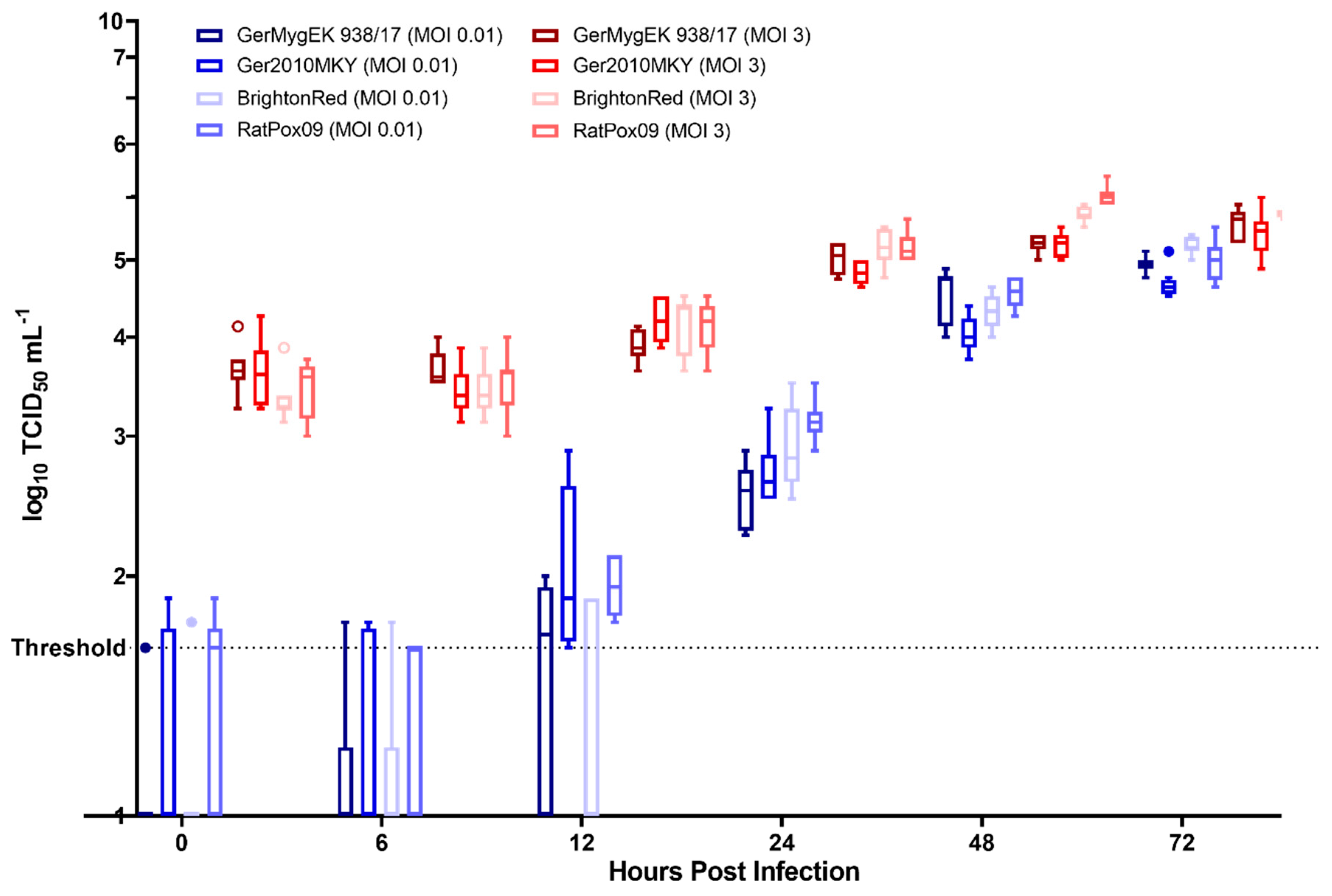
3.1. In Vitro Characterization: Virus Growth Kinetics
3.2. In Vivo Characterization: Animal Experiments and Sample Analyses
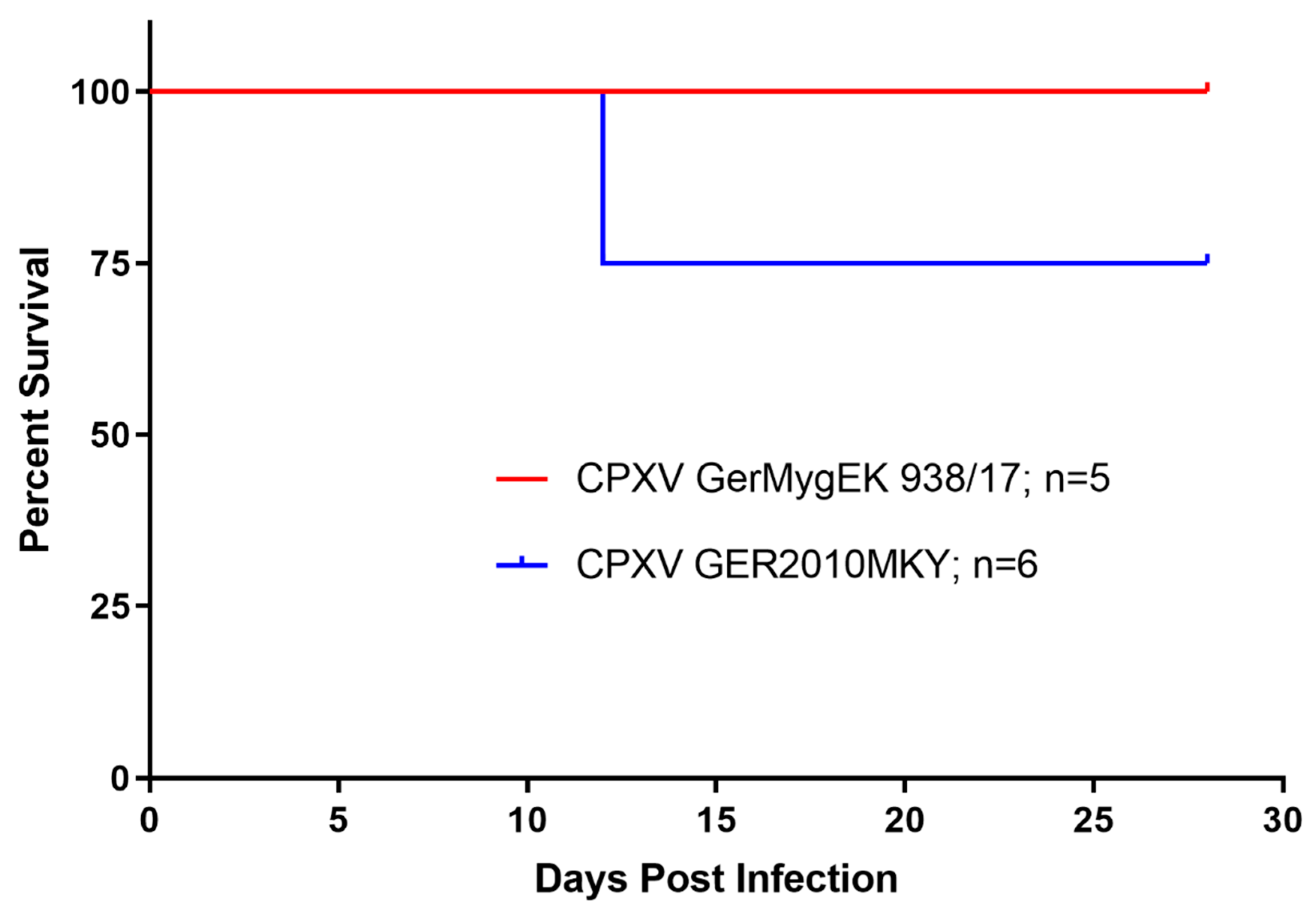
3.2.1. Morbidity and Mortality: Weight Loss in Bank Voles in the CPXV Ger2010MKY Group
3.2.2. Virus Shedding: Infected Bank Voles Excrete CPXV
3.2.3. Viral Load in Organ Samples: CPXV Positive Turbinate Sample of a Bank Vole at 28 dpi
3.2.4. Serology: CPXV-Specific Seroconversion in Bank Voles with High Titers
4. Discussion
Author Contributions
Funding
Acknowledgments
Conflicts of Interest
References
- Essbauer, S.; Pfeffer, M.; Meyer, H. Zoonotic poxviruses. Vet. Microbiol. 2010, 140, 229–236. [Google Scholar] [CrossRef] [PubMed]
- Vorou, R.M.; Papavassiliou, V.G.; Pierroutsakos, I.N. Cowpox virus infection: an emerging health threat. Curr. Opin. Infect. Dis. 2008, 21, 153–156. [Google Scholar] [CrossRef] [PubMed]
- Switaj, K.; Kajfasz, P.; Kurth, A.; Nitsche, A. Cowpox after a cat scratch—Case report from Poland. Ann. Agric. Environ. Med. 2015, 22, 456–458. [Google Scholar] [CrossRef] [PubMed]
- McInerney, J.; Papasouliotis, K.; Simpson, K.; English, K.; Cook, S.; Milne, E.; Gunn-Moore, D.A. Pulmonary cowpox in cats: five cases. J. Feline Med. Surg. 2016, 18, 518–525. [Google Scholar] [CrossRef] [Green Version]
- Becker, C.; Kurth, A.; Hessler, F.; Kramp, H.; Gokel, M.; Hoffmann, R.; Kuczka, A.; Nitsche, A. Cowpox virus infection in pet rat owners: not always immediately recognized. Dtsch. Arztebl. Int. 2009, 106, 329–334. [Google Scholar] [CrossRef]
- Elsendoorn, A.; Agius, G.; Le Moal, G.; Aajaji, F.; Favier, A.L.; Wierzbicka-Hainault, E.; Beraud, G.; Flusin, O.; Crance, J.M.; Roblot, F. Severe ear chondritis due to cowpox virus transmitted by a pet rat. J. Infect. 2011, 63, 391–393. [Google Scholar] [CrossRef]
- Vogel, S.; Sardy, M.; Glos, K.; Korting, H.C.; Ruzicka, T.; Wollenberg, A. The Munich outbreak of cutaneous cowpox infection: transmission by infected pet rats. Acta Derm. Venereol. 2012, 92, 126–131. [Google Scholar] [CrossRef] [Green Version]
- Prkno, A.; Hoffmann, D.; Goerigk, D.; Kaiser, M.; van Maanen, A.C.F.; Jeske, K.; Jenckel, M.; Pfaff, F.; Vahlenkamp, T.W.; Beer, M.; et al. Epidemiological Investigations of Four Cowpox Virus Outbreaks in Alpaca Herds, Germany. Viruses 2017, 9, 344. [Google Scholar] [CrossRef] [Green Version]
- Kurth, A.; Wibbelt, G.; Gerber, H.P.; Petschaelis, A.; Pauli, G.; Nitsche, A. Rat-to-elephant-to-human transmission of cowpox virus. Emerg. Infect. Dis. 2008, 14, 670–671. [Google Scholar] [CrossRef]
- Kalthoff, D.; Bock, W.I.; Huhn, F.; Beer, M.; Hoffmann, B. Fatal cowpox virus infection in cotton-top tamarins (Saguinus oedipus) in Germany. Vector Borne Zoonotic Dis. 2014, 14, 303–305. [Google Scholar] [CrossRef]
- Matz-Rensing, K.; Ellerbrok, H.; Ehlers, B.; Pauli, G.; Floto, A.; Alex, M.; Czerny, C.P.; Kaup, F.J. Fatal poxvirus outbreak in a colony of New World monkeys. Vet. Pathol. 2006, 43, 212–218. [Google Scholar] [CrossRef] [PubMed]
- Ashford, R.W. When is a reservoir not a reservoir? Emerg. Infect. Dis. 2003, 9, 1495–1496. [Google Scholar] [CrossRef] [PubMed] [Green Version]
- Popova, A.Y.; Maksyutov, R.A.; Taranov, O.S.; Tregubchak, T.V.; Zaikovskaya, A.V.; Sergeev, A.A.; Vlashchenko, I.V.; Bodnev, S.A.; Ternovoi, V.A.; Alexandrova, N.S.; et al. Cowpox in a human, Russia, 2015. Epidemiol. Infect. 2017, 145, 755–759. [Google Scholar] [CrossRef] [PubMed] [Green Version]
- Fassbender, P.; Zange, S.; Ibrahim, S.; Zoeller, G.; Herbstreit, F.; Meyer, H. Generalized Cowpox Virus Infection in a Patient with HIV, Germany, 2012. Emerg. Infect. Dis. 2016, 22, 553–555. [Google Scholar] [CrossRef] [Green Version]
- Kinnunen, P.M.; Holopainen, J.M.; Hemmila, H.; Piiparinen, H.; Sironen, T.; Kivela, T.; Virtanen, J.; Niemimaa, J.; Nikkari, S.; Jarvinen, A.; et al. Severe Ocular Cowpox in a Human, Finland. Emerg. Infect. Dis. 2015, 21, 2261–2263. [Google Scholar] [CrossRef] [Green Version]
- Cohen, J. Bioterrorism: Smallpox vaccinations: How much protection remains? Science 2001, 294, 985. [Google Scholar] [CrossRef]
- Mortimer, P.P. The new cell culture smallpox vaccine should not be offered to the general population. Rev. Med. Virol. 2003, 13, 17–20. [Google Scholar] [CrossRef]
- Shchelkunov, S.N. An increasing danger of zoonotic orthopoxvirus infections. PLoS Pathog. 2013, 9, e1003756. [Google Scholar] [CrossRef] [Green Version]
- Dictionary, O.E. Art, n.1; Oxford University Press: Oxford, UK, 2019. [Google Scholar]
- Haydon, D.T.; Cleaveland, S.; Taylor, L.H.; Laurenson, M.K. Identifying reservoirs of infection: a conceptual and practical challenge. Emerg. Infect. Dis. 2002, 8, 1468–1473. [Google Scholar] [CrossRef]
- Marennikova, S.S.; Shelukhina, E.M.; Efremova, E.V. New outlook on the biology of cowpox virus. Acta Virol. 1984, 28, 437–444. [Google Scholar]
- Tsanava, S.A.; Marennikova, S.S.; Sakvarelidze, L.A.; Shelukhina, E.M.; Ianova, N.N. Isolation of cowpox virus from the red-tailed jird. Vopr. Virusol. 1989, 34, 95–97. [Google Scholar] [PubMed]
- Bennett, M.; Crouch, A.J.; Begon, M.; Duffy, B.; Feore, S.; Gaskell, R.M.; Kelly, D.F.; McCracken, C.M.; Vicary, L.; Baxby, D. Cowpox in British voles and mice. J. Comp. Pathol. 1997, 116, 35–44. [Google Scholar] [CrossRef]
- Feore, S.M.; Bennett, M.; Chantrey, J.; Jones, T.; Baxby, D.; Begon, M. The effect of cowpox virus infection on fecundity in bank voles and wood mice. Proc. Biol. Sci. 1997, 264, 1457–1461. [Google Scholar] [CrossRef] [PubMed]
- Kinnunen, P.M.; Henttonen, H.; Hoffmann, B.; Kallio, E.R.; Korthase, C.; Laakkonen, J.; Niemimaa, J.; Palva, A.; Schlegel, M.; Ali, H.S.; et al. Orthopox virus infections in Eurasian wild rodents. Vector Borne Zoonotic Dis. 2011, 11, 1133–1140. [Google Scholar] [CrossRef]
- Fischer, S.; Franke, A.; Imholt, C.; Gethmann, J.; Spierling, N.G.; Jacob, J.; Beer, M.; Hoffmann, D.; Ulrich, R.G. Patchy Occurrence of Cowpox Virus in Voles from Germany. Vector Borne Zoonotic Dis. 2020. [Google Scholar] [CrossRef]
- Hoffmann, D.; Franke, A.; Jenckel, M.; Tamosiunaite, A.; Schluckebier, J.; Granzow, H.; Hoffmann, B.; Fischer, S.; Ulrich, R.G.; Höper, D.; et al. Out of the Reservoir: Phenotypic and Genotypic Characterization of a Novel Cowpox Virus Isolated from a Common Vole. J. Virol. 2015, 89, 10959–10969. [Google Scholar] [CrossRef] [Green Version]
- Franke, A.; Pfaff, F.; Jenckel, M.; Hoffmann, B.; Höper, D.; Antwerpen, M.; Meyer, H.; Beer, M.; Hoffmann, D. Classification of Cowpox Viruses into Several Distinct Clades and Identification of a Novel Lineage. Viruses 2017, 9, 142. [Google Scholar] [CrossRef] [Green Version]
- Breithaupt, A.; Kalthoff, D.; Deutskens, F.; König, P.; Hoffmann, B.; Beer, M.; Meyer, H.; Teifke, J.P. Clinical course and pathology in rats (Rattus norvegicus) after experimental cowpox virus infection by percutaneous and intranasal application. Vet. Pathol. 2012, 49, 941–949. [Google Scholar] [CrossRef]
- Kalthoff, D.; König, P.; Meyer, H.; Beer, M.; Hoffmann, B. Experimental cowpox virus infection in rats. Vet. Microbiol. 2011, 153, 382–386. [Google Scholar] [CrossRef]
- Franke, A.; Ulrich, R.G.; Weber, S.; Osterrieder, N.; Keller, M.; Hoffmann, D.; Beer, M. Experimental Cowpox Virus (CPXV) Infections of Bank Voles: Exceptional Clinical Resistance and Variable Reservoir Competence. Viruses 2017, 9, 391. [Google Scholar] [CrossRef] [Green Version]
- Jeske, K.; Weber, S.; Pfaff, F.; Imholt, C.; Jacob, J.; Beer, M.; Ulrich, R.G.; Hoffmann, D. Molecular Detection and Characterization of the First Cowpox Virus Isolate Derived from a Bank Vole. Viruses 2019, 11, 1075. [Google Scholar] [CrossRef] [PubMed] [Green Version]
- Essbauer, S.; Meyer, H. Genus Orthopoxvirus: Cowpox virus. In Poxviruses; Mercer, A.A., Schmidt, A., Weber, O., Eds.; Birkhäuser Basel: Basel, Switzerland, 2007; pp. 75–88. [Google Scholar]
- Xu, Z.; Zikos, D.; Tamosiunaite, A.; Klopfleisch, R.; Osterrieder, N.; Tischer, B.K. Identification of 10 cowpox virus proteins that are necessary for induction of hemorrhagic lesions (red pocks) on chorioallantoic membranes. J. Virol. 2014, 88, 8615–8628. [Google Scholar] [CrossRef] [PubMed] [Green Version]
- Kärber, G. Beitrag zur kollektiven Behandlung pharmakologischer Reihenversuche. Naunyn Schmiedebergs Arch. Exp. Pathol. Pharmakol. 1931, 162, 480–483. [Google Scholar] [CrossRef]
- Spearman, C. The method of ‘right and wrong cases’(‘constant stimuli’) without Gauss’s formulae. Br. J. Psychol. 1908, 2, 227–242. [Google Scholar] [CrossRef]
- Scaramozzino, N.; Ferrier-Rembert, A.; Favier, A.L.; Rothlisberger, C.; Richard, S.; Crance, J.M.; Meyer, H.; Garin, D. Real-time PCR to identify variola virus or other human pathogenic orthopox viruses. Clin. Chem. 2007, 53, 606–613. [Google Scholar] [CrossRef]
- Chantrey, J.; Meyer, H.; Baxby, D.; Begon, M.; Bown, K.J.; Hazel, S.M.; Jones, T.; Montgomery, W.I.; Bennett, M. Cowpox: reservoir hosts and geographic range. Epidemiol. Infect. 1999, 122, 455–460. [Google Scholar] [CrossRef] [Green Version]
- Tamosiunaite, A.; Hoffmann, D.; Franke, A.; Schluckebier, J.; Tauscher, K.; Tischer, B.K.; Beer, M.; Klopfleisch, R.; Osterrieder, N. Histopathological and Immunohistochemical Studies of Cowpox Virus Replication in a Three-Dimensional Skin Model. J. Comp. Pathol. 2016, 155, 55–61. [Google Scholar] [CrossRef]
- Tikhonova, G.N.; Tikhonov, I.A.; Osipova, O.V.J.B.B. Some behavioral characteristics of common voles Microtus arvalis arvalis and Microtus arvalis obscurus in family groups under experimental conditions. Boil. Bull. 2008, 35, 482–488. [Google Scholar] [CrossRef]
- Magwire, M.M.; Fabian, D.K.; Schweyen, H.; Cao, C.; Longdon, B.; Bayer, F.; Jiggins, F.M. Genome-wide association studies reveal a simple genetic basis of resistance to naturally coevolving viruses in Drosophila melanogaster. PLoS Genet. 2012, 8, e1003057. [Google Scholar] [CrossRef]
- van Sluijs, L.; Pijlman, G.P.; Kammenga, J.E. Why do Individuals Differ in Viral Susceptibility? A Story Told by Model Organisms. Viruses 2017, 9, 284. [Google Scholar] [CrossRef] [Green Version]
- Liem, J.; Liu, J. Stress Beyond Translation: Poxviruses and More. Viruses 2016, 8, 169. [Google Scholar] [CrossRef] [PubMed] [Green Version]
- Szulc-Dabrowska, L.; Struzik, J.; Cymerys, J.; Winnicka, A.; Nowak, Z.; Toka, F.N.; Gierynska, M. The in Vitro Inhibitory Effect of Ectromelia Virus Infection on Innate and Adaptive Immune Properties of GM-CSF-Derived Bone Marrow Cells Is Mouse Strain-Independent. Front. Microbiol. 2017, 8, 2539. [Google Scholar] [CrossRef] [PubMed] [Green Version]
- Buller, R.M.; Bhatt, P.N.; Wallace, G.D. Evaluation of an enzyme-linked immunosorbent assay for the detection of ectromelia (mousepox) antibody. J. Clin. Microbiol. 1983, 18, 1220–1225. [Google Scholar] [CrossRef] [PubMed] [Green Version]
- Reames, H.R. Pathogenesis and Immunity in Ectromelia Virus Infection of the Nasal Mucosa of the Rat. J. Infect. Dis. 1940, 66, 254–262. [Google Scholar] [CrossRef]
- Tamosiunaite, A.; Weber, S.; Schippers, T.; Franke, A.; Xu, Z.; Jenckel, M.; Pfaff, F.; Hoffmann, D.; Newell, M.; Tischer, B.K.; et al. What a difference a gene makes - identification of virulence factors of Cowpox virus. J. Virol. 2019, 94. [Google Scholar] [CrossRef] [PubMed]
- Carroll, D.S.; Emerson, G.L.; Li, Y.; Sammons, S.; Olson, V.; Frace, M.; Nakazawa, Y.; Czerny, C.P.; Tryland, M.; Kolodziejek, J.; et al. Chasing Jenner’s vaccine: revisiting cowpox virus classification. PLoS ONE 2011, 6, e23086. [Google Scholar] [CrossRef] [PubMed] [Green Version]
- Mauldin, M.R.; Antwerpen, M.; Emerson, G.L.; Li, Y.; Zoeller, G.; Carroll, D.S.; Meyer, H. Cowpox virus: What’s in a Name? Viruses 2017, 9, 101. [Google Scholar] [CrossRef]


| Cowpox Virus (CPXV) | Animals | Experiment Design | |||||||
|---|---|---|---|---|---|---|---|---|---|
| Phylogenetic Lineage | Strain | Origin | Species | Number of Animals per Group | Application Route | Dose of Inoculum/ Animal | Duration of the Experiment | Nasal/buccal Swabs | Study |
| CPXV-like 3 | GerMygEK 938/17 | Bank vole | Bank vole | 5* | oronasal | 105.5 TCID50 | 28 dpi | Every 2nd day | This study |
| Common vole | 6 | oronasal | 105.5 TCID50 | 28 dpi | Every 2nd day | This study | |||
| Wistar rat | 10 | oronasal | 105.5 TCID50 | 28 dpi | Every 2nd day | This study | |||
| CPXV-like 3 | Ger2010MKY | Cotton-top tamarin | Bank vole | 6 | oronasal | 105.5 TCID50 | 28 dpi | Every 2nd day | This study |
| Common vole | 6 | oronasal | 105.5 TCID50 | 28 dpi | Every 2nd day | This study | |||
| Wistar rat | 4 | oronasal | 104/106 TCID50 | 28 dpi | Every 2nd day | [10] | |||
| CPXV Strain | Species | Dissection | Tissue | ||||||
|---|---|---|---|---|---|---|---|---|---|
| Turbinate | Trachea | Lung | Liver | Spleen | Skin | ||||
| GerMygEK 938/17 | Bank vole | 28 dpi | 0/5* | 0/5 | 0/5 | 0/5 | 0/5 | 0/5 | |
| Common vole | 28 dpi | 0/6 | 0/6 | 0/6 | 0/6 | 0/6 | 0/6 | ||
| Wistar rat | 28 dpi | 0/10 | 0/10 | 0/10 | 0/10 | 0/10 | 0/10 | ||
| Ger2010MKY | Bank vole | 12 dpi# | 2/2 | 2/2 | 1/2 | 0/2 | 0/2 | 2/2 | |
| Cq value (TCID50 mL−1) | 24.8 (103.25) 26.3 (103.25) | 34.2 36.1 | 32.2 | 35.1 36 | |||||
| 28 dpi | 1/4 | 0/4 | 0/4 | 0/4 | 0/4 | 0/4 | |||
| Cq value (TCID50 mL−1) | 22.9 (103.75) | ||||||||
| Common vole | 28 dpi | 0/6 | 0/6 | 0/6 | 0/6 | 0/6 | 0/6 | ||
| CPXV Strain | Species | Number of Animals per Group | Number of Serum/Lavage Samples | Dilution | |||||||
|---|---|---|---|---|---|---|---|---|---|---|---|
| 1:20 | 1:40 | 1:80 | 1:160 | 1:320 | 1:640 | 1:1280 | 1:2560 | ||||
| seroreactive | seropositive | ||||||||||
| GerMygEK 938/17 | Bank vole | 5* | Serum 4 Lavage 5 | 4/4 1/5# | 4/4 0/5# | 4/4 | 4/4 | 3/4 | 2/4 | 0/4 | 0/4 |
| Common vole | 6 | Serum 6 Lavage 6 | 0/6 0/6 | 0/6 0/6 | |||||||
| Wistar rat | 10 | Serum 10 | 10/10 | 10/10 | 10/10 | 10/10 | 10/10 | 10/10 | 6/10 | 0/10 | |
| Ger2010MKY | Bank vole | 6 | Serum 4 Lavage 6 | 4/4 5/6## | 4/4 2/6## | 4/4 | 4/4 | 4/4 | 2/4 | 1/4 | 0/4 |
| Common vole | 6 | Serum 6 Lavage 6 | 0/60/6 | 0/6 0/6 | |||||||
| Cowpox Virus (CPXV) | Animals Inoculated | Results of Animal Experiments | Study | |||||||
|---|---|---|---|---|---|---|---|---|---|---|
| Phylogenetic Lineage | Strain / Origin | Species | Dose of Inoculum/ Animal | Clinical Signs | Virus Shedding | Viral DNA in Organs (28 dpi) | Seroconversion | Mortality | ||
| Common vole reservoir | CPXV-like 2 | FM2292 / Common vole | Common vole | 104 TCID50 | none | 2/3 | none | 66% | 0% | [27] |
| 106 TCID50 | respiratory | 3/3 | 1/3 (nasal septum) | 100% | 33% | [27] | ||||
| Bank vole | 105 TCID50 | none | none | none | 60% | 0% | [31] | |||
| Wistar rat | 104 TCID50 | Respiratory Pox-like lesions | 4/4 | none | 100% | 0% | [27] | |||
| 106 TCID50 | Respiratory Pox-like lesions | 3/3 | none | 100% | 0% | [27] | ||||
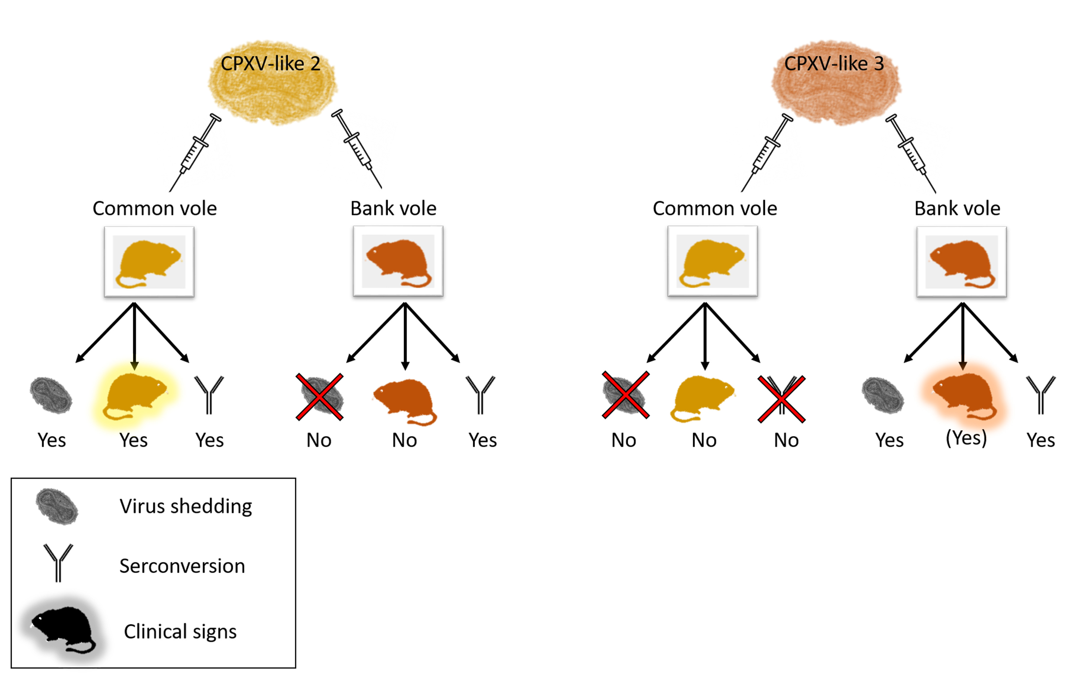
| Bank vole reservoir | CPXV-like 3 | GerMygEK 938/17 / Bank vole | Bank vole | 105.5 TCID50 | none | 5/5 | none | 100% | 0% | This study |
| Common vole | 105.5 TCID50 | none | none | none | 0% | 0% | This study | |||
| Wistar rat | 105.5 TCID50 | none | none | none | 100% | 0% | This study | |||
| Ger2010MKY / Cotton-top tamarin | Bank vole | 105.5 TCID50 | Weight loss up to 25% | 6/6 | 1/6 (nasal septum) | 83% | 33% | This study | ||
| Common vole | 105.5 TCID50 | none | none | none | 0% | 0% | This study | |||
| Wistar rat | 104 TCID50 | none | none | none | 25% | 0% | [10] | |||
| 106 TCID50 | none | none | none | 25% | 0% | [10] | ||||
© 2020 by the authors. Licensee MDPI, Basel, Switzerland. This article is an open access article distributed under the terms and conditions of the Creative Commons Attribution (CC BY) license (http://creativecommons.org/licenses/by/4.0/).
Share and Cite
Weber, S.; Jeske, K.; Ulrich, R.G.; Imholt, C.; Jacob, J.; Beer, M.; Hoffmann, D. In Vivo Characterization of a Bank Vole-Derived Cowpox Virus Isolate in Natural Hosts and the Rat Model. Viruses 2020, 12, 237. https://0-doi-org.brum.beds.ac.uk/10.3390/v12020237
Weber S, Jeske K, Ulrich RG, Imholt C, Jacob J, Beer M, Hoffmann D. In Vivo Characterization of a Bank Vole-Derived Cowpox Virus Isolate in Natural Hosts and the Rat Model. Viruses. 2020; 12(2):237. https://0-doi-org.brum.beds.ac.uk/10.3390/v12020237
Chicago/Turabian StyleWeber, Saskia, Kathrin Jeske, Rainer G. Ulrich, Christian Imholt, Jens Jacob, Martin Beer, and Donata Hoffmann. 2020. "In Vivo Characterization of a Bank Vole-Derived Cowpox Virus Isolate in Natural Hosts and the Rat Model" Viruses 12, no. 2: 237. https://0-doi-org.brum.beds.ac.uk/10.3390/v12020237