Baseline Amino Acid Substitutions in the NS5A ISDR and PKR Binding Domain of Hepatitis C and Different Fibrosis Levels and Levels of Development of Hepatocellular Carcinoma in Patients Treated with DAAs

 , and
, and
Abstract
:1. Introduction
2. Materials and Methods
2.1. Patient and Clinical Characteristics
2.2. HCV-RNA Quantification and Sequencing
2.3. Statistical Analysis
3. Results
Sequence Analysis of Other Viral Regions
4. Discussion
Author Contributions
Funding
Acknowledgments
Conflicts of Interest
References
- Shiratori, Y.; Imazeki, F.; Moriyama, M.; Yano, M.; Arakawa, Y.; Yokosuka, O.; Kuroki, T.; Nishiguchi, S.; Sata, M.; Yamada, G.; et al. Histologic improvement of fibrosis in patients with hepatitis C who have sustained response to interferon therapy. Ann. Intern. Med. 2000, 132, 517–524. [Google Scholar] [CrossRef]
- Persico, M.; Aglitti, A.; Milella, M.; Coppola, C.; Messina, V.; Claar, E.; Gentile, I.; Sogari, F.; Pierri, P.; Surace, L.A.; et al. Real-life glecaprevir/pibrentasvir in a large cohort of patients with hepatitis C virus infection: The MISTRAL Study. Liver Int. 2019, 39, 1852–1859. [Google Scholar] [CrossRef]
- Toyoda, H.; Tada, T.; Yasuda, S.; Mizuno, K.; Ito, T.; Kumada, T. Dynamic Evaluation of Liver Fibrosis to Assess the Risk of Hepatocellular Carcinoma in Patients with Chronic Hepatitis C Who Achieved Sustained Virologic Response. Clin. Infect. Dis. 2019. [Google Scholar] [CrossRef] [PubMed]
- Ogawa, E.; Furusyo, N.; Kajiwara, E.; Takahashi, K.; Nomura, H.; Maruyama, T.; Tanabe, Y.; Satoh, T.; Nakamuta, M.; Kotoh, K.; et al. Efficacy of pegylated interferon alpha-2b and ribavirin treatment on the risk of hepatocellular carcinoma in patients with chronic hepatitis C: A prospective, multicenter study. J. Hepatol. 2013, 58, 495–501. [Google Scholar] [CrossRef] [PubMed]
- Van der Meer, A.J.; Wedemeyer, H.; Feld, J.J.; Hansen, B.E.; Manns, M.P.; Zeuzem, S.; Janssen, H.L. Is there sufficient evidence to recommend antiviral therapy in hepatitis C? J. Hepatol. 2014, 60, 191–196. [Google Scholar] [CrossRef] [Green Version]
- Dandachi, D.; Hassan, M.; Kaseb, A.; Angelidakis, G.; Torres, H.A. Hepatitis C virus-associated hepatocellular carcinoma as a second primary malignancy: Exposing an overlooked presentation of liver cancer. J. Hepatocell. Carcinoma 2018, 5, 81–86. [Google Scholar] [CrossRef] [PubMed] [Green Version]
- Aleman, S.; Rahbin, N.; Weiland, O.; Davidsdottir, L.; Hedenstierna, M.; Rose, N.; Verbaan, H.; Stål, P.; Carlsson, T.; Norrgren, H.; et al. A risk for hepatocellular carcinoma persists long-term after sustained virologic response in patients with hepatitis C-associated liver cirrhosis. Clin. Infect. Dis. 2013, 57, 230–236. [Google Scholar] [CrossRef]
- Ioannou, G.N.; Green, P.K.; Beste, L.A.; Mun, E.J.; Kerr, K.F.; Berry, K. Development of models estimating the risk of hepatocellular carcinoma after antiviral treatment for hepatitis C. J. Hepatol. 2018, 69, 1088–1098. [Google Scholar] [CrossRef]
- Toyoda, H.; Kumada, T.; Tada, T.; Kiriyama, S.; Tanikawa, M.; Hisanaga, Y.; Kanamori, A.; Kitabatake, S.; Ito, T. Risk factors of hepatocellular carcinoma development in noncirrhotic patients with sustained virologic response for chronic hepatitis C virus infection. J. Gastroenterol. Hepatol. 2015, 30, 1183–1189. [Google Scholar] [CrossRef]
- Yasuda, S.; Ishigami, M.; Ishizu, Y.; Kuzuya, T.; Honda, T.; Hayashi, K.; Toyoda, H.; Kumada, T.; Hirooka, Y.; Goto, H. Substitutions in interferon sensitivity-determining region and hepatocarcinogenesis after hepatitis C virus eradication. J. Gastroenterol. Hepatol. 2018, 33, 1904–1911. [Google Scholar] [CrossRef]
- Patiño-Galindo, J.Á.; González-Candelas, F. Comparative analysis of variation and selection in the HCV genome. Infect. Genet. Evol. 2017, 49, 104–110. [Google Scholar] [CrossRef] [Green Version]
- Cuevas, J.M.; Gonzalez, M.; Torres-Puente, M.; Jiménez-Hernández, N.; Bracho, M.A.; García-Robles, I.; González-Candelas, F.; Moya, A. The role of positive selection in hepatitis C virus. Infect. Genet. Evol. 2009, 9, 860–866. [Google Scholar] [CrossRef] [PubMed]
- Muñoz de Rueda, P.; Fuentes Rodríguez, J.M.; Quiles Pérez, R.; Gila Medina, A.; Martín Álvarez, A.B.; Casado Ruíz, J.; Ruíz Extremera, A.; Salmerón, J. Hepatitis C virus NS5A region mutation in chronic hepatitis C genotype 1 patients who are nonresponders to two or more treatments and its relationship with response to a new treatment. World J. Gastroenterol. 2017, 23, 4538–4547. [Google Scholar] [CrossRef] [PubMed]
- Polyak, S.J.; Paschal, D.M.; McArdle, S.; Gale, M.J., Jr.; Moradpour, D.; Gretch, D.R. Characterization of the effects of hepatitis C virus nonstructural 5A protein expression in human cell lines and on interferon-sensitive virus replication. Hepatology 1999, 29, 1262–1271. [Google Scholar] [CrossRef] [PubMed]
- Hung, C.H.; Chen, C.H.; Lee, C.M.; Wu, C.M.; Hu, T.H.; Wang, J.H.; Yen, Y.H.; Lu, S.N. Association of amino acid variations in the NS5A and E2-PePHD region of hepatitis C virus 1b with hepatocellular carcinoma. J. Viral. Hepatol. 2008, 15, 58–65. [Google Scholar] [CrossRef] [PubMed]
- Gale, M., Jr.; Blakely, C.M.; Kwieciszewski, B.; Tan, S.L.; Dossett, M.; Tang, N.M.; Korth, M.J.; Polyak, S.J.; Gretch, D.R.; Katze, M.G. Control of PKR protein kinase by hepatitis C virus nonstructural 5A protein: Molecular mechanisms of kinase regulation. Mol. Cell. Biol. 1998, 18, 5208–5218. [Google Scholar] [CrossRef] [Green Version]
- Paolucci, S.; Fiorina, L.; Piralla, A.; Gulminetti, R.; Novati, S.; Barbarini, G.; Sacchi, P.; Gatti, M.; Dossena, L.; Baldanti, F.M. Naturally occurring mutations to HCV protease inhibitors in treatment-naïve patients. Virol. J. 2012, 9, 245. [Google Scholar] [CrossRef] [Green Version]
- Paolucci, S.; Premoli, M.; Novati, S.; Gulminetti, R.; Maserati, R.; Barbarini, G.; Sacchi, P.; Piralla, A.; Sassera, D.; De Marco, L.; et al. Baseline and Breakthrough Resistance Mutations in HCV Patients Failing DAAs. Sci. Rep. 2017, 7, 16017. [Google Scholar] [CrossRef] [Green Version]
- Sarrazin, C. The importance of resistance to direct antiviral drugs in HCV infection in clinical practice. J. Hepatol. 2016, 64, 486–504. [Google Scholar] [CrossRef]
- Pawlotsky, J.M. Hepatitis C Virus Resistance to Direct-Acting Antiviral Drugs in Interferon-Free Regimens. Gastroenterology 2016, 151, 70–86. [Google Scholar] [CrossRef] [Green Version]
- Sorbo, M.C.; Cento, V.; Di Maio, V.C.; Howe, A.Y.M.; Garcia, F.; Perno, C.F.; Ceccherini-Silberstein, F. Hepatitis C virus drug resistance associated substitutions and their clinical relevance: Update2018. Drug Resist. Update 2018, 37, 17–39. [Google Scholar] [CrossRef] [PubMed]
- Galati, G.; Muley, M.; Viganò, M.; Iavarone, M.; Vitale, A.; Dell’Unto, C.; Lai, Q.; Cabibbo, G.; Sacco, R.; Villa, E.; et al. Occurrence of hepatocellular carcinoma after direct-acting antiviral therapy for hepatitis C virus infection: Literature review and risk analysis. Expert. Opin. Drug Saf. 2019, 8, 1–8. [Google Scholar] [CrossRef] [PubMed]
- Zou, W.Y.; Choi, K.; Kramer, J.R.; Yu, X.; Cao, Y.; El-Serag, H.B.; Kanwal, F. Risk of Hepatocellular Cancer Recurrence in Hepatitis C Virus+ Patients Treated with Direct-Acting. Antiviral Agents. Dig. Dis. Sci. 2019, 64, 3328–3336. [Google Scholar] [CrossRef] [PubMed]
- Giménez-Barcons, M.; Franco, S.; Suárez, Y.; Forns, X.; Ampurdanès, S.; Puig-Basagoiti, F.; Barrera, J.M.; Llovet, J.M.; Bruix, J.; Sánchez-Tapias, J.M.; et al. High amino acid variability within the NS5A of hepatitis C virus (HCV) is associated with hepatocellular carcinoma in patients with HCV-1b-related cirrhosis. Hepatology 2001, 34, 158–167. [Google Scholar] [CrossRef]
- Petsaris, O.; Vallet, S.; Le Guillou-Guillemette, H.; Veillon, P.; Gouriou, S.; Barbier, G.; Nousbaum, J.B.; Saliou, P.; NKontchou, G.; Trinchet, J.C.; et al. Duplication of the V3 domain in hepatitis C virus (1b) NS5A protein: Clonal analysis and physicochemical properties related to hepatocellular carcinoma occurrence. J. Clin. Virol. 2016, 74, 19–25. [Google Scholar] [CrossRef]
- Goh, K.C.; deVeer, M.J.; Williams, B.R. The protein kinase PKR is required for p38 MAPK activation and the innate immune response to bacterial endotoxin. EMBO J. 2000, 19, 4292–4297. [Google Scholar]
- Deb, A.; Haque, S.J.; Mogensen, T.; Silverman, R.H.; Williams, B.R. RNA-dependent protein kinase PKR is required for activation of NF-kappa B by IFN-gamma in a STAT1-independent pathway. J. Immunol. 2001, 166, 6170–6180. [Google Scholar] [CrossRef] [Green Version]
- Watanabe, T.; Hiasa, Y.; Tokumoto, Y.; Hirooka, M.; Abe, M.; Ikeda, Y.; Matsuura, B.; Chung, R.T.; Onji, M. Protein kinase R modulates c-Fos and c-Jun signaling to promote proliferation of hepatocellular carcinoma with hepatitis C virus infection. PLoS ONE 2013, 8, e67750. [Google Scholar] [CrossRef] [Green Version]
- Liu, S.; Ansari, I.H.; Das, S.C.; Pattnaik, A.K. Insertion and deletion analyses identify regions of nonstructural protein 5A of Hepatitis C virus that are dispensable for viral genome replication. J. Gen. Virol. 2006, 87, 323–327. [Google Scholar] [CrossRef]
- Sugiyama, R.; Murayama, A.; Nitta, S.; Yamada, N.; Tasaka-Fujita, M.; Masaki, T.; Aly, H.H.; Shiina, M.; Ryo, A.; Ishii, K.; et al. Interferon sensitivity-determining region of hepatitis C virus influences virus production and interferon signaling. Oncotarget 2017, 9, 5627–5640. [Google Scholar] [CrossRef] [Green Version]
- Raimondi, S.; Bruno, S.; Mondelli, M.U.; Maisonneuve, P. Hepatitis C virus genotype 1b as a risk factor for hepatocellular carcinoma development: A meta-analysis. J. Hepatol. 2009, 50, 1142–1154. [Google Scholar] [CrossRef] [PubMed]


| Univariate | Multivariate | |||||
|---|---|---|---|---|---|---|
| F0–F2 (180) | F3–F4 (136) | OR (95%IC) | p Value | OR (95%IC) | P Value | |
| Age in years (SD) | 62.24 (±13.0) | 63.69 (±14.0) | 1.0 (0.9–1.1) | 0.34 | ||
| Gender (male) (%) | 86 (47.7) | 83 (61.0) | 1.7 (1.1–2.7) | 0.019 | ||
| ALT (IU/L) (IQR) | 30 (20–46.5) | 62.5 (39.5–100) | 1.0 (0.9–1.0) | 0.16 | ||
| AST (IU/L) (IQR) | 28 (23–40) | 58.5 (34.5–96.5) | 1.0 (1.0–1.0) | <0.001 | ||
| PLT (104/mm3) (SD) | 199.78 (±72.9) | 160.25 (±59.6) | 0.9 (0.9–0.9) | <0.001 | 0.99 (0.98–0.99) | <0.001 |
| γGTP (IU/L) (IQR) | 28.5 (18–46) | 65 (34–105) | 1.0 (1.0–1.0) | <0.001 | 1.01 (1.00–1,01) | 0.002 |
| Log10 HCV load (UI/mL) (SD) | 5.71 (±0.89) | 5.4 (±0.95) | 0.7 (0.5–0.8) | 0.003 | 0.65 (0.49–0.85) | 0.002 |
| Genotype 1a (%) | 33 (18.3) | 19 (13.9) | 0.6 (0.3–1.1) | 0.45 | ||
| Genotype 1b (%) | 57 (31.6) | 45 (33.1) | 0.7 (0.3–1.4) | |||
| Genotype 2c (%) | 57 (31.6) | 38 (27.9) | 0.8 (0.4–1.4) | |||
| Genotype 3a (%) | 19 (10.5) | 23 (16.9) | 1.5 (0.7–3.1) | |||
| Genotype 4d (%) | 14 (7.7) | 11 (8.1) | 0.9 (0.4–2.4) | |||
| ISDR ≥ 3 (%) | 36 (20) | 54 (39.7) | 2.6 (1.5–4.3) | <0.001 | 2.14 (1.2–3.7) | 0.007 |
| PKR-bd ≥ 6 (%) | 12 (6.6) | 38 (27.9) | 5.4 (2.7–10.8) | <0.001 | 4.8 (2.2–10.2) | <0.001 |
| Ins/del (%) | 5 (2.7) | 11 (8.0) | 3.1 (1.0–9.1) | 0.04 | ||
| ISDR Mutations | PKR-bd Mutations | |||||
|---|---|---|---|---|---|---|
| <3 (226) | ≥3 (90) | p Value | <6 (235) | ≥6 (81) | p Value | |
| Age in years (SD) | 61.5 (±13) | 66 (±14) | 0.006 | 60.6 (±13) | 69.3 (±13) | <0.001 |
| Gender (male) (%) | 119 (52.7) | 52 (56.5) | 0.70 | 128 (54.5) | 41 (50.6) | 0.61 |
| ALT (IU/L) (IQR) | 39.5 (25–64) | 44.5 (23–87) | 0.31 | 38 (23–64) | 53 (25–98) | 0.01 |
| AST (IU/L) (IQR) | 33 (26–55) | 48.5 (25.3–78.8) | 0.03 | 32 (25–52) | 52 (29–91) | <0.001 |
| PLT (104/mm3) (SD) | 187.4 (±73.1) | 171.2 (±61) | 0.13 | 185.3 (±71.1) | 175.3 (±67.2) | 0.50 |
| γGTP (IU/L) (IQR) | 36.5 (22–63) | 57.5 (24.3–108.3) | 0.01 | 36 (21–64) | 59 (25–100) | 0.003 |
| Log10 HCV load (UI/mL) (SD) | 5.6 (±0.9) | 5.5 (±0.9) | 0.71 | 5.7 (±0.9) | 5.3 (±0.9) | 0.002 |
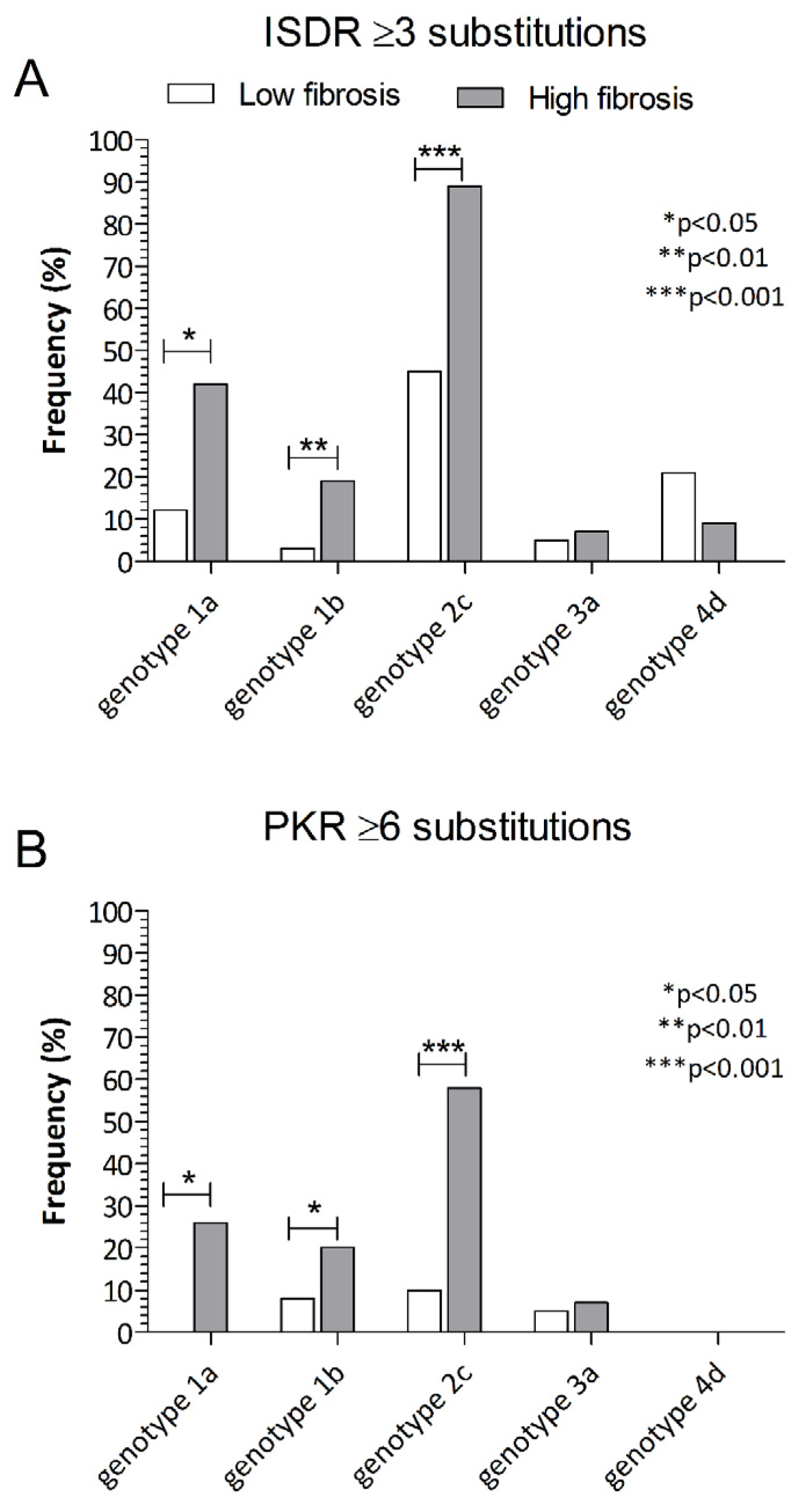
| Genotype 1a (%) | 40 (17.7) | 12 (13.3) | <0.001 | 45 (19.1) | 7 (8.6) | <0.001 |
| Genotype 1b (%) | 91 (40.3) | 11 (12.2) | 74 (31.5) | 28 (34.6) | ||
| Genotype 2c (%) | 35 (15.5) | 60 (66.7) | 53 (22.5) | 42 (51.8) | ||
| Genotype 3a (%) | 39 (17.2) | 3 (3.3) | 39 (16.6) | 3 (3.7) | ||
| Genotype 4d (%) | 21 (9.3) | 4 (4.4) | 24 (10.2) | 1 (1.2) | ||
| Advanced fibrosis (F3-F4) (%) | 82 (36.3) | 54 (60.1) | <0.001 | 82 (34.9) | 54 (66.7) | <0.001 |
| NonHCC | HCC | p Value | |
|---|---|---|---|
| Number of patients | 299 | 17 | |
| Age in years (SD) | 62.5 (±13.2) | 66 (±10) | 0.05 |
| Gender (male) (%) | 158 (52.84) | 11 (64.7) | 0.07 |
| ALT (IU/L) (IQR) | 38 (23–67) | 59 (52–102) | <0.001 |
| AST (IU/L) (IQR) | 34.5 (25–62) | 70 (48–105) | <0.001 |
| PLT (104/mm3) (SD) | 185 (±70) | 136 (±56) | <0.001 |
| γGTP (IU/L) (IQR) | 38 (21–71) | 76 (37–114) | 0.02 |
| Log10 HCV load (UI/mL) (SD) | 5.62 (±0.91) | 4.76 (±0.9) | <0.001 |
| Genotype 1a (%) | 52 (17.4) | 0 (0) | <0.001 |
| Genotype 1b (%) | 94 (31.4) | 8 (47.0) | |
| Genotype 2c (%) | 90 (30.1) | 5 (29.4) | |
| Genotype 3a (%) | 38 (12.7) | 4 (23.5) | |
| Genotype 4d (%) | 25 (8.3) | 0 (0) | |
| ISDR ≥3 (%) | 83 (27.7) | 7 (41.2) | 0.27 |
| PKR-bd ≥6 (%) | 46 (15.4) | 7 (41.2) | 0.01 |
| Ins/del (%) | 16 (4.4) | 0 (0) | 0.37 |
| Advanced fibrosis (F3–F4) (%) | 121 (40.4) | 15 (88.2) | <0.001 |
© 2020 by the authors. Licensee MDPI, Basel, Switzerland. This article is an open access article distributed under the terms and conditions of the Creative Commons Attribution (CC BY) license (http://creativecommons.org/licenses/by/4.0/).
Share and Cite
Paolucci, S.; Piralla, A.; Novazzi, F.; Fratini, A.; Maserati, R.; Gulminetti, R.; Novati, S.; Barbarini, G.; Sacchi, P.; De Silvestri, A.; et al. Baseline Amino Acid Substitutions in the NS5A ISDR and PKR Binding Domain of Hepatitis C and Different Fibrosis Levels and Levels of Development of Hepatocellular Carcinoma in Patients Treated with DAAs. Viruses 2020, 12, 255. https://0-doi-org.brum.beds.ac.uk/10.3390/v12030255
Paolucci S, Piralla A, Novazzi F, Fratini A, Maserati R, Gulminetti R, Novati S, Barbarini G, Sacchi P, De Silvestri A, et al. Baseline Amino Acid Substitutions in the NS5A ISDR and PKR Binding Domain of Hepatitis C and Different Fibrosis Levels and Levels of Development of Hepatocellular Carcinoma in Patients Treated with DAAs. Viruses. 2020; 12(3):255. https://0-doi-org.brum.beds.ac.uk/10.3390/v12030255
Chicago/Turabian StylePaolucci, Stefania, Antonio Piralla, Federica Novazzi, Alice Fratini, Renato Maserati, Roberto Gulminetti, Stefano Novati, Giorgio Barbarini, Paolo Sacchi, Annalisa De Silvestri, and et al. 2020. "Baseline Amino Acid Substitutions in the NS5A ISDR and PKR Binding Domain of Hepatitis C and Different Fibrosis Levels and Levels of Development of Hepatocellular Carcinoma in Patients Treated with DAAs" Viruses 12, no. 3: 255. https://0-doi-org.brum.beds.ac.uk/10.3390/v12030255