Article

Identification of African Swine Fever Virus Transcription within Peripheral Blood Mononuclear Cells of Acutely Infected Pigs

by
Ann Sofie Olesen
1,2,†,
Miyako Kodama
3,†,
Louise Lohse
2,
Francesc Accensi
4,5,
Thomas Bruun Rasmussen
2,
Christina M. Lazov
1,
Morten T. Limborg
3,
M. Thomas P. Gilbert
3,
Anette Bøtner
1,2 and
Graham J. Belsham
1,*
1
Section of Veterinary Clinical Microbiology, Department of Veterinary and Animal Sciences, University of Copenhagen, 1870 Frederiksberg, Denmark
2
Section for Veterinary Virology, Department of Virus & Microbiological Special Diagnostics, Statens Serum Institut, 2300 Copenhagen, Denmark
3
Center for Evolutionary Hologenomics, The Globe Institute, University of Copenhagen, 1350 Copenhagen, Denmark
4
Centre de Recerca en Sanitat Animal (IRTA-CReSA), Campus de la Universitat Autònoma de Barcelona, 08193 Bellaterra, Spain
5
Departament de Sanitat i d’Anatomia Animals, Facultat de Veterinària, Universitat Autònoma de Barcelona, 08193 Bellaterra, Spain
*
Author to whom correspondence should be addressed.
These authors contributed equally.
Submission received: 26 October 2021 / Revised: 16 November 2021 / Accepted: 18 November 2021 / Published: 22 November 2021
(This article belongs to the Section Animal Viruses)

Abstract

:
African swine fever virus (ASFV) has become widespread in Europe, Asia and elsewhere, thereby causing extensive economic losses. The viral genome includes nearly 200 genes, but their expression within infected pigs has not been well characterized previously. In this study, four pigs were infected with a genotype II strain (ASFV POL/2015/Podlaskie); blood samples were collected before inoculation and at both 3 and 6 days later. During this period, a range of clinical signs of infection became apparent in the pigs. From the blood, peripheral blood mononuclear cells (PBMCs) were isolated. The transcription of the ASFV genes was determined using RNAseq on poly(A)+ mRNAs isolated from these cells. Only very low levels of virus transcription were detected in the PBMCs at 3 days post-inoculation (dpi) but, at 6 dpi, extensive transcription was apparent. This was co-incident with a large increase in the level of ASFV DNA within these cells. The pattern of the virus gene expression was very reproducible between the individual pigs. Many highly expressed genes have undefined roles. Surprisingly, some genes with key roles in virus replication were expressed at only low levels. As the functions of individual genes are identified, information about their expression becomes important for understanding their contribution to virus biology.

1. Introduction

African swine fever virus (ASFV) is the sole member of the Asfarviridae family. The virus has a large, linear dsDNA genome (ca. 170–190 kbp, depending on the strain) that includes nearly 200 genes (reviewed in [1]). This virus infects domestic pigs together with a range of wildlife species (family Suidae), including bush pigs and warthogs in Africa, while wild boar are important hosts in Europe and Asia [2]. In addition, ASFV can replicate within soft ticks (genus Ornithodoros) and is unique in being the only known DNA arbovirus. A sylvatic cycle involving replication in soft ticks and warthogs occurs in Africa [2]; the infection is largely asymptomatic in the warthogs but becomes apparent when domestic pigs become involved. Outside of Africa, the transmission of the virus is believed to occur mainly by the direct or indirect contact between infected pigs, generally without the involvement of soft ticks; however, some aspects of its transmission are poorly understood [3].
Many (at least 24) different genotypes of the virus exist in Africa; these are distinguished based on the sequence of the VP72 gene [4,5,6,7,8]. In 1957 and 1960, excursions of a genotype I ASFV from Africa into Europe (Portugal) occurred, and the virus (e.g., the Ba71 strain) was present in the Iberian peninsula until the 1990s [9]. In 2007, a genotype II virus entered into Georgia (in the Caucasus region), and, subsequently, African swine fever (ASF) has become widespread within neighboring countries, such as Russia and those in Eastern Europe. It has also spread into Western Europe, including Belgium and, during 2020, Germany [10]. Furthermore, in 2018, essentially the same virus was reported from China, the world’s largest pig producer [10,11], and quickly moved into many countries in the vicinity (e.g., Vietnam, Korea and Cambodia) and to the Philippines. In 2021, the virus has been introduced into pigs in the Dominican Republic and Haiti [10]; thus, this virus is a global concern.
Infection with ASFV can result in very high levels of case fatality and, thus, has major economic importance. There are no commercially available approved vaccines or antiviral agents to control the disease, so the control measures rely on the culling of infected animals, restrictions on animal movement and high biosecurity [12,13].
Highly virulent isolates of ASFV often cause a peracute to acute disease progression with high fever (>41 °C) and a range of clinical signs, including anorexia and lethargy, which occur within a few days of infection [14,15,16]. ASFV replicates within the cytoplasm of infected cells and encodes its own RNA polymerase and transcription factors. Genes can be expressed at different stages of the virus life cycle, e.g., early (prior to DNA replication) or late (following DNA replication). The open reading frames (ORFs) are closely spaced on the viral genome and are transcribed from the two different strands of the DNA [17,18]. The mRNA transcripts are capped at their 5′-termini and are post-transcriptionally modified at their 3′-termini to generate a poly(A) tail by a virus encoded capping enzyme and poly(A) polymerase, respectively (reviewed in [1]). An initial analysis of the virus gene expression, using the total RNA extracted from the whole blood of pigs infected with the ASFV Georgia 2007/1 virus, has been performed [19] but showed very variable levels of gene expression between animals. Furthermore, a detailed analysis of the ASFV transcription has been undertaken using the cell culture adapted Ba71V strain (genotype I) of ASFV within Vero cells (derived from the African green monkey kidney) [18]. Analyses of the transcription within porcine peripheral blood macrophages and porcine pulmonary alveolar macrophages (PPAM), infected in vitro with isolates of ASFV, have also been reported previously [20,21].
In the pig, the initial sites of virus replication during a natural infection include the pharyngeal tonsils, and the secondary sites include the spleen, lymph nodes and liver (reviewed in [1]). More specifically, the virus primarily replicates in the cells of the monocyte macrophage lineage [22]. In a virus-infected animal, it is not possible to achieve the synchronous infection of all the cells within the animal (c.f. all the cells within a flask). However, in order to follow the time course of infection, in this study, we have examined the expression of virus genes from within the peripheral blood mononuclear cell (PBMC) population (including lymphocytes, monocytes and macrophages) harvested from individual animals at 3 and 6 days post-inoculation (dpi) with ASFV/POL/2015/Podlaskie. This has allowed the transcription of the ASFV genes to be assessed within key target cells of the natural host animal during the progression of individual pigs from being uninfected to being diseased. As the functions of the ASFV genes become known, then information about their expression should assist in the understanding of their contribution to virus biology.

2. Materials and Methods

2.1. Pigs

Four male pigs, eight weeks of age, were included in this study. The pigs were obtained from a conventional Spanish swine herd (Landrace × Large White). On arrival at the research facility, one week before the start of the experiment, all pigs were found to be healthy by veterinary inspection. Water and a commercial diet for weaned pigs were provided ad libitum.
Animal care and maintenance, experimental procedures and euthanasia were conducted in accordance with EU legislation on animal experimentation (EU Directive 2010/63/EU). The experiment was performed within high containment facilities at the Centre de Recerca en Sanitat Animal (IRTA-CReSA, Barcelona, Spain).

2.2. Challenge Virus

For the experimental infection, ASFV was isolated from spleen material obtained from a dead wild boar in 2015 in the Podlaskie voivodeship (province), Poland, as previously described [16]. This virus is designated here as ASFV POL/2015/Podlaskie, the genome has been sequenced [23] and it is very closely related to the updated ASFV Georgia_2007/1 sequence (GenBank Accession no. FR682468.2, [24]). Briefly, clarified spleen suspension was passaged twice in PPAM and the titer of the second passage was then determined by end-point titration in PPAM [16]. For intranasal inoculation of pigs, the second passage virus was diluted in phosphate buffered saline (PBS) to a final concentration of 4 log10 50% tissue culture infectious doses (TCID50) per 2 mL, as used previously [16]. Back titration of the inoculum was carried out in PPAM to confirm the administered dose.

2.3. Study Design

Upon arrival at the research facility, the four pigs were housed together in a high containment stable unit (BSL-3). After an acclimatization period of one week, the pigs were inoculated intranasally with 2 mL virus suspension containing 4 log10 TCID50 of the ASFV POL/2015/Podlaskie (see [16]). The time course of the infection in the pigs was followed from their clinical signs, rectal temperatures and using laboratory analyses as described in the following sections.

2.4. Clinical Examination and Euthanasia

Clinical scores and rectal temperatures were recorded from individual pigs on each day. A total clinical score was calculated per day based on a modified system from that described previously [16], omitting food intake as it was available ad libitum; also, the rectal temperatures were recorded separately and were not used as part of the clinical score. The total clinical scores were calculated as the sum of scores given in eight categories (see Table 1). This allowed a maximum total clinical score of 31.
The pigs were euthanized after they reached the humane end-points set in the study, which occurred at 6 days post-infection (dpi), by intravascular injection of Pentobarbital following deep anesthesia.

2.5. Sampling from the Inoculated Pigs

EDTA-stabilized blood (EDTA blood) samples were collected prior to inoculation at 0 dpi, at 3 dpi and at 6 dpi, just prior to euthanasia. EDTA blood samples were processed directly for isolation of PBMCs (see below).

2.6. PBMC Isolation and Processing

Using fresh EDTA blood samples (4 mL) from each pig, PBMCs were isolated using the Histopaque® system (Sigma-Aldrich, St. Louis, MO, USA). The PBMC fraction samples were lysed by addition of TrizolTM Reagent (ThermoFisher Scientific, Waltham, MA, USA), and the samples were stored frozen at −80 °C until further processing.

2.7. RNA Purification

Total RNA was extracted from the PBMCs in TrizolTM Reagent, (ThermoFisher Scientific) using the Direct-zolTM RNA MiniPrep kit (Zymo Research, Irvine, CA, USA). This purification system includes a DNAse I digestion to remove host and viral DNA. Analysis of the RNA transcripts was performed using poly(A)+ selected mRNAs. These samples include both viral and host mRNAs, but the selection removes most ribosomal RNA, which were then sequenced (following reverse transcription using random primers, second strand synthesis, adaptor ligation and PCR-amplification) by BGI Europe Genome Center (Copenhagen, Denmark) (termed RNA-T on DNBseq with ca. 40 million reads per sample).

2.8. ASFV DNA Detection by Quantitative Real-Time Polymerase Chain Reaction (qPCR)

Following chloroform-mediated phase separation of the remaining volume of PBMCs lysed in TrizolTM Reagent (ThermoFisher Scientific), DNA purification was performed on the interphase material using the MagNA Pure 96 system (Roche, Basel, Switzerland). The presence of ASFV DNA was determined by qPCR assays employing 45 cycles [16,25]. Results are presented as viral genome copy numbers/per mL EDTA blood calculated by reference to a standard curve based on a 10-fold dilution series of a pVP72 plasmid (prepared from a cloned PCR product amplified with primers dCCCGGTCCGAAGCGCGCTTTCCCGGGATGGCATCAGGAGGAGCTTTTTG and dCGAAAGCGGCCGCGGGATCGACTAGTCTATTAGGTACTGTAACGCAGCAC; the sequences in italics match exactly to the ASFV p72 coding sequence from GenBank Accession no. MH681419.1) using as the template DNA extracted from a spleen sample of a pig infected with ASFV POL/2015/Podlaskie [16]).

2.9. Data Analysis

Mapping of Sequence Reads to the Pig Genome and to the ASFV Genome

Sequence reads (27–47 million per sample) were initially mapped using STAR v. 2.7.0 [26] to the USMARCv1.0 pig genome assembly (Accession no. PRJNA392765), which was derived from a male pig within a population that was approximately one-half Landrace, one-quarter Duroc and one-quarter Yorkshire (see [27]). The analysis of changes in expression of the pig genes within these infected pigs will be reported separately. The unmapped reads were then mapped to the updated ASFV Georgia_2007/1 genome (GenBank Accession no. FR682468.2) using BWA v.0.7.10 [28]; note that one correction to the annotation for the D205R gene was made (using the coding sequence as nt 138482 to 139234). Reads were mapped to individual virus genes using featureCounts, which is a part of the Subread package, v.2.0.3 [29]. The counts were then standardized, when indicated, to take account of the library size and the length of each gene from each sample (gene length corrected trimmed mean of M-values (GeTMM)) [30].
The reads that did not map to the pig genome were also examined using Kaiju [31] to determine the origin of the reads. The parameters used were: minimum match length (11), minimum match score (75), allowed mismatches (5) and maximum E-value (0.01).

3. Results

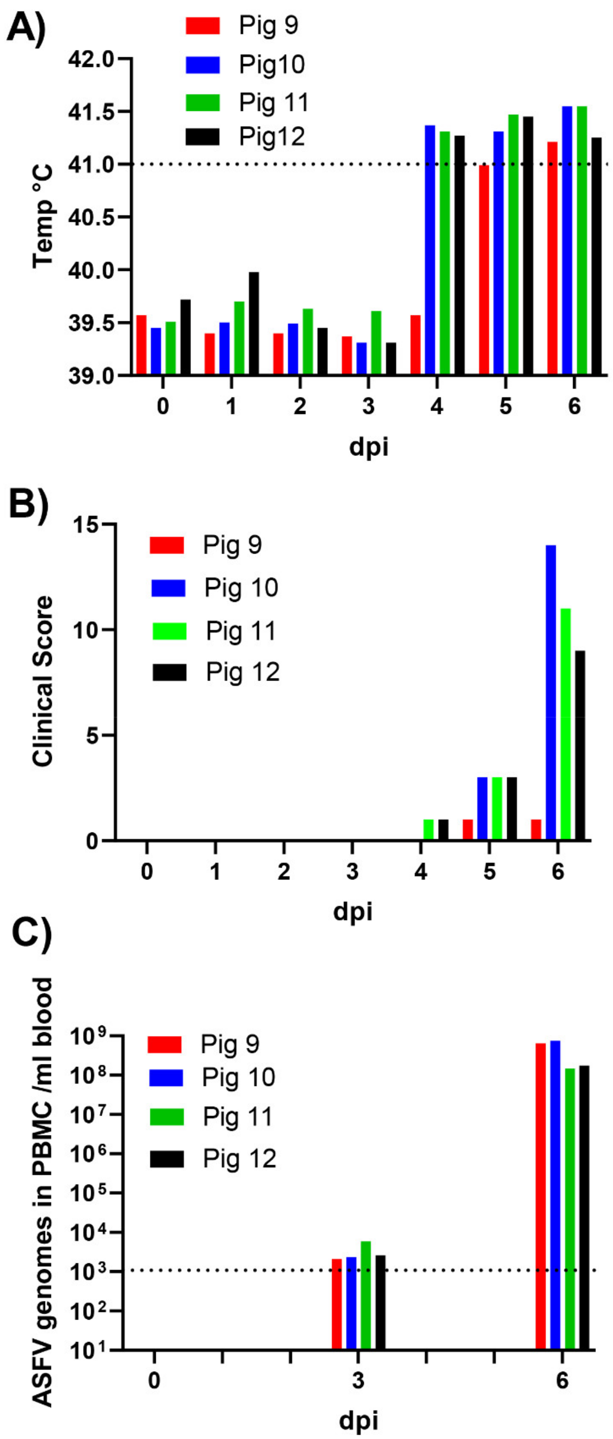
3.1. Course of Infection in the Inoculated Pigs

Following intranasal inoculation on day 0, three out of the four pigs (numbers 10, 11 and 12), at 4 dpi, had high fever (rectal temperature above 41 °C; see Figure 1A). Furthermore, at 5 and 6 dpi, all four pigs had high fever. Clinical signs of infection also became apparent and included depression, anorexia, mildly labored breathing, hyperemia of the skin and cyanosis on the ears and distal limbs, plus blood in feces (pig 10). At 6 dpi, all the inoculated pigs were euthanized since pigs 10, 11 and 12 had reached the pre-determined humane endpoint and the remaining animal, pig 9, was euthanized to avoid having a solitary animal. The clinical scores for the four inoculated pigs through the course of the infection are depicted in Figure 1B. All four pigs showed at least some clinical signs of disease, in addition to the elevated temperature by 5 dpi, which had become much more apparent in pigs 10, 11 and 12 at 6 dpi, just prior to euthanasia.

3.2. Virus Derived RNA Transcript Analysis from PBMCs of ASFV-Inoculated Pigs

Blood samples (12 in total) were collected separately from the four pigs on day 0 (prior to inoculation), at 3 dpi and 6 dpi (on the day of euthanasia). The PBMC fraction was isolated from the EDTA blood samples, and the level of ASFV DNA in the nucleic acids isolated from the aliquots of the PBMCs was determined by qPCR (Figure 1C). Only low levels of ASFV DNA were detectable in the PBMCs at 3 dpi, but much higher levels (>1000-fold) were present at 6 dpi in all four pigs (Figure 1C). The total RNA was also extracted from the PBMC samples and freed of most of the DNA from the host and the virus. The poly(A)+ RNA (mRNAs) was then selected, reverse transcribed and sequenced (as described in Materials and Methods). The reads (ca. 27–47 million per sample) were initially mapped to the pig genome (resulting in 81–92% of the reads being uniquely mapped, see Table 2), and the unmapped reads were then mapped to the ASFV Georgia_2007/1 sequence (see Table 2). As expected, no reads mapping to the ASFV genome were present in any of the samples collected on day 0, prior to inoculation. At 3 dpi, the PBMCs from pigs 10, 11 and 12 each only generated between 600–850 reads (less than 0.0025% of the total reads) mapping to the ASFV genome, with no ASFV-derived reads detected in pig 9 (Table 2). However, in contrast, at 6 dpi, between 871,681 and 1,945,879 reads (ca. 2–4% of the total reads) from the PBMCs mapped to the ASFV genome from each of the four pigs (Table 2); the lowest numbers of ASFV reads were found in pigs 9 and 10. For pigs 11 and 12, about 40% of the reads that did not map to the pig genome did map to the ASFV genome. Thus, in parallel with the appearance of clinical signs between 3 and 6 dpi and the large increase in the levels of ASFV DNA in the PBMCs of the inoculated pigs (see Figure 1), a huge increase (>1000-fold) in the level of ASFV RNA transcripts within the PBMCs was apparent during this time period.

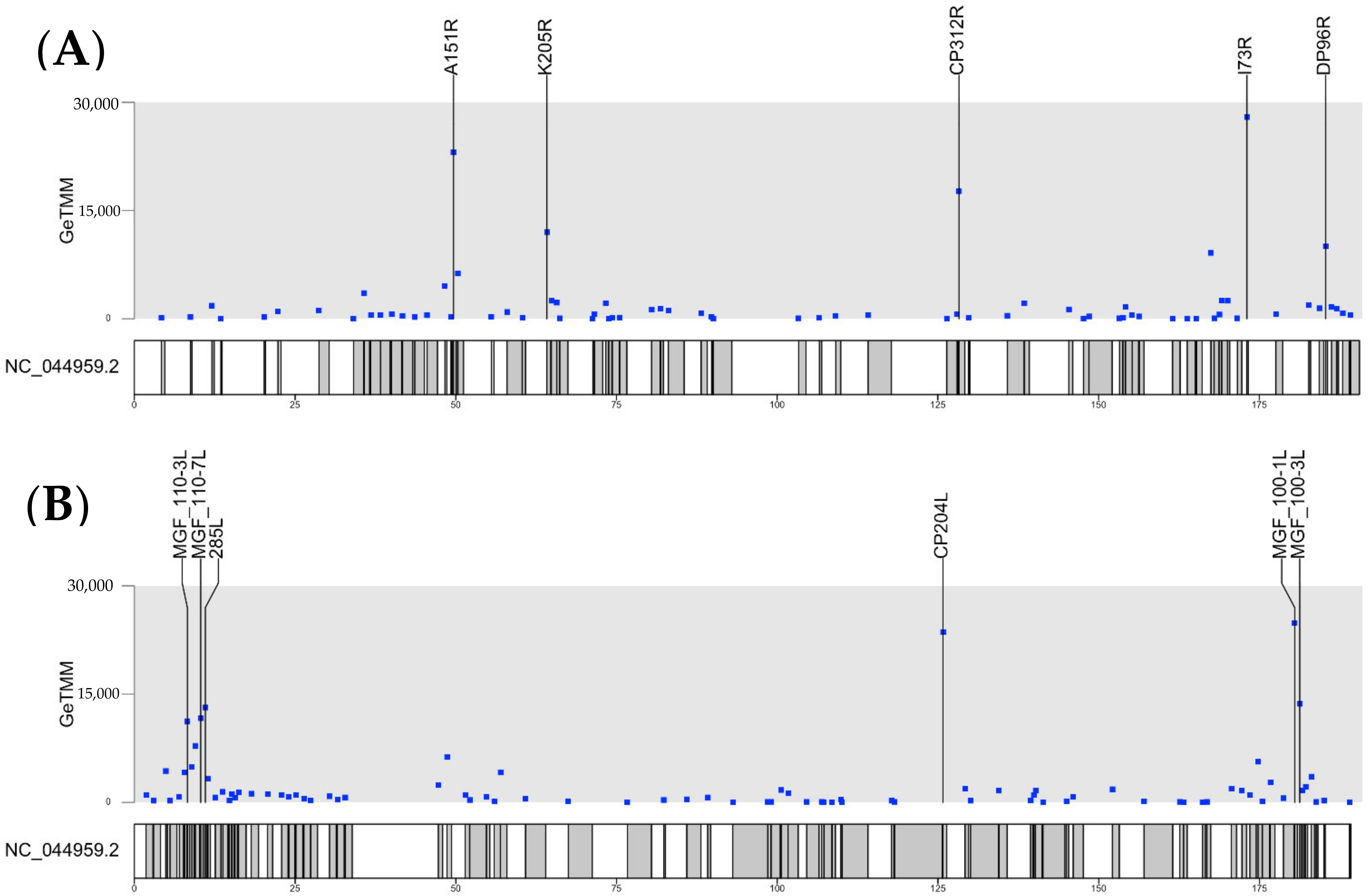
3.3. Transcription of Individual ASFV Genes

In general, there was a very good agreement between the pattern of expression of the individual ASFV genes obtained for the PBMCs from each of the different pigs, although the actual levels varied to some extent (see Supplementary Table S1). In particular, the highest levels of virus gene expression were observed in pigs 11 and 12, and the levels of ASFV transcripts present in these cells from pigs 9 and 10 were, on average, about 50% lower. The most highly expressed genes were C312R, CP204L, MGF 100-1L and A151R (see Table 3), while some genes were expressed at a much lower level, i.e., less than 1% of these most highly expressed genes (see Table 4 and Supplementary Table S1). Interestingly, all the identified ASFV coding genes, with ORFs > 180 nt in length, were expressed to some extent (see Figure 2 and Supplementary Tables S1 and S2). Note that there are some very short genes, designated pNG1-7, that were recently identified [18] from the Ba71V genome that were not specifically included here, but several of them correspond to features annotated in the ASFV Georgia_2007/1 genome as ASFV G ACD 00xx0 (see [18]). Some of these genes (e.g., ASFV G ACD 00350 and ASFV G ACD 00600) were expressed (see Supplementary Table S1). The gene designated as pNG4 (in the Ba71V genome) does not seem to have a corresponding gene in the ASFV Georgia_2007/1 genome. Some annotated features within the ASFV Georgia/2007 genome were present within the reads derived from other annotated gene transcripts (e.g., “polyC regions” (nt 14225 to 14237 (within the MGF 110-10-L-MGF110-14L fusion ORF) and nt 15666 to 15682 (within the MGF 110-13Lb ORF) plus a “polyG region” (nt 19993 to 20008, within the ASFV G ACD 00350 ORF)). However, another “polyG region” (nt 17624 to 17632) was expressed at quite high levels (even in the list of total reads not adjusted for the length of the transcript, see Supplementary Table S1). This sequence is present within a very short open reading frame (encoding a 15-residue peptide, MFDLSSILIRGGGPY) that was not annotated previously. The two genes flanking this polyG region were detected at lower levels (see Supplementary Table S1) and, hence, it does not seem possible to account for these reads as read-through from adjacent genes. Three so-called “hypothetical” genes (nt 19411 to 19506, nt 51223 to 51337 and nt 182044 to 182151, see Supplementary Table S1) were detected with many reads. However, the genes annotated as “ASFV G ACD 01760 no indication” (nt 175922 to 176006) and “DP63R no indication” (nt 178506 to 178652) were present within the transcripts from the I177L and MGF 360-16R genes, respectively, and are not listed separately.
Potentially, some sequence reads could be derived from residual ASFV DNA in the samples, even after the DNAseI treatment and purification of the poly(A)+ RNA. However, it appears that the number of such reads should be very low since a number of genes are only included in a small number of reads (<50, see Table 4); this should be compared to >100,000 reads for some of the highly expressed genes (Table 3). The reads derived from genomic DNA should be derived similarly from all the regions of the genome.
We have also “standardized” the number of reads to take into account the size differences between the libraries generated prior to sequencing and the lengths of the different genes. This process had little overall effect on the pattern of highly expressed genes (see Supplementary Table S2). The most highly expressed genes, based on the total reads as listed in Table 3, are also high on the list of the ASFV genes expressed that is shown in Supplementary Table S2, following standardization, e.g., CP312R, CP204L, MGF 100-1L, A151R, K205R and I73R (see also Table 5).
The distribution of RNA transcripts across the genome, derived from the two different strands of the genomic DNA, is shown in Figure 2. Most of the highly expressed transcripts are derived from genes that are well-separated across the genome, and the highly expressed transcripts are copied from each of the strands of the genome. As may be expected, the different strands of the genome are either read in one direction or the other; this prevents the production of mRNAs that are complementary to each other and would form dsRNAs. Two genes, A151R and MGF360-15R, which are highly expressed (see Table 3) are adjacent to each other and transcribed in the same direction (see [17]). However, it is noteworthy that, after the standardization of the number of reads, to take account of the gene length, the apparent level of MGF360-15R expression is less markedly high (Table 5) and does not feature among the most highly expressed genes shown in Figure 2.
The small number of ASFV reads detected in the pigs at 3 dpi (Supplementary Table S1) precludes the accurate quantification of their relative gene expression. However, it is apparent that the few reads that were observed at 3 dpi corresponded to transcripts that were highly expressed at 6 dpi (e.g., see Table 5). It seems most likely that the viral RNA reads observed in the PBMCs at 3 dpi represent the infection of a very small proportion of the cells, consistent with the low levels of ASFV DNA present within the PBMCs at this stage of the infection. It is not possible to differentiate “early” and “late” transcription within the pigs (c.f. in cell culture, [18]) as it cannot be expected that a synchronous state of infection can be achieved. In the RNAseq analysis performed here, the use of alternative transcription start sites has not yet been explored.

4. Discussion

The intranasal inoculation of pigs with ASFV is an efficient and consistent means of initiating infection (as seen previously [16]). Each of the inoculated pigs became infected and followed a similar course of disease. Pig 9 was slightly delayed in showing clinical signs, and both pigs 9 and 10 had slightly lower levels of the ASFV derived transcripts within their PBMCs, but it is expected that there will be some differences between animals.
Overall, we have obtained very consistent results among the four pigs infected with the ASFV POL/2015/Podlaskie strain for the expression of each ASFV gene within the PBMCs; this cell population includes the major target cell types (monocytes and macrophages) for this virus. The virus strain used here is very similar to the other closely related genotype II viruses currently circulating in Europe and Asia.
A complete listing of the standardized gene expression data is provided in Supplementary Table S2. In studies described previously by Jaing et al. [19], three pigs were infected (by intranasal instillation) with the ASFV Georgia 2007/1 strain using 104 TCID50. The pigs were euthanized on days 7–10 after infection; each of the pigs were shown to have ASFV DNA in the blood from 7 dpi and displayed clinical signs of infection from that time. Total RNA was extracted from whole blood and used for the transcriptomic analysis; only about 0.1% of the reads mapped to the ASFV genome. This is a much lower level than observed here (up to 4.2%, see Table 1); presumably, the much higher proportion of ASFV derived reads that we obtained is largely due to the selection of poly(A)+ mRNAs from the PBMC fraction prior to the sequencing (thus removing many reads derived from other RNAs (e.g., ribosomal RNA). About 1–2 million reads that mapped to the ASFV genome were analyzed here from each of the inoculated pigs at 6 dpi. Another advantage obtained from selecting the poly(A)+ mRNA is that reads derived from residual ASFV DNA should be further diminished beyond that achieved by DNAseI treatment alone, which may not be completely effective [32]. However, some reads may be derived from residual ASFV DNA (there are only short regions of the genome that are not included in any reads), along with other incompletely digested host DNA, e.g., pig mitochondrial DNA. In addition, it is worth noting that <1% of the reads (about 250,000 per sample) do not map to the pig genome nor to ASFV but map to bacterial genome sequences, as determined using Kaiju [31]. It seems likely that this results from bacteria being engulfed by the porcine macrophages and then residual DNA being present within these cells.
A key feature of the data presented here is the close correspondence between the results observed for each of the four pigs. Thus, the most highly expressed ASFV genes observed in one pig were also highly expressed in the other three pigs (Table 3). Indeed, the relative order of expression of the top 20 genes was very similar in each of the four pigs. Furthermore, genes that were expressed in one pig at a low level were also expressed at a low level in the other animals (Table 4). This consistency in the gene expression contrasts with the much more variable results reported previously by Jaing et al. [19]. For example, surprisingly, for nine of the seventeen genes indicated in their study as being highly expressed overall, in one of the three pigs examined, apparently no reads corresponding to these highly expressed genes were detected. Indeed, for the most abundantly expressed transcript overall, encoding MGF-360-15R (an inhibitor of interferon-β induction, [33]), the fragments per kilobase per million mapped fragments (FPKM) values obtained for the three pigs were: 1,190,000; 0 (zero) and 19,979, respectively, showing great variation. This gene was the sixth or seventh most highly expressed gene in each of the four pigs studied here (see Table 3), with the total reads ranging from 23,647 to 49,844 (see Supplementary Table S1). Following the standardization (to account for differences in library size and for the lengths of the genes), this gene was fifteenth in the list of highly expressed genes (Table 5) and, thus, it does not feature among the most highly expressed genes (see Figure 2).
The highly expressed genes of ASFV are mainly located in different regions across the genome and are derived from each strand. There can be a concern that, if the transcription termination signals are not 100% efficient, then read-through into the adjacent genes could affect the apparent level of transcription of the genes adjacent to the highly expressed genes. For the most highly expressed genes that we have analyzed, this could only be an issue for the A151R gene (fourth most highly expressed) and the adjacent MGF360-15R gene (sixth or seventh most highly expressed, see Table 3) since these genes are both transcribed in the same direction. However, after the normalization of the gene reads to take into account the length of the ORF, the expression of the MGF360-15R gene does not appear to be very high (see Table 5 and Figure 2). It seems unlikely that the reads corresponding to the MGF 360-15R gene were greatly affected by the read-through from the higher number of reads derived from the A151R gene unless the transcription termination process was very inefficient. Furthermore, the studies by Cackett et al. [18] indicated a good correlation beyond the transcription measured by RNA-seq (as used here) and the 5′-end cap analysis gene expression sequencing (CAGE-seq). The latter methodology is independent of any read-through transcription. Finally, there was a good correspondence between the genes found to be highly expressed in the pigs, as shown here (see Table 3 and Table 5 and Supplementary Table S1), and those found to be highly expressed during both the early (at 5 h post-infection, prior to DNA replication) and late (at 16 h, after DNA replication) stages of infection in Ba71V-infected Vero cells [18]. Thus, the genes CP312R, CP204L, A151R, K205R and I73R were all found to be highly expressed in both studies. There was also good overall correspondence to the results from Jaing et al. [19] within the pigs, although the higher degree of variability of the signal (as mentioned above) makes for less precise quantification. Many of the ASFV genes are poorly characterized [17], and this applies to the highly expressed genes as well as those expressed at a lower level.
As the functions of the various ASFV genes become known, information about the expression of the genes in different cell types will be important for understanding the biology of the virus. Interestingly, most of the major components of ASF virions (as described previously [34]) are not translated from among the most highly expressed genes. Only the dUTPase component of the virion (from the E165R gene) is produced from among the top 20 expressed transcripts (see Table 3). The genes encoding the major capsid protein (p72 expressed from B646L), the outer envelope protein CD2v (from E402R) plus the two polyproteins pp220 (from CP2475L) and pp65 (from CP530R) do not appear in the list of the top 20 expressed genes. It is noteworthy that the S273R gene, which expresses a protease that processes the two polyproteins (pp220 and pp65) to major components of the virion [34], is expressed at very low levels (Table 4). Clearly, the level of protein expression does not only reflect the level of the mRNA; it will also depend on the stability of the proteins and the translational efficiency of the individual mRNAs. It can be expected that proteins with catalytic functions, e.g., the ASFV protease (S273R), may be required at a lower level than the structural protein precursor substrates.
Overall, it seems that all open reading frames encoding products longer than 60 amino acids (i.e., >180 nt, as annotated in Accession no. FR682468.2) are expressed within the pigs. It could have been possible that some ASFV genes were only expressed in soft ticks as part of its sylvatic cycle, but this does not appear to be the case. However, for the genes that are expressed at very low levels (see Table 3), it is formally possible that these reads are due to the presence of residual ASFV DNA. Clearly, there could be quantitative differences in the pattern of gene expression from the virus when it replicates in the ticks compared to that observed in pigs.
It is apparent that the ASFV must have replicated somewhere in the pigs during the time period from inoculation until after 3 dpi since the virus must have spread to the PBMCs to enable their infection. Following the intranasal infection, it is believed that the primary ASFV replication occurs within the tonsil and regional lymph nodes before being spread, through the lymphatic system and the blood, to other replication sites (e.g., the spleen and liver (see [22]). Further studies should focus on these primary sites of infection and also other sites of subsequent infection. Furthermore, the host responses to the ASFV infection can also be addressed using the PBMCs (as here) and other cells that become infected.

Supplementary Materials

The following are available online at https://0-www-mdpi-com.brum.beds.ac.uk/article/10.3390/v13112333/s1, Supplementary Table S1: Total numbers of reads for all annotated features in the ASFV genome throughout the course of infection; Supplementary Table S2: Ordered list of standardized number of reads for genes with ORFs > 180 nt expressed in PBMCs at 3 and 6 dpi.

Author Contributions

Conceptualization, A.S.O., M.T.P.G., A.B. and G.J.B.; methodology, A.S.O., M.K., L.L, F.A., M.T.L., A.B. and G.J.B.; formal analysis, A.S.O., M.K. and C.M.L.; investigation, A.S.O., L.L. and A.B.; data curation, A.S.O. and M.K.; writing—original draft preparation, G.J.B.; writing—review and editing, all authors; visualization, M.K. and G.J.B.; supervision, L.L, F.A., T.B.R., M.T.P.G., A.B. and G.J.B.; funding acquisition, M.T.L. All authors have read and agreed to the published version of the manuscript.

Funding

This study was funded in part through the University of Copenhagen and the Statens Serum Institut and also by a ‘Danish National Research Foundation 143 Award’.

Institutional Review Board Statement

This study was approved by the Ethical and Animal Welfare Committee of the Generalitat de Catalunya (Autonomous Government of Catalonia; permit number: CEA-OH/10869/1).

Informed Consent Statement

Not applicable.

Data Availability Statement

The data analyzed in this study are included within the paper and its Supplementary Information (Tables S1 and S2).

Acknowledgments

We are very grateful to the staff at IRTA-CReSA who were involved in this study. Especially, we owe great thanks to Guillermo Cantero, Iván Cordon, Joanna Wiacek, María Jesús Navas, Marta Muñoz, Samanta Giler, Xavier Abad and also to Preben Normann (U. of C.) for their technical assistance.

Conflicts of Interest

The authors declare no conflict of interest. The funders had no role in the design of the study; in the collection, analyses, or interpretation of data; in the writing of the manuscript or in the decision to publish the results.

References

  1. Understanding and Combatting African Swine Fever. 2021, pp. 25–61. Available online: https://agritrop.cirad.fr/598144/7/ID598144.pdf (accessed on 26 October 2021).
  2. Dixon, L.; Sun, H.; Roberts, H. African swine fever. Antivir. Res. 2019, 165, 34–41. [Google Scholar] [CrossRef] [PubMed]
  3. Olesen, A.S.; Belsham, G.J.; Rasmussen, T.B.; Lohse, L.; Bødker, R.; Halasa, T.; Boklund, A.; Bøtner, A. Potential routes for indirect transmission of African swine fever virus into domestic pig herds. Transbound. Emerg. Dis. 2020, 67, 1472–1484. [Google Scholar] [CrossRef]
  4. Achenbach, J.E.; Gallardo, C.; Pelegrin, E.N.; Rivera-Arroyo, B.; Degefa-Negi, T.; Arias, M.; Jenberie, S.; Mulisa, D.D.; Gizaw, D.; Gelaye, E.; et al. Identification of a New Genotype of African Swine Fever Virus in Domestic Pigs from Ethiopia. Transbound. Emerg. Dis. 2017, 64, 1393–1404. [Google Scholar] [CrossRef] [PubMed]
  5. Bastos, A.D.S.; Penrith, M.-L.; Crucière, C.; Edrich, J.L.; Hutchings, G.; Roger, F.; Couacy-Hymann, E.; Thomson, G.R. Genotyping field strains of African swine fever virus by partial p72 gene characterisation. Arch. Virol. 2003, 148, 693–706. [Google Scholar] [CrossRef] [PubMed]
  6. Boshoff, C.; Bastos, A.; Gerber, L.; Vosloo, W. Genetic characterisation of African swine fever viruses from outbreaks in southern Africa (1973–1999). Vet. Microbiol. 2007, 121, 45–55. [Google Scholar] [CrossRef] [Green Version]
  7. Lubisi, B.A.; Bastos, A.; Dwarka, R.M.; Vosloo, W. Molecular epidemiology of African swine fever in East Africa. Arch. Virol. 2005, 150, 2439–2452. [Google Scholar] [CrossRef]
  8. Quembo, C.J.; Jori, F.; Vosloo, W.; Heath, L. Genetic characterization of African swine fever virus isolates from soft ticks at the wildlife/domestic interface in Mozambique and identification of a novel genotype. Transbound. Emerg. Dis. 2018, 65, 420–431. [Google Scholar] [CrossRef] [Green Version]
  9. Costard, S.; Wieland, B.; De Glanville, W.; Jori, F.; Rowlands, R.; Vosloo, W.; Roger, F.; Pfeiffer, D.; Dixon, L.K. African swine fever: How can global spread be prevented? Philos. Trans. R. Soc. B Biol. Sci. 2009, 364, 2683–2696. [Google Scholar] [CrossRef] [Green Version]
  10. OIE WAHIS Interface. 2021. Available online: https://wahis.oie.int/#/dashboards/country-or-disease-dashboard (accessed on 19 August 2021).
  11. FAO. ASF Situation in Asia Update. Available online: http://www.fao.org/ag/againfo/programmes/en/empres/ASF/situation_update.html (accessed on 1 June 2020).
  12. Busch, F.; Haumont, C.; Penrith, M.-L.; Laddomada, A.; Dietze, K.; Globig, A.; Guberti, V.; Zani, L.; Depner, K. Evidence-Based African Swine Fever Policies: Do We Address Virus and Host Adequately? Front. Vet. Sci. 2021, 8, 224. [Google Scholar] [CrossRef]
  13. Sánchez-Cordón, P.J.; Montoya, M.; Reis, A.L.; Dixon, L.K. African swine fever: A re-emerging viral disease threatening the global pig industry. Vet. J. 2018, 233, 41–48. [Google Scholar] [CrossRef]
  14. Gallardo, C.; Soler, A.; Nieto, R.; Cano, C.; Pelayo, V.; Sánchez, M.A.; Pridotkas, G.; Fernandez-Pinero, J.; Briones, V.; Arias, M. Experimental Infection of Domestic Pigs with African Swine Fever Virus Lithuania 2014 Genotype II Field Isolate. Transbound. Emerg. Dis. 2015, 64, 300–304. [Google Scholar] [CrossRef] [PubMed]
  15. Guinat, C.; Reis, A.L.; Netherton, C.L.; Goatley, L.; Pfeiffer, D.U.; Dixon, L. Dynamics of African swine fever virus shedding and excretion in domestic pigs infected by intramuscular inoculation and contact transmission. Vet. Res. 2014, 45, 1–9. [Google Scholar] [CrossRef]
  16. Olesen, A.S.; Lohse, L.; Boklund, A.; Halasa, T.; Gallardo, C.; Pejsak, Z.; Belsham, G.; Rasmussen, T.B.; Bøtner, A. Transmission of African swine fever virus from infected pigs by direct contact and aerosol routes. Vet. Microbiol. 2017, 211, 92–102. [Google Scholar] [CrossRef] [Green Version]
  17. Dixon, L.K.; Chapman, D.A.; Netherton, C.L.; Upton, C. African swine fever virus replication and genomics. Virus Res. 2013, 173, 3–14. [Google Scholar] [CrossRef] [PubMed]
  18. Cackett, G.; Matelska, D.; Sýkora, M.; Portugal, R.; Malecki, M.; Bähler, J.; Dixon, L.; Werner, F. The African Swine Fever Virus Transcriptome. J. Virol. 2020, 94, e00119-20. [Google Scholar] [CrossRef] [Green Version]
  19. Jaing, C.; Rowland, R.R.R.; Allen, J.; Certoma, A.; Thissen, J.B.; Bingham, J.; Rowe, B.; White, J.; Wynne, J.; Johnson, D.; et al. Gene expression analysis of whole blood RNA from pigs infected with low and high pathogenic African swine fever viruses. Sci. Rep. 2017, 7, 1–14. [Google Scholar] [CrossRef]
  20. Zhu, J.J.; Ramanathan, P.; Bishop, E.A.; O’Donnell, V.; Gladue, D.P.; Borca, M.V. Mechanisms of African swine fever virus pathogenesis and immune evasion inferred from gene expression changes in infected swine macrophages. PLoS ONE 2019, 14, e0223955. [Google Scholar] [CrossRef]
  21. Ju, X.; Li, F.; Li, J.; Wu, C.; Xiang, G.; Zhao, X.; Nan, Y.; Zhao, D.; Ding, Q. Genome-wide transcriptomic analysis of highly virulent African swine fever virus infection reveals complex and unique virus host interaction. Vet. Microbiol. 2021, 261, 109211. [Google Scholar] [CrossRef] [PubMed]
  22. Gómez-Villamandos, J.; Bautista, M.; Cordón, P.S.; Carrasco, L. Pathology of African swine fever: The role of monocyte-macrophage. Virus Res. 2013, 173, 140–149. [Google Scholar] [CrossRef] [PubMed]
  23. Olesen, A.S.; Lohse, L.; Dalgaard, M.D.; Woźniakowski, G.; Belsham, G.J.; Bøtner, A.; Rasmussen, T.B. Complete genome sequence of an African swine fever virus (ASFV POL/2015/Podlaskie) determined directly from pig erythrocyte-associated nucleic acid. J. Virol. Methods 2018, 261, 14–16. [Google Scholar] [CrossRef] [PubMed] [Green Version]
  24. Forth, J.H.; Forth, L.F.; King, J.; Groza, O.; Hübner, A.; Olesen, A.S.; Höper, D.; Dixon, L.K.; Netherton, C.L.; Rasmussen, T.B.; et al. A Deep-Sequencing Workflow for the Fast and Efficient Generation of High-Quality African Swine Fever Virus Whole-Genome Sequences. Viruses 2019, 11, 846. [Google Scholar] [CrossRef] [PubMed] [Green Version]
  25. Tignon, M.; Gallardo, C.; Iscaro, C.; Hutet, E.; Van der Stede, Y.; Kolbasov, D.; De Mia, G.M.; Le Potier, M.-F.; Bishop, R.P.; Arias, M.; et al. Development and inter-laboratory validation study of an improved new real-time PCR assay with internal control for detection and laboratory diagnosis of African swine fever virus. J. Virol. Methods 2011, 178, 161–170. [Google Scholar] [CrossRef] [PubMed]
  26. Dobin, A.; Davis, C.A.; Schlesinger, F.; Drenkow, J.; Zaleski, C.; Jha, S.; Batut, P.; Chaisson, M.; Gingeras, T.R. STAR: Ultrafast universal RNA-seq aligner. Bioinformatics 2013, 29, 15–21. [Google Scholar] [CrossRef] [PubMed]
  27. Warr, A.; Affara, N.; Aken, B.; Beiki, H.; Bickhart, D.M.; Billis, K.; Chow, W.; Eory, L.; Finlayson, H.A.; Flicek, P.; et al. An improved pig reference genome sequence to enable pig genetics and genomics research. GigaScience 2020, 9, giaa051. [Google Scholar] [CrossRef] [PubMed]
  28. Li, H. Aligning sequence reads, clone sequences and assembly contigs with BWA-MEM. arXiv 2013, arXiv:1303.3997. [Google Scholar]
  29. Liao, Y.; Smyth, G.K.; Shi, W. The R package Rsubread is easier, faster, cheaper and better for alignment and quantification of RNA sequencing reads. Nucleic Acids Res. 2019, 47, e47. [Google Scholar] [CrossRef] [Green Version]
  30. Smid, M.; van de Werken, H.J.G.; van Riet, J.; van Galen, A.; de Weerd, V.; van der Vlugt-Daane, M.; Bril, S.I.; Lalmahomed, Z.S.; Kloosterman, W.P.; Wilting, S.M.; et al. Gene length corrected trimmed mean of M-values (GeTMM) processing of RNA-seq data performs similarly in intersample analyses while improving intrasample comparisons. BMC Bioinform. 2018, 19, 236. [Google Scholar] [CrossRef] [Green Version]
  31. Menzel, P.; Ng, K.; Krogh, A. Fast and sensitive taxonomic classification for metagenomics with Kaiju. Nat. Commun. 2016, 7, 11257. [Google Scholar] [CrossRef] [Green Version]
  32. Lazov, C.; Belsham, G.; Bøtner, A.; Rasmussen, T. Full-Genome Sequences of Alphacoronaviruses and Astroviruses from Myotis and Pipistrelle Bats in Denmark. Viruses 2021, 13, 1073. [Google Scholar] [CrossRef]
  33. Correia, S.; Ventura, S.; Parkhouse, R.M. Identification and utility of innate immune system evasion mechanisms of ASFV. Virus Res. 2013, 173, 87–100. [Google Scholar] [CrossRef]
  34. Alejo, A.; Matamoros, T.; Guerra, M.; Andrés, G. A Proteomic Atlas of the African Swine Fever Virus Particle. J. Virol. 2018, 92, e01293-18. [Google Scholar] [CrossRef] [PubMed] [Green Version]
Figure 1. Time course of infection of pigs with ASFV. Pigs were inoculated intranasally with ASFV/POL/2015/Podlaskie at 0 dpi, and then their rectal temperatures were taken on a daily basis (A) and clinical signs of disease (for 8 parameters) were also scored (B). Blood samples were collected at 0 dpi (prior to inoculation), at 3 dpi and at 6 dpi; the animals were euthanized at 6 dpi. PBMCs were purified from the blood samples (4 mL). The presence of ASFV DNA in the PBMCs (C) was quantified by qPCR and values converted to gene copy numbers/mL blood by reference to a standard curve. Levels below 103 ASFV genomes/mL (indicated by dashed line) were outside of the linear range of the assay.
Viruses 13 02333 g001
Figure 2. Analysis of ASFV gene transcription within PBMCs of ASFV-infected pigs at 6 dpi. The number of ASFV mRNA derived reads from PBMCs of infected pigs collected at 6 dpi were standardized according to the size of the library and the length of the ORFs (for ORFs >180 nt) and plotted (as GeTMM values) along the entire ASFV genome for the two separate strands of the ASFV genome (panel (A), genes transcribed from left to right and panel (B), genes transcribed from right to left). The ASFV genes with the highest GeTMM values are indicated.
Viruses 13 02333 g002
Table 1. Description of clinical score system.
FeatureScoreDescription
Alertness and recumbency0Alert
1Depressed/lethargic
2Only gets up when touched
4Gets up slowly when touched
6Remains recumbent when touched
Body condition0Normal, full stomach
1Empty stomach, sunken flanks
2Empty stomach, sunken flanks, loss of muscle mass
3Emaciated
Skin0Normal
1Minimal area of the skin with observed bleeding (<10% of the body)
2Moderate area of the skin with observed bleeding (10–25% of the body)
3Generalized skin bleeding (>25% of the body)
Joints0No joint swelling
1Swelling
4Severe swelling and lameness
Respiration0Normal
1Mildly labored
2Labored +/− cough
3Severely labored
Eyes0Normal
1Small amount of exudate
2Moderate amount of exudate
Gastrointestinal and urinary tracts0No diarrhea
1Mild diarrhea for less than 24 h
3Diarrhea for more than 24 h or vomiting
4Bloody diarrhea or blood in urine
Neurology0No symptoms
3Hesitant, unsteady walk, crossing-over of legs is corrected slowly
4Pronounced ataxia
6Paralysis or convulsions
This is modified from the system described previously [16] since feed was available ad libitum and rectal temperatures were recorded separately.
Table 2. Poly(A)+ mRNA derived sequence reads from PBMCs of pigs infected with ASFV.
Pig Number (Sampling Day)Number of Input ReadsNumber of Reads Uniquely Mapped on Pig GenomeUniquely Mapped Reads (%) on Pig GenomeNumber of Reads Mapped to ASFV
Genome
Proportion of ASFV Reads/Total Reads (%)
Pig 9 (0 dpi)319790212886382690.2600
Pig 9 (3 dpi)318071262920723891.8300
Pig 9 (6 dpi)400934033547211588.478716812.17
Pig 10 (0 dpi)326177532966878990.9600
Pig 10 (3 dpi)467640304309523192.156380.0014
Pig 10 (6 dpi)270958892428816689.649155073.38
Pig 11 (0 dpi)459535823977142086.5500
Pig 11 (3 dpi)465619594169620789.556160.0013
Pig 11 (6 dpi)459704364037297987.8219392874.21
Pig 12 (0 dpi)463269243760995281.1800
Pig 12 (3 dpi)414067443654931888.278510.0021
Pig 12 (6 dpi)459152794120322589.7419458794.23
Table 3. Total numbers of reads for annotated ASFV genome features that are highly expressed in PBMCs from infected pigs at 6 dpi.
Gene or Feature
Name
CDS Start (nt)CDS
End
(nt)
Pig9-
6 dpi
Pig10-
6 dpi
Pig11-
6 dpi
Pig12-
6 dpi
1 Mean
Pigs 11–12
CP312R1282771292006639878462138910144010141460
CP204L1257831263675913750204128247133890131069
MGF 100-1L180479180904505663018310807798680103379
A151R49652501074519845603965658994593255
K205R64174647912990634834648726580565339
I73R1730881733062388724501591675480956988
MGF 360-15R50346512152427523647473504984448597
MGF 110-5L-6L9490101071874020840428204604844434
A240L48633493431835315318418824588243882
MGF 110-7L10314107272078020790430904263242861
E165R1674681679651709520759409144137441144
MGF 100-3L1812691815771571618157360294220939119
F334L56956579601715317920381033904438574
MGF 110-3L823986131620122633358423404534944
285L11042113261717514716347623286833815
I215L1747941754321634114199323613273632549
MGF 505-3R35760366021383611627255673031727942
DP96R1853391856291210710550275162814127829
ASFV G ACD 00600480004815299027709226752256322619
K196R6511365703981214044221632218322173
1: Average number of reads from pigs 11 and 12 at 6 dpi were used to place genes into order. The values given are the total number of sequence reads mapped per annotated region (not standardized for gene length or total number of reads) in the updated ASFV Georgia_2007/1 genome sequence (GenBank Acc. No. FR682468.2). Note that the start and end of the CDS are indicated by their position in the genome independently of the orientation of the gene.
Table 4. Total number of reads for annotated ASFV genome features expressed at low levels in PBMC from infected pigs at 6 dpi.
Gene or Feature
Name
CDS
Start (nt)
CDS
End (nt)
Pig9-
6 dpi
Pig10-
6 dpi
Pig11-
6 dpi
Pig12-
6 dpi
1 Mean
Pigs 11–12
Gene Product
Properties (If Known)
ASFV G ACD 01960187401187532141114292254273
ASFV G ACD 00190124561258181122265276271
E423R16380316507493126240285263
H171R153250153765100117264245255
ASFV G ACD 0009076477760107150229236233
L11L1838211841027579233204219
B407L10726110849952119189185187
O61R12979512998060126197177187P12 attachment protein
B117L1069071072546681196165181TR containing protein
MGF 505-2R340933567366100197155176
B318L96276972325169152182167Prenyltransferase
E301R1652251661303474146159153Proliferating cell nuclear antigen-like protein
EP153R73808742847738139129134C-type lectin-like
ASFV G ACD 010209290193059473714088114
B119L959369629538397510992FAD-dependent thiol oxidase
B175L10852710905428478210091Late TF VLTF-2
S273R1476701484912037708980SUMO-1-like protease
E183L1632181637722846846977P54, Virus entry
ASFV G ACD 018701826041827412012856676
S183L1470581476091844557666
E146L1661641666042545695663PSP
ASFV G ACD 0036020169202851414564852
ASFV G ACD 0021013461136521817295140
ASFV G ACD 0024014570146801311264033
EP84R7130671560713132620PSP
1: The values given are the total number of sequence reads mapped per annotated feature (not standardized for gene length or total number of reads) in the updated ASFV Georgia 2007/1 genome sequence (Acc. No. FR682468.2). Gene product functions (where known) are taken from Dixon et al. [17] and Cackett et al. [18]. PSP = putative signal peptide, TR = transmembrane region.
Table 5. Most highly expressed ASFV genes (>180 nt) following standardization.
1. Gene NameGene Product
Properties
(If Known)
StartEndPig11-
3 dpi
Pig12-
3 dpi
Pig9-
6 dpi
Pig10-
6 dpi
Pig11-
6 dpi
Pig12-
6 dpi
Mean Pigs
11 and 12
I73RTandem repeat sequence1730881733069131523622260375122937133441
MGF 100-1L 1804791809049161654414066351472712431136
CP204LP32 (P30) phosphoprotein1257831263679161408017027303512678328567
A151RRedox pathway49652501078131381219851293332309326213
CP312RImmunodominant protein128277129200681000316837208011822719514
MGF 100-3L 18126918157747709511676161681600916088
285L 110421132638840910263169171352015219
K205RIn virus factories641746479138674011182145321245913495
MGF 110-7L 10314107277369969970144201205913239
DP96R 1853391856296458057205131141133612225
MGF 110-3L 8239861344602311986132451063411940
E165RdUTPase167468167965354783827311378972510551
MGF 110-5L-6L 9490101073442236690959287199155
A240LThymidylate kinase48633493431335944273815375497851
MGF 360-15R 50346512152338845390753167017116
I215LUbiquitin conjugating enzyme1747941754322335614408701059946502
L83L 487851233527752009634351855764
MGF 110-4LHas KDEL-like domain892793012326894421654246845613
A104RHistone-like48322486362425323481611949435531
F334LRibonucleotide reductase subunit56956579602223763535524545434894
K196RThymidine kinase65113657031223124715519243924792
MGF 110-2L 782881422124863884469442984496
1: The total numbers of reads per gene were standardized according to the length of the open reading frame and for the size of the library. Only genes with a coding sequence > 180 nt (encoding > 60 amino acids) were included. The genes are listed according to the mean value for the reads from pigs 11 and 12 at 6 dpi. For comparison, the number of reads generated from the PBMCs at 3 dpi are also indicated for pigs 11 and 12 (for pigs 9 and 10, there were zero (0) or between 0 and 8 reads for each of these genes on this day, respectively). A complete listing of the ASFV genes expressed at 3 and 6 dpi is given in the Supplementary Tables S1 and S2. Gene product properties are from Dixon et al. [17] and Jaing et al. [19].
Publisher’s Note: MDPI stays neutral with regard to jurisdictional claims in published maps and institutional affiliations.

