
Mosquito-Borne Viruses and Non-Human Vertebrates in Australia: A Review


1
Children’s Medical Research Institute, Westmead, NSW 2145, Australia
2
Mosquito Control Laboratory, QIMR Berghofer Medical Research Institute, Herston, QLD 4006, Australia
3
Environmental Futures Research Institute, Griffith University, Gold Coast, QLD 4222, Australia
4
Biology Department, Stanford University, Stanford, CA 94305, USA
5
School of Science, Western Sydney University, Hawkesbury, Locked bag 1797, Penrith, NSW 2751, Australia
*
Author to whom correspondence should be addressed.
Viruses 2021, 13(2), 265; https://0-doi-org.brum.beds.ac.uk/10.3390/v13020265
Submission received: 21 December 2020
/
Revised: 2 February 2021
/
Accepted: 3 February 2021
/
Published: 9 February 2021
(This article belongs to the Special Issue Emerging Wildlife Viral Diseases)
Abstract
:Mosquito-borne viruses are well recognized as a global public health burden amongst humans, but the effects on non-human vertebrates is rarely reported. Australia, houses a number of endemic mosquito-borne viruses, such as Ross River virus, Barmah Forest virus, and Murray Valley encephalitis virus. In this review, we synthesize the current state of mosquito-borne viruses impacting non-human vertebrates in Australia, including diseases that could be introduced due to local mosquito distribution. Given the unique island biogeography of Australia and the endemism of vertebrate species (including macropods and monotremes), Australia is highly susceptible to foreign mosquito species becoming established, and mosquito-borne viruses becoming endemic alongside novel reservoirs. For each virus, we summarize the known geographic distribution, mosquito vectors, vertebrate hosts, clinical signs and treatments, and highlight the importance of including non-human vertebrates in the assessment of future disease outbreaks. The mosquito-borne viruses discussed can impact wildlife, livestock, and companion animals, causing significant changes to Australian ecology and economy. The complex nature of mosquito-borne disease, and challenges in assessing the impacts to non-human vertebrate species, makes this an important topic to periodically review.
1. Introduction
Mosquito-borne diseases pose a great risk to public health threatening more than half the human population, and many non-human vertebrates [1]. In Australia, mosquito-borne diseases are currently a major focus due to public health concerns, which have been amplified by the potential effects of climate change and urbanization (see [2] for a review). Exacerbating the effects of climate change is the rapid and sustained increase in global trade and the alteration of the physical environment as a result of urbanization, deforestation, and agricultural expansion. Such changes have been highly favorable to urban vectors of foreign viruses including dengue, chikungunya, and Zika virus [3], whereas increased air travel and trade has led to a proliferation of viruses and their vectors globally [4,5]. However, the impacts of these global phenomena on non-human vertebrate hosts (species which may be susceptible to infection) and the consequences to the maintenance and transmission of mosquito-borne viruses remains poorly understood.
Australia has a diverse climatic range and environmental bioregions, which promote unique and endemic faunal diversity. This, in combination with a number of introduced vectors and pathogens, has resulted in the emergence of novel disease transmission pathways, including those of regional and global importance. Additionally, due to the dispersal of Australian animals, distinct patterns in evolution of mosquito-borne viruses have been found. The viruses across Australia either (i) evolve slowly and uniformly, (ii) have significant divergence due to single nucleotide changes, or (iii) have multiple lineages that are periodically redistributed over the Australian continent [6]. While studies on the distribution of mosquitoes and human infection rates are important, it is essential to consider the complex relationship between vectors and non-human vertebrates in various regions throughout Australia. Here, we review evidence for the impacts of Australian mosquito-borne viruses (introduced and endemic) on non-human vertebrates (including native and domestic species), and the role that these vertebrates may play in disease and transmission cycles. The aim of this review is to assess the impact of mosquito-borne diseases to Australian animals and the implications of the rapid change in Australia due to urbanization and climate change. We conclude with a discussion of exotic mosquito-borne diseases that threaten the unique fauna of Australia.
2. Mosquitoes: The Link between Vertebrate Host and Disease
There are more than 300 species of mosquitoes identified in Australia and almost 100 of these are capable of transmitting pathogens to wildlife and domestic animals (see Table 1), of which Aedes, Culex, and Anopheles species are the most common genera of vectors [7]. Feeding preferences by these species are highly variable with some species (such as Aedes aegypti) reported to have host specific feeding patterns, while other species (such as Aedes vigilax) exhibit more generalist feeding behaviors [8], both of which can play an important role as bridging vectors [9,10]. Individual feeding patterns are often dependent on host abundance and availability, both of which are strongly linked to habitat identity, and which can change annually and seasonally depending on the biology and ecology of individual host species (see [11] for a review). To date, vertebrate blood-meal hosts have been identified for a variety of taxonomic groups, including Carnivora (e.g., cats, dogs, and foxes), Aves (birds), Diprotodontia (e.g., possums and macropods), Artiodactyla (e.g., cattle, sheep, pigs, and goats), and Equidae (horses), with individual vector species displaying a trade-off between host preference and host availability [11]. For example, in rural Queensland, Australia, bloodmeal origins for Culex annulirostris, were dominated by cattle [12], but in Sydney, a highly urbanized city, Cx. annulirostris bloodmeal origins were mostly from birds, rodents and rabbits [8]. Thus, the ecologies of Australian mosquito-borne viruses can be driven by complex interactions between vector species, host availability, and host preference, which may be a derivative of habitat or climate. This will ultimately drive the spread of pathogens.
3. Uniquely Australian Vertebrate Hosts
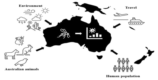
Australia provides a unique opportunity to investigate the transmission of viruses between vertebrates and mosquitoes. The long geographic isolation of Australia has led to the co-evolution of viruses, mosquitoes, and endemic vertebrate hosts, which offer unique insights into immunology and physiology. Over the last 200 years, Australia has also experienced the introduction of viruses and domestic species, and an expansion of urbanization, all of which have shifted the dynamics of disease and ecology among native species. This crockpot of co-evolution and introductions mean that Australian fauna have highly heterogenic roles for transmitting different viruses within the community (Figure 1), or are affected by viruses in different ways (i.e., asymptomatically vs. symptomatically; Table 1).
Marsupials
There are more than 300 extant marsupial species globally, of which close to 70% occur on the Australian continent (the mainland, Tasmania, New Guinea, and nearby islands), representing the most diverse extant marsupial radiation [71]. Many species exhibit unique physiological characteristics, such as adaptations to specific climatic envelopes, which have allowed them to succeed in even the harshest of Australian environments. However, immunological characteristics of marsupials may increase their susceptibility to infection for mosquito-borne viruses. When compared to eutherian mammals, neonatal marsupials are born without histological mature immune tissues [72,73] and are therefore unable to mount specific immune responses and are presumably highly reliant on maternal and innate immune strategies. Furthermore, some studies have reported marsupial immune systems are slower to mount some specific immune responses and occur at lower levels than those mounted by eutherian species [74,75]. However, the impact of such differences on disease susceptibility are poorly understood and the many similarities between eutherian and marsupial immune systems [76] cannot be overlooked (Figure 1).
The immediate threat of land use and climate change on the survival of many Australian marsupial species highlights a need to better understand the impacts disease on marsupial health and reproductive fitness. Additionally, we need to consider the ecological impacts of exotic mosquito introductions and range expansions on native species. The proliferation of global trade and travel makes the introduction exotic mosquito species highly likely (see [77,78] for reviews), whereas climate change is already changing native species distributions [3].
4. Ross River Virus
Ross River virus (RRV; belonging to the family Togaviridae and genus Alphavirus) is the most common arboviral disease in Australia [79,80,81]. Human infection can lead to chronic polyarthritis, with some symptoms lasting more than a year [82]. Although predominantly endemic to Australia, emerging evidence suggests RRV circulation is occurring in Pacific Island countries, including Fiji [83], American Samoa [84], Cook Islands [85], and French Polynesia [86]. Identifying the cause of RRV outbreaks is critical in Australia, as human infections are reported nationwide. Australia has approximately 5000 RRV human infection cases reported annually [23]. RRV is maintained in the environment through complicated transmission dynamics with multiple vectors and hosts. Since it was first identified in 1948, RRV has been isolated from more than 40 different mosquito species [87]. Under laboratory investigations the most prominent vectors are thought to be Aedes camptorhynchus, Aedes notoscriptus, Ae. Vigilax, and Cx. annulirostris based on their ability to amplify RRV in their saliva and transmit RRV in mouse-models [19,88].
Of the 81 non-human vertebrate species that have been serologically tested in Australia, 60 have had positive antibodies to RRV including domestic and livestock species including dogs (Canis lupus familiaris), cattle (Bos taurus), and horses (Equus caballus), and native birds, marsupials (possums and macropods), and flying foxes (Pteropus spp.) [20]. Recently, a sero-survey of koalas in Queensland, Australia found more than 80% have been exposed to RRV [89]. Not all of these species are thought to be important as reservoirs of RRV. Under experimental infection conditions, marsupials develop one of the highest-longest lasting viraemias [90]. Given the extensive length of marsupial viremias, it formed the basis of a long-held dogma that marsupials are better reservoirs than eutherian mammals and birds. The absence of marsupials in some Pacific Islands where local transmission of RRV is reported suggests that other species likely act as reservoirs. There is evidence that suggests birds may be important. Firstly, experimentally infected little corellas (Cacatua sanguinea) infected 14% of susceptible mosquito vectors, despite developing a relatively low-short lived viraemia [90]. Secondly, the first isolates of RRV from three bird species, magpie larks (Grallina cyanoleuca), Australian brown flycatcher (Microeca leucophaea), and masked finch (Poephila personata) [19], demonstrating that the virus circulates among bird populations. Overall, RRV in birds has been largely understudied and requires further investigation. Another understudied, but potential reservoirs of RRV are murids. Murids demonstrate moderate viraemia under experimental infection conditions [90] and one modelling study found house mouse (Mus musculus) abundance closely correlated with human notifications of RRV in Victoria [91].
Horses are the only species other than humans that have been reported to have clinical symptoms associated with RRV, including joint swelling and muscle stiffness [92]. Serological surveys of horses in Australia have varied between 26% [21] and 91% [93] being seropositive for RRV. Horse populations in Australia are estimated to exceed 1.2 million individuals and the thoroughbred industry was estimated to be $6.3 billion alone in 2001 [94]. In 2011, a national outbreak of equine diseases show high RRV infections in horses showing neurological or muscular symptoms [22]. As such, RRV can have large economic and ethical implications for horses in Australia and potentially internationally. Further studies are needed to determine the true burden of RRV in horse populations, and to develop appropriate treatments and preventative measures.
Future investigations of RRV in non-human vertebrates would benefit from additional field surveillance to identify ecological traits (such as habitat, seasonality, feeding behavior, and reproduction) that may be important for ongoing transmission [95,96]. There is some evidence to suggest that landscapes with proximity close to water reservoirs and the presence of some marsupial reservoirs is considered high risk for RRV disease transmission [97]. Further, animals with longer gestation periods, have dietary specialization and small population density are also more likely to have a history of RRV infection, suggesting that these animals should be monitored prior to an outbreak [98]. More localised research is needed to determine the significance of these trends in a given area.
5. Barmah Forest Virus
Barmah Forest virus (BFV) is a zoonotic alphavirus with humans infections reported nationally. In humans, BFV presents with similar clinical signs as RRV including polyarthritis, rash, fever, and myalgia [99]. BFV was first isolated from Cx. annulirostris mosquitoes in northern Victoria in 1974 [100]. However, given the similarity in clinical symptoms to RRV [80], it is likely that human cases of BFV may have been previously misdiagnosed and believed to be RRV. On average, there are 2400 notifications of BFV in Australia annually, with the majority of cases reported in Queensland [23]. The 2012–2013 period marked the largest BFV epidemic on record in Australia, with more than 2223 notifications in Queensland and 1024 notifications in Western Australia [23].
BFV has been isolated from a number of wild-caught mosquito species, including Cx. annulirostris [101] and Ae. vigilax [101,102,103,104]. Other insect vectors include the biting midge (Culicoides marksi) [105,106,107]. Vector competence studies found that an urban freshwater species, Ae. notoscriptus, was moderately susceptible to infection with transmission occurring between days 5 and 12, and an average transmission rate of 45% [108]. Ae. vigilax and Aedes procax have also demonstrated a high susceptibility to infection under vector competence studies [24], but Cx. annulirostris is a relatively ineffective vector of BFV with infection not exceeding 8% [109].
Evidence for BFV transmission in non-human vertebrates is limited. Serological investigations have found exposure of BFV in a diversity of non-human vertebrates. Moderate exposure has been reported in eastern grey kangaroos (Macropus giganteus) (44%) [25] and cattle (29%) [110], and low seropositivity in common brushtail possums (Trichosurus vulpecula) (10.7%) [21], koalas (Phascolarctos cinereus) (9%) [25], quokkas (Setonix brachyurus) (3.2%), domestic cats (Felis catus) (2%) [27], domestic dogs (Canis lupus familiaris) (1.3%) [27], and horses (1.2%) [70]. Despite a relatively large number of bush rats (Rattus fuscipes) and swamp rats (Rattus lutreolus) being tested, no exposure to BFV was found in these species [26]. Only common brushtail possums, dogs and cats have been experimentally infected with BFV, all of which demonstrated poor capability as amplifiers. Two of the 10 possums developed an immune response to the infection and had detectable antibodies for at least 45 days following the infection, however the species did not develop sufficient viraemia to infect susceptible mosquitoes [42]. Similarly, none of the 10 dogs or cats developed a detectable viraemia for BFV, and just one dog and three cats developed antibodies post infection [111]. Future serological, experimental and modelling studies on the non-human vertebrates of BFV would greatly improve current understandings for this medically important arbovirus.
6. Sindbis Virus
Sindbis virus (SINV) is one of the most commonly isolated arboviruses in Australian mosquitoes, despite rare instances of human infection. There are two different genotypes of the Sindbis virus: (i) the Oriental/Australian strain circulating throughout most of Australia (excluding Tasmania) and other surrounding countries including Malaysia and Papua New Guinea; and (ii) a strain endemic to southwestern Australia, with the Oriental/Australian SINV strain first isolated in 1960 from Cx. annulirostris in far north Queensland [28]. More recently, a new strain of SINV endemic to the south-west region of Western Australia, which differs in nucleotide sequences from the Oriental/Australian strain and Paleoarctic/Ethiopian strain by 25.4–28.9% and 16.8–19.4%, respectively [30]. The higher similarity between the endemic south-west Western Australia isolates and Paleoartic/Ethiopian strain suggested that this particular strain was imported by a traveler or migratory bird, and selective pressures in that region resulted in a new SINV strain.
Isolations of SINV have been found in Cx. annulirostris, Aedes normanensis, Ae. camptorhynchus [29], and Aedes pseudonormanensis [30]. There is evidence for the vertical transmission of SINV, particularly in Ae. camptorhynchus [29]. It has been reported that SINV infection is higher in Ae. aegypti mosquitoes that were reared at 30 °C compared to 20 °C [112], suggesting that warmer temperatures as a result of climate change could potentially increase SINV transmission.
Migratory birds are considered the main amplifying host for SINV, particularly those that have migration patterns connecting Australia [30], United Kingdom [113], northern Europe [114], South Africa [115], and China [116]. Birds that are vectors for SINV have persistent infections without any clinical symptoms, and are therefore healthy and able to travel between countries [113]. Although birds are considered one of the primary vectors for SINV, they do not necessarily contribute to outbreaks [116]. Additionally, many endemic birds found in south-west Western Australia are sedentary, suggesting that the new south-west Western Australia isolate is maintained through birds that do not travel long distances or vertebrate hosts available in its surrounding areas which includes marsupials [30]. Antibodies to SINV have been found present in one chuditch (Dasyurus geoffroi), emus (Dromaius novaehollandiae), European rabbits (Oryctolagus cuniculus), and horses in Australia [31].
7. Murray Valley Encephalitis Virus
Murray Valley encephalitis virus (MVEV) human infections are often asymptomatic, however approximately 1:150 to 1:1000 MVEV infections result in symptomatic encephalitic disease, which may include neurological features [117,118]. Although cases of MVEV in humans are not common, its high mortality/morbidity rate relative to other circulating flaviviruses is cause for concern when outbreaks occur. Several large outbreaks and epidemics have been reported since the early 20th century following its isolation from a human in 1951 [118]. To date, cases of MVEV have been reported in most Australian states, with four major outbreaks on the east coast, including two human MVEV isolations in Papua New Guinea in 1956 [119] and 1960 [120]. Despite this large historical distribution, MVEV is only considered endemic across northern Australia and Papua New Guinea [118].
MVEV has been isolated from Culex sitiens and other Culicine mosquitoes [36] but the major vector implicated in MVEV transmission is Cx. annulirostris [35]. Vector competence studies found that Cx. annulirostris from two different colonies (Queensland and Victoria) have been shown to transmit MVEV at a 75–100% success rate, even at temperatures as low as 20 °C [121]. Culex pipiens quinquefasciatus has also been assessed as a potential vector for MVEV, but had a poor average infection rate of 12.9% [122].
Mammalian species likely play an important role in the secondary transmission MVEV. MVEV was implicated in the 2011 national equine outbreak, though the number of infections is less compared to RRV and West Nile virus (WNV) [22]. That same year, seroconversion in sentinel chickens along Murray River was detected after high rainfall and flooding, indicating an increased risk of MVEV infections [117]. Of the currently investigated marsupial species, western grey kangaroos (Macropus fuliginosus) may play a role as important reservoirs as they develop sufficient viraemia to infect Cx. annulirostris mosquitoes, whereas agile wallabies (Macropus agilis) do not as they do not develop high enough or long lasting viraemias [37]. MVEV transmission without detectable viremia in several other marsupial species may occur although it is unknown whether this contributes significantly to the maintenance of MVEV in the wild [37].
Following a major outbreak of MVEV in the Murray Valley in 1951, serological investigations were undertaken for a number of other wild and domestic vertebrate species [123,124]. These early investigations found that waterbirds are commonly infected with MVEV. Further serological investigations in waterbirds following a 1974 outbreak of MVEV in south-western New South Wales and northern Victoria found that Ciconiiformes, particularly rufous night herons (Nycticorax caledonicus), had the highest seropositivity rate (55%) [34]. Australian avian species are also implicated in the transmission of MVEV, including galahs (Eolophus roseicapilla), sulphur-crested cockatoos (Cacatua galerita), and Pacific black ducks (Anas superciliosis). These bird species can develop a moderate viraemia lasting 1–9 days and infect up to 50% of susceptible Cx. annulirostris following a blood meal [38]. Thus, MVEV is speculated to be maintained in an enzootic cycle largely involving waterfowl and ornithophilic mosquitoes in the north of Western Australia and the Top End of the Northern Territory [118,125]. However, the importance of marsupials and other non-avian vertebrates in the transmission of MVEV warrants further investigation.
8. West Nile Virus
West Nile virus (WNV) was first isolated in 1937 in Uganda, Africa, and currently circulates throughout the Americas, Europe, and Asia [126]. The virus is thought to have been introduced into Australia, possibly through travellers from Europe and the transportation of convicts [127]. WNV Kunjin strain (WNVKUN) is endemic to tropical northern Australia and was first isolated from Cx. annulirostris in 1960 in northern Queensland [128]. Overall, WNVKUN has been isolated from all Australian states [42,43] and is especially prevalent around tropical areas in northern Queensland and the Northern Territory [62], though incidents of the disease are rarely reported in humans. WNVKUN has consistently been isolated from Cx. annulirostris since 1984 and is found in all states in Australia [44], but the virus has also been isolated from Cx. australicus, Cx squamosus, Cx. quinquefasciatus, Ae. tremulus, Ae. alternans, Ae. nomenensis, Ae. Vigilax, and Anopheles amictus [45,46].
In 2011, a new strain of WNVKUN, WNVKUN2011, was characterized due to an unprecedented outbreak in horses [42]. Clinical signs were recorded in more than 1000 horses with a fatality rate of 10–15% [42]. Previous WNVKUN infections usually occurred when flooding occurs due to high rainfall that supported mosquito and waterbird populations; however, even though high rainfall did occur during the outbreak, mosquito populations remained small in many of the affected areas suggesting that WNVKUN2011 is more virulent compared to WNVKUN [22,42]. Additionally, it was found that Cx. annulirostris transmitted WNVKUN2011 more efficiently compared to other WNVKUN strains [48].
Historically, mammals have been thought to be dead-end hosts because of their short-term, low viremia [129]. However, more recent studies have shown that this may not be the case with some wild mammals having high seroprevalence. In the United States, urban mosquitoes such as Aedes albopictus and wild mammals are increasingly implicated in maintenance of WNV and may be establishing a unique transmission cycle that does not involve birds [130,131,132]. Wild mammals implicated in WNV transmission include the Virginian opossum (Didelphis virginiana), certain species of tree squirrel (Sciurus spp.), eastern chipmunks (Tamias striatus), and eastern cottontail rabbits (Sylvilagus floridanus) [133,134,135,136]. Although these overseas studies have not investigated the Australian strain, WNVKUN, it is important to be aware of these transmission dynamics, as Australian mammals could be used as indicators for future transmission studies should overseas strains enter Australia.
It is well-known that WNV is spread by migratory birds (see [43] for a review), with the rufous night heron considered one of the main reservoirs [137], however a study found one Australian white ibis (Threskiornis moluccus) had antibodies to the virus [47]. Australian white ibis are common in urban areas around Australia, which suggests that they could be effective reservoirs for zoonotic pathogens. WNVKUN have been exposed to house sparrows (Passer domesticus), however, was shown to be non-virulent as opposed to the West Nile virus Afro-European and North American strain [49]. With no other comparative viremia profiles among urban Australian birds, the ibis should be considered a potential host in future studies. The role of other Australian fauna in the transmission WNVKUN is poorly understood and further investigations are necessary.
9. Japanese Encephalitis Virus
Japanese Encephalitis virus (JEV) is an acute arbovirus disease associated with encephalitis. The first outbreak was detected in Torres Strait in 1995 after the viral isolation of two Badu Island residents. Cx. annulirostris was implicated as the major vector for the JEV outbreak as mosquito surveys revealed that their populations bred near the Badu community in pigpens and JEV isolations were only found in Cx. annulirostris at the time [53]. Enzootic transmission is suspected to occur between migratory birds, frugivorous bats, or mosquitoes [53,138]. Most humans exhibit little to no symptoms, but some can develop encephalitis and 25% of these cases can be fatal [139], making JEV a significant public health threat where endemic.
In northern Australia and Papua New Guinea, JEV occurs sporadically due to migratory birds, travelling mosquitoes, and the close proximity of domestic pigs to humans [53,54]. Domestic pigs are considered one of the main reservoirs for JEV, alongside frugivorous bats that are suspected to be involved in the introduction of JEV into Australia [138], particularly because they have high titres of JEV antibodies in other countries [140]. Black flying foxes (Pteropus alecto) are common throughout north and eastern Australia [141]. Experimental JEV inoculations in black flying fox resulted in no JEV symptoms and only one (out of five) inoculated individuals showed low-level viremia, despite detection of anti-JEV antibodies in all individuals. While little to no viraemia were detected in all individuals infected, some were still capable of infecting Cx. annulirostris, suggesting the potential of black flying fox populations to cause JEV outbreaks [60]. As these bats show no detectable viremia but still infect vectors it suggests that their potential role in the transmission of JEV should be more thoroughly investigated. Moreover as bats are known hosts of many other viruses that are pathogenic to a wide range of mammals including humans, and exhibit no clinical symptoms, future studies should further investigate bats as potential reservoirs for JEV and other arboviruses (see [142,143] for reviews). Furthermore, additional studies including the other three species of Australian flying fox, the little red (Pteropus scapulatus), spectacled (P. conspicillatus), and grey-headed (P. poliocephalus) may provide insights into the potential of future viral outbreaks given the wide distribution of flying foxes in northern and eastern Australia, as observed in studies on Hendra virus [144]. This includes areas where the distribution of competent vector species and flying foxes overlap, particularly given bats can transmit JEV with no detectable viremia [138]. Studies should also investigate flying foxes, and other potential reservoir species, in terms of physiological stress. For example, McMichael, et al. [145] indicated an indirect association between lower temperatures and physiological stress in black flying foxes and increased Hendra virus infection and excretion. These factors may also have implications for arbovirus outbreaks due to reservoir species biology.
10. Kokobera and Related Viruses
There are currently five known members of the Kokobera virus (KOKV) group which are native to Australia [128]. These include the KOKV (isolated in 1960, Queensland), Stratford (STRV) (isolated in 1961, Cairns), New Mapoon (NMV) (isolated in 1998, Cape York Peninsula), Bainyik (BAIV) (previously strain MK7979; isolated in 1966, Papua New Guinea), and Torres (previously strain TS5273; isolated in 2000, Torres Strait) viruses [146]. Compared to the prototype strain (KOKV), other members in the Kokobera virus group have different antigenic profiles based on their monoclonal antibody binding patterns [146,147].
Altogether, the KOKV group has many different mosquito vectors. STRV alone has been isolated in six mosquito vectors, which include five Aedes spp. (Aedes aculeatus, Ae. alternans, Ae. notoscriptus, Aedes procax, and Ae. vigilax) and Anopheles annulipes [63]. STRV was first isolated in Ae. vigilax from Cairns [90]. Similar to KOKV, the NMV was also first isolated from Cx. annulirostris [146]. BAIV and Torres virus were isolated from a pool of mosquitoes [148,149].
An experimental study found that only BAIV produced signs of encephalitis in mice, and while virus were detected up to day 3 post-infection, hardly any were detected in the organs of the experimental mice [65]. When inoculated directly into mouse brain, all viruses in the Kokobera virus group (except NMV) caused mortality in mice suggesting the possibility of replication in the brain and high neurovirulence [65]. Antibodies to KOKV have been found in kangaroos, horses, cattle, and wallabies in Australia [17,64]. While considered endemic to certain regions of Australia, KOKV antibodies have previously been found in Indonesian cattle from Java and Bali [150]. Marsupials (specifically macropods) are suspected to be an important reservoir for KOKV and STRV, particularly because these viruses remained largely limited to Australia even after European colonization and consistent to the main distribution of macropods [151,152]. Horses could also be implicated in the transmission of KOKV as KOKV group-specific antibodies have been detected in Australian horses, and it is currently unknown whether viruses in the KOKV group is associated with known equine disease [64,65]. At this stage, all five members of the KOKV group will need further characterization and definition. Recently, the complete coding sequences of STRV, BAIV and Torres virus have become publicly available and is first step towards understanding the virus’ virulence [153].
11. Gan Gan and Trubanaman Viruses
Bunyaviruses are negative-stranded RNA viruses, consisting of three RNA segments which are named small, medium and large due to the length of nucleotides [154]. The Gan Gan virus (GGV) and Trubanaman virus (TRUV) were first isolated in 1966 (isolate MRM3630) [68] and 1970 (isolate NB6057) [67], respectively. Both viruses were only genetically characterized recently in 2016 [154]. Currently, GGV and TRUV have only been reported in Queensland, New South Wales and Western Australia [66].
GGV and TRUV cause polyarthritic illness, with symptoms similar to RRV. Because of its similarity to RRV, it is speculated that RRV-like infections that have negative serology for RRV may have been GGV or TRUV infections [154]. Generally, GGV neutralizing antibodies have higher titres and prevalences in humans compared to TRUV in most reported areas [26,70,155] except for Cape York Peninsula in Queensland [154]]. Humans are most likely a dead-end host as no horizontal or vertical transmissions have been reported [156]. There are currently no treatments for GGV or TRUV.
Bunyaviruses have multiple vectors that include arthropods, murids and bats; however, mosquitoes are the main vectors for GGV and TRUV [154]. Isolation of TRUV has been reported in An. annulipes [66] and Cx. annulirostris [62] mosquitoes, whereas GGV is most commonly isolated from Ae. vigilax [67], but has also been isolated in Cx. annulirostris [68] and Anopheles meraukensis [69].
In general, there is limited knowledge of the clinical signs and symptoms of GGV and TRUV and its effect on vertebrates, however serological studies have found antibodies in a broad range of mammals. A report in 1991 found GGV and TRUV antibodies in macropods, cattle, and horses in New South Wales [26]. Antibodies to GGV were also found in one out of 76 bush rats (Rattus fuscipes), suggesting that the virus is able to infect murids [26]. Antibodies to TRUV have been found in western grey kangaroos, feral pigs, rabbits, European red foxes (Vulpes vulpes), quokkas (Setonix brachyurus), and horses [70]. Despite exposure across a broad range of vertebrate species, research has indicated that macropods are a key host for both viruses [26,70]. The wide range of species reported to be seropositive for these bunyaviruses may reflect the wide ranging host range of host mosquitoes in Australia [26,70].
12. A Changing Australia and Its Consequences
The infection, amplification and transmission of the pathogens mentioned are often affected by environmental and climatic change (see [157] for a review). A study collating 19 articles concerning the impact of climate change on RRV outbreaks demonstrated that the complex ecology, interactions between social and environmental factors, and climate change and socioeconomic development needs to be considered when trying to understand the ecology of RRV and prevent/reduce viral transmission [158]. Climate change can influence mosquito and wildlife distribution directly and/or indirectly by changing behaviors or movements. For example, while rainfall is predicted to decrease in certain areas of Australia, sea levels are predicted to increase which can potentially create another source of water for mosquito breeding [158]. This potential environmental change should be considered particularly for viruses that can be transmitted by various mosquito species. While the population of some mosquito species will decrease, others may consequentially thrive. Additionally, humans respond to climate change by altering their surroundings, which could influence the survival of wildlife and distribution of mosquito species depending on their ability to adapt. Human land-use change is one of the primary drivers of a range of infectious disease outbreaks and modifiers of the transmission of endemic infections [159]. Anthropophilic mosquito species such as Aedes aegypti often increase in response to urbanization, particularly taking advantage of man-made objects and preferentially feeding on human hosts [160].
Australia also has one of the highest extinction rates of mammalian fauna in the world [161,162]. The surviving Australian species are currently threatened by competition and predation from a range of introduced mammalian species, the low levels of conservation funding compared to other countries and the effects of climate change. The problem with losing biodiversity in Australia is that it can result in the loss of a “dilution effect”, which predicts that high host species richness can lower pathogen transmission [163]. This particularly applies to vectors that feed on multiple host species varying in their competence for a particular pathogen. For example, lower incidence of human WNV and Lyme disease has been observed in areas of the United States with greater host diversity [164,165]. Thus, continued population decline, and loss of species represents a significant public health threat in Australia.
There is also concern for the transmission of mosquito-borne diseases between countries. Other than migrating animal reservoirs such as birds and bats, increased human movements are now influencing mosquito and mosquito-borne disease distribution. Many mosquito species have been found to survive long-distance flights, including Anopheles mosquito species which transmits malaria [137,166]. Global travel and trade also enables the establishment of exotic zoonotic pathogens due to the availability of suitable vectors and hosts in many different countries [167]. It is therefore important to discuss the potential effects of such changes in Australia, particularly for future disease management purposes.
12.1. Climate and Its Significance to Australia’s Unique Vertebrate Communities
Climatically, higher temperatures have swept the whole of Australia and have created a dry landscape that is prone to bushfires. Bushfires have occurred in areas unaccustomed to fires, and are predicted to be more severe and frequent in the future [168]. Unexpected fires cause stress on wildlife, triggering immunosuppression, which increases the chances of infectious diseases [169]. Most recently in 2019, New South Wales, Queensland, South Australia and Victoria experienced intense bushfires [170], and undoubtedly have led to a decrease in wildlife populations [171]. Previous intense bushfires have caused devastating impacts on various marsupials including the koala [172], quokka [173], and possums [174].
Drought is a long term trend that is a natural part of the Australian hydroclimate; however, in addition to natural drought, the continuously changing agriculture and infrastructure landscape and societal context leads to a limited time to learn, adapt and prepare for droughts (see [175] for a review). While it is true that egg laying by mosquitoes decline during droughts, some mosquito species are able to retain their eggs for extended periods allowing them to search out remnant water sources during prolonged periods of drought [176]. Drought conditions may also increase the vector competence of Cx. quinquefasciatus for WNV by altering the immune response against the virus [177]. Increased temperatures associated with drought may also extend the length of disease transmission by increasing the normal seasonal activity of major vector species. For example in urban environments, Culex mosquitoes have been shown to breed earlier and extend their breeding season due to an increase in environmental temperatures [178]. Drought also leads to humans storing more water containers around houses, leading to increased mosquito breeding and disease outbreaks, particularly diseases associated with container-inhabiting species like dengue, chikungunya, and Zika [179,180].
Increased drought will also likely affect the abundance and distribution of competent vector species. In Australia, repeated drought events have decreased the survival and reproductive fitness of some smaller marsupials. After experiencing drought, the female agile antechinus (Antechinus agilis) survival and number of young per litter decreased and some females failed to give birth [181]. The brush-tailed phascogale (Phascogale tapoatafa) delayed births by increasing period of sperm storage beyond the drought and while beneficial, would decrease populations if the drought was long-term [182]. Bigger marsupials such as kangaroos change their distribution depending on drought and rainfall, with red kangaroo (Macropus rufus) populations moving long distances and aggregating at areas with a higher quality food supply and water [183]. Higher temperatures also lead to heat stress, which coincides with larger admissions of Australian birds and marsupials into veterinary clinics [184]. It is also important to mention that some Australian animals have a proven ability to cope with higher temperatures, however many also suffer from heat stress [185].
12.2. Climate and Its Significance to Mosquitoes
Climatic factors influence mosquito breeding and disease transmission [9]. The Australian continent has increased in temperature by 0.9 °C between 1910 to 2011, which is higher than the global temperature increase of 0.7 °C [186]. Predicted climate change in Australia will likely increase the distribution of Australian vector-borne diseases such as RRV [187]. Disease distribution also relies on the type of vector and its efficiency in spreading disease. For example, the Ae. aegypti population in Australia migrated from Western Australia, Northern Territory and New South Wales to Queensland, which led to repeated outbreaks of dengue [188]. Thus, the spread of disease will ultimately rely upon the distribution of suitable vector species. The distribution of the principal vectors of dengue, malaria, and other global vector-borne diseases are projected to increase considerably under current climate change scenarios as warming temperatures will allow them to spread to areas previously unsuitable for survival [189,190,191]. If correct, such increases will surely result in the spread of disease to previously uninfected areas. Although transmission of vector-borne diseases can be limited by seasonal temperature change in temperate environments [192], many mosquito species have proven highly adaptable to survive in areas of lower humidity [193] and even in areas that under winter [194].
Rising sea levels in response to climate change will dramatically change shoreline hydrology, causing marshes and seagrass beds to migrate landward, and will push salinity up the estuary [195]. The landward expansion of saline habitat may increase the risk of vector-borne disease outbreaks in many regions of the world by increasing the distribution of salt-associated mosquito species [196]. In Australia, the major salt marsh mosquitoes Ae. vigilax and Ae. camptorhynchus are important vectors of RRV and BFV [16] and any increase in their distributions represents a significant public health threat [197]. In addition to increased disease risk, the landward expansion of saline environments will increase the already extreme biting nuisance potential of these species [198,199] decreasing the quality of life in may coastal areas.
12.3. Urbanization and Habitat Fragmentation
Ecological change resulting from land-use modification often leads to the transmission of infectious diseases from wild animals to humans [200], and Australia is no exception. Vector-borne disease outbreaks from Australian wildlife almost always involve the installation of wetlands, encroachment of residential developments on reclaimed coastal wetlands or remote locations and deforestation (see [201] for a review). Although urbanization has led to the decline of certain marsupial populations due to decreased habitat [202,203], the impacts of such reductions on endemic arboviruses is not currently known.
Deforestation also leads to changes in wildlife movements, either away from the development, or adapting to human settlement. Whereas, urbanization decreases host species richness, as only some are capable of adaptation. Marsupials such as koalas have been historically and significantly affected by changing landscapes. As koalas are specialized feeders of predominantly Eucalyptus and Corymbia species, their diet also varies within regional areas due to different soil characteristics, tree structures, leaf water, and chemical content [204,205], which makes it difficult for koalas to adapt with the cumulative threats from environmental and landscape changes. They are also threatened by disease (i.e., chlamydial infections and koala retrovirus) and stress from habitat fragmentation or clearing, is expected to result in population decline (see [206] for a review). Some koala populations persist in urban landscapes where resources are available; however, patchy resources also increase their risk of death [207]. Reduced nutritional and population health likely compromise immunological fitness [208] and enhance the potential of some koala species to act as reservoirs or reduce the removal of certain pathogens from a host.
Even marsupials that are found in high densities in urban landscapes have been affected by urbanization and human population growth. The eastern grey kangaroo declined in overall population by 42% in south east Queensland, with a further decline anticipated with the increase of humans [202]. Additionally, land clearing and timber harvesting have also had an impact on the structure and distribution of various marsupial species because of the change in predation and food availability. For example, eastern grey kangaroos prefer the relatively open foraging sites for grazing and swamp wallabies prefer dense vegetation sites for feeding, suggesting that there will be changes of marsupial distribution depending on specific preferences of the species (see [209] for a review). Koalas from south east Queensland, Australia are more exposed to major RRV mosquito vector, Cx. annulirostris, because of their confinement to edges of permanent wetlands that are not suitable for urban development [89]. However, while some Australian wildlife species are struggling to adapt to rapid environmental and climatic change, some are proliferating. Possums, for example, have been seen thriving in urban environments as they are more tolerant of disturbances compared to other marsupials [203,210]. While this is good for the maintenance of the possum population, it is also suspected that they might cause disease outbreaks in urban areas due to the lack of biodiversity and the close proximity to humans or domestic animals [211].
13. Conclusions
This review summarises the current literature on mosquito-borne viruses in non-human species in Australia. Mosquito-borne viruses threaten both human and non-human vertebrate health; as such it is critical to periodically review the impacts of mosquito-borne pathogens in non-human species. Since Australian marsupials are considered key hosts for endemic mosquito-borne diseases such as RRV and BFV, these vertebrates could potentially be reservoirs for introduced mosquito-borne diseases. Alternatively, Australia’s diverse faunal species could prevent the spread of disease, reinforcing the importance of studying the role non-human vertebrates’ play in mosquito-borne disease transmission, although this diversity is currently under threat. We emphasise that it is important to consider the impact of non-human vertebrates on mosquito-borne diseases, particularly in mosquito control strategies and predicting future disease outbreaks. Environmental conditions in prediction studies are important, but there have been circumstances where they do not significantly affect the distribution of mosquitoes or contribute to disease outbreaks in Australia [212,213]. However, there is a possibility that the environment may influence animal behavior, further influencing the spread of disease. Understanding the complexity of factors that influence the transmission of mosquito-borne diseases will help us develop strategies to minimize the risk of outbreaks.
Author Contributions
Conceptualization, O.T.W.O. and J.M.O.; writing—original draft preparation, O.T.W.O. and J.M.O.; writing—review and editing, O.T.W.O., E.B.S., B.J.J., and J.M.O.; visualization, O.T.W.O.; supervision, J.M.O. All authors have read and agreed to the published version of the manuscript.
Funding
E.B.S. was supported by the National Institute of General Medical Sciences (R35GM133439).
Institutional Review Board Statement
Not applicable.
Informed Consent Statement
Not applicable.
Data Availability Statement
Data sharing not applicable.
Conflicts of Interest
The authors declare no conflict of interest.
References
- Gubler, D.J. Human arbovirus infections worldwide. Ann. N. Y. Acad. Sci. 2001, 951, 13–24. [Google Scholar] [CrossRef]
- Russell, R.C. Mosquito-borne disease and climate change in Australia: Time for a reality check. Aust. J. Entomol. 2009, 48, 1–7. [Google Scholar] [CrossRef]
- Kraemer, M.U.; Sinka, M.E.; Duda, K.A.; Mylne, A.Q.; Shearer, F.M.; Barker, C.M.; Moore, C.G.; Carvalho, R.G.; Coelho, G.E.; Van Bortel, W. The global distribution of the arbovirus vectors Aedes aegypti and Ae. albopictus. eLife 2015, 4, e08347. [Google Scholar] [CrossRef]
- Canyon, D.; Speare, R.; Naumann, I.; Winkel, K. Environmental and economic costs of invertebrate invasions in Australia. In Biological Invasions: Economic and Environmental Cost of Alien Plant, Animal and Microbe Species; Pimental, D., Ed.; CRC Press: London, UK, 2002. [Google Scholar]
- Mwebaze, P.; Bennett, J.; Beebe, N.W.; Devine, G.J.; De Barro, P. Economic valuation of the threat posed by the establishment of the Asian Tiger Mosquito in Australia. Environ. Resour. Econ. 2018, 71, 357–379. [Google Scholar] [CrossRef]
- Mackenzie, J.S.; Poidinger, M.; Lindsay, M.D.; Hall, R.A.; Sammels, L.M. Molecular epidemiology and evolution of mosquito-borne flaviviruses and alphaviruses enzootic in Australia. In Molecular Evolution of Viruses—Past and Present; Becker, Y., Ed.; Springer: Boston, MA, USA, 1996; pp. 153–165. [Google Scholar]
- Webb, C.; Doggett, S.; Russell, R. A Guide to Mosquitoes of Australia; CSIRO Publishing: Canberra, Australia, 2016. [Google Scholar]
- Jansen, C.C.; Webb, C.E.; Graham, G.C.; Craig, S.B.; Zborowski, P.; Ritchie, S.A.; Russell, R.C.; Van den Hurk, A.F. Blood sources of mosquitoes collected from urban and peri-urban environments in eastern Australia with species-specific molecular analysis of avian blood meals. Am. J. Trop. Med. Hyg. 2009, 81, 849–857. [Google Scholar] [CrossRef] [PubMed] [Green Version]
- Reiter, P. Climate change and mosquito-borne disease. Environ. Health Perspect. 2001, 109, 141. [Google Scholar] [PubMed] [Green Version]
- Stephenson, E.B.; Murphy, A.K.; Jansen, C.C.; Peel, A.J.; McCallum, H. Interpreting mosquito feeding patterns in Australia through an ecological lens: An analysis of blood meal studies. Parasit. Vectors 2019, 12, 156. [Google Scholar] [CrossRef] [PubMed] [Green Version]
- Takken, W.; Verhulst, N.O. Host preferences of blood-feeding mosquitoes. Annu. Rev. Entomol. 2013, 58, 433–453. [Google Scholar] [CrossRef] [Green Version]
- Gyawali, N.; Taylor-Robinson, A.W.; Bradbury, R.S.; Huggins, D.W.; Hugo, L.E.; Lowry, K.; Aaskov, J.G. Identification of the source of blood meals in mosquitoes collected from north-eastern Australia. Parasit. Vectors 2019, 12, 198. [Google Scholar] [CrossRef]
- Russell, R.C.; Dwyer, D.E. Arboviruses associated with human disease in Australia. Microbes Infect. 2000, 2, 1693–1704. [Google Scholar] [CrossRef]
- Flies, E.J.; Lau, C.L.; Carver, S.; Weinstein, P. Another Emerging Mosquito-Borne Disease? Endemic Ross River Virus Transmission in the Absence of Marsupial Reservoirs. Bioscience 2018, 68, 288–293. [Google Scholar] [CrossRef]
- Old, J.M.; Deane, E.M. Antibodies to the Ross River virus in captive marsupials in urban areas of eastern New South Wales, Australia. J. Wildl. Dis. 2005, 41, 611–614. [Google Scholar] [CrossRef] [PubMed] [Green Version]
- Boyd, A.M.; Hall, R.A.; Gemmell, R.T.; Kay, B.H. Experimental infection of Australian brushtail possums, Trichosurus vulpecula (Phalangeridae: Marsupialia), with Ross River and Barmah Forest viruses by use of a natural mosquito vector system. Am. J. Trop. Med. Hyg. 2001, 65, 777–782. [Google Scholar] [CrossRef] [Green Version]
- Doherty, R.; Standfast, H.; Domrow, R.; Wetters, E.; Whitehead, R.; Carley, J. Studies of the epidemiology of arthropod-borne virus infections at Mitchell River Mission, Cape York Peninsula, North Queensland IV. Arbovirus infections of mosquitoes and mammals, 1967–1969. Trans. R. Soc. Trop. Med. Hyg. 1971, 65, 504–513. [Google Scholar] [CrossRef]
- Potter, A.; Johansen, C.A.; Fenwick, S.; Reid, S.A.; Lindsay, M.D. The seroprevalence and factors associated with Ross River virus infection in western grey kangaroos (Macropus fuliginosus) in Western Australia. Vector Borne Zoonotic Dis. 2014, 14, 740–745. [Google Scholar] [CrossRef] [Green Version]
- Russell, R.C. Ross River virus: Ecology and distribution. Annu. Rev. Entomol. 2002, 47, 1–31. [Google Scholar] [CrossRef]
- Stephenson, E.B.; Peel, A.J.; Reid, S.A.; Jansen, C.C.; McCallum, H. The non-human reservoirs of Ross River virus: A systematic review of the evidence. Parasit. Vectors 2018, 11, 188. [Google Scholar] [CrossRef] [PubMed] [Green Version]
- Kay, B.H.; Boyd, A.M.; Ryan, P.A.; Hall, R.A. Mosquito feeding patterns and natural infection of vertebrates with Ross River and Barmah Forest viruses in Brisbane, Australia. Am. J. Trop. Med. Hyg. 2007, 76, 417–423. [Google Scholar] [CrossRef] [PubMed]
- Roche, S.; Wicks, R.; Garner, M.; East, I.; Paskin, R.; Moloney, B.; Carr, M.; Kirkland, P. Descriptive overview of the 2011 epidemic of arboviral disease in horses in Australia. Aust. Vet. J. 2013, 91, 5–13. [Google Scholar] [CrossRef]
- National Notifiable Diseases Surveillance System. Available online: http://www9.health.gov.au/cda/source/cda-index.cfm (accessed on 30 January 2021).
- Kay, B.H.; Ryan, P.A. Vector Competence of Mosquitoes (Diptera: Culicidae) from Maroochy Shire, Australia, for Barmah Forest Virus. J. Med. Entomol. 1999, 36, 856–860. [Google Scholar]
- Aldred, J.; Campbell, J.; Mitchell, G.; Davis, G.; Elliott, J. Involvement of Wildlife in the Natural Cycle of Ross River and Barmah Forest Viruses. In Proceedings of the Wildlife Disease Association Meeting, Mallacoota, Melbourne, Australia, 12–16 March 1991. [Google Scholar]
- Vale, T.; Spratt, D.; Cloonan, M. Serological evidence of arbovirus infection in native and domesticated mammals on the south coast of New South Wales. Aust. J. Zool. 1991, 39, 1–7. [Google Scholar] [CrossRef]
- Boyd, A.; Kay, B. Assessment of the potential of dogs and cats as urban reservoirs of Ross River and Barmah Forest viruses. Aust. Vet. J. 2002, 80, 83–86. [Google Scholar] [CrossRef]
- Olson, K.; Trent, D. Genetic and antigenic variations among geographical isolates of Sindbis virus. J. Gen. Virol. 1985, 66, 797–810. [Google Scholar] [CrossRef] [PubMed]
- Dhileepan, K.; Azuolas, J.; Gibson, C. Evidence of vertical transmission of Ross River and Sindbis viruses (Togaviridae: Alphavirus) by mosquitoes (Diptera: Culicidae) in southeastern Australia. J. Med. Entomol. 1996, 33, 180–182. [Google Scholar] [CrossRef] [PubMed]
- Sammels, L.M.; Lindsay, M.D.; Poidinger, M.; Coelen, R.J.; Mackenzie, J.S. Geographic distribution and evolution of Sindbis virus in Australia. J. Gen. Virol. 1999, 80, 739–748. [Google Scholar] [CrossRef] [Green Version]
- Johansen, C.; Power, S.; Broom, A. Determination of mosquito (Diptera: Culicidae) bloodmeal sources in Western Australia: Implications for arbovirus transmission. J. Med. Entomol. 2009, 46, 1167–1175. [Google Scholar] [CrossRef] [PubMed] [Green Version]
- Cordova, S.; Smith, D.; Broom, A.; Lindsay, M.; Dowse, G.; Beers, M. Murray Valley encephalitis in Western Australia in 2000, with evidence of southerly spread. Commun. Dis. Intell. 2000, 24, 368–372. [Google Scholar]
- Hobson-Peters, J.; Yam, A.W.Y.; Lu, J.W.F.; Setoh, Y.X.; May, F.J.; Kurucz, N.; Walsh, S.; Prow, N.A.; Davis, S.S.; Weir, R. A new insect-specific flavivirus from northern Australia suppresses replication of West Nile virus and Murray Valley encephalitis virus in co-infected mosquito cells. PLoS ONE 2013, 8, e56534. [Google Scholar] [CrossRef]
- Marshall, I.D.; Brown, B.K.; Keith, K.; Gard, G.P.; Thibos, E. Variation in arbovirus infection rates in species of birds sampled in a serological survey during an encephalitis epidemic in the Murray Valley of south-eastern Australia, February 1974. Immunol. Cell Biol. 1982, 60, 471. [Google Scholar] [CrossRef]
- Kay, B.; Fanning, I.; Carley, J. The vector competence of Australian Culex annulirostris with Murray Valley encephalitis and Kunjin viruses. Aust. J. Exp. Biol. Med. Sci. 1984, 62, 641–650. [Google Scholar] [CrossRef]
- Johansen, C.A.; Susai, V.; Hall, R.A.; Mackenzie, J.S.; Clark, D.C.; May, F.J.; Hemmerter, S.; Smith, D.W.; Broom, A.K. Genetic and phenotypic differences between isolates of Murray Valley encephalitis virus in Western Australia, 1972–2003. Virus Genes 2007, 35, 147–154. [Google Scholar] [CrossRef]
- Kay, B.; Young, P.; Hall, R.; Fanning, I. Experimental infection with Murray Valley encephalitis virus. Pigs, cattle, sheep, dogs, rabbits, macropods and chickens. Aust. J. Exp. Biol. Med. Sci. 1985, 63, 109–126. [Google Scholar] [CrossRef] [PubMed]
- Kay, B.; Hall, R.; Fanning, I.; Young, P. Experimental infection with Murray Valley Encephalitis virus: Galahs, sulphur-crested cockatoos, corellas, black ducks and wild mice. Aust. J. Exp. Biol. Med. Sci. 1985, 63, 599–606. [Google Scholar] [CrossRef]
- Carver, S.; Bestall, A.; Jardine, A.; Ostfeld, R.S. Influence of hosts on the ecology of arboviral transmission: Potential mechanisms influencing dengue, Murray Valley encephalitis, and Ross River virus in Australia. Vector Borne Zoonotic Dis. 2009, 9, 51–64. [Google Scholar] [CrossRef] [PubMed] [Green Version]
- Gordon, A.N.; Marbach, C.R.; Oakey, J.; Edmunds, G.; Condon, K.; Diviney, S.M.; Williams, D.T.; Bingham, J. Confirmed case of encephalitis caused by Murray Valley encephalitis virus infection in a horse. J. Vet. Diagn. Investig. 2012, 24, 431–436. [Google Scholar] [CrossRef] [Green Version]
- Holmes, J.; Gilkerson, J.; El Hage, C.; Slocombe, R.; Muurlink, M. Murray Valley encephalomyelitis in a horse. Aust. Vet. J. 2012, 90, 252–254. [Google Scholar] [CrossRef]
- Frost, M.J.; Zhang, J.; Edmonds, J.H.; Prow, N.A.; Gu, X.; Davis, R.; Hornitzky, C.; Arzey, K.E.; Finlaison, D.; Hick, P. Characterization of virulent West Nile virus Kunjin strain, Australia, 2011. Emerg. Infect. Dis. 2012, 18, 792. [Google Scholar] [CrossRef]
- Jansen, C.C.; Ritchie, S.A.; van den Hurk, A.F. The Role of Australian Mosquito Species in the Transmission of Endemic and Exotic West Nile Virus Strains. Int. J. Environ. Res. Public Health 2013, 10, 3735–3752. [Google Scholar] [CrossRef]
- Scherret, J.H.; Poidinger, M.; Mackenzie, J.S.; Broom, A.K.; Deubel, V.; Lipkin, W.I.; Briese, T.; Gould, E.A.; Hall, R.A. The Relationships between West Nile and Kunjin Viruses. Emerg. Infect. Dis. 2001, 7, 697–705. [Google Scholar] [CrossRef]
- Prow, N.A. The Changing Epidemiology of Kunjin Virus in Australia. Int. J. Environ. Res. Public Health 2013, 10, 6255–6272. [Google Scholar] [CrossRef] [PubMed]
- Jansen, C.C.; Webb, C.E.; Northill, J.A.; Ritchie, S.A.; Russell, R.C.; Hurk, A.F.V.D. Vector Competence of Australian Mosquito Species for a North American Strain of West Nile Virus. Vector Borne Zoonotic Dis. 2008, 8, 805–812. [Google Scholar] [CrossRef]
- Maute, K.; Webb, C.; Phalen, D.; Martin, J.; Hobson-Peters, J.; Major, R.E.; French, K. Clean bill of health? Towards an understanding of health risks posed by urban ibis. J. Urban Ecol. 2019, 5, juz006. [Google Scholar] [CrossRef]
- van den Hurk, A.F.; Hall-Mendelin, S.; Webb, C.E.; Tan, C.S.E.; Frentiu, F.D.; Prow, N.A.; Hall, R.A. Role of enhanced vector transmission of a new West Nile virus strain in an outbreak of equine disease in Australia in 2011. Parasit. Vectors 2014, 7, 586. [Google Scholar] [CrossRef]
- Langevin, S.A.; Brault, A.C.; Panella, N.A.; Bowen, R.A.; Komar, N. Variation in virulence of West Nile virus strains for house sparrows (Passer domesticus). Am. J. Trop. Med. Hyg. 2005, 72, 99–102. [Google Scholar] [CrossRef] [Green Version]
- Tiawsirisup, S.; Platt, K.B.; Tucker, B.J.; Rowley, W.A. Eastern cottontail rabbits (Sylvilagus floridanus) develop West Nile virus viremias sufficient for infecting select mosquito species. Vector Borne Zoonotic Dis. 2005, 5, 342–350. [Google Scholar] [CrossRef] [PubMed]
- Suen, W.; Prow, N.; Wang, W.; Broad, N.; Hall, R.; Kirkland, P.; Bielefeldt-Ohmann, H. The establishment of a rabbit model to elucidate the mechanism of neuroinvasion by an emergent Australian West Nile virus, In Proceedings of the 7th Australasian Virology Society Meeting, Queenstown, New Zealand, 8–11 December 2013.
- Austgen, L.E.; Bowen, R.A.; Bunning, M.L.; Davis, B.S.; Mitchell, C.J.; Chang, G.-J.J. Experimental infection of cats and dogs with West Nile virus. Emerg. Infect. Dis. 2004, 10, 82. [Google Scholar] [CrossRef] [PubMed]
- Hanna, J.N.; Ritchie, S.A.; Phillips, D.A.; Shield, J.; Bailey, M.C.; Mackenzie, J.S.; Poidinger, M.; McCall, B.J.; Mills, P.J. An outbreak of Japanese encephalitis in the Torres Strait, Australia, 1995. Med. J. Aust. 1996, 165, 256–260. [Google Scholar] [CrossRef] [PubMed]
- Hanna, J.N.; Ritchie, S.A.; Hills, S.L.; van den Hurk, A.F.; Phillips, D.A.; Pyke, A.T.; Lee, J.M.; Johansen, C.A.; Mackenzie, J.S. Japanese encephalitis in north Queensland, Australia, 1998. Med. J. Aust. 1999, 170, 533–536. [Google Scholar] [CrossRef]
- Burke, D.; Leake, C. Japanese encephalitis. In Arboviruses: Epidemiology and Ecology; Monath, T., Ed.; CRC Press: Boca Raton, FL, USA, 1988. [Google Scholar]
- Ompusunggu, S.; Hills, S.L.; Maha, M.S.; Moniaga, V.A.; Susilarini, N.K.; Widjaya, A.; Sasmito, A.; Suwandono, A.; Sedyaningsih, E.R.; Jacobson, J.A. Confirmation of Japanese encephalitis as an endemic human disease through sentinel surveillance in Indonesia. Am. J. Trop. Med. Hyg. 2008, 79, 963–970. [Google Scholar] [CrossRef]
- Halstead, S.B.; Jacobson, J. Japanese encephalitis. Adv. Virus Res. 2003, 61, 103–138. [Google Scholar]
- van den Hurk, A.F.; Ritchie, S.A.; Johansen, C.A.; Mackenzie, J.S.; Smith, G.A. Domestic pigs and Japanese encephalitis virus infection, Australia. Emerg. Infect. Dis. 2008, 14, 1736. [Google Scholar] [CrossRef] [PubMed]
- Lim, S.I.; Kweon, C.H.; Tark, D.S.; Kim, S.H.; Yang, D.K. Sero-survey on Aino, Akabane, Chuzan, bovine ephemeral fever and Japanese encephalitis virus of cattle and swine in Korea. J. Vet. Sci. 2007, 8, 45–49. [Google Scholar] [CrossRef] [Green Version]
- van den Hurk, A.F.; Smith, C.S.; Field, H.E.; Smith, I.L.; Northill, J.A.; Taylor, C.T.; Jansen, C.C.; Smith, G.A.; Mackenzie, J.S. Transmission of Japanese encephalitis virus from the black flying fox, Pteropus alecto, to Culex annulirostris mosquitoes, despite the absence of detectable viremia. Am. J. Trop. Med. Hyg. 2009, 81, 457–462. [Google Scholar] [CrossRef] [Green Version]
- Russell, R. Arboviruses and their vectors in Australia: An update on the ecology and epidemiology of some mosquito-borne arboviruses. Rev. Med. Vet. Entomol. 1995, 83, 141–158. [Google Scholar]
- Mackenzie, J.; Lindsay, M.; Coelen, R.; Broom, A.; Hall, R.; Smith, D. Arboviruses causing human disease in the Australasian zoogeographic region. Arch. Virol. 1994, 136, 447–467. [Google Scholar] [CrossRef] [PubMed]
- Toi, C.S.; Webb, C.E.; Haniotis, J.; Clancy, J.; Doggett, S.L. Seasonal activity, vector relationships and genetic analysis of mosquito-borne Stratford virus. PLoS ONE 2017, 12, e0173105. [Google Scholar] [CrossRef] [Green Version]
- Doherty, R.L.; Carley, J.; Gorman, B.M.; Buchanan, P.; Welch, J.; Whitehead, R. Studies of arthropod-borne virus infections in Queensland: IV. Further serological investigations of antibodies to group B arboviruses in man and animals. Aust. J. Exp. Biol. Med. Sci. 1964, 42, 149–164. [Google Scholar] [CrossRef]
- May, F.J.; Clark, D.C.; Pham, K.; Diviney, S.M.; Williams, D.T.; Field, E.J.; Kuno, G.; Chang, G.-J.; Cheah, W.Y.; Setoh, Y.X.; et al. Genetic divergence among members of the Kokobera group of flaviviruses supports their separation into distinct species. J. Gen. Virol. 2013, 94, 1462–1467. [Google Scholar] [CrossRef]
- Briese, T.; Williams, D.T.; Kapoor, V.; Diviney, S.M.; Certoma, A.; Wang, J.; Johansen, C.A.; Chowdhary, R.; Mackenzie, J.S.; Lipkin, W.I. Analysis of arbovirus isolates from Australia identifies novel bunyaviruses including a Mapputta group virus from Western Australia that links Gan Gan and Maprik viruses. PLoS ONE 2016, 11, e0164868. [Google Scholar] [CrossRef]
- Gard, G.; Marshall, I.D.; Woodroofe, G.M. Annually recurrent epidemic polyarthritis and Ross River virus activity in a coastal area of New South Wales: II. Mosquitoes, viruses, and wildlife. Am. J. Trop. Med. Hyg. 1973, 22, 551–560. [Google Scholar] [CrossRef] [PubMed]
- Doherty, R.; Whitehead, R.; Wetters, E.J.; Gorman, B. Studies of the epidemiology of arthropod-borne virus infections at Mitchell River Mission, Cape York Peninsula, North Queensland: II. Arbovirus infections of mosquitoes, man and domestic fowls, 1963–1966. Trans. R. Soc. Trop. Med. Hyg. 1968, 62, 430–438. [Google Scholar] [CrossRef]
- Weir, R.P. Classification and Identification of Viruses Isolated from Mosquitoes in the Northern Territory, 1982–1992, Using a Range of Techniques. Ph.D. Thesis, University of Sydney, Sydney, Australia, 2002. [Google Scholar]
- Johansen, C.A.; Mackenzie, J.S.; Smith, D.W.; Lindsay, M.D.A. Prevalence of neutralising antibodies to Barmah Forest, Sindbis and Trubanaman viruses in animals and humans in the south-west of Western Australia. Aust. J. Zool. 2005, 53, 51–58. [Google Scholar] [CrossRef]
- Johnson, C.N.; Isaac, J.L.; Fisher, D.O. Rarity of a top predator triggers continent-wide collapse of mammal prey: Dingoes and marsupials in Australia. Proc. R. Soc. Lond. B Biol. Sci. 2006, 274, 341–346. [Google Scholar] [CrossRef]
- Old, J.M.; Deane, E.M. The detection of mature T-and B-cells during development of the lymphoid tissues of the tammar wallaby (Macropus eugenii). J. Anat. 2003, 203, 123–131. [Google Scholar] [CrossRef] [PubMed]
- Old, J.M.; Selwood, L.; Deane, E.M. The appearance and distribution of mature T and B cells in the developing immune tissues of the stripe-faced dunnart (Sminthopsis macroura). J. Anat. 2004, 205, 25–33. [Google Scholar] [CrossRef]
- Wilson, D.; Fox, D.; Rowlands, D., Jr. Proliferative reactivity of opossum peripheral blood leukocytes to allogeneic cells, mitogens, and specific antigens. Transplantation 1976, 21, 164–167. [Google Scholar] [CrossRef]
- Stone, W.H.; Bruun, D.A.; Foster, E.B.; Manis, G.S.; Hoffman, E.S.; Saphire, D.G.; VandeBerg, J.L.; Infante, A.J. Absence of a significant mixed lymphocyte reaction in a marsupial (Monodelphis domestica). Comp. Med. 1998, 48, 184–189. [Google Scholar]
- Belov, K.; Miller, R.D.; Old, J.M.; Young, L.J. Marsupial immunology bounding ahead. Aust. J. Zool. 2013, 61, 24–40. [Google Scholar] [CrossRef]
- Mackenzie, J.S.; Gubler, D.J.; Petersen, L.R. Emerging flaviviruses: The spread and resurgence of Japanese encephalitis, West Nile and dengue viruses. Nat. Med. 2004, 10, S98. [Google Scholar] [CrossRef] [PubMed]
- Mackenzie, J.S.; van den Hurk, A.F. The risks to Australia from emerging and exotic arboviruses. Microbiol. Aust. 2018, 63, 24–27. [Google Scholar] [CrossRef]
- Lindsay, M.; Oliveira, N.; Jasinska, E.; Johansen, C.; Harrington, S.; Wright, A.; Smith, D. An outbreak of Ross River virus disease in Southwestern Australia. Emerg. Infect. Dis. 1996, 2, 117. [Google Scholar] [CrossRef] [Green Version]
- Kelly-Hope, L.A.; Purdie, D.M.; Kay, B.H. Ross River virus disease in Australia, 1886–1998, with analysis of risk factors associated with outbreaks. J. Med. Entomol. 2004, 41, 133–150. [Google Scholar] [CrossRef] [PubMed]
- Contigiani, M.S.; Diaz, L.A. Togaviridae. In Arthropod Borne Diseases; Marcondes, C., Ed.; Springer: Berlin/Heidelberg, Germany, 2017; pp. 115–135. [Google Scholar]
- Suhrbier, A.; La Linn, M. Clinical and pathologic aspects of arthritis due to Ross River virus and other alphaviruses. Curr. Opin. Rheumatol. 2004, 16, 374–379. [Google Scholar] [CrossRef]
- Togami, E.; Gyawali, N.; Ong, O.; Kam, M.; Cao-Lormeaud, V.-M.; Aubry, M.; Ko, A.I.; Nilles, E.J.; Collins-Emerson, J.M.; Devine, G.J.; et al. First evidence of concurrent enzootic and endemic transmission of Ross River virus in the absence of marsupial reservoirs in Fiji. Int. J. Infect. Dis. 2020, 96, 94–96. [Google Scholar] [CrossRef] [PubMed]
- Sammels, L.M.; Coelen, R.J.; Lindsay, M.D.; Mackenzie, J.S. Geographic distribution and evolution of Ross River virus in Australia and the Pacific Islands. Virology 1995, 212, 20–29. [Google Scholar] [CrossRef] [Green Version]
- Rosen, L.; Gubler, D.J.; Bennett, P.H. Epidemic polyarthritis (Ross River) virus infection in the Cook Islands. Am. J. Trop. Med. Hyg. 1981, 30, 294–1302. [Google Scholar] [CrossRef]
- Aubry, M.; Finke, J.; Teissier, A.; Roche, C.; Broult, J.; Paulous, S.; Desprès, P.; Cao-Lormeau, V.-M.; Musso, D. Silent circulation of ross river virus in French Polynesia. Int. J. Infect. Dis. 2015, 37, 19–24. [Google Scholar] [CrossRef] [Green Version]
- Harley, D.; Sleigh, A.; Ritchie, S. Ross River virus transmission, infection, and disease: A cross-disciplinary review. Clin. Microbiol. Rev. 2001, 14, 909–932. [Google Scholar] [CrossRef] [PubMed] [Green Version]
- Claflin, S.B.; Webb, C.E. Ross River virus: Many vectors and unusual hosts make for an unpredictable pathogen. PLoS Pathog. 2015, 11, e1005070. [Google Scholar] [CrossRef] [Green Version]
- Johnson, B.J.; Robbins, A.; Gyawali, N.; Ong, O.; Loader, J.; Murphy, A.K.; Hanger, J.; Devine, G.J. The environmental and ecological determinants of elevated Ross River Virus exposure in koalas residing in urban coastal landscapes. Sci. Rep. 2021. under review. [Google Scholar]
- Kay, B.; Hall, R.; Fanning, I.; Mottram, P.; Young, P.; Pollitt, C. Experimental infection of vertebrates with Murray Valley encephalitis and Ross River viruses. Arbovirus Res. Aust. 1986, 4, 71–75. [Google Scholar]
- Carver, S.; Sakalidis, V.; Weinstein, P. House mouse abundance and Ross River virus notifications in Victoria, Australia. Int. J. Infect. Dis. 2008, 12, 528–533. [Google Scholar] [CrossRef] [Green Version]
- El-Hage, C.M.; Bamford, N.J.; Gilkerson, J.R.; Lynch, S.E. Ross River Virus Infection of Horses: Appraisal of Ecological and Clinical Consequences. J. Equine Vet. Sci. 2020, 93, 103143. [Google Scholar] [CrossRef]
- Gummow, B.; Tan, R.; Joice, R.; Burgess, G.; Picard, J. Seroprevalence and associated risk factors of mosquito-borne alphaviruses in horses in northern Queensland. Aust. Vet. J. 2018, 96, 243–251. [Google Scholar] [CrossRef] [PubMed] [Green Version]
- Gordon, J. The Horse Industry; Rural Industries Research and Development Corporation: Kingston, Australia, 2001; pp. 1–7. [Google Scholar]
- Skinner, E.B.; Murphy, A.; Jansen, C.C.; Shivas, M.A.; McCallum, H.; Onn, M.B.; Reid, S.A.; Peel, A.J. Associations Between Ross River Virus Infection in Humans and Vector-Vertebrate Community Ecology in Brisbane, Australia. Vector Borne Zoonotic Dis. 2020, 20, 680–691. [Google Scholar] [CrossRef] [PubMed]
- Skinner, E.B.; Rudd, P.A.; Peel, A.J.; McCallum, H.; Reid, S.A.; Herrero, L.J. Species Traits and Hotspots Associated with Ross River Virus Infection in Nonhuman Vertebrates in South East Queensland. Vector Borne Zoonotic Dis. 2020, 21, 50–58. [Google Scholar] [CrossRef]
- Walsh, M.G.; Webb, C. Hydrological features and the ecological niches of mammalian hosts delineate elevated risk for Ross River virus epidemics in anthropogenic landscapes in Australia. Parasit. Vectors 2018, 11, 192. [Google Scholar] [CrossRef]
- Walsh, M.G. Ecological and life history traits are associated with Ross River virus infection among sylvatic mammals in Australia. BMC Ecol. 2019, 19, 2. [Google Scholar] [CrossRef] [PubMed] [Green Version]
- Flaxman, J.P.; Smith, D.W.; Mackenzie, J.S.; Fraser, J.; Bass, S.P.; Hueston, L.; Lindsay, M.D.; Cunningham, A.L. A comparison of the diseases caused by Ross River virus and Barmah Forest virus. Med. J. Aust. 1998, 169, 159–163. [Google Scholar] [CrossRef] [PubMed]
- Marshall, I.D.; Woodroofe, G.M.; Hirsch, S. Viruses recovered from mosquitoes and wildlife serum collected in the Murray Valley of south-eastern Australia, February 1974, during an epidemic of encephalitis. Aust. J. Exp. Biol. Med. Sci. 1982, 60, 457–470. [Google Scholar] [CrossRef] [PubMed]
- Kizu, J.; Neuman, C.; Le Grand, L.; Liu, W. Discovery of Cocirculating Ross River Virus and Barmah Forest Virus At Wide Bay Military Training Area, Northeastern Australia. J. Am. Mosq. Control Assoc. 2019, 35, 220–223. [Google Scholar] [CrossRef] [PubMed] [Green Version]
- Lindsay, M.D.; Johansen, C.A.; Wallace, M.J.; Mackenzie, J.S.; Smith, D.W. An outbreak of Barmah Forest virus disease in the south-west of Western Australia. Med. J. Aust. 1995, 162, 291–294. [Google Scholar] [CrossRef]
- Doggett, S.L.; Russell, R.C.; Clancy, J.; Haniotis, J.; Cloonan, M.J. Barmah Forest virus epidemic on the south coast of New South Wales, Australia, 1994–1995: Viruses, vectors, human cases, and environmental factors. J. Med. Entomol. 1999, 36, 861–868. [Google Scholar] [CrossRef]
- Ryan, P.; Do, K.-A.; Kay, B. Definition of Ross River virus vectors at Maroochy shire, Australia. J. Med. Entomol. 2000, 37, 146–152. [Google Scholar] [CrossRef] [PubMed]
- Hawkes, R.A.; Nairn, H.M.; Myrick, B.M.; Ramsay, L.G. Barmah Forest virus infections in humans in New South Wales. Med. J. Aust. 1987, 146, 569–573. [Google Scholar] [CrossRef]
- Merianos, A.; Farland, A.; Patel, M.; Currie, B.; Whelan, P.; Dentith, H.; Smith, D. A concurrent outbreak of Barmah Forest and Ross River virus disease in Nhulunbuy, Northern Territory. Commun. Dis. Intell. 1992, 16, 110–111. [Google Scholar]
- Whelan, P.; Weir, R. The isolation of alpha and flavi viruses from mosquitoes in the Northern Territory 1982–1992. Arbovirus Res. Aust. 1993, 6, 270–278. [Google Scholar]
- Kay, B.H.; Watson, T.M. Vector Competence of Aedes notoscriptus (Diptera: Culicidae) for Barmah Forest Virus and of Aedes aegypti (Diptera: Culicidae) for Dengue 1–4 Viruses in Queensland, Australia. J. Med. Entomol. 1999, 36, 508–514. [Google Scholar]
- Boyd, A.M.; Kay, B.H. Vector Competence of Aedes aegypti, Culex sitiens, Culex annulirostris, and Culex quinquefasciatus (Diptera: Culicidae) for Barmah Forest Virus. J. Med. Entomol. 2000, 37, 660–663. [Google Scholar] [CrossRef] [Green Version]
- Van Buynder, P.; Sam, G.; Russel, R. Barmah Forest virus epidemic on the south coast of New South Wales. Commun. Dis. Intell. 1995, 19, 188–191. [Google Scholar]
- Boyd, A.; Kay, B. Solving the urban puzzle of Ross River and Barmah Forest viruses. Arbrovirus Res. Aust. 2001, 8, 14–22. [Google Scholar]
- Muturi, E.J.; Alto, B.W. Larval environmental temperature and insecticide exposure alter Aedes aegypti competence for arboviruses. Vector Borne Zoonotic Dis. 2011, 11, 1157–1163. [Google Scholar] [CrossRef]
- Buckley, A.; Dawson, A.; Moss, S.R.; Hinsley, S.A.; Bellamy, P.E.; Gould, E.A. Serological evidence of West Nile virus, Usutu virus and Sindbis virus infection of birds in the UK. J. Gen. Virol. 2003, 84, 2807–2817. [Google Scholar] [CrossRef]
- Kurkela, S.; Rätti, O.; Huhtamo, E.; Uzcátegui, N.Y.; Nuorti, J.P.; Laakkonen, J.; Manni, T.; Helle, P.; Vaheri, A.; Vapalahti, O. Sindbis virus infection in resident birds, migratory birds, and humans, Finland. Emerg. Infect. Dis. 2008, 14, 41. [Google Scholar] [CrossRef]
- McIntosh, B.; Madsen, W.; Dickinson, D. Ecological studies on Sindbis and West Nile viruses in South Africa. VI. The antibody response of wild birds. S. Afr. J. Med. Sci. 1969, 34, 83–91. [Google Scholar]
- Hesson, J.C.; Lundström, J.O.; Tok, A.; Östman, Ö.; Lundkvist, Å. Temporal variation in Sindbis virus antibody prevalence in bird hosts in an endemic area in Sweden. PLoS ONE 2016, 11, e0162005. [Google Scholar]
- Knox, J.; Cowan, R.; Doyle, J.; Ligtermoet, M.; Archer, J.; Burrow, J.; Tong, S.; Currie, B.; Mackenzie, J.; Smith, D. Murray Valley encephalitis: A review of clinical features, diagnosis and treatment. Med. J. Aust. 2012, 196, 322–326. [Google Scholar] [CrossRef] [PubMed] [Green Version]
- Selvey, L.A.; Dailey, L.; Lindsay, M.; Armstrong, P.; Tobin, S.; Koehler, A.P.; Markey, P.G.; Smith, D.W. The Changing Epidemiology of Murray Valley Encephalitis in Australia: The 2011 Outbreak and a Review of the Literature. PLoS Negl. Trop. Dis. 2014, 8, e2656. [Google Scholar] [CrossRef] [PubMed] [Green Version]
- French, E.; Anderson, S.; Price, A.; Rhodes, F. Murray Valley Encephalitis in New Guinea. Am. J. Trop. Med. Hyg. 1957, 6, 827–834. [Google Scholar] [CrossRef] [PubMed]
- Essed, W.; Van Tongeren, H. Arthropod-Borne Virus Infections in Western New Guinea. I. Report of a Case of Murray Valley Encephalitis in a Papuan Woman. Trop. Geogr. Med. 1965, 17, 52–55. [Google Scholar]
- Kay, B.H.; Fanning, I.D.; Mottram, P. The vector competence of Culex annulirostris, Aedes sagax and Aedes alboannulatus for Murray Valley encephalitis virus at different temperatures. Med. Vet. Entomol. 1989, 3, 107–112. [Google Scholar] [CrossRef]
- Kay, B.; Fanning, I.; Carley, J. Vector competence of Culex pipiens quinquefasciatus for Murray Valley encephalitis, Kunjin, and Ross River viruses from Australia. Am. J. Trop. Med. Hyg. 1982, 31, 844–848. [Google Scholar] [CrossRef] [PubMed]
- Anderson, S. Murray Valley encephalitis: A survey of avian sera, 1951–1952. J. Med. 1953, 1, 573–576. [Google Scholar] [CrossRef]
- Miles, J.; Howes, D. Observations on virus encaphalitis in South Australia. Med. J. Aust. 1953, 1, 7–12. [Google Scholar] [CrossRef] [PubMed]
- Cella, E.; Gabrielli, I.; Zehender, G.; Giovanetti, M.; Presti, A.L.; Lai, A.; Dicuonzo, G.; Angeletti, S.; Salemi, M.; Ciccozzi, M. Phylogeny of Murray Valley encephalitis virus in Australia and Papua New Guinea. Asian Pac. J. Trop. Med. 2016, 9, 385–389. [Google Scholar] [CrossRef] [Green Version]
- Hayes, C.G. West Nile Virus: Uganda, 1937, to New York City, 1999. Ann. N. Y. Acad. Sci. 2001, 951, 25–37. [Google Scholar] [CrossRef]
- May, F.J.; Davis, C.T.; Tesh, R.B.; Barrett, A.D. Phylogeography of West Nile virus: From the cradle of evolution in Africa to Eurasia, Australia, and the Americas. J. Virol. 2011, 85, 2964–2974. [Google Scholar] [CrossRef] [PubMed] [Green Version]
- Doherty, R.L.; Carley, J.; Mackerras, M.J.; Marks, E.N. Studies of arthropod-borne virus infections in Queensland: III. Isolation and characterization of virus strains from wild-caught mosquitoes in Northern Queensland. Aust. J. Exp. Biol. Med. Sci. 1963, 41, 17–39. [Google Scholar] [CrossRef] [PubMed]
- Blitvich, B.J. Transmission dynamics and changing epidemiology of West Nile virus. Anim. Health Res. Rev. 2008, 9, 71–86. [Google Scholar] [CrossRef]
- Root, J.J. West Nile virus associations in wild mammals: A synthesis. Arch. Virol. 2013, 158, 735–752. [Google Scholar] [CrossRef]
- Gómez, A.; Kilpatrick, A.M.; Kramer, L.D.; Dupuis, A.P. Land use and West Nile virus seroprevalence in wild mammals. Emerg. Infect. Dis. 2008, 14, 962. [Google Scholar] [CrossRef]
- Goodman, H.; Egizi, A.; Fonseca, D.M.; Leisnham, P.T.; LaDeau, S.L. Primary blood-hosts of mosquitoes are influenced by social and ecological conditions in a complex urban landscape. Parasit. Vectors 2018, 11, 218. [Google Scholar] [CrossRef]
- Root, J.J.; Hall, J.S.; Mclean, R.G.; Marlenee, N.L.; Beaty, B.J.; Gansowski, J.; Clark, L. Serologic evidence of exposure of wild mammals to flaviviruses in the central and eastern United States. Am. J. Trop. Hyg. Med. 2005, 72, 622–630. [Google Scholar] [CrossRef] [Green Version]
- Docherty, D.E.; Samuel, M.D.; Nolden, C.A.; Egstad, K.F.; Griffin, K.M. West Nile virus antibody prevalence in wild mammals, Southern Wisconsin. Emerg. Infect. Dis. 2006, 12, 1982. [Google Scholar] [CrossRef] [Green Version]
- Bentler, K.T.; Hall, J.S.; Root, J.J.; Klenk, K.; Schmit, B.; Blackwell, B.F.; Ramey, P.C.; Clark, L. Serologic evidence of West Nile virus exposure in North American mesopredators. Am. J. Trop. Hyg. Med. 2007, 76, 173–179. [Google Scholar] [CrossRef] [Green Version]
- Lamglait, B.; Lair, S. Fatal West Nile virus infection in a Virginia opossum (Didelphis virginiana) with pulmonary lepidic-predominant adenocarcinoma. J. Wildl. Dis. 2019, 55, 990–994. [Google Scholar] [CrossRef]
- Russell, R. Transport of insects of public health importance on international aircraft. Travel Med. Int. 1989, 7, 26–31. [Google Scholar]
- Ritchie, S.A.; Rochester, W. Wind-blown mosquitoes and introduction of Japanese encephalitis into Australia. Emerg. Infect. Dis. 2001, 7, 900. [Google Scholar] [CrossRef] [PubMed]
- Fischer, M.; Hills, S.; Staples, E.; Johnson, B.; Yaich, M.; Solomon, T. Japanese encephalitis prevention and control: Advances, challenges, and new initiatives. In Emerging Infections 8; Scheld, W., Hammer, S., Hughes, J.M., Eds.; American Society of Microbiology: Washington, DC, USA, 2008; pp. 93–124. [Google Scholar]
- Cui, J.; Counor, D.; Shen, D.; Sun, G.; He, H.; Deubel, V.; Zhang, S. Detection of Japanese encephalitis virus antibodies in bats in Southern China. Am. J. Trop. Med. Hyg. 2008, 78, 1007–1011. [Google Scholar] [CrossRef] [PubMed]
- Webb, N.; Tidemann, C. Mobility of Australian flying-foxes, Pteropus spp.(Megachiroptera): Evidence from genetic variation. Proc. R. Soc. Lond. B Biol. Sci. 1996, 263, 497–502. [Google Scholar]
- Calisher, C.H.; Childs, J.E.; Field, H.E.; Holmes, K.V.; Schountz, T. Bats: Important reservoir hosts of emerging viruses. Clin. Microbiol. Rev. 2006, 19, 531–545. [Google Scholar] [CrossRef] [PubMed] [Green Version]
- Wong, S.; Lau, S.; Woo, P.; Yuen, K.Y. Bats as a continuing source of emerging infections in humans. Rev. Med. Virol. 2007, 17, 67–91. [Google Scholar] [CrossRef]
- Field, H.; de Jong, C.; Melville, D.F.; Smith, C.; Smith, I.; Broos, A.; Kung, N.Y.; McLaughlin, A.; Zeddeman, A. Hendra virus infection dynamics in Australian fruit bats. PLoS ONE 2011, 6, e28678. [Google Scholar] [CrossRef] [PubMed]
- McMichael, L.; Eson, D.; Smith, C.; Mayer, D.; Smith, I.; Kopp, S.; Meers, J.; Field, H. Physiological stress and Hendra virus in flying foxes (Pteropus spp.), Australia. PLoS ONE 2017, 12, e182171. [Google Scholar]
- Nisbet, D.J.; Lee, K.J.; van den Hurk, A.F.; Johansen, C.A.; Kuno, G.; Chang, G.-J.J.; Mackenzie, J.S.; Ritchie, S.A.; Hall, R.A. Identification of new flaviviruses in the Kokobera virus complex. J. Gen. Virol. 2005, 86, 121–124. [Google Scholar] [CrossRef] [PubMed]
- Hall, R.A.; Burgess, G.W.; Kay, B.H.; Clancy, P. Monoclonal antibodies to Kunjin and Kokobera viruses. Immunol. Cell Biol. 1991, 69, 47–49. [Google Scholar] [CrossRef]
- Poidinger, M.; Hall, R.A.; Mackenzie, J.S. Molecular characterization of the Japanese encephalitis serocomplex of the flavivirus genus. Virology 1996, 218, 17–421. [Google Scholar] [CrossRef]
- Johansen, C.A.; Nisbet, D.J.; Foley, P.N.; Van Den Hurk, A.F.; Hall, R.A.; Mackenzie, J.S.; Ritchie, S.A. Flavivirus isolations from mosquitoes collected from Saibai Island in the Torres Strait, Australia, during an incursion of Japanese encephalitis virus. Med. Vet. Entomol. 2004, 18, 281–287. [Google Scholar] [CrossRef]
- Miura, Y.; Inaba, Y.; Tsuda, T.; Tokuhisa, S.; Sato, K.; Akashi, H.; Matumoto, M. A Survey of Antibodies to Arthropod-Borne Viruses in Indonesian Cattle. Jpn. J. Vet. Res. 1982, 44, 857–863. [Google Scholar] [CrossRef] [Green Version]
- Mackenzie, J.; Williams, D. The zoonotic flaviviruses of Southern, South-Eastern and Eastern Asia, and Australasia: The potential for emergent viruses. Zoonoses Public Health 2009, 56, 338–356. [Google Scholar] [CrossRef]
- Blasi, A.; Presti, A.L.; Cella, E.; Angeletti, S.; Ciccozzi, M. The phylogenetic and evolutionary history of Kokobera virus. Asian Pac. J. Trop. Med. 2016, 9, 968–972. [Google Scholar] [CrossRef] [Green Version]
- Warrilow, D.; Hall-Mendelin, S.; Hobson-Peters, J.; Prow, N.A.; Allcock, R.; Hall, R.A. Complete coding sequences of three members of the Kokobera group of flaviviruses. Genome Announc. 2014, 2, e00890–e14. [Google Scholar] [CrossRef] [PubMed] [Green Version]
- Gauci, P.J.; McAllister, J.; Mitchell, I.R.; Weir, R.P.; Melville, L.F.; Gubala, A.J. Genomic characterisation of Trubanaman and Gan Gan viruses, two bunyaviruses with potential significance to public health in Australia. Virol. Rep. 2016, 6, 1–10. [Google Scholar] [CrossRef] [Green Version]
- Boughton, C.; Hawkes, R.; Naim, H. Arbovirus infection in humans in NSW: Seroprevalence and pathogenicity of certain Australian bunyaviruses. Aust. N. Z. J. Med. 1990, 20, 51–55. [Google Scholar] [CrossRef] [PubMed]
- Walter, C.T.; Barr, J.N. Recent advances in the molecular and cellular biology of bunyaviruses. J. Gen. Virol. 2011, 92, 2467–2484. [Google Scholar] [CrossRef]
- Bartlow, A.W.; Manore, C.; Xu, C.; Kaufeld, K.A.; Del Valle, S.; Ziemann, A.; Fairchild, G.; Fair, J.M. Forecasting zoonotic infectious disease response to climate change: Mosquito vectors and a changing environment. Vet. Sci. 2019, 6, 40. [Google Scholar] [CrossRef] [Green Version]
- Yu, W.; Dale, P.; Turner, L.; Tong, S. Projecting the impact of climate change on the transmission of Ross River virus: Methodological challenges and research needs. Epidemiol. Infect. 2014, 142, 2013–2023. [Google Scholar] [CrossRef] [PubMed] [Green Version]
- Patz, J.A.; Graczyk, T.K.; Geller, N.; Vittor, A.Y. Effects of environmental change on emerging parasitic diseases. Int. J. Parasitol. 2000, 30, 1395–1405. [Google Scholar] [CrossRef] [Green Version]
- Scott, T.W.; Clark, G.G.; Lorenz, L.H.; Amerasinghe, P.H.; Reiter, P.; Edman, J.D. Detection of multiple blood feeding in Aedes aegypti (Diptera: Culicidae) during a single gonotrophic cycle using a histologic technique. J. Med. Entomol. 1993, 30, 94–99. [Google Scholar] [CrossRef] [PubMed]
- Loehle, C.; Eschenbach, W. Historical bird and terrestrial mammal extinction rates and causes. Divers. Distrib. 2012, 18, 84–91. [Google Scholar] [CrossRef]
- Urban, M.C. Accelerating extinction risk from climate change. Science 2015, 348, 571–573. [Google Scholar] [CrossRef] [Green Version]
- Schmidt, K.A.; Ostfeld, R.S. Biodiversity and the dilution effect in disease ecology. Ecology 2001, 82, 609–619. [Google Scholar] [CrossRef]
- Bradley, C.A.; Altizer, S. Urbanization and the ecology of wildlife diseases. Trends Ecol. Evol. 2007, 22, 95–102. [Google Scholar] [CrossRef] [PubMed]
- LoGiudice, K.; Ostfeld, R.S.; Schmidt, K.A.; Keesing, F. The ecology of infectious disease: Effects of host diversity and community composition on Lyme disease risk. Proc. Natl. Acad. Sci. USA 2003, 100, 567–571. [Google Scholar] [CrossRef] [Green Version]
- Russell, R. Survival of insects in the wheel bays of a Boeing 747B aircraft on flights between tropical and temperate airports. Bull. World Health Organ. 1987, 65, 659. [Google Scholar]
- Gratz, N.G.; Steffen, R.; Cocksedge, W. Why aircraft disinsection? Bull. World Health Organ. 2000, 78, 995–1004. [Google Scholar]
- Baldwin, C.; Ross, H. Our warming climate and fire’s role in the Australian landscape. Australas. J. Environ. Manag. 2019, 26, 305–310. [Google Scholar] [CrossRef]
- Biondi, M.; Zannino, L.-G. Psychological stress, neuroimmunomodulation, and susceptibility to infectious diseases in animals and man: A review. Psychother. Psychosom. 1997, 66, 3–26. [Google Scholar] [CrossRef]
- Pickrell, J. Australian blazes will ‘reframe our understanding of bushfire’. Science 2019, 366, 937. [Google Scholar] [CrossRef] [PubMed]
- Behera, S.; Sahu, S.; Pradhan, B. Bushfire Study: Causes, effects and risks to the ecosystem due to changes in climate. Int. J. Mod. Agric. 2020, 9, 579–589. [Google Scholar]
- Wallis, R.L. Koalas Phascolarctos cinereus in Framlingham Forest, south-west Victoria: Introduction, translocation and the effects of a bushfire. Vic. Nat. 2013, 130, 37. [Google Scholar]
- Bain, K.; Halley, M.; Barton, B.; Wayne, A.; McGilvray, A.; Wilson, I.; Wayne, J. Survival of Quokkas in the 2015 Northcliffe Bush Fire: Understanding the Impact of Intense and Broadscale Fire on an Important Population of Quokkas in the Southern Forest of Western Australia; WWF Australia: Wembley, Australia, 2016; p. 34. [Google Scholar]
- Russell, B.; Smith, B.; Augee, M. Changes to a population of common ringtail possums (Pseudocheirus peregrinus) after bushfire. Wildl. Res. 2003, 30, 389–396. [Google Scholar] [CrossRef]
- Kiem, A.S.; Johnson, F.; Westra, S.; van Dijk, A.; Evans, J.P.; O’Donnell, A.; Rouillard, A.; Barr, C.; Tyler, J.; Thyer, M. Natural hazards in Australia: Droughts. Clim. Chang. 2016, 139, 37–54. [Google Scholar] [CrossRef]
- Johnson, B.J.; Fonseca, D.M. The effects of forced-egg retention on the blood-feeding behavior and reproductive potential of Culex pipiens (Diptera: Culicidae). J. Insect Physiol. 2014, 66, 53–58. [Google Scholar] [CrossRef]
- Smartt, C.T.; Richards, S.L.; Anderson, S.L.; Vitek, C.J. Effects of forced egg retention on the temporal progression of West Nile virus infection in Culex pipiens quinquefasciatus (Diptera: Culicidae). Environ. Entomol. 2010, 39, 190–194. [Google Scholar] [CrossRef]
- Townroe, S.; Callaghan, A. British container breeding mosquitoes: The impact of urbanisation and climate change on community composition and phenology. PLoS ONE 2014, 9, e95325. [Google Scholar] [CrossRef] [PubMed] [Green Version]
- Beebe, N.W.; Cooper, R.D.; Mottram, P.; Sweeney, A.W. Australia’s dengue risk driven by human adaptation to climate change. Plos Negl. Trop. Dis. 2009, 3, e429. [Google Scholar] [CrossRef]
- Epstein, P. The ecology of climate change and infectious diseases: Comment. Ecology 2010, 91, 925–928. [Google Scholar] [CrossRef] [PubMed]
- Parrott, M.L.; Ward, S.J.; Temple-Smith, P.D.; Selwood, L. Effects of drought on weight, survival and breeding success of agile antechinus (Antechinus agilis), dusky antechinus (A. swainsonii) and bush rats (Rattus fuscipes). Wildl. Res. 2007, 34, 437–442. [Google Scholar] [CrossRef]
- Rhind, S. Reproductive demographics among brush-tailed phascogales (Phascogale tapoatafa) in south-western Australia. Wildl. Res. 2002, 29, 247–257. [Google Scholar] [CrossRef]
- Pople, A.R.; Phinn, S.R.; Menke, N.; Grigg, G.C.; Possingham, H.P.; McAlpine, C. Spatial patterns of kangaroo density across the South Australian pastoral zone over 26 years: Aggregation during drought and suggestions of long distance movement. J. Appl. Ecol. 2007, 44, 1068–1079. [Google Scholar] [CrossRef]
- Taylor-Brown, A.; Booth, R.; Gillett, A.; Mealy, E.; Ogbourne, S.M.; Polkinghorne, A.; Conroy, G.C. The impact of human activities on Australian wildlife. PLoS ONE 2019, 14, e0206958. [Google Scholar] [CrossRef] [Green Version]
- McKechnie, A.E.; Wolf, B.O. The physiology of heat tolerance in small endotherms. Physiology 2019, 34, 302–313. [Google Scholar] [CrossRef] [PubMed]
- Cleugh, H.; Cleugh, H.; Smith, M.S.; Battaglia, M.; Graham, P. Climate Change: Science and Solutions for Australia; CSIRO Publishing: Collinwood, Australia, 2011. [Google Scholar]
- Mackenzie, J.; Lindsay, M.; Broom, A. Climate changes and vector-borne diseases: Potential consequences for human health. In Health in the Greenhouse: The Medical and Environmental Health Effects of Global Climate Change; Ewan, C.E., Bryant, E.A., Calvert, C.D., Carrick, J.A., Eds.; Australian Government Publishing Service: Canberra, Australia, 1993; pp. 229–234. [Google Scholar]
- Hanna, J.N.; Ritchie, S.A. Outbreaks of dengue in north Queensland, 1990–2008. Commun. Dis. Intell. 2009, 33, 32. [Google Scholar]
- Williams, C.R.; Mincham, G.; Ritchie, S.A.; Viennet, E.; Harley, D. Bionomic response of Aedes aegypti to two future climate change scenarios in far north Queensland, Australia: Implications for dengue outbreaks. Parasit. Vectors 2014, 7, 47. [Google Scholar] [CrossRef] [PubMed] [Green Version]
- Samy, A.M.; Elaagip, A.H.; Kenawy, M.A.; Ayres, C.F.; Peterson, A.T.; Soliman, D.E. Climate change influences on the global potential distribution of the mosquito Culex quinquefasciatus, vector of West Nile virus and lymphatic filariasis. PLoS ONE 2016, 11, e0163863. [Google Scholar] [CrossRef]
- Ren, Z.; Wang, D.; Ma, A.; Hwang, J.; Bennett, A.; Sturrock, H.J.; Fan, J.; Zhang, W.; Yang, D.; Feng, X. Predicting malaria vector distribution under climate change scenarios in China: Challenges for malaria elimination. Sci. Rep. 2016, 6, 1–13. [Google Scholar] [CrossRef] [PubMed] [Green Version]
- Brady, O.J.; Golding, N.; Pigott, D.M.; Kraemer, M.U.; Messina, J.P.; Reiner, R.C., Jr.; Scott, T.W.; Smith, D.L.; Gething, P.W.; Hay, S.I. Global temperature constraints on Aedes aegypti and Ae. albopictus persistence and competence for dengue virus transmission. Parasit. Vectors 2014, 7, 338. [Google Scholar] [CrossRef] [Green Version]
- Faull, K.J.; Williams, C.R. Intraspecific variation in desiccation survival time of Aedes aegypti (L.) mosquito eggs of Australian origin. J. Vector Ecol. 2015, 40, 292–300. [Google Scholar] [CrossRef] [Green Version]
- Costa, E.A.P.A.; Santos, E.M.M.; Correia, J.C.; Albuquerque, C.M.R. Impact of small variations in temperature and humidity on the reproductive activity and survival of Aedes aegypti (Diptera, Culicidae). Rev. Bras. Entomol. 2010, 54, 488–493. [Google Scholar] [CrossRef] [Green Version]
- Parker, V.T.; Boyer, K.E. Sea-level rise and climate change impacts on an urbanized Pacific Coast estuary. Wetlands 2019, 39, 1219–1232. [Google Scholar] [CrossRef]
- Ramasamy, R.; Surendran, S.N. Possible impact of rising sea levels on vector-borne infectious diseases. BMC Infect. Dis. 2011, 11, 18. [Google Scholar] [CrossRef] [Green Version]
- Van Schie, C.; Spafford, H.; Carver, S.; Weinstein, P. Salinity tolerance of Aedes camptorhynchus (Diptera: Culicidae) from two regions in southwestern Australia. Austral. Entomol. 2009, 48, 293–299. [Google Scholar] [CrossRef]
- Ryan, P.; Alsemgeest, D.; Gatton, M.; Kay, B. Ross River virus disease clusters and spatial relationship with mosquito biting exposure in Redland Shire, southern Queensland, Australia. J. Med. Entomol. 2006, 43, 1042–1059. [Google Scholar] [CrossRef]
- Johnson, B.J.; Manby, R.; Devine, G.J. What happens on islands, doesn’t stay on islands: Patterns of synchronicity in mosquito nuisance and host-seeking activity between a mangrove island and adjacent coastal development. Urban Ecosyst. 2020, 23, 1321–1333. [Google Scholar] [CrossRef]
- Taylor, L.H.; Latham, S.M.; Woolhouse, M.E. Risk factors for human disease emergence. Philos. Trans. R. Soc. Lond. B Biol. Sci. 2001, 356, 983–989. [Google Scholar] [CrossRef]
- McFarlane, R.; Sleigh, A.; McMichael, A. Land-use change and emerging infectious disease on an island continent. Int. J. Environ. Res. Public Health 2013, 10, 2699–2719. [Google Scholar] [CrossRef] [PubMed] [Green Version]
- Brunton, E.A.; Srivastava, S.K.; Schoeman, D.S.; Burnett, S. Quantifying trends and predictors of decline in eastern grey kangaroo (Macropus giganteus) populations in a rapidly urbanising landscape. Pac. Conserv. Biol. 2018, 24, 63–73. [Google Scholar] [CrossRef]
- Isaac, B.; White, J.; Ierodiaconou, D.; Cooke, R. Simplification of arboreal marsupial assemblages in response to increasing urbanization. PLoS ONE 2014, 9, e91049. [Google Scholar] [CrossRef] [PubMed] [Green Version]
- Moore, B.D.; Wallis, I.R.; Wood, J.T.; Foley, W.J. Foliar nutrition, site quality, and temperature influence foliar chemistry of tallowwood (Eucalyptus microcorys). Ecol. Monogr. 2004, 74, 553–568. [Google Scholar] [CrossRef]
- Lunney, D.; Crowther, M.S.; Wallis, I.; Foley, W.J.; Lemon, J.; Wheeler, R.; Madani, G.; Orscheg, C.; Griffith, J.E.; Krockenberger, M. Koalas and climate change: A case study on the Liverpool Plains, north-west New South Wales. In Wildlife and Climate Change: Towards Robust Conservation Strategies for Australian Fauna; Lunney, D., Hutchings, P., Eds.; Royal Zoological Society of New South Wales: Mosman, Australia, 2012; pp. 150–168. [Google Scholar]
- Adams-Hosking, C.; McBride, M.F.; Baxter, G.; Burgman, M.; De Villiers, D.; Kavanagh, R.; Lawler, I.; Lunney, D.; Melzer, A.; Menkhorst, P. Use of expert knowledge to elicit population trends for the koala (Phascolarctos cinereus). Divers. Distrib. 2016, 22, 249–262. [Google Scholar] [CrossRef] [Green Version]
- De Oliveira, S.; Murray, P.; De Villiers, D.; Baxter, G. Ecology and movement of urban koalas adjacent to linear infrastructure in coastal south-east Queensland. Aust. Mammal. 2014, 36, 45–54. [Google Scholar] [CrossRef] [Green Version]
- Tarlinton, R.; Meers, J.; Hanger, J.; Young, P. Real-time reverse transcriptase PCR for the endogenous koala retrovirus reveals an association between plasma viral load and neoplastic disease in koalas. J. Gen. Virol. 2005, 86, 783–787. [Google Scholar] [CrossRef]
- Williamson, K.; Doherty, H.; Di Stefano, J. Changes in the relative density of swamp wallabies (Wallabia bicolor) and Eastern grey kangaroos (Macropus giganteus) in response to timber harvesting and wildfire. In New Advances and Contributions to Forestry Research; Oteng-Amoako, A.A., Ed.; InTech: Rijeka, Croatia, 2012; pp. 101–116. [Google Scholar]
- Hillman, A.E.; Lymbery, A.J.; Elliot, A.D.; Ash, A.L.; Thompson, R.A. Parasitic infections of brushtail possums Trichosurus vulpecula in urbanised environments and bushland in the greater Perth region, Western Australia. Wildl. Biol. 2018, 1. [Google Scholar] [CrossRef] [Green Version]
- Soulsbury, C.D.; White, P.C. Human-wildlife interactions in urban areas: A review of conflicts, benefits and opportunities. Wildl. Res. 2016, 42, 541–553. [Google Scholar] [CrossRef] [Green Version]
- Lindsay, M.; Mackenzie, J. Vector-borne viral diseases and climate change in the Australia region: Major concerns and public health response. In Climate Change and Human Health in the Asia-Pacific Region; Curson, P., Guest, C., Jackson, E., Eds.; Australian Medical Association and Greenpeace International: Canberra, Australia, 1996; pp. 47–62. [Google Scholar]
- Kay, B.; Jennings, C. Enhancement or modulation of the vector competence of Ochlerotatus vigilax (Diptera: Culicidae) for Ross River virus by temperature. J. Med. Entomol. 2002, 39, 99–105. [Google Scholar] [CrossRef] [PubMed]
Figure 1.
Relationship between Australian wildlife, mosquitoes, and humans. Often mosquito-borne diseases are often spread from human to human via the bite of a mosquito; however, consideration of Australian wildlife is important as they are often hosts for these pathogens. There are also many factors that can determine the spread of mosquito-borne disease, including the change in wildlife distribution, which should be considered when developing strategies to minimize the spread of disease.


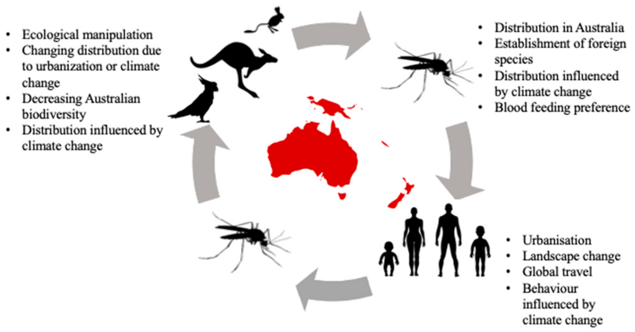
Table 1.
A summary of pathogen, pathogen distribution, mosquito vectors, and symptomatic a and asymptomatic b host distribution in Australia. Dengue and Rift Valley fever are not listed as there have been no known non-human vertebrate hosts in Australia.
Table 1.
A summary of pathogen, pathogen distribution, mosquito vectors, and symptomatic a and asymptomatic b host distribution in Australia. Dengue and Rift Valley fever are not listed as there have been no known non-human vertebrate hosts in Australia.
| Pathogen | Pathogen Distribution in Australia | Mosquito Vectors | Asymptomatic Hosts of Pathogen in Australia | Symptomatic Hosts of Pathogen in Australia |
|---|---|---|---|---|
| Ross River virus (RRV) | All of Australia [13] | Aedes and Culex mosquitoes, particularly Aedes vigilax, Aedes camptohynchus, and Culex annulirostris [14] | Marsupials: wallabies, wallaroos [15], common brushtail possums [16], eastern grey kangaroos [17], western grey kangaroos [18] Australian birds: little corella, magpie larks, Australian brown flycatcher, masked finch [19,20] Wild eutherian mammals: rodents, Pteropus spp. [20] Domestic mammals: cattle, dogs [20], cats [21] | Domestic mammals: horses [20,22] |
| Barmah Forest virus (BFV) | All of Australia [23] | Culex annuliristris, Aedes normanensis, Aedes vigilax and Aedes procax [24] | Marsupials: eastern grey kangaroo [25], koalas [25] and brushtail possums [21]. Wild eutherian mammals: Australian bush rats and swamp rats [26] Domestic mammals: cats, dogs, horses [27] | |
| Sindbis virus (SINV) | Most of Australia (excluding Tasmania) [28] | Culex annulirostris, Aedes normanensis, Aedes camptorhynchus [29], Aedes pseudonormanensis [30] | Marsupials: chudditch [31] Wild eutherian mammals: European rabbits [31] Domestic mammals: horses [31] Birds: Emus [31] | |
| Murray Valley encephalitis virus (MVEV) | Western Australia [32], Northern Territory [33], New South Wales, and Victoria [34] | Culex annulirostris [35], Culex sitiens and other Culicine mosquitoes [36] | Marsupials: eastern grey kangaroos [37], western grey kangaroos, agile wallabies [38] Australian birds: galahs, sulphur-crested cockatoos [38], chickens [39] Wild eutherian mammals: rabbits [37], wild mice [38] Domestic mammals: dogs, sheep, pigs, cattle [37] Water birds: rufous night herons [34], Pacific black ducks [38] | Domestic mammals: horses [22,40,41] |
| West Nile virus (WNV) | All of Australia [42,43] | Mainly isolated from Culex annulirostris [44]. Other Culex species, Aedes species and Anopheles amictus can also transmit the virus [45,46] | Marsupials: western grey kangaroos, agile wallabies [37] Australian bird: Australian white ibis [47] Ardeid birds: herons, egrets [48] Introduced bird: house sparrow [49]. | Wild eutherian mammals: rabbits [50,51] Domestic mammals: horses [22,42], cats (mild) [52] |
| Japanese encephalitis virus (JEV) | Torres Strait (incursion) [53], North Peninsula Area and mainland [54] | Culex annulirostris [53]. | Ardeid birds: herons, egrets [55,56,57] Domestic mammals: pigs [58], horses [59] | Wild eutherian mammals: Frugivorous bats i.e., black flying fox [60] Other birds: pigeons, sparrows, ducks, chickens [60] |
| Kokobera (KOKV) and related viruses | Queensland, New South Wales, Northern Territory, Western Australia, and Papua New Guinea [61,62] | Aedes species including Aedes aculeatus, Aedes alternas, Aedes notoscriptus, Aedes procax, Aedes vigilax and Anopheles annulipes [63] | Marsupials: mainly kangaroos and wallabies [17,64]. Domestic mammals: cattle [26] | Domestic mammals: horses [64,65] It is unknown whether horses are affected by Kokobera and related viruses, as it could be associated with a known equine disease. |
| Gan Gan (GGV) and Trubanaman viruses (TRUV) | Queensland, New South Wales, and Western Australia [66] | Aedes vigilax (GGV) [67], Culex annulirostris (GGV and TRUV) [62,68], Anopheles annulipes (TRUV) [66] and Anopheles meraukensis [69] | Marsupials: eastern grey kangaroos (GGV, TRUV), red-necked wallaby (GGV, TRUV) [26], western grey kangaroos (TRUV) [70] Wild eutherian mammals: Australian bush rat (GGV) [26], feral pigs (TRUV), rabbits (TRUV), foxes (TRUV), quokkas (TRUV) [70] Domestic mammals: sheep (GGV), horses (GGV, TRUV), cattle (GGV) [26,70] |
a A asymptomatic host would be a reservoir host species that does not experience the symptoms of disease but is able to produce sufficient pathogen levels to infect mosquitoes that blood-feed on that animal. b A symptomatic host would be a non-reservoir host species that show symptoms of the disease and also produces sufficient pathogen levels to infect mosquitoes that blood-feed on that animal.
Publisher’s Note: MDPI stays neutral with regard to jurisdictional claims in published maps and institutional affiliations. |
© 2021 by the authors. Licensee MDPI, Basel, Switzerland. This article is an open access article distributed under the terms and conditions of the Creative Commons Attribution (CC BY) license (http://creativecommons.org/licenses/by/4.0/).
Share and Cite
MDPI and ACS Style
Ong, O.T.W.; Skinner, E.B.; Johnson, B.J.; Old, J.M. Mosquito-Borne Viruses and Non-Human Vertebrates in Australia: A Review. Viruses 2021, 13, 265. https://0-doi-org.brum.beds.ac.uk/10.3390/v13020265
AMA Style
Ong OTW, Skinner EB, Johnson BJ, Old JM. Mosquito-Borne Viruses and Non-Human Vertebrates in Australia: A Review. Viruses. 2021; 13(2):265. https://0-doi-org.brum.beds.ac.uk/10.3390/v13020265
Chicago/Turabian StyleOng, Oselyne T. W., Eloise B. Skinner, Brian J. Johnson, and Julie M. Old. 2021. "Mosquito-Borne Viruses and Non-Human Vertebrates in Australia: A Review" Viruses 13, no. 2: 265. https://0-doi-org.brum.beds.ac.uk/10.3390/v13020265
Note that from the first issue of 2016, this journal uses article numbers instead of page numbers. See further details here.