
Post-COVID-19 Arthritis and Sacroiliitis: Natural History with Longitudinal Magnetic Resonance Imaging Study in Two Cases and Review of the Literature

 ,
,
Abstract
:1. Introduction
2. Case Reports
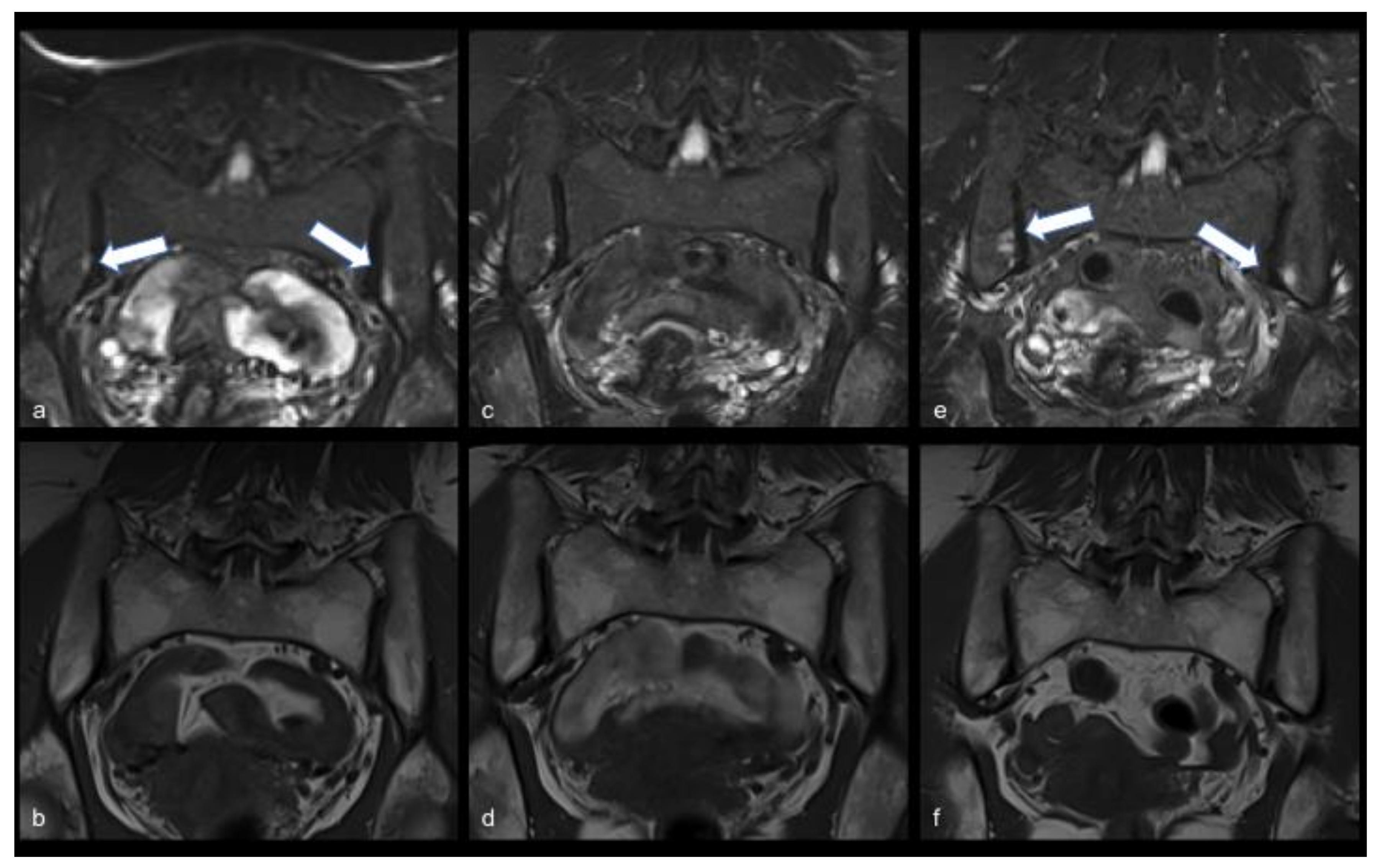
2.1. Case Report 1
2.2. Case Report 2
3. Materials and Methods
4. Results
5. Discussion
Author Contributions
Funding
Institutional Review Board Statement
Informed Consent Statement
Data Availability Statement
Acknowledgments
Conflicts of Interest
References
- Baj, J.; Karakuła-Juchnowicz, H.; Teresiński, G.; Buszewicz, G.; Ciesielka, M.; Sitarz, E.; Forma, A.; Karakuła, K.; Flieger, W.; Portincasa, P.; et al. COVID-19: Specific and Non-Specific Clinical Manifestations and Symptoms: The Current State of Knowledge. J. Clin. Med. 2020, 9, 1753. [Google Scholar] [CrossRef] [PubMed]
- Cascella, M.; Rajnik, M.; Cuomo, A.; Dulebohn, S.C.; Di Napoli, R. Features, Evaluation, and Treatment of Coronavirus (COVID-19). In StatPearls; StatPearls Publishing: Treasure Island, FL, USA, 2021. [Google Scholar]
- Chen, N.; Zhou, M.; Dong, X.; Qu, J.; Gong, F.; Han, Y.; Qiu, Y.; Wang, J.; Liu, Y.; Wei, Y.; et al. Epidemiological and Clinical Characteristics of 99 Cases of 2019 Novel Coronavirus Pneumonia in Wuhan, China: A Descriptive Study. Lancet 2020, 395, 507–513. [Google Scholar] [CrossRef] [Green Version]
- Cevik, M.; Kuppalli, K.; Kindrachuk, J.; Peiris, M. Virology, Transmission, and Pathogenesis of SARS-CoV-2. BMJ 2020, 371, m3862. [Google Scholar] [CrossRef] [PubMed]
- Pascarella, G.; Strumia, A.; Piliego, C.; Bruno, F.; Del Buono, R.; Costa, F.; Scarlata, S.; Agrò, F.E. COVID-19 Diagnosis and Management: A Comprehensive Review. J. Intern. Med. 2020, 288, 192–206. [Google Scholar] [CrossRef] [PubMed]
- Kamal, M.; Abo Omirah, M.; Hussein, A.; Saeed, H. Assessment and Characterisation of Post-COVID-19 Manifestations. Int. J. Clin. Pract. 2020, 75, e13746. [Google Scholar] [CrossRef]
- Kanwar, D.; Baig, A.M.; Wasay, M. Neurological Manifestations of COVID-19. J. Pak. Med. Assoc. 2020, 70 (Suppl. S3), S101–S103. [Google Scholar] [CrossRef]
- Abu-Rumeileh, S.; Abdelhak, A.; Foschi, M.; Tumani, H.; Otto, M. Guillain-Barré Syndrome Spectrum Associated with COVID-19: An up-to-Date Systematic Review of 73 Cases. J. Neurol. 2020, 268, 1133–1170. [Google Scholar] [CrossRef]
- El Sayed, S.; Shokry, D.; Gomaa, S.M. Post-COVID-19 Fatigue and Anhedonia: A Cross-Sectional Study and Their Correlation to Post-Recovery Period. Neuropsychopharmacol. Rep. 2020, 41, 50–55. [Google Scholar] [CrossRef]
- Wiersinga, W.J.; Rhodes, A.; Cheng, A.C.; Peacock, S.J.; Prescott, H.C. Pathophysiology, Transmission, Diagnosis, and Treatment of Coronavirus Disease 2019 (COVID-19): A Review. JAMA 2020, 324, 782–793. [Google Scholar] [CrossRef]
- Yelin, D.; Wirtheim, E.; Vetter, P.; Kalil, A.C.; Bruchfeld, J.; Runold, M.; Guaraldi, G.; Mussini, C.; Gudiol, C.; Pujol, M.; et al. Long-Term Consequences of COVID-19: Research Needs. Lancet Infect. Dis. 2020, 20, 1115–1117. [Google Scholar] [CrossRef]
- Gupta, A.; Madhavan, M.V.; Sehgal, K.; Nair, N.; Mahajan, S.; Sehrawat, T.S.; Bikdeli, B.; Ahluwalia, N.; Ausiello, J.C.; Wan, E.Y.; et al. Extrapulmonary Manifestations of COVID-19. Nat. Med. 2020, 26, 1017–1032. [Google Scholar] [CrossRef]
- Talarico, R.; Stagnaro, C.; Ferro, F.; Carli, L.; Mosca, M. Symmetric Peripheral Polyarthritis Developed during SARS-CoV-2 Infection. Lancet Rheumatol. 2020, 2, e518–e519. [Google Scholar] [CrossRef]
- Gasparotto, M.; Framba, V.; Piovella, C.; Doria, A.; Iaccarino, L. Post-COVID-19 Arthritis: A Case Report and Literature Review. Clin. Rheumatol. 2021, 40, 3357–3362. [Google Scholar] [CrossRef]
- Liew, I.Y.; Mak, T.M.; Cui, L.; Vasoo, S.; Lim, X.R. A Case of Reactive Arthritis Secondary to Coronavirus Disease 2019 Infection. J. Clin. Rheumatol. 2020, 26, 233. [Google Scholar] [CrossRef]
- Yokogawa, N.; Minematsu, N.; Katano, H.; Suzuki, T. Case of Acute Arthritis Following SARS-CoV-2 Infection. Ann. Rheum. Dis. 2020, 80, e101. [Google Scholar] [CrossRef]
- Di Carlo, M.; Tardella, M.; Salaffi, F. Can SARS-CoV-2 Induce Reactive Arthritis? Clin. Exp. Rheumatol. 2021, 39 (Suppl. S128), 25–26. [Google Scholar]
- Ono, K.; Kishimoto, M.; Shimasaki, T.; Uchida, H.; Kurai, D.; Deshpande, G.A.; Komagata, Y.; Kaname, S. Reactive Arthritis after COVID-19 Infection. RMD Open 2020, 6, e001350. [Google Scholar] [CrossRef]
- Schett, G.; Manger, B.; Simon, D.; Caporali, R. COVID-19 Revisiting Inflammatory Pathways of Arthritis. Nat. Rev. Rheumatol. 2020, 16, 465–470. [Google Scholar] [CrossRef]
- Jali, I. Reactive Arthritis After COVID-19 Infection. Cureus 2020, 12, e11761. [Google Scholar] [CrossRef]
- Danssaert, Z.; Raum, G.; Hemtasilpa, S. Reactive Arthritis in a 37-Year-Old Female With SARS-CoV2 Infection. Cureus 2020, 12, e9698. [Google Scholar] [CrossRef]
- Fragata, I.; Mourão, A.F. Coronavirus Disease 19 (COVID-19) Complicated with Post-Viral Arthritis. Acta Reumatol. Port. 2020, 45, 278–280. [Google Scholar] [PubMed]
- Jovani, V.; Pascual, E.; Vela, P.; Andrés, M. Acute Arthritis Following SARS-CoV-2 Infection. J. Med. Virol. 2021, 93, 661. [Google Scholar] [CrossRef] [PubMed]
- Hønge, B.L.; Hermansen, M.-L.F.; Storgaard, M. Reactive Arthritis after COVID-19. BMJ Case Rep. 2021, 14. [Google Scholar] [CrossRef]
- Wendling, D.; Verhoeven, F.; Chouk, M.; Prati, C. Can SARS-CoV-2 Trigger Reactive Arthritis? Jt. Bone Spine 2021, 88, 105086. [Google Scholar] [CrossRef] [PubMed]
- Davido, B.; Seang, S.; Tubiana, R.; de Truchis, P. Post-COVID-19 Chronic Symptoms: A Postinfectious Entity? Clin. Microbiol. Infect. 2020, 26, 1448–1449. [Google Scholar] [CrossRef]
- Saricaoglu, E.M.; Hasanoglu, I.; Guner, R. The First Reactive Arthritis Case Associated with COVID-19. J. Med. Virol. 2021, 93, 192–193. [Google Scholar] [CrossRef]
- López-González, M.-D.-C.; Peral-Garrido, M.L.; Calabuig, I.; Tovar-Sugrañes, E.; Jovani, V.; Bernabeu, P.; García-Sevila, R.; León-Ramírez, J.-M.; Moreno-Perez, O.; Boix, V.; et al. Case Series of Acute Arthritis during COVID-19 Admission. Ann. Rheum. Dis. 2020, 80, e58. [Google Scholar] [CrossRef]
- Parisi, S.; Borrelli, R.; Bianchi, S.; Fusaro, E. Viral Arthritis and COVID-19. Lancet Rheumatol. 2020, 2, e655–e657. [Google Scholar] [CrossRef]
- Alivernini, S.; Cingolani, A.; Gessi, M.; Paglionico, A.; Pasciuto, G.; Tolusso, B.; Fantoni, M.; Gremese, E. Comparative Analysis of Synovial Inflammation after SARS-CoV-2 Infection. Ann. Rheum. Dis. 2020, 80, e91. [Google Scholar] [CrossRef]
- De Stefano, L.; Rossi, S.; Montecucco, C.; Bugatti, S. Transient Monoarthritis and Psoriatic Skin Lesions Following COVID-19. Ann. Rheum. Dis. 2020. [Google Scholar] [CrossRef]
- Schenker, H.M.; Hagen, M.; Simon, D.; Schett, G.; Manger, B. Reactive Arthritis and Cutaneous Vasculitis after SARS-CoV-2 Infection. Rheumatology 2021, 60, 479–480. [Google Scholar] [CrossRef]
- Salvatierra, J.; Martínez-Peñalver, D.; Salvatierra-Velasco, L. CoVid-19 Related Dactyitis. Jt. Bone Spine 2020, 87, 660. [Google Scholar] [CrossRef]
- Novelli, L.; Motta, F.; Ceribelli, A.; Guidelli, G.M.; Luciano, N.; Isailovic, N.; Vecellio, M.; Caprioli, M.; Clementi, N.; Clementi, M.; et al. A Case of Psoriatic Arthritis Triggered by SARS-CoV-2 Infection. Rheumatology 2021, 60, e21–e23. [Google Scholar] [CrossRef]
- Baimukhamedov, C.; Barskova, T.; Matucci-Cerinic, M. Arthritis after SARS-CoV-2 Infection. Lancet Rheumatol. 2021, 3, e324–e325. [Google Scholar] [CrossRef]
- Shokraee, K.; Moradi, S.; Eftekhari, T.; Shajari, R.; Masoumi, M. Reactive Arthritis in the Right Hip Following COVID-19 Infection: A Case Report. Trop. Dis. Travel Med. Vaccines 2021, 7, 18. [Google Scholar] [CrossRef]
- Cincinelli, G.; Di Taranto, R.; Orsini, F.; Rindone, A.; Murgo, A.; Caporali, R. A Case Report of Monoarthritis in a COVID-19 Patient and Literature Review. Medicine 2021, 100, e26089. [Google Scholar] [CrossRef]
- El Hasbani, G.; Jawad, A.; Uthman, I. Axial and Peripheral Spondyloarthritis Triggered by Sars-Cov-2 Infection: A Report of Two Cases. Reumatismo 2021, 73, 59–63. [Google Scholar] [CrossRef]
- Coath, F.L.; Mackay, J.; Gaffney, J.K. Axial Presentation of Reactive Arthritis Secondary to COVID-19 Infection. Rheumatology 2021, 60, e232–e233. [Google Scholar] [CrossRef]
- Braun, J.; Kingsley, G.; van der Heijde, D.; Sieper, J. On the Difficulties of Establishing a Consensus on the Definition of and Diagnostic Investigations for Reactive Arthritis. Results and Discussion of a Questionnaire Prepared for the 4th International Workshop on Reactive Arthritis, Berlin, Germany, July 3–6, 1999. J. Rheumatol. 2000, 27, 2185–2192. [Google Scholar]
- Campos-Correia, D.; Sudoł-Szopińska, I.; Diana Afonso, P. Are We Overcalling Sacroiliitis on MRI? Differential Diagnosis That Every Rheumatologist Should Know—Part I. Acta Reumatol. Port. 2019, 44, 29–41. [Google Scholar]
- De Winter, J.; de Hooge, M.; van de Sande, M.; de Jong, H.; van Hoeven, L.; de Koning, A.; Berg, I.J.; Ramonda, R.; Baeten, D.; van der Heijde, D.; et al. Magnetic Resonance Imaging of the Sacroiliac Joints Indicating Sacroiliitis According to the Assessment of SpondyloArthritis International Society Definition in Healthy Individuals, Runners, and Women With Postpartum Back Pain. Arthritis Rheumatol. 2018, 70, 1042–1048. [Google Scholar] [CrossRef] [PubMed]
- Slobodin, G.; Rimar, D.; Boulman, N.; Kaly, L.; Rozenbaum, M.; Rosner, I.; Odeh, M. Acute Sacroiliitis. Clin. Rheumatol. 2016, 35, 851–856. [Google Scholar] [CrossRef] [PubMed]
- Silva Andrade, B.; Siqueira, S.; de Assis Soares, W.R.; de Souza Rangel, F.; Santos, N.O.; Dos Santos Freitas, A.; Ribeiro da Silveira, P.; Tiwari, S.; Alzahrani, K.J.; Góes-Neto, A.; et al. Long-COVID and Post-COVID Health Complications: An Up-to-Date Review on Clinical Conditions and Their Possible Molecular Mechanisms. Viruses 2021, 13, 700. [Google Scholar] [CrossRef] [PubMed]
- Burgio, S.; Conway de Macario, E.; Macario, A.J.; Cappello, F. SARS-CoV-2 in Patients with Cancer: Possible Role of Mimicry of Human Molecules by Viral Proteins and the Resulting Anti-Cancer Immunity. Cell Stress Chaperones 2021, 26, 611–616. [Google Scholar] [CrossRef]
- Bachmaier, K.; Penninger, J.M. Chlamydia and Antigenic Mimicry. Curr. Top. Microbiol. Immunol. 2005, 296, 153–163. [Google Scholar] [CrossRef]
- Sedlackova, L.; Nguyen, T.T.H.; Zlacka, D.; Sosna, A.; Hromadnikova, I. Cell Surface and Relative MRNA Expression of Heat Shock Protein 70 in Human Synovial Cells. Autoimmunity 2009, 42, 17–24. [Google Scholar] [CrossRef]
- Sedlackova, L.; Sosna, A.; Vavrincova, P.; Frýdl, J.; Guerriero, V.; Raynes, D.A.; Hromadnikova, I. Heat Shock Protein Gene Expression Profile May Differentiate between Rheumatoid Arthritis, Osteoarthritis, and Healthy Controls. Scand. J. Rheumatol. 2011, 40, 354–357. [Google Scholar] [CrossRef]
- Schmitt, S.K. Reactive Arthritis. Infect. Dis. Clin. N. Am. 2017, 31, 265–277. [Google Scholar] [CrossRef]
- García-Kutzbach, A.; Chacón-Súchite, J.; García-Ferrer, H.; Iraheta, I. Reactive Arthritis: Update 2018. Clin. Rheumatol. 2018, 37, 869–874. [Google Scholar] [CrossRef]
- Selmi, C.; Gershwin, M.E. Diagnosis and Classification of Reactive Arthritis. Autoimmun. Rev. 2014, 13, 546–549. [Google Scholar] [CrossRef]
- Manasson, J.; Shen, N.; Garcia Ferrer, H.R.; Ubeda, C.; Iraheta, I.; Heguy, A.; Von Feldt, J.M.; Espinoza, L.R.; Garcia Kutzbach, A.; Segal, L.N.; et al. Gut Microbiota Perturbations in Reactive Arthritis and Postinfectious Spondyloarthritis. Arthritis Rheumatol. 2018, 70, 242–254. [Google Scholar] [CrossRef] [Green Version]
- Qaiyum, Z.; Lim, M.; Inman, R.D. The Gut-Joint Axis in Spondyloarthritis: Immunological, Microbial, and Clinical Insights. Semin. Immunopathol. 2021, 43, 173–192. [Google Scholar] [CrossRef]
- Quartuccio, L.; Sonaglia, A.; McGonagle, D.; Fabris, M.; Peghin, M.; Pecori, D.; De Monte, A.; Bove, T.; Curcio, F.; Bassi, F.; et al. Profiling COVID-19 Pneumonia Progressing into the Cytokine Storm Syndrome: Results from a Single Italian Centre Study on Tocilizumab versus Standard of Care. J. Clin. Virol. 2020, 129, 104444. [Google Scholar] [CrossRef]
- Quartuccio, L.; Fabris, M.; Sonaglia, A.; Peghin, M.; Domenis, R.; Cifù, A.; Curcio, F.; Tascini, C. Interleukin 6, Soluble Interleukin 2 Receptor Alpha (CD25), Monocyte Colony-Stimulating Factor, and Hepatocyte Growth Factor Linked with Systemic Hyperinflammation, Innate Immunity Hyperactivation, and Organ Damage in COVID-19 Pneumonia. Cytokine 2021, 140, 155438. [Google Scholar] [CrossRef]
- Benucci, M.; Damiani, A.; Infantino, M.; Manfredi, M.; Quartuccio, L. Old and New Antirheumatic Drugs for the Treatment of COVID-19. Jt. Bone Spine 2020, 87, 195–197. [Google Scholar] [CrossRef]
- Henderson, L.A.; Canna, S.W.; Schulert, G.S.; Volpi, S.; Lee, P.Y.; Kernan, K.F.; Caricchio, R.; Mahmud, S.; Hazen, M.M.; Halyabar, O.; et al. On the Alert for Cytokine Storm: Immunopathology in COVID-19. Arthritis Rheumatol. 2020, 72, 1059–1063. [Google Scholar] [CrossRef] [Green Version]
- Zhou, Q.; Vadakekolathu, J.; Watad, A.; Sharif, K.; Russell, T.; Rowe, H.; Khan, A.; Millner, P.A.; Loughenbury, P.; Rao, A.; et al. SARS-CoV-2 Infection Induces Psoriatic Arthritis Flares and Enthesis Resident Plasmacytoid Dendritic Cell Type-1 Interferon Inhibition by JAK Antagonism Offer Novel Spondyloarthritis Pathogenesis Insights. Front. Immunol. 2021, 12, 635018. [Google Scholar] [CrossRef]
- Yokota, S.; Miyamae, T.; Kuroiwa, Y.; Nishioka, K. Novel Coronavirus Disease 2019 (COVID-19) and Cytokine Storms for More Effective Treatments from an Inflammatory Pathophysiology. J. Clin. Med. 2021, 10, 801. [Google Scholar] [CrossRef]
- Vieira, M.; Maalouf, G.; Hasan, M.; Le Joncour, A.; Karkeni, E.; Idir, M.; Amelin, D.; Salem, J.-E.; Gougis, P.; Lacorte, J.-M.; et al. Cytokine Profile as a Prognostic Tool in Coronavirus Disease 2019. Comment on “Urgent Avenues in the Treatment of COVID-19: Targeting Downstream Inflammation to Prevent Catastrophic Syndrome” by Quartuccio et al. Joint Bone Spine. 2020;87:191-93. Jt. Bone Spine 2021, 88, 105074. [Google Scholar] [CrossRef]
- Alunno, A.; Najm, A.; Machado, P.M.; Bertheussen, H.; Burmester, G.R.; Carubbi, F.; De Marco, G.; Giacomelli, R.; Hermine, O.; Isaacs, J.D.; et al. EULAR Points to Consider on Pathophysiology and Use of Immunomodulatory Therapies in COVID-19. Ann. Rheum. Dis. 2021, 80, 698–706. [Google Scholar] [CrossRef]
- Joo, Y.B.; Lim, Y.-H.; Kim, K.-J.; Park, K.-S.; Park, Y.-J. Respiratory Viral Infections and the Risk of Rheumatoid Arthritis. Arthritis Res. Ther. 2019, 21, 199. [Google Scholar] [CrossRef] [PubMed] [Green Version]
- Tripathy, A.; Swain, N.; Gupta, B. The COVID-19 Pandemic: An Increased Risk of Rheumatoid Arthritis. Future Virol. 2021, 16. [Google Scholar] [CrossRef] [PubMed]
- Liu, Y.; Sawalha, A.H.; Lu, Q. COVID-19 and Autoimmune Diseases. Curr. Opin. Rheumatol. 2021, 33, 155–162. [Google Scholar] [CrossRef] [PubMed]
- Ferri, C.; Giuggioli, D.; Raimondo, V.; L’Andolina, M.; Tavoni, A.; Cecchetti, R.; Guiducci, S.; Ursini, F.; Caminiti, M.; Varcasia, G.; et al. COVID-19 and Rheumatic Autoimmune Systemic Diseases: Report of a Large Italian Patients Series. Clin. Rheumatol. 2020, 39, 3195–3204. [Google Scholar] [CrossRef]


| Feature | Patient 1 | Patient 2 |
|---|---|---|
| Age, year | 58 | 53 |
| Gender | Female | Female |
| Comorbidity | Autoimmune hypothyroidism | Autoimmune hypothyroidism |
| First COVID-related symptoms | End of March | End of March |
| First sacroiliitis symptoms | End of April | End of April |
| Improvement of peripheral pain and inflammatory back pain | End of May | End of May |
| Nasopharyngeal SARS-CoV-2 POSITIVE buffer (date, month/day) | 03/27, 04/04, 04/07, 04/17 | 03/27, 04/10, 04/17 |
| Nasopharyngeal SARS-CoV-2 NEGATIVE buffer (date) | 04/24, 04/28, 05/12, 05/19 | 04/24, 04/28, 05/12, 05/19 |
| Drugs for the infection | HCQ and azithromycin | HCQ and azithromycin |
| Treatment of the sacroiliitis | NSAIDs | NSAIDs |
| Clinical outcome | Recovery | Recovery |
| First MRI | June 2020, bilateral sacroiliitis | June 2020 unilateral sacroiliitis |
| Second MRI | July 2020, bilateral sacroiliitis | July 2020 unilateral sacroiliitis |
| Third MRI | January 2021, bilateral sacroiliitis | February 2021, unilateral sacroiliitis |
| LABORATORY FEATURES | Patient 1 | Patient 2 | ||||
|---|---|---|---|---|---|---|
| First Visit (June 20) | Second Visit (July 20) | Last Visit (March 21) | First Visit (June 20) | Second Visit (July 20) | Last Visit (March 21) | |
| Serology (UA/mL) (Negative < 8; Borderline 8–12; Positive > 12) | IgG 65.4 UA/mL IgM 2.76 UA/mL | IgG 75 UA/mL, IgM 12.4 UA/mL | ND | IgG 32.2 UA/mL, IgM 1.9 UA/mL | IgG 17.3 UA/mL, IgM 0.9 UA/mL | ND |
| Fibrinogen, mg/dL (normal range 180–380) | 248 | 295 | 250 | 272 | 406 | 247 |
| CRP, mg/L (normal range 0–5.00) | 1.20 | 2.24 | 1.41 | 1.49 | 4.56 | 2.61 |
| IL1 beta, pg/mL (normal range < 0.001) | 0. | 0. | ND | 0. | 0 | ND |
| IL6, pg/mL (normal range < 7) | 0.9 | 2 | 2 | 1.3 | 2 | 2 |
| IL2R, pg/mL (normal range 440–1435) | 1346 | 1446 | ND | 904 | 835 | ND |
| IL8, pg/mL (normal range 6.7–16.2) | 12.6 | 13.9 | ND | 19.2 | 53 | ND |
| IL10, pg/mL (normal range 1.8–3.8) | 1.8 | 2 | ND | 2.1 | 2 | ND |
| TNF alpha, pg/mL (normal range 7.8–12.2) | 8.9 | 13.2 | ND | 11 | 66 | ND |
| IFN gamma, pg/mL (normal range < 0.99) | 0 | 1.3 | ND | 0 | 0 | ND |
| IP10, pg/mL (normal range 37.2–222) | 112 | 127 | ND | 90.6 | 96 | ND |
| Haemoglobin, g/dL | 13.7 | 13.4 | 13.9 | 13.8 | 13.1 | 13.3 |
| Leucocyte count, cell/mm3 | 3970 | 4050 | 4400 | 3960 | 4970 | 5070 |
| Platelet count, cell/mm3 | 197,000 | 223,000 | 202,000 | 321,000 | 305,000 | 357,000 |
| Antinuclear antibody (ANA) | Negative | ND | Negative | Negative | ND | Negative |
| Anti-SSA/SSB | Negative | ND | ND | Negative | ND | ND |
| Rheumatoid factor (RF) | Negative | ND | ND | Negative | ND | ND |
| CPK, IU/L (normal range 26–170) | 187 | 266 | 171 | 101 | 147 | 102 |
| Sex and Age | Onset | Articular Involvement | SF Analysis | Presence of Crystals | SF Culture | SF RT-PCR for SARS-CoV2 | RF and/or ACPA | ANA | HLA-B 27 | Synovial Biopsy | Imaging | Therapy | Outcome | |
|---|---|---|---|---|---|---|---|---|---|---|---|---|---|---|
| Talarico et al., 2020 [13] | M 45 | at COVID-19 diagnosis | polyarticular (bilateral MCP and PIP, right wrist) | NA | NA | NA | NA | neg | NA | NA | NA | US: slight effusion of the involved joints | oral steroid treatment | arthralgia after steroid suspension |
| Gasparotto et al., 2020 [14] | M 60 | 32 days after COVID-19 diagnosis | right knee and ankle | yes | no | neg | neg | neg | neg | neg | NA | oral NSAIDs | recovered | |
| Liew et al., 2020 [15] | M 47 | at COVID-19 diagnosis | right knee | yes | no | (neg GRAM stain) | neg | NA | NA | NA | NA | X-rays: no erosion | oral NSAIDs and intra-articular steroid | long-term data not available |
| Yokogawa et al., 2020 [16] | M 57 | 15 days after diagnosis | right knee | yes | no | NA | neg | neg | neg | neg | NA | NA | no therapy | recovered |
| Di Carlo et al., 2020 [17] | M 55 | 4 weeks after COVID-19 infection | right ankle, subtalar joint synovitis | NA | NA | NA | NA | NA | NA | neg | NA | US: subtalar joint synovitis | oral steroid treatment | recovered |
| Ono et al., 2020 [18] | M 50 | 21 days after COVID-19 diagnosis | left and right ankle | yes | no | neg | NA | neg | neg | neg | NA | X-rays: no erosion | oral NSAIDs and intra-articular steroid | moderate improvement, long-term data not available |
| Jali et al., 2020 [20] | F 39 | 3 weeks after infection | DIP and PIP bilateral hands | NA | NA | NA | NA | neg | neg | NA | NA | X-rays: normal | oral NSAIDs | recovered |
| Danssaert et al., 2020 [21] | F 37 | 12 days after COVID-19 diagnosis | tendonitis of the ll, lll, lV right hand extensor | NA | NA | NA | NA | neg | neg | NA | NA | MRI inflammation around the extensor involved | topical NSAIDs, oral opioid gabapentin | improvement without complete recovery, long-term data not available |
| Fragata et al., 2020 [22] | F 41 | 4 weeks after COVID-19 symptoms | polyarticular (small joints of the right hand) | NA | NA | NA | NA | neg | neg | NA | NA | NA | oral NSAIDs and oral prednisolone | recovered |
| Langhoff Hønge et al., 2020 [24] | M 53 | 4 days after hospital discharge for severe respiratory insufficiency | polyarticular (right knee bilateral ankles, lateral side of the left foot) | yes | no | neg | NA | neg | NA | neg | NA | X-rays: fluid accumulation on the right knee | oral NSAIDs and oral prednisolone | recovered |
| Saricaoglu et al., 2020 [27] | M 73 | subacute | left l MTP, PIP, right ll PIP, DIP | NA | NA | NA | NA | neg | neg | NA | NA | X-rays: no erosion | oral NSAIDS | recovered |
| Parisi et al., 2020 [29] | F 58 | 25 days after COVID-19 symptoms | ankle arthritis | NA | NA | NA | NA | neg | neg | neg | NA | NA | oral NSAIDs | recovery from symptoms, US still positive after one month |
| Alivernini et al., 2020 [30] | M 61 | at COVID-19 diagnosis | polyarticular | yes | no | NA | NA | neg | neg | NA | Inflammatory infiltrates in the synovia | US: synovitis | baricitinib and oral prednisone | recovered |
| De Stefano et al., 2020 [31] | M 30 | 10 days post COVID-19 symptoms resolution | right elbow | yes | no | NA | NA | neg | neg | neg | NA | US: synovitis PD+ | oral NSAIDs | recovered |
| Schenker et al., 2020 [32] | F 65 | 10 days after COVID-19 symptoms cessation | polyarticular (bilateral ankles knee and wrists joints) | NA | NA | NA | NA | neg | neg | pos | NA | NA | prednisolone | immediate steroid response, follow-up not available |
| Salvatierra et al., 2020 [33] | F 16 | 3 weeks after COVID-19 symptoms | II, IV and V left toes dactylitis | NA | NA | NA | NA | neg | neg | neg | NA | NA | oral NSAIDs | recovered |
| Shokraee et al., 2021 [36] | F 58 | 2 weeks after COVID-19 symptoms initiation | right hip | NA | NA | NA | NA | NA | NA | NA | NA | US: articular effusion and synovium thickness MRI: fluid rim | oral NSAIDs and prednisolone | recovered |
| Cincinelli et al., 2021 [37] | M 27 | 2 weeks after COVID-19 diagnosis | I MCF | NA | NA | NA | NA | NA | NA | NA | NA | NA | oral NSAIDs and oral prednisolone | recovered |
| El Hasbani et al. (case 2) [38] | M 57 | One month after COVID-19 infection | left wrist | NA | NA | NA | NA | neg | neg | pos | NA | MRI: wrist synovitis | oral prednisolone and oral NSAIDs | recovered |
Publisher’s Note: MDPI stays neutral with regard to jurisdictional claims in published maps and institutional affiliations. |
© 2021 by the authors. Licensee MDPI, Basel, Switzerland. This article is an open access article distributed under the terms and conditions of the Creative Commons Attribution (CC BY) license (https://creativecommons.org/licenses/by/4.0/).
Share and Cite
Colatutto, D.; Sonaglia, A.; Zabotti, A.; Cereser, L.; Girometti, R.; Quartuccio, L. Post-COVID-19 Arthritis and Sacroiliitis: Natural History with Longitudinal Magnetic Resonance Imaging Study in Two Cases and Review of the Literature. Viruses 2021, 13, 1558. https://0-doi-org.brum.beds.ac.uk/10.3390/v13081558
Colatutto D, Sonaglia A, Zabotti A, Cereser L, Girometti R, Quartuccio L. Post-COVID-19 Arthritis and Sacroiliitis: Natural History with Longitudinal Magnetic Resonance Imaging Study in Two Cases and Review of the Literature. Viruses. 2021; 13(8):1558. https://0-doi-org.brum.beds.ac.uk/10.3390/v13081558
Chicago/Turabian StyleColatutto, Donatella, Arianna Sonaglia, Alen Zabotti, Lorenzo Cereser, Rossano Girometti, and Luca Quartuccio. 2021. "Post-COVID-19 Arthritis and Sacroiliitis: Natural History with Longitudinal Magnetic Resonance Imaging Study in Two Cases and Review of the Literature" Viruses 13, no. 8: 1558. https://0-doi-org.brum.beds.ac.uk/10.3390/v13081558