Article

Detection and Complete Genome Analysis of Circoviruses and Cycloviruses in the Small Indian Mongoose (Urva auropunctata): Identification of Novel Species

1
Department of Biomedical Sciences, Ross University School of Veterinary Medicine, Basseterre P.O. Box 334, Saint Kitts and Nevis, West Indies
2
College of Animal Biotechnology, Guru Angad Dev Veterinary and Animal Science University, Ludhiana 141001, India
*
Author to whom correspondence should be addressed.
Submission received: 26 July 2021 / Revised: 23 August 2021 / Accepted: 24 August 2021 / Published: 27 August 2021
(This article belongs to the Special Issue Emerging Wildlife Viral Diseases)

Abstract

:
Fecal samples from 76 of 83 apparently healthy small Indian mongooses (Urva auropunctata) were PCR positive with circovirus/cyclovirus pan-rep (replicase gene) primers. In this case, 30 samples yielded high quality partial rep sequences (~400 bp), of which 26 sequences shared maximum homology with cycloviruses from an arthropod, bats, humans or a sheep. Three sequences exhibited maximum identities with a bat circovirus, whilst a single sequence could not be assigned to either genus. Using inverse nested PCRs, the complete genomes of mongoose associated circoviruses (Mon-1, -29 and -66) and cycloviruses (Mon-20, -24, -32, -58, -60 and -62) were determined. Mon-1, -20, -24, -29, -32 and -66 shared <80% maximum genome-wide pairwise nucleotide sequence identities with circoviruses/cycloviruses from other animals/sources, and were assigned to novel circovirus, or cyclovirus species. Mon-58, -60 and -62 shared maximum pairwise identities of 79.90–80.20% with human and bat cycloviruses, which were borderline to the cut-off identity value for assigning novel cycloviral species. Despite high genetic diversity, the mongoose associated circoviruses/cycloviruses retained the various features that are conserved among members of the family Circoviridae, such as presence of the putative origin of replication (ori) in the 5′-intergenic region, conserved motifs in the putative replication-associated protein and an arginine rich region in the amino terminus of the putative capsid protein. Since only fecal samples were tested, and mongooses are polyphagous predators, we could not determine whether the mongoose associated circoviruses/cycloviruses were of dietary origin, or actually infected the host. To our knowledge, this is the first report on detection and complete genome analysis of circoviruses/cycloviruses in the small Indian mongoose, warranting further studies in other species of mongooses.

1. Introduction

Viruses belonging to the family Circoviridae (genera Circovirus and Cyclovirus) contain a covalently closed, circular, single-stranded DNA genome (~1.7–2.1 kb in size) [1,2]. The circovirus and cyclovirus genomes have an ambisense organization, consisting of at least two inversely arranged open reading frames (ORFs) that encode the replication-associated protein (Rep) and the capsid protein (Cp) [1,2]. In circoviruses, the ORF coding for the Rep is organized on the virion-sense (positive-sense) strand, whilst the cycloviral Rep is encoded by the complementary (anti-sense) strand of a double-stranded DNA replicative form. The Rep is the most conserved protein in circoviruses/cycloviruses and contains sequence motifs that are characteristic of proteins participating in rolling-circle replication (RCR) [1,2]. On the other hand, the Cp has been found to be significantly more divergent and is characterized by the presence of an arginine/basic amino acid (aa) rich region in the amino terminus that might be involved in DNA binding activity [1,2,3,4,5].
Circovirus and cyclovirus genomes contain two intergenic regions (IR) that are located between the initiation codons (5′-IR) and between the stop codons (3′-IR) of the rep and cp genes, although cycloviruses lacking the 3′-IR have also been reported [1,2,3,4,5]. The 5′-IR contains the origin of replication (ori), characterized by a conserved nonanucleotide motif (nAnTATTAC, where ‘n’ represents any nucleotide (nt)) at the apex of a stem-loop structure [1,2,6,7]. The 3′-IR is shorter than 5′-IR, and consistently smaller in cycloviruses than those observed in circoviruses [1,2,3,4,5].
Based on genome-wide pairwise nt sequence identities and phylogenetic analysis, the International Committee on Taxonomy of Viruses (ICTV) has recognized at least 49 and 52 species within the genera Circovirus and Cyclovirus, respectively [https://talk.ictvonline.org/ictv-reports/ictv_online_report/ssdna-viruses/w/circoviridae, accessed 10 July 2021]. Circoviruses and cycloviruses have been reported in various mammals, birds and arthropods [1,2,3,4,5,8,9,10,11,12,13,14,15,16,17,18,19,20,21,22,23]. Circoviruses have also been detected in fish and reptiles [2,23,24,25,26,27,28]. Despite identification in a wide variety of animals, only some circovirus species have been associated with clinical conditions [15,29,30,31,32,33,34,35], notable among which are porcine circovirus 2 associated diseases in pigs [17,29] and beak and feather disease virus associated psittacine beak and feather disease in wild and captive psittacine birds [30]. Furthermore, circovirus infection has been related to lymphopenia and immunosuppression [1,2]. On the other hand, since the current members of the genus Cyclovirus were discovered by molecular methods, their definitive host/s and pathogenesis are largely unknown [1,2,4], although cycloviruses have been detected in humans with paraplegia [36] and pneumonia [19]. The detection of many of the cycloviruses in the gut/fecal samples also indicate a possible origin from consumed food, or enteric parasites [1,2,4,21].
Mongooses are small terrestrial carnivores that belong to the family Herpestidae [37,38]. Due to their invasive and scavenging behavior, and proximity to humans and other animals, mongooses can serve as potential carriers of viral pathogens [37,39,40]. However, to date, limited virological studies have been conducted in mongoose populations. Mongooses are recognized as an important enzootic reservoir of the rabies virus, especially in the Caribbean region [39], and have been shown to be a reservoir animal for hepatitis E virus on Okinawa Island, Japan [40]. Other viruses reported in mongooses include Carnivore protoparvovirus 1, cowpox virus, feline panleukopenia virus, picobirnavirus and thogoto virus [41,42,43,44,45]. Among the eukaryotic circular Rep-encoding single-stranded DNA (CRESS DNA) viruses, four novel gemycircularviruses were detected in an Egyptian mongoose (Herpestes ichneumon) [46]. In the present study, we report for the first-time detection and complete genomic analysis of circoviruses and cycloviruses in the small Indian mongoose (Urva auropunctata).

2. Materials and Methods

2.1. Ethics Statement

This study received approval from the Institutional Animal Care and Use Committee (IACUC) of the Ross University School of Veterinary Medicine, St. Kitts and Nevis (IACUC protocol title: trapping and necropsy for mongoose microbial ecology study. Approved IACUC protocol number: 17.04.13, dated 13 April 2017).

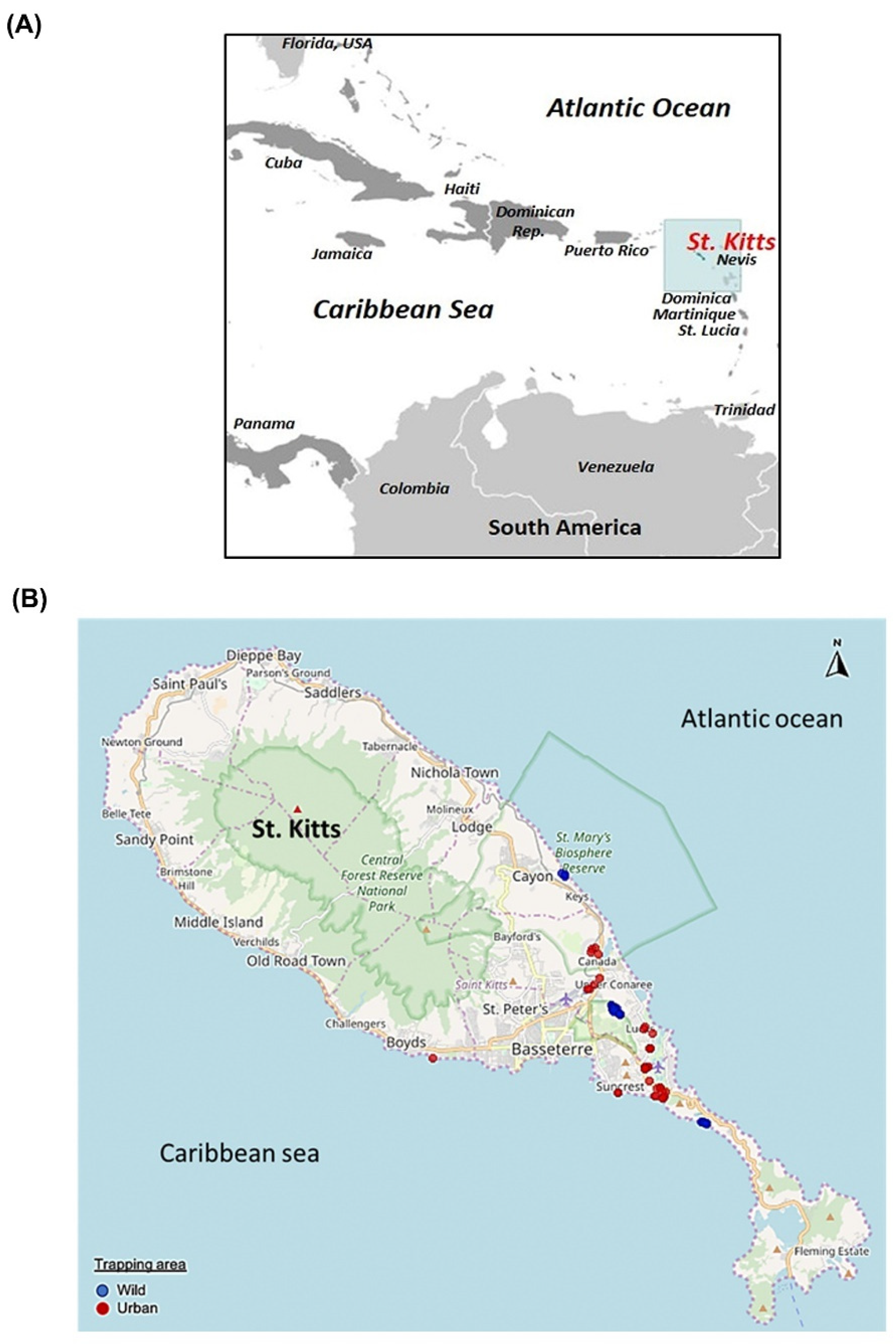
2.2. Sample Collection

During April–July 2017, non-diarrheic fecal samples were obtained from the rectum and distal part of the colon of 83 small Indian mongooses that were trapped, euthanized and necropsied under sterile conditions for a gut microbiome study on the Caribbean island of St. Kitts [47]. The samples were kept at −80 °C until further analyses.

2.3. Amplification of Viral DNA

Viral DNA was extracted from the fecal samples using the QIAamp Fast DNA Stool Mini Kit (Qiagen Sciences, Germantown, MD, USA) following the instructions provided by the manufacturer. The samples were screened for the presence of circoviruses and cycloviruses by nested PCR assays using pan-rep primers (primers CV-F1, CV-R1, CV-F2 and CV-R2, targeting a short stretch (~400 bp) of the Rep-encoding ORF), as described previously [3]. Additional primers were designed from the partial Rep-encoding ORF sequences and used in inverse nested PCRs to amplify the complete genomes of the mongoose associated circovirus and cyclovirus (Supplementary material S1). PCRs were performed using the Platinum™ Taq DNA Polymerase (Invitrogen™, Thermo Fisher Scientific Corporation, Waltham, MA, USA) according to the manufacturer’s instructions. Sterile water was used as a negative control in all PCR reactions.

2.4. Nucleotide Sequencing

The PCR amplicons were purified using the Wizard® SV Gel and PCR Clean-Up kit (Promega, Madison, WI, USA) following the instructions provided by the manufacturer. Nucleotide sequences were obtained using the ABI Prism Big Dye Terminator Cycle Sequencing Ready Reaction Kit (Applied Biosystems, Foster City, CA, USA) on an ABI 3730XL Genetic Analyzer (Applied Biosystems, Foster City, CA, USA).

2.5. Sequence Analysis

Homology search for related nt and deduced aa sequences were performed using the standard BLASTN and BLASTP program (Basic Local Alignment Search Tool, www.ncbi.nlm.nih.gov/blast, accessed on 22 June 2021), respectively. Putative ORFs encoding the viral Rep and Cp were identified using the ORF finder (https://0-www-ncbi-nlm-nih-gov.brum.beds.ac.uk/orffinder/, accessed on 20 June 2021), whilst those with a putative intron between the Rep coding sequences (CDS) were determined by BLASTN analysis with the CDS feature. Pairwise sequence (%) identities for the complete viral genomes, and the putative Rep and Cp were determined using the MUSCLE algorithm in the SDTv1.2 program, as described previously [2,48]. On the other hand, pairwise identities between the partial Rep-encoding ORF sequences were calculated using the MUSCLE alignment program (https://www.ebi.ac.uk/Tools/msa/muscle/, accessed on 23 June 2021) and the ‘align two or more sequences’ option of BLASTN program (https://blast.ncbi.nlm.nih.gov/, accessed on 23 June 2021). The maps of the circular viral genomes were constructed with the ‘Draw Custom Plasmid Map’ program (https://www.rf-cloning.org/savvy.php, accessed on 20 June 2021). The putative stem-loop structure was identified in the viral genome using the mFold program [49].
Multiple alignments of nt and deduced aa sequences were carried out using the MUSCLE algorithm embedded in the MEGA7 software [50]. Phylogenetic analysis was performed by the maximum likelihood (ML) method using the MEGA7 software [50], with the GTR+G model of substitution and 1000 bootstrap replicates, as described previously [2]. The complete genomes of the mongoose associated circoviruses and cycloviruses were investigated for recombination events using the RDP4 program with default parameters [51]. A circovirus/cyclovirus sequence was determined as a recombinant if it was supported by two, or more than two detection methods (3Seq, BOOTSCAN, CHIMERA, GENECONV, MAXCHI, RDP and SISCAN) with a highest acceptable p-value of p < 0.01 with Bonferroni’s correction [17,51].

2.6. GenBank Accession Numbers

The GenBank accession numbers for the mongoose associated CRESS DNA viral sequences determined in this study are MZ382570-MZ382599.

3. Results and Discussion

3.1. Detection of Circoviruses and Cycloviruses in the Small Indian Mongoose

The small island of St. Kitts (~69 square miles, human population of ~35,000) is inhabited by a large population of the small Indian mongoose (~45,000) that dwell in wild and urban habitats (Figure 1A,B) [44,52], [https://www.sknbs.org/about-bureau/about-st-kitts-and-nevis/, accessed on 14 July 2021]. In the present study, single fecal samples from 76 (91.56%) of the 83 small Indian mongooses yielded the expected ~400 bp amplicon with circovirus/cyclovirus pan-rep primers in screening PCR assays. In this case, 39 of the 76 positive samples showed strong PCR amplification and were sequenced for the partial rep gene. By BLASTN analysis, all the mongoose-associated partial CRESS DNA viral sequences shared maximum homology with published circovirus, or cyclovirus rep gene sequences, except for Mon-66, which shared maximum pairwise nt sequence identities of 63.37% with that of an unclassified CRESS DNA virus (GenBank accession number KY487932) from a wastewater sample (Table 1). However, based on analysis of the complete genome sequence, Mon-66 was assigned to the genus Circovirus (Table 2). Nine of the partial rep sequences lacked high quality and were excluded from further analysis.
Based on BLASTN analysis and pairwise nt sequence identities of partial rep, the mongoose associated partial CRESS DNA viral sequences were classified into at least 8 putative groups (designated as I-VIII) (Table 1). Mongoose associated partial rep sequences sharing >95% pairwise identities between themselves were assigned to the same group (Table 1). Group-I sequences shared maximum pairwise identities of 77.65–79.20% with circovirus isolate C072 that was detected in the intestinal sample from a vesper bat (Myotis fimbriatus) in China (Table 1). Group II-VII exhibited maximum homology with cyclovirus rep sequences (Table 1). Group-II and -VI sequences shared maximum homology (pairwise identities of 97.62–98.31% and 77.47–82.21%, respectively) with cycloviruses from bats (Table 1). Group-III and -V consisted of a single rep sequence each, and shared maximum pairwise identities of 94.25% and 93.11% with a cockroach associated cyclovirus and a sheep associated cyclovirus, respectively (Table 1). Group-IV was the largest, consisting of 15 partial rep sequences that shared maximum pairwise identities of 95.37–97.48% with human cyclovirus TN2 (detected in fecal sample of a healthy child who came in contact with a non-polio-infected acute flaccid paralysis patient [3]). Group-VII sequences shared maximum homology (pairwise identities of 91.84–92.07%) with human cyclovirus VS5700009 that was detected in the serum of a patient with unexplained paraplegia [36].
Even though we reported high rates of detection of CRESS DNA viral sequences that exhibited maximum homology with circoviruses, or cycloviruses, all the fecal samples were obtained from apparently healthy mongooses, indicating a lack of association between these viruses and clinical conditions. Circoviruses appear to primarily infect vertebrates, whilst those detected in hematophagous arthropod vectors might actually be viruses of mammals, or birds that these insects feed upon [2,5,21,23]. On the other hand, cycloviruses have been reported in a wide array of both invertebrates and mammals [2,5,21,23]. Based on these observations, it has been proposed that cycloviruses are more diverse and widespread than circoviruses [2,5,21,23]. In the present study, ~90% (35/39) of the partial rep sequences shared maximum pairwise identities with cycloviruses, which constituted 6 of the 8 putative groups of diverse CRESS DNA viral sequences (Table 1). Since only fecal samples were analyzed, and the small Indian mongoose has been known to feed on small mammals, reptiles, birds, bird and reptile eggs, crustaceans, insects and human waste [53], we could not determine whether the mongoose associated CRESS DNA viruses replicated in the host or were of dietary origin.
Based on the detection of closely related circoviruses/cycloviruses in different animal species, especially in tissues, some studies have proposed interspecies transmission events within the family Circoviridae [2,3,5,54]. In this study, 15 (group-IV) and 2 (group-II) of the mongoose associated partial rep sequences were closely related (>95% pairwise nt identities) to cycloviruses from humans and bats, respectively (Table 1). St. Kitts island has a sizeable bat population [55], and the small Indian mongoose has often been seen in close proximity to humans [44], offering an ideal environment for interspecies transmission events. However, we could not obtain the full-length genome sequences for group-II viruses, whilst that of complete genome of human cyclovirus TN2 (closely related to group-IV sequences) was not available in the GenBank database. Furthermore, caution should be exercised whilst commenting on cross-species transmission of circoviruses/cycloviruses from fecal samples, as they may have a dietary origin [2,3,5,54], especially in mongooses that have a wide-range of feeding habits [53]. Interestingly, one of the mongoose associated partial rep sequences (group-III) shared maximum homology with a cockroach associated cyclovirus, corroborating previous observations that cyclovirus sequences detected in vertebrate samples might be actually those from viruses of arthropods [21,23] (Table 1).

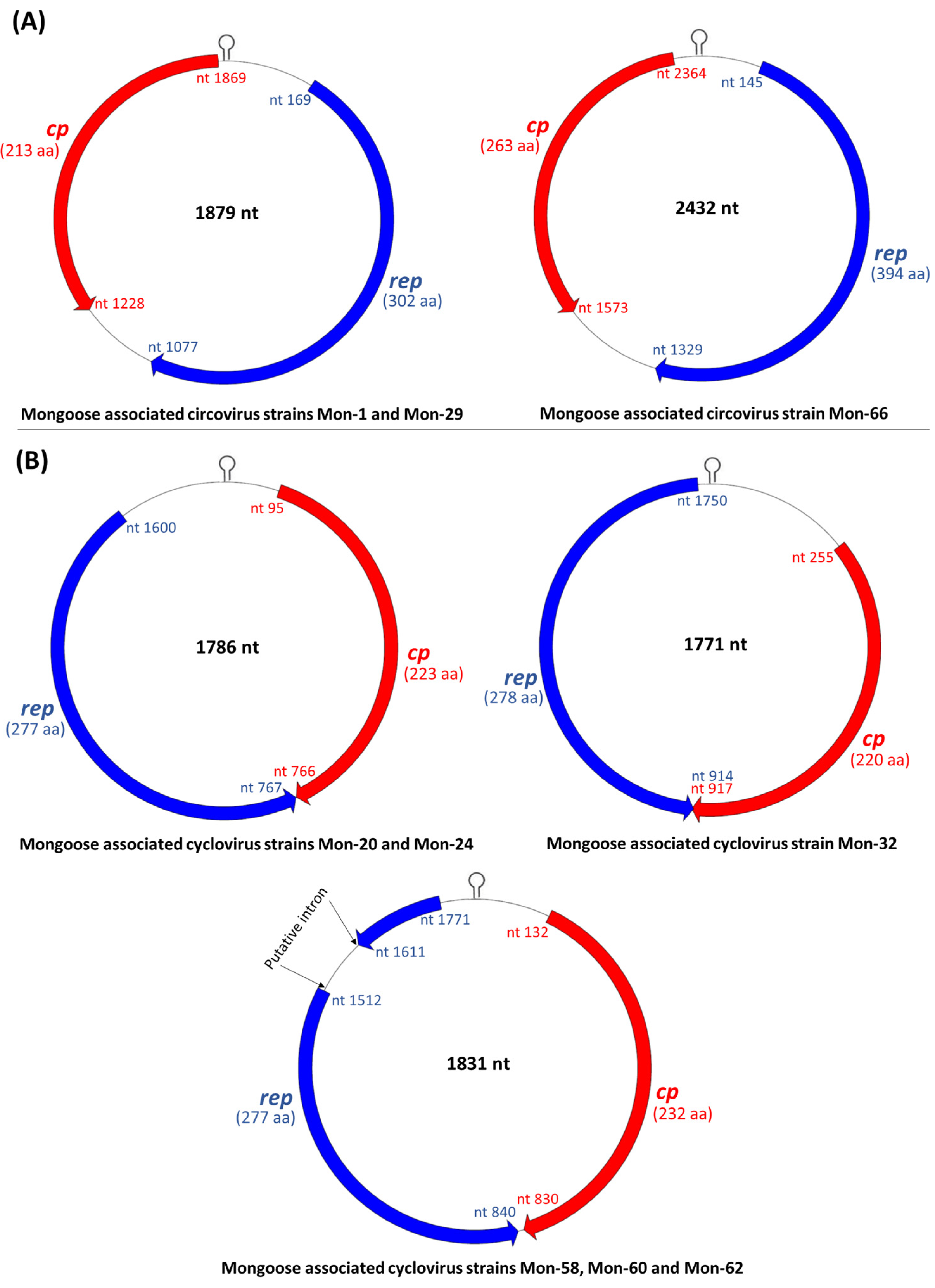
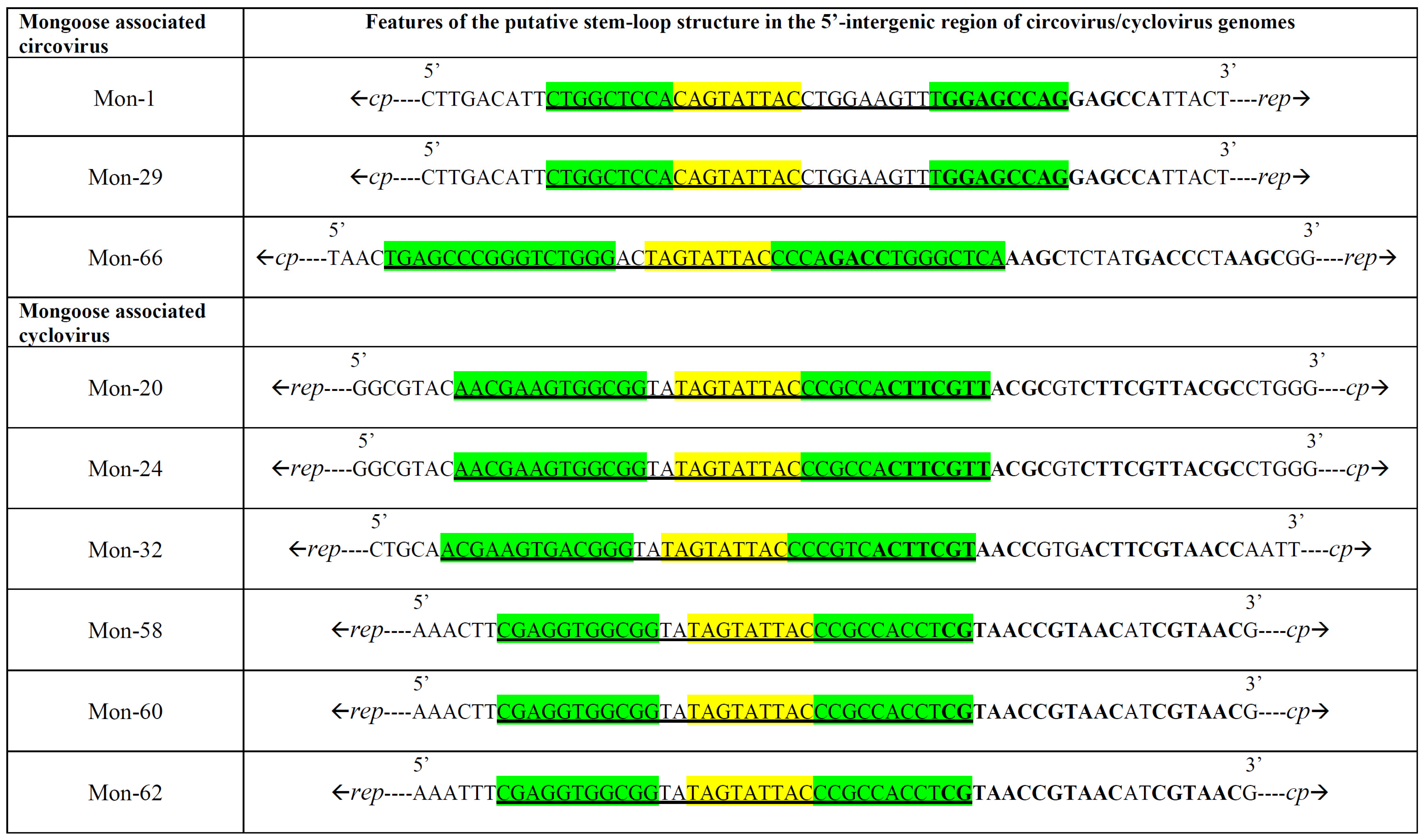
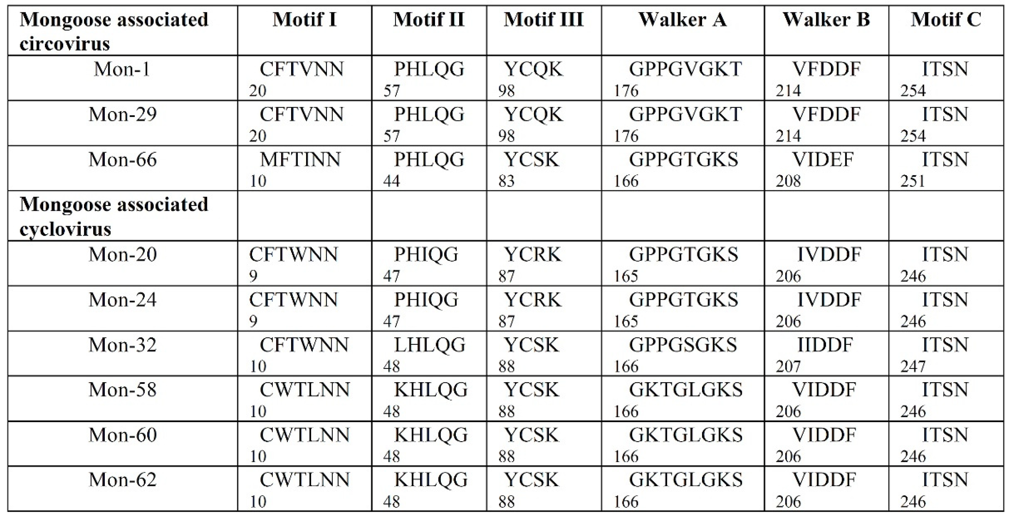
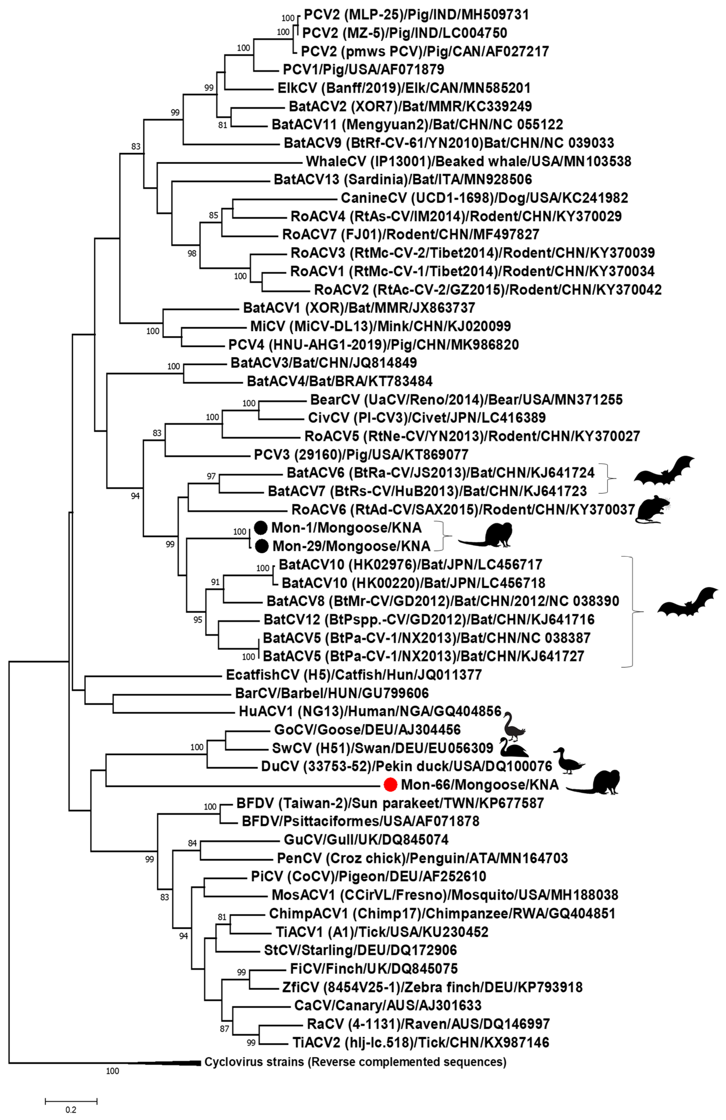
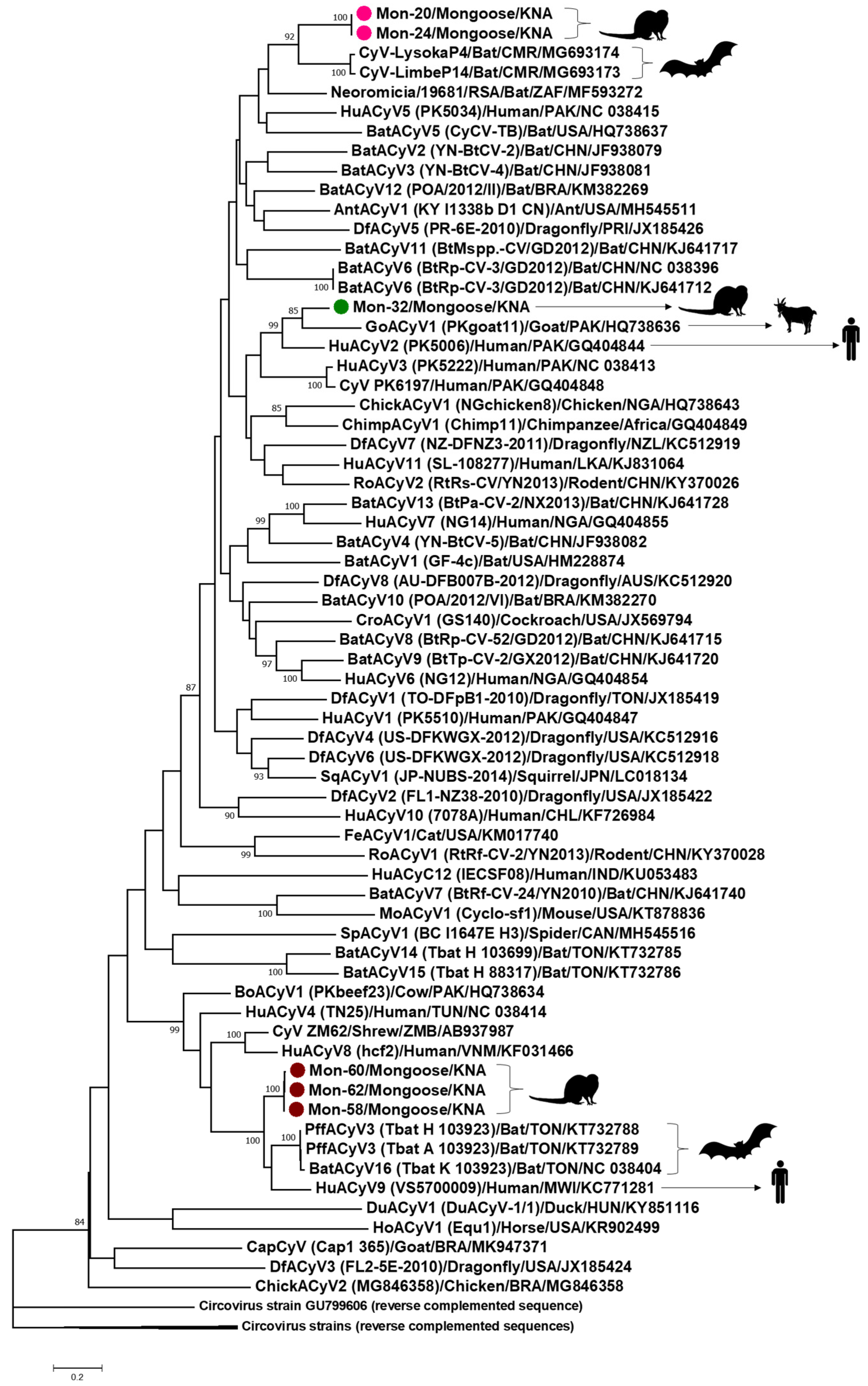
3.2. Analysis of the Complete Genomes of Mongoose Associated Circoviruses and Cycloviruses

Since the ICTV classification scheme for the family Circoviridae is based on genome-wide pairwise identities [1,2], attempts were made to determine the complete genome sequences of mongoose associated CRESS DNA viruses representing the putative groups I-VIII (Table 1). Using an inverse nested PCR assay, we obtained the full-length genome sequences of 2 group-I (Mon-1 and -29), a single group-IV (Mon-32), 2 group-VI (Mon-20 and -24), 3 group-VII (Mon-58, -60 and -62) and a single group-VIII (Mon-66) viruses (Table 1 and Table 2), whilst the complete genomes of group-II, -III and -V viruses could not be amplified. The genomic organization of the mongoose associated circoviruses and cycloviruses are shown in Figure 2. The complete genome sequences of the mongoose associated circoviruses and cycloviruses (collectively referred to as the ‘Mon sequences’) retained the various features that are conserved in members of the genera Circovirus and Cyclovirus, respectively, within the family Circoviridae (Figure 2, Figure 3 and Figure 4; Supplementary material S2) [1,2].
All the Mon sequences contained the putative ori in the 5′-IR, marked by the presence of the conserved nonanucleotide motif ((C/T)AGTATTAC) at the apex of a potential stem-loop structure (Figure 2 and Figure 3). Following ICTV guidelines [1,2], the first nt of the nonanucleotide motif was considered as ‘position one’ of the Mon sequences. Mon-1, -29 and -66 contained the putative ori in the Rep-encoding strand, whilst the putative ori was located in the Cp coding strand of Mon-20, -24, -32, -58, -60 and -62 (Figure 2). The 3′-IR was absent, or smaller in Mon-20, -24, -32, -58, -60 and -62 than those observed in Mon-1, -29 and -66 (Figure 2). Based on these observations, genome-wide pairwise identities, and phylogenetic analysis, Mon-1, -29 and -66 were classified as circoviruses, whilst Mon-20, -24, -32, -58, -60 and -62 were assigned to the genus Cyclovirus (Figure 2, Figure 3, Figure 4, Figure 5, Figure 6; Table 2, Table 3, Table 4; Supplementary Materials S2–S5).
Corroborating previous observations [1,2], the putative Rep of the mongoose associated circoviruses and cycloviruses retained the conserved RCR (motifs I through III) and superfamily 3 helicase (Walker A and B, and motif C) motifs (Figure 4). On the other hand, the putative Cp, although much more divergent than the Rep (Table 3 and Table 4), contained the conserved arginine rich region at the amino terminus, with the exception of Mon-66 (Supplementary material S2). Since recombinants have been reported in both circoviruses and cycloviruses [4,17,29,56,57,58], the Mon sequences were evaluated for potential recombination using the RDP4 program. However, we did not obtain reliable evidence for recombination events in the Mon sequences, except for Mon-32, which was identified by several models in the RDP4 program as minor parent to goat associated cyclovirus 1 (isolate PKgoat11, GenBank accession number HQ738636), whilst the major parent was unknown (Supplementary material S6).
The complete genomes of mongoose associated circoviruses Mon-1 and -29 were 1879 bp in size (Figure 2), which was comparable to those observed in most circoviruses [2]. Mon-1 and -29 were closely related to each other (Figure 5; Table 2, Table 3, Table 4; Supplementary Materials S3), and shared maximum pairwise identities of 67.40% and 67.20%, respectively, with that of bat circovirus isolate BtPspp.-CV (GenBank accession number KJ641716) from China [59], followed by identities of 66.90% and 66.60%, respectively, with bat associated circovirus 10 (isolate HK02976, GenBank accession number LC456717) from Japan (Table 2) [60]. Phylogenetically, the complete genome sequences of Mon-1 and -29 formed a distinct cluster within a clade that mostly consisted of circoviruses from bats, including isolates BtPspp.-CV and HK02976 (Figure 5). These observations were corroborated by analysis of the putative proteins (pairwise identities of Rep and Cp, and phylogenetic analysis of Rep) of Mon-1 and -29 (Table 3 and Table 4; Supplementary material S5).
The complete genome of Mon-66 was 2432 nt long (Figure 2), which was larger than those of most circoviruses [1,2]. Even though the partial rep sequence of Mon-66 exhibited maximum homology with that of an unclassified CRESS DNA virus (Table 1), the complete genome of Mon-66 shared maximum pairwise identities of 60.70% with that of porcine circovirus 2 isolate MZ-5 (GenBank accession number LC004750) from India (Table 2). Since the genome-wide pairwise identity cut-off value for assigning a sequence to a genus within the family Circoviridae is 55% [1,2], Mon-66 was classified as a circovirus. On the other hand, the Rep and Cp of Mon-66 shared maximum deduced aa identities of 51.20% and 44.40% with that of unclassified CRESS DNA viruses from a wild bird (MW182878) and a wastewater sample (KY487977), respectively (Table 3 and Table 4). Phylogenetically, Mon-66 formed an isolated branch within the clade of circoviruses (Figure 5; Supplementary material S5).
The complete genomes of the mongoose associated cycloviruses were 1771 nt, 1786 nt or 1831 nt long (Figure 2), which was within the size-range for cyclovirus genomes [1,2]. Mon-20 and -24 were fully identical in their complete genome sequences, and shared maximum homology (pairwise identities of 72.10%) with that of bat cyclovirus isolate CyV-LysokaP4 (GenBank accession number MG693174) from Cameroon (Table 2; Figure 6) [61]. Pairwise identities of the Rep and Cp, and phylogenetic analysis of Rep of Mon-20 and -24 also revealed similar findings (Table 3 and Table 4; Supplementary material S5).
Mon-32 exhibited maximum pairwise identity of 77.30% with the complete genome of goat cyclovirus isolate PKgoat11, followed by 75.30% with that of human cyclovirus isolate PK5006 (GQ404844) (Table 2). Cyclovirus isolates PKgoat11 and PK5006 were detected in the same study from Pakistan [3]. The Rep and Cp of Mon-32 exhibited maximum pairwise deduced aa identity of 87.10% and 51.60% with that of isolates PKgoat11 and PK5006, respectively (Table 3 and Table 4). Phylogenetically, Mon-32 clustered with isolate PKgoat11 within a clade that also consisted of PK5006 (Figure 6; Supplementary material S5).
The Rep-encoding ORF of Mon-58, -60 and -62 was interrupted by a putative intron (nt 1610–nt 1511) with a canonical splice donor site (GT) and splice acceptor site (AG) (Supplementary material S7). The complete genomes of Mon-58, -60 and -62 shared 99.30–99.50% pairwise identities between themselves, and maximum identities of 79.90–80.20% with those of human cyclovirus isolate VS570000 (GenBank accession number KC771281) from the CSF of a paraplegic patient in Malawi [36] and Pacific flying fox associated cyclovirus-3 isolate Tbat_H_103923 (KT732788) from the feces of a bat in Tonga (Table 2) [62]. Mon-58, -60 and -62 shared maximum pairwise identities of 92.40% with the Rep of isolate VS570000 (Table 3), and 56.10–56.60% with the Cp of isolate Tbat_H_103923 (Table 4). By phylogenetic analysis, Mon-58, -60 and -62 clustered together near bat associated cycloviruses (including isolate Tbat_H_103923) [62] and the human cyclovirus isolate VS570000 (Figure 6; Supplementary material S5) [36].
The ICTV has recommended a species demarcation threshold of 80% genome-wide pairwise nt sequence identity for members of the family Circoviridae [1,2]. Based on the ICTV classification system, mongoose associated circoviruses Mon-1/Mon-29 and Mon-66 qualify as novel species within the genus Circovirus, whilst mongoose associated cycloviruses Mon-20/Mon-24 and Mon-32 represent new species in the genus Cyclovirus (Table 2; Supplementary Materials S3 and S4). On the other hand, the maximum pairwise identities (79.90–80.20%) of mongoose associated cycloviruses Mon-58, -60 and -62 with cycloviruses from other animals/sources were borderline to the cut-off identity value for assigning novel cycloviral species (Table 2; Supplementary material S4).

3.3. Conclusions

Taken together, our findings suggest that CRESS DNA viruses are widely circulating in the small Indian mongoose population on the island of St. Kitts. However, in the absence of sampling from tissues, and considering that mongooses are polyphagous predators, we could not determine whether the circoviruses/cycloviruses in fecal samples of apparently healthy mongooses were of dietary origin, or actually infected the host. Based on the classification scheme proposed by the ICTV Circoviridae study group [1,2], we identified 2 novel species in each of the genera Circovirus and Cyclovirus, further expanding the genetic diversity of these viruses. However, despite high genetic diversity, the mongoose associated circoviruses/cycloviruses retained the various features that are conserved among members of the family Circoviridae. Studies aimed at detection of viral DNA in tissues, screening for virus-specific antibodies, in-vitro replication of virus in mongoose cells and virus inoculation in gnotobiotic animals are required to gain a proper understanding of circovirus/cyclovirus infection in mongooses. To our knowledge, this is the first report on detection and complete genome analysis of circoviruses and cycloviruses in the small Indian mongoose, warranting further studies in other species of mongooses.

Supplementary Materials

The following are available online at https://0-www-mdpi-com.brum.beds.ac.uk/article/10.3390/v13091700/s1, Supplementary material S1: Additional primers used in inverse nested PCRs to amplify the complete genomes of mongoose associated circoviruses and cycloviruses. Supplementary material S2: The presence of the arginine rich region in amino terminus of putative capsid proteins of mongoose associated circoviruses (Mon-1 and -29) and cycloviruses (Mon-20, -24, -32, -58, -60 and -62). Supplementary material S3: Two-dimensional graphical representation of pairwise nucleotide sequence (%) identities between the complete genomes of circoviruses detected in various animal species. Supplementary material S4: Two-dimensional graphical representation of pairwise nucleotide sequence (%) identities between the complete genomes of cycloviruses detected in various animal species. Supplementary material S5: Phylogenetic analysis of the putative replication-associated proteins (Rep) of mongoose associated circoviruses (A), and cycloviruses (B), Supplementary material S6: Identification of inter-species recombination event involving mongoose associated cyclovirus strain Mon-32, as determined by the Recombination Detection Program (RDP v.4.101). Supplementary material S7: BLASTN (with coding sequence (CDS) feature) analysis showing the alignment of the putative replication associated protein (Rep) CDS (shown with brown line) of mongoose associated cyclovirus Mon-58 (Query sequence, Rep CDS: join nt 1771-nt 1611, nt 1512-nt 840) with that of Pacific flying fox associated cyclovirus-3 isolate Tbat_H_103923 (GenBank accession number KT732788) (Subject (Sbjct) sequence, Rep CDS: join nt 1788-nt 1628, nt 1515-nt 843).

Author Contributions

Conceptualization, K.G. and S.G.; collected samples, A.A.M.J.B.; secured funding, S.G.; contributed reagents, S.G.; screened samples, K.G. and S.G.; performed laboratory work, K.G. and S.G.; performed data analysis, K.G. and S.G.; wrote the manuscript, K.G. and S.G.; edited and finalized the manuscript, A.A.M.J.B., K.G., S.G. and Y.S.M. All authors have read and agreed to the published version of the manuscript.

Funding

The present study was funded by intramural grant # Viruses 41001-21 entitled ‘Detection and molecular characterization of viruses in pigs and wildlife in the Caribbean and Central America from the One Health Center for Zoonoses and Tropical Veterinary Medicine, Ross University School of Veterinary Medicine, St. Kitts and Nevis.

Institutional Review Board Statement

This study was approved by the Institutional Animal Care and Use Committee (IACUC) of the Ross University School of Veterinary Medicine, St. Kitts and Nevis (IACUC protocol title: trapping and necropsy for mongoose microbial ecology study. Approved IACUC protocol number: 17.04.13, dated 13 April 2017).

Informed Consent Statement

Not applicable.

Acknowledgments

We would like to thank DVM research assistant/volunteers KC Hill, Vania Monsul and Elizabeth Coloian from the Ross University School of Veterinary Medicine (RUSVM), St. Kitts and Nevis, for assisting with sampling/laboratory work.

Conflicts of Interest

The authors declare no conflict of interest.

References

  1. Breitbart, M.; Delwart, E.; Rosario, K.; Segalés, J.; Varsani, A. ICTV virus taxonomy profile: Circoviridae. J. Gen. Virol. 2017, 98, 1997–1998. [Google Scholar] [CrossRef]
  2. Rosario, K.; Breitbart, M.; Harrach, B.; Segalés, J.; Delwart, E.; Biagini, P.; Varsani, A. Revisiting the taxonomy of the family Circoviridae: Establishment of the genus Cyclovirus and removal of the genus Gyrovirus. Arch. Virol. 2017, 162, 1447–1463. [Google Scholar] [CrossRef] [Green Version]
  3. Li, L.; Kapoor, A.; Slikas, B.; Bamidele, O.S.; Wang, C.; Shaukat, S.; Masroor, M.A.; Wilson, M.L.; Ndjango, J.-B.N.; Peeters, M.; et al. Multiple Diverse Circoviruses Infect Farm Animals and Are Commonly Found in Human and Chimpanzee Feces. J. Virol. 2010, 84, 1674–1682. [Google Scholar] [CrossRef] [Green Version]
  4. Zhao, L.; Rosario, K.; Breitbart, M.; Duffy, S. Eukaryotic Circular Rep-Encoding Single-Stranded DNA (CRESS DNA) Viruses: Ubiquitous Viruses with Small Genomes and a Diverse Host Range. Adv. Virus Res. 2019, 103, 71–133. [Google Scholar] [CrossRef]
  5. Delwart, E.; Li, L. Rapidly expanding genetic diversity and host range of the Circoviridae viral family and other Rep encoding small circular ssDNA genomes. Virus Res. 2012, 164, 114–121. [Google Scholar] [CrossRef] [PubMed] [Green Version]
  6. Steinfeldt, T.; Finsterbusch, T.; Mankertz, A. Demonstration of Nicking/Joining Activity at the Origin of DNA Replication Associated with the Rep and Rep′ Proteins of Porcine Circovirus Type 1. J. Virol. 2006, 80, 6225–6234. [Google Scholar] [CrossRef] [PubMed] [Green Version]
  7. Cheung, A.K. Identification of an octanucleotide motif sequence essential for viral protein, DNA, and progeny virus biosynthesis at the origin of DNA replication of porcine circovirus type 2. Virology 2004, 324, 28–36. [Google Scholar] [CrossRef] [PubMed] [Green Version]
  8. Alex, C.E.; Fahsbender, E.; Altan, E.; Bildfell, R.; Wolff, P.; Jin, L.; Black, W.; Jackson, K.; Woods, L.; Munk, B.; et al. Viruses in unexplained encephalitis cases in american black bears (ursus americanus). PLoS ONE 2020, 15, e0244056. [Google Scholar] [CrossRef] [PubMed]
  9. Fisher, M.; Harrison, T.M.R.; Nebroski, M.; Kruczkiewicz, P.; Rothenburger, J.L.; Ambagala, A.; Macbeth, B.; Lung, O. Discovery and comparative genomic analysis of elk circovirus (elkcv), a novel circovirus species and the first reported from a cervid host. Sci. Rep. 2020, 10, 19548. [Google Scholar] [CrossRef]
  10. Payne, N.; Kraberger, S.; Fontenele, R.S.; Schmidlin, K.; Bergeman, M.H.; Cassaigne, I.; Culver, M.; Varsani, A.; Van Doorslaer, K. Novel circoviruses detected in feces of sonoran felids. Viruses 2020, 12, 1027. [Google Scholar] [CrossRef] [PubMed]
  11. Tisza, M.J.; Pastrana, D.V.; Welch, N.L.; Stewart, B.; Peretti, A.; Starrett, G.J.; Pang, Y.S.; Krishnamurthy, S.R.; Pesavento, P.A.; McDermott, D.H.; et al. Discovery of several thousand highly diverse circular DNA viruses. Elife 2020, 9, e51971. [Google Scholar] [CrossRef] [PubMed]
  12. Nishizawa, T.; Sugimoto, Y.; Takeda, T.; Kodera, Y.; Hatano, Y.; Takahashi, M.; Okamoto, H. Identification and full-genome characterization of novel circoviruses in masked palm civets (Paguma larvata). Virus Res. 2018, 258, 50–54. [Google Scholar] [CrossRef] [PubMed]
  13. Nebbak, A.; Monteil-Bouchard, S.; Berenger, J.M.; Almeras, L.; Parola, P.; Desnues, C. Virome diversity among mosquito populations in a sub-urban region of Marseille, France. Viruses 2021, 13, 768. [Google Scholar] [CrossRef]
  14. Wu, Z.; Lu, L.; Du, J.; Yang, L.; Ren, X.; Liu, B.; Jiang, J.; Yang, J.; Dong, J.; Sun, L.; et al. Comparative analysis of rodent and small mammal viromes to better understand the wildlife origin of emerging infectious diseases. Microbiome 2018, 6, 178. [Google Scholar] [CrossRef]
  15. Hui, A.; Altan, E.; Slovis, N.; Fletcher, C.; Deng, X.; Delwart, E. Circovirus in blood of a febrile horse with hepatitis. Viruses 2021, 13, 944. [Google Scholar] [CrossRef]
  16. Zhang, H.H.; Hu, W.Q.; Li, J.Y.; Liu, T.N.; Zhou, J.Y.; Opriessnig, T.; Xiao, C.T. Novel circovirus species identified in farmed pigs designated as porcine circovirus 4, hunan province, china. Transbound. Emerg. Dis. 2020, 67, 1057–1061. [Google Scholar] [CrossRef] [PubMed]
  17. Kleymann, A.; Soto, E.; Illanes, O.; Malik, Y.S.; Fuentealba, C.; Ghosh, S. High rates of detection and complete genomic analysis of porcine circovirus 2 (PCV2) in the Lesser Antilles island of St. Kitts: Identification of PCV2b-PCV2d recombinants. Transbound. Emerg. Dis. 2020, 67, 2282–2289. [Google Scholar] [CrossRef]
  18. Bandoo, R.A.; Bautista, J.; Lund, M.; Newkirk, E.; Squires, J.; Varsani, A.; Kraberger, S. Identification of novel circovirus and anelloviruses from wolverines using a non-invasive faecal sampling approach. Infect. Genet. Evol. 2021, 93, 104914. [Google Scholar] [CrossRef]
  19. Prades, Y.; Pizarro, R.; Ruiz, M.; Moreno, C.; Avendaño, L.F.; Luchsinger, V. Cyclovirus detection in Chilean adults with and without community-acquired pneumonia. J. Med. Virol. 2021, 93, 4786–4793. [Google Scholar] [CrossRef]
  20. Sasaki, M.; Orba, Y.; Ueno, K.; Ishii, A.; Moonga, L.; Hangombe, B.M.; Mweene, A.S.; Ito, K.; Sawa, H. Metagenomic analysis of the shrew enteric virome reveals novel viruses related to human stool-associated viruses. J. Gen. Virol. 2015, 96, 440–452. [Google Scholar] [CrossRef]
  21. Rosario, K.; Mettel, K.A.; Benner, B.E.; Johnson, R.; Scott, C.; Yusseff-Vanegas, S.Z.; Baker, C.C.M.; Cassill, D.L.; Storer, C.; Varsani, A.; et al. Virus discovery in all three major lineages of terrestrial arthropods highlights the diversity of single-stranded DNA viruses associated with invertebrates. PeerJ 2018, 6, e5761. [Google Scholar] [CrossRef] [PubMed]
  22. Liu, Q.; Wang, H.; Ling, Y.; Yang, S.X.; Wang, X.C.; Zhou, R.; Xiao, Y.Q.; Chen, X.; Yang, J.; Fu, W.G.; et al. Viral metagenomics revealed diverse CRESS-DNA virus genomes in faeces of forest musk deer. Virol. J. 2020, 17, 61. [Google Scholar] [CrossRef] [Green Version]
  23. Dennis, T.P.W.; Flynn, P.J.; de Souza, W.M.; Singer, J.B.; Moreau, C.S.; Wilson, S.J.; Gifford, R.J. Insights into Circovirus Host Range from the Genomic Fossil Record. J. Virol. 2018, 92, e00145-18. [Google Scholar] [CrossRef] [PubMed] [Green Version]
  24. Altan, E.; Kubiski, S.V.; Burchell, J.; Bicknese, E.; Deng, X.; Delwart, E. The first reptilian circovirus identified infects gut and liver tissues of black-headed pythons. Vet. Res. 2019, 50, 35. [Google Scholar] [CrossRef] [PubMed] [Green Version]
  25. Chang, W.S.; Li, C.X.; Hall, J.; Eden, J.S.; Hyndman, T.H.; Holmes, E.C.; Rose, K. Meta-transcriptomic discovery of a divergent circovirus and a chaphamaparvovirus in captive reptiles with proliferative respiratory syndrome. Viruses 2020, 12, 1073. [Google Scholar] [CrossRef] [PubMed]
  26. Lorincz, M.; Cságola, A.; Farkas, S.L.; Székely, C.; Tuboly, T. First detection and analysis of a fish circovirus. J. Gen. Virol. 2011, 92, 1817–1821. [Google Scholar] [CrossRef] [PubMed]
  27. Lorincz, M.; Dan, A.; Lang, M.; Csaba, G.; Toth, A.G.; Szekely, C.; Csagola, A.; Tuboly, T. Novel circovirus in european catfish (Silurus glanis). Arch. Virol. 2012, 157, 1173–1176. [Google Scholar] [CrossRef] [Green Version]
  28. Doszpoly, A.; Tarján, Z.L.; Glávits, R.; Müller, T.; Benko, M. Full genome sequence of a novel circo-like virus detected in an adult European eel Anguilla anguilla showing signs of cauliflower disease. Dis. Aquat. Organ. 2014, 109, 107–115. [Google Scholar] [CrossRef] [Green Version]
  29. Opriessnig, T.; Karuppannan, A.K.; Castro, A.M.M.G.; Xiao, C.T. Porcine circoviruses: Current status, knowledge gaps and challenges. Virus Res. 2020, 286, 198044. [Google Scholar] [CrossRef]
  30. Fogell, D.J.; Martin, R.O.; Groombridge, J.J. Beak and feather disease virus in wild and captive parrots: An analysis of geographic and taxonomic distribution and methodological trends. Arch. Virol. 2016, 161, 2059–2074. [Google Scholar] [CrossRef] [Green Version]
  31. Soike, D.; Köhler, B.; Albrecht, K. A circovirus-like infection in geese related to a runting syndrome. Avian Pathol. 1999, 28, 199–202. [Google Scholar] [CrossRef]
  32. Stenzel, T.; Koncicki, A. The epidemiology, molecular characterization and clinical pathology of circovirus infections in pigeons—Current knowledge. Vet. Q. 2017, 37, 166–174. [Google Scholar] [CrossRef]
  33. Li, L.; McGraw, S.; Zhu, K.; Leutenegger, C.M.; Marks, S.L.; Kubiski, S.; Gaffney, P.; Cruz, F.N.D.; Wang, C.; Delwart, E.; et al. Circovirus in tissues of dogs with vasculitis and hemorrhage. Emerg. Infect. Dis. 2013, 19, 534–541. [Google Scholar] [CrossRef] [PubMed]
  34. Van Kruiningen, H.J.; Heishima, M.; Kerr, K.M.; Garmendia, A.E.; Helal, Z.; Smyth, J.A. Canine circoviral hemorrhagic enteritis in a dog in Connecticut. J. Vet. Diagn. Investig. 2019, 31, 732–736. [Google Scholar] [CrossRef] [PubMed]
  35. Lian, H.; Liu, Y.; Li, N.; Wang, Y.; Zhang, S.; Hu, R. Novel circovirus from mink, China. Emerg. Infect. Dis. 2014, 20, 1548–1550. [Google Scholar] [CrossRef]
  36. Smits, S.L.; Zijlstra, E.E.; van Hellemond, J.J.; Schapendonk, C.M.E.; Bodewes, R.; Schürch, A.C.; Haagmans, B.L.; Osterhaus, A.D.M.E. Novel cyclovirus in human cerebrospinal fluid, Malawi, 2010–2011. Emerg. Infect. Dis. 2013, 19, 1511–1513. [Google Scholar] [CrossRef] [PubMed]
  37. Barun, A.; Hanson, C.C.; Campell, K.J.; Simberlo, D. A review of small Indian mongoose management and eradications on islands. In Island Invasives: Eradication and Management; Veitch, C.R., Clout, M.N., Towns, D.R., Eds.; IUCN: Gland, Switzerland, 2011; pp. 17–25. [Google Scholar]
  38. Veron, G.; Jennings, A.P. Javan mongoose or small Indian mongoose—Who is where? Mamm. Biol. 2017, 87, 62–70. [Google Scholar] [CrossRef]
  39. Sauvé, C.C.; Rees, E.E.; Gilbert, A.T.; Berentsen, A.R.; Allibert, A.; Leighton, P.A. Modeling mongoose rabies in the caribbean: A model—Guided fieldwork approach to identify research priorities. Viruses 2021, 13, 323. [Google Scholar] [CrossRef]
  40. Nidaira, M.; Takahashi, K.; Ogura, G.; Taira, K.; Okano, S.; Kudaka, J.; Itokazu, K.; Mishiro, S.; Nakamura, M. Detection and phylogenetic analysis of Hepatitis E Viruses from mongooses in Okinawa, Japan. J. Vet. Med. Sci. 2012, 74, 1665–1668. [Google Scholar] [CrossRef] [Green Version]
  41. Chang, A.M.; Chen, C.C. Molecular characteristics of Carnivore protoparvovirus 1 with high sequence similarity between wild and domestic carnivores in Taiwan. Pathogens 2021, 10, 671. [Google Scholar] [CrossRef]
  42. Schmiedeknecht, G.; Eickmann, M.; Köhler, K.; Herden, C.E.; Kolesnikova, L.; Förster, C.; Burkhardt, E.H.; König, M.; Thiel, M.; Reinacher, M. Fatal cowpox virus infection in captive Banded mongooses (Mungos mungo). Vet. Pathol. 2010, 47, 547–552. [Google Scholar] [CrossRef] [Green Version]
  43. Duarte, M.D.; Henriques, A.M.; Barros, S.C.; Fagulha, T.; Mendonça, P.; Carvalho, P.; Monteiro, M.; Fevereiro, M.; Basto, M.P.; Rosalino, L.M.; et al. Snapshot of Viral Infections in Wild Carnivores Reveals Ubiquity of Parvovirus and Susceptibility of Egyptian Mongoose to Feline Panleukopenia Virus. PLoS ONE 2013, 8, e59399. [Google Scholar] [CrossRef]
  44. Kleymann, A.; Becker, A.A.M.J.; Malik, Y.S.; Kobayashi, N.; Ghosh, S. Detection and molecular characterization of picobirnaviruses (PBVs) in the mongoose: Identification of a novel PBV using an alternative genetic code. Viruses 2020, 12, 99. [Google Scholar] [CrossRef] [Green Version]
  45. Ogen-Odoi, A.; Miller, B.R.; Happ, C.M.; Maupin, G.O.; Burkot, T.R. Isolation of thogoto virus (Orthomyxoviridae) from the banded mongoose, Mongos mungo (herpestidae), in Uganda. Am. J. Trop. Med. Hyg. 1999, 60, 439–440. [Google Scholar] [CrossRef] [Green Version]
  46. Conceição-Neto, N.; Zeller, M.; Heylen, E.; Lefrère, H.; Mesquita, J.R.; Matthijnssens, J. Fecal virome analysis of three carnivores reveals a novel nodavirus and multiple gemycircularviruses. Virol. J. 2015, 12, 79. [Google Scholar] [CrossRef] [PubMed] [Green Version]
  47. Becker, A.A.M.J.; Hill, K.C.; Butaye, P. Unraveling the gut microbiome of the invasive small Indian mongoose (Urva auropunctata) in the Caribbean. Microorganisms 2021, 9, 465. [Google Scholar] [CrossRef] [PubMed]
  48. Muhire, B.M.; Varsani, A.; Martin, D.P. SDT: A virus classification tool based on pairwise sequence alignment and identity calculation. PLoS ONE 2014, 9, e108277. [Google Scholar] [CrossRef]
  49. Zuker, M. Mfold web server for nucleic acid folding and hybridization prediction. Nucleic Acids Res. 2003, 31, 3406–3415. [Google Scholar] [CrossRef] [PubMed]
  50. Kumar, S.; Stecher, G.; Tamura, K. MEGA7: Molecular Evolutionary Genetics Analysis Version 7.0 for Bigger Datasets. Mol. Biol. Evol. 2016, 33, 1870–1874. [Google Scholar] [CrossRef] [PubMed] [Green Version]
  51. Martin, D.P.; Murrell, B.; Golden, M.; Khoosal, A.; Muhire, B. RDP4: Detection and analysis of recombination patterns in virus genomes. Virus Evol. 2015, 1, vev003. [Google Scholar] [CrossRef] [Green Version]
  52. Cheng, T.; Halper, B.; Siebert, J.; Cruz-Martinez, L.; Chapwanya, A.; Kelly, P.; Ketzis, J.K.; Vessell, J.; Köster, L.; Yao, C. Parasites of small Indian mongoose, Herpestes auropunctatus, on St. Kitts, West Indies. Parasitol. Res. 2018, 117, 989–994. [Google Scholar] [CrossRef] [Green Version]
  53. Louppe, V.; Leroy, B.; Herrel, A.; Veron, G. The globally invasive small Indian mongoose Urva auropunctata is likely to spread with climate change. Sci. Rep. 2020, 10, 7461. [Google Scholar] [CrossRef] [PubMed]
  54. Li, L.; Shan, T.; Soji, O.B.; Alam, M.M.; Kunz, T.H.; Zaidi, S.Z.; Delwart, E. Possible cross-species transmission of circoviruses and cycloviruses among farm animals. J. Gen. Virol. 2011, 92, 768–772. [Google Scholar] [CrossRef]
  55. Reeves, W.K.; Beck, J.; Orlova, M.V.; Daly, J.L.; Pippin, K.; Revan, F.; Loftis, A.D. Ecology of Bats, Their Ectoparasites, and Associated Pathogens on Saint Kitts Island. J. Med. Entomol. 2016, 53, 1218–1225. [Google Scholar] [CrossRef]
  56. Dayaram, A.; Potter, K.A.; Moline, A.B.; Rosenstein, D.D.; Marinov, M.; Thomas, J.E.; Breitbart, M.; Rosario, K.; Argüello-Astorga, G.R.; Varsani, A. High global diversity of cycloviruses amongst dragonflies. J. Gen. Virol. 2013, 94, 1827–1840. [Google Scholar] [CrossRef] [Green Version]
  57. Heath, L.; Martin, D.P.; Warburton, L.; Perrin, M.; Horsfield, W.; Kingsley, C.; Rybicki, E.P.; Williamson, A.-L. Evidence of Unique Genotypes of Beak and Feather Disease Virus in Southern Africa. J. Virol. 2004, 78, 9277–9284. [Google Scholar] [CrossRef] [PubMed] [Green Version]
  58. Kaszab, E.; Lengyel, G.; Marton, S.; Dán, Á.; Bányai, K.; Fehér, E. Occurrence and genetic diversity of CRESS DNA viruses in wild birds: A Hungarian study. Sci. Rep. 2020, 10, 7036. [Google Scholar] [CrossRef] [PubMed]
  59. Wu, Z.; Yang, L.; Ren, X.; He, G.; Zhang, J.; Yang, J.; Qian, Z.; Dong, J.; Sun, L.; Zhu, Y.; et al. Deciphering the bat virome catalog to better understand the ecological diversity of bat viruses and the bat origin of emerging infectious diseases. ISME J. 2016, 10, 609–620. [Google Scholar] [CrossRef] [Green Version]
  60. Matsumoto, T.; Sato, M.; Nishizono, A.; Ahmed, K. A novel bat-associated circovirus identified in northern Hokkaido, Japan. Arch. Virol. 2019, 164, 2179–2182. [Google Scholar] [CrossRef]
  61. Yinda, C.K.; Ghogomu, S.M.; Conceição-Neto, N.; Beller, L.; Deboutte, W.; Vanhulle, E.; Maes, P.; Van Ranst, M.; Matthijnssens, J. Cameroonian fruit bats harbor divergent viruses, including rotavirus H, bastroviruses, and picobirnaviruses using an alternative genetic code. Virus Evol. 2018, 4, vey008. [Google Scholar] [CrossRef] [Green Version]
  62. Male, M.F.; Kraberger, S.; Stainton, D.; Kami, V.; Varsani, A. Cycloviruses, gemycircularviruses and other novel replication-associated protein encoding circular viruses in Pacific flying fox (Pteropus tonganus) faeces. Infect. Genet. Evol. 2016, 39, 279–292. [Google Scholar] [CrossRef] [PubMed]
Figure 1. (A) Geographical location of the Caribbean island of St. Kitts. The map in Figure 1A was obtained from https://www.cia.gov/library/publications/the-world-factbook (accessed on 1 April 2021). (B) Map of St. Kitts showing the mongoose trapping sites. The trapping sites in the wild and urban habitats are shown with blue and red, respectively. The figure originally appeared in Kleymann et al., 2020 [44], and was used here with permission from the corresponding author of the publication [44].
Viruses 13 01700 g001
Figure 2. Genome organization of the mongoose associated circoviruses (A) and cycloviruses (B). The inversely arranged major open reading frames encoding the putative replication associated (Rep) and capsid (Cp) proteins are shown with blue and red arrows, respectively. The putative origin of replication (ori) characterized by a nonanucleotide motif at the apex of a stem-loop structure is marked in the 5′-intergenic region. The size of the Rep and Cp are shown in parenthesis. nt: nucleotide; aa: amino acid.
Viruses 13 01700 g002
Figure 3. Potential stem-loop structures found in the 5′-intergenic region of mongoose associated circovirus and cyclovirus genomes. The putative stem-loop sequence is underlined. The nonanucleotide motif is shown with yellow. The complementary regions of the nucleotide sequence constituting the ‘putative stem’ are highlighted with green. The tandem repeat motifs are shown with bold font. rep: replicase gene; cp: capsid gene.
Viruses 13 01700 g003
Figure 4. Presence of the conserved rolling circle replication (motifs I through III) and superfamily 3 helicase (Walker A and B, and motif C) motifs in the putative replication-associated proteins (Rep) of mongoose associated circovirus and cyclovirus. The number below the motif sequence indicates the position of the amino acid residue in the respective putative Rep protein.
Viruses 13 01700 g004
Figure 5. Phylogenetic analysis of the complete genomes of mongoose associated circoviruses (Mon-1, -29 and -66) with those of other circoviruses. The virus name/source (detected in animal species)/country are shown for the Mon sequences, while the species, or virus name (isolate)/source (detected in animal species)/country/GenBank accession number have been mentioned for the other circovirus sequences. The tree was constructed by the maximum likelihood (ML) method, with the GTR+G model of substitution and 1000 bootstrap replicates and rooted after using cyclovirus reverse complemented sequences as the outgroup. Scale bar, 0.2 substitutions per nucleotide. Bootstrap values of <80 is not shown. Mon-1 and -29 are shown with black circles, whilst Mon-66 is highlighted with a red circle. BarCV: barbel circovirus; BatACV: bat associated circovirus; BFDV: beak and feather disease virus; CaCV: canary circovirus; ChimpACV: chimpanzee associated circovirus; CivCV: civet circovirus; CV: circovirus; DuCV: duck circovirus; EcatfishCV: European catfish circovirus; ElkCV: elk circovirus; FiCV: finch circovirus; GoCV: goose circovirus; GuCV: gull circovirus; HuACV: human associated circovirus; MiCV: mink circovirus; MosACV: mosquito associated circovirus; PenCV: penguin circovirus; PCV: porcine circovirus; PiCV: pigeon circovirus; RaCV: raven circovirus; RoACV: rodent associated circovirus; StCV: starling circovirus; SwCV: swan circovirus; TiACV: tick associated circovirus; ZfiCV: zebra fish circovirus.
Viruses 13 01700 g005
Figure 6. Phylogenetic analysis of the complete genomes of mongoose associated cycloviruses (Mon-20, -24, -32, -58, -60 and -62) with those of other cycloviruses. The virus name/source (detected in animal species)/country are shown for the Mon sequences, while the species, or virus name (isolate)/source (detected in animal species)/country/GenBank accession number have been mentioned for the other cyclovirus sequences. The tree was constructed by the maximum likelihood (ML) method, with the GTR+G model of substitution and 1000 bootstrap replicates and rooted after using circovirus reverse complemented sequences as the outgroup. Scale bar, 0.2 substitutions per nucleotide. Bootstrap values of <80 are not shown. Mon-20 and -24 are shown with pink circles, Mon-32 is highlighted with a green circle, whilst Mon-58, -60 and -62 are indicated with brown circles. AntACyV: ant associated cylcovirus; BatACyV: bat associated cyclovirus; BoACyV: bovine associated cyclovirus; ChickACyV: chicken associated cyclovirus; ChimpACyV: chimpanzee associated cyclovirus; CroACyV: cockroach associated cyclovirus; CyV: cyclovirus; DfACyV: dragonfly associated cycovirus; DuACyV: duck associated cyclovirus; FeACyV: feline associated cyclovirus; GoACyV: goat associated cyclovirus; HoACyV: horse associated cyclovirus; HuACyV: human associated cyclovirus; MoACyV: mouse associated cyclovirus; PffACyV: Pacific flying fox associated cyclovirus; RoACyV: rodent associated cyclovirus; SpACyV: spider associated cyclovirus; SqACyV: squirrel associated cyclovirus.
Viruses 13 01700 g006
Table 1. Pairwise identities of partial circular Rep-encoding single-stranded (CRESS) DNA viral sequences detected in the small Indian mongoose (Urva auropunctata) with those reported from other animal species/environmental samples. Based on BLASTN analysis and pairwise nucleotide (nt) sequence identities, the mongoose associated partial CRESS DNA viral sequences were classified into at least 8 putative groups (designated as I–VIII). Mongoose associated viral sequences sharing >95% nt sequence identities between themselves were assigned to the same group and are highlighted with the same color.
CRESS DNA Viral Sequence from the Small Indian MongoosePutative
Group
Length of High Quality nt Sequence Analyzed 1GenBank Accession NumberMaximum Pairwise nt Sequence (%) Identity with Cognate CRESS Viral DNA Sequence (Virus Name/Detected in Animal, or Environment/Country/Year of Detection/GenBank Accession Number) from Other Animal Species, or Environment 2
Mon-1 3I358 ntMZ38257077.65% with Bat circovirus isolate C072/Bat/China/2015/KX834490 3
Mon-2II463 ntMZ38257997.62% with Pacific flying fox associated cyclovirus-3/Bat/Tonga/2015/KT732789
Mon-3III364 ntMZ38258094.25% with Cockroach-associated cyclovirus/Palmetto bug/USA/2011/JX569794
Mon-6IV363 ntMZ38258196.69% with Cyclovirus TN2/Human/Tunisia/2007/GQ404904
Mon-10IV390 ntMZ38258297.18% with Cyclovirus TN2/Human/Tunisia/2005/GQ404904
Mon-14IV381 ntMZ38258397.38% with Cyclovirus TN2/Human/Tunisia/2005/GQ404904
Mon-16V363 ntMZ38258493.11% with Cyclovirus NG_sheep50/Sheep/Nigeria/2009/GQ404982
Mon-18IV324 ntMZ38258595.37% with Cyclovirus TN2/Human/Tunisia/2005/GQ404904
Mon-20 3VI364 ntMZ38257377.47% with Bat cyclovirus isolate CyV-LimbeP14/Bat/Cameroon/2013/MG693173 3
Mon-22IV343 ntMZ38258696.79% with Cyclovirus TN2/Human/Tunisia/2005/GQ404904
Mon-24 3VI364 ntMZ38257477.47% with Bat cyclovirus isolate CyV-LimbeP14/Bat/Cameroon/2013/MG693173 3
Mon-25IV372 ntMZ38258797.04% with Cyclovirus TN2/Human/Tunisia/2005/GQ404904
Mon-29 3I358 ntMZ38257177.65% with Bat circovirus isolate C072/Bat/China/2015/KX834490 3
Mon-32 3IV363 ntMZ38257297.25% with Cyclovirus TN2/Human/Tunisia/2005/GQ404904 3
Mon-33I327 ntMZ38258879.20% with Bat circovirus isolate C072/Bat/China/2015/KX834490
Mon-36VI281 ntMZ38258982.21% with Bat cyclovirus isolate CyV-LimbeP14/Bat/Cameroon/2013/MG693173
Mon-37IV374 ntMZ38259096.79% with Cyclovirus TN2/Human/Tunisia/2005/GQ404904
Mon-39IV350 ntMZ38259196.86% with Cyclovirus TN2/Human/Tunisia/2005/GQ404904
Mon-41IV354 ntMZ38259297.18% with Cyclovirus TN2/Human/Tunisia/2005/GQ404904
Mon-44IV357 ntMZ38259397.48% with Cyclovirus TN2/Human/Tunisia/2005/GQ404904
Mon-45VI275 ntMZ38259481.45% with Bat cyclovirus isolate CyV-LimbeP14/Bat/Cameroon/2013/MG693173
Mon-56IV366 ntMZ38259597.27% with Cyclovirus TN2/Human/Tunisia/2005/GQ404904
Mon-58 3VII428 ntMZ38257591.84% with Human cyclovirus VS5700009/Human/Malawi/2010-2011/KC771281 3
Mon-59II472 ntMZ38259698.31% with Pacific flying fox associated cyclovirus-3/Bat/Tonga/2015/KT732789
Mon-60 3VII428 ntMZ38257691.84% with Human cyclovirus VS5700009/Human/Malawi/2010-2011/KC771281 3
Mon-61IV354 ntMZ38259796.61% with Cyclovirus TN2/Human/Tunisia/2005/GQ404904
Mon-62 3VII428 ntMZ38257792.07% with Human cyclovirus VS5700009/Human/Malawi/2010-2011/KC771281 3
Mon-66 3VIII370 ntMZ38257863.37% with Uncultured virus clone CG263/Environmental sample/USA/2015/KY487932 3
Mon-71IV284 ntMZ38259897.18% with Cyclovirus TN2/Human/Tunisia/2005/GQ404904
Mon-76IV374 ntMZ38259997.33% with Cyclovirus TN2/Human/Tunisia/2005/GQ404904
1 Partial Rep coding sequences were obtained using the primer CV-R2 as described by Li et al. [3]. Short lengths were trimmed from the end regions to obtain high quality nt sequences. 2 Pairwise nt sequence identities were determined using the MUSCLE alignment program (https://www.ebi.ac.uk/Tools/msa/muscle/, accessed 23 June 2021) and the ‘align two or more sequences’ option of BLASTN program (https://blast.ncbi.nlm.nih.gov/, accessed on 23 June 2021). 3 The viral strain was molecularly characterized for the complete genome. The nt sequence identity shown here is that based on the partial Rep coding sequence.
Table 2. Maximum/significant pairwise nucleotide (nt) sequence identities of the complete genomes of mongoose associated circoviruses and cycloviruses between themselves and with those from other animal species.
Mongoose associated circovirusGenBank accession numberMaximum/Significant Pairwise nt Sequence (%) Identities 1
Between mongoose associated circovirusesWith circovirus (Strain Name/Detected in Animal Species/Country/Year/GenBank Accession Number) from other animal species
Mon-1MZ38257099.60% with Mon-2967.40% with Bat circovirus isolate BtPspp.-CV/GD2012/Bat/China/2012/KJ641716
66.90% with Bat associated circovirus 10, isolate HK02976/Bat/Japan/2013/LC456717
Mon-29MZ38257199.60% with Mon-167.20% with Bat circovirus isolate BtPspp.-CV/GD2012/Bat/China/2012/KJ641716
66.60% with Bat associated circovirus 10, isolate HK02976/Bat/Japan/2013/LC456717
Mon-66MZ38257858.60% with Mon-29
58.50% with Mon-1
60.70% with Porcine circovirus 2 isolate MZ-5/Pig/India/2013/LC004750
60.50% with Porcine circovirus 2 strain MLP-25/Pig/India/2016/MH509731
Mongoose associated cyclovirus Between mongoose associated
cycloviruses
With cyclovirus (Strain name/Detected in animal species/Country/Year/GenBank accession number) from other animal species
Mon-20MZ382573100% between Mon-20 and Mon-2472.10% with Bat cyclovirus isolate CyV-LysokaP4/Bat/Cameroon/2013/MG693174
71.50% with Bat cyclovirus isolate CyV-LimbeP14/Bat/Cameroon/2013/MG693173
Mon-24MZ382574
Mon-32MZ38257265.70% with Mon-20 and Mon-2477.30% with Cyclovirus isolate PKgoat11/Goat/Pakistan/2009/HQ738636
75.30% with Cyclovirus isolate PK5006/Human/Pakistan/2007/GQ404844
Mon-58MZ38257599.30% with Mon-60 and Mon-6280.20% with Human cyclovirus isolate VS570000/Human/Malawi/2010-2011/KC771281
80.20% with Pacific flying fox associated cyclovirus-3 isolate Tbat_H_103923/Bat/Tonga/2015/KT732788
Mon-60MZ38257699.50% with Mon-62
99.30% with Mon-58
79.90% with Human cyclovirus isolate VS570000/Human/Malawi/2010-2011/KC771281
79.90% with Pacific flying fox associated cyclovirus-3 isolate Tbat_H_103923/Bat/Tonga/2015/KT732788
Mon-62MZ38257799.50% with Mon-60
99.30% with Mon-58
80.00% with Pacific flying fox associated cyclovirus-3 isolate Tbat_H_103923/Bat/Tonga/2015/KT732788
79.90% with Human cyclovirus isolate VS570000/Human/Malawi/2010-2011/KC771281
1 The pairwise nt sequence (%) identities were determined using the Sequence Demarcation Tool Version 1.2 (SDTv1.2) with the MUSCLE alignment algorithm, as described previously [2].
Table 3. Maximum deduced amino acid (aa) sequence identities of the putative replication associated proteins (Rep) of mongoose associated circoviruses and cycloviruses between themselves and with those from other animal species.
Mongoose associated circovirusMaximum/Significant Pairwise Deduced aa Sequence (%) Identities 1
Between mongoose
associated circoviruses
With circovirus/CRESS DNA Virus (Strain Name/Detected in Animal Species/Country/Year/GenBank Accession Number) from other animal species
Mon-199.70% with Mon-2971.50% with Bat associated circovirus 10, isolate HK02976/Bat/Japan/2013/LC456717
Mon-2999.70% with Mon-171.20% with Bat associated circovirus 10, isolate HK02976/Bat/Japan/2013/LC456717
Mon-6640.00% with Mon-1
39.60% with Mon-29
51.20% with Syrmaticus reevesii CRESS-DNA-virus sp. isolate phe68cre9/Wild bird/China/2018/MW182878
Mongoose associated cyclovirusBetween mongoose
associated cycloviruses
With cyclovirus (Strain name/Detected in animal species/Country/Year/GenBank accession number) from other animal species
Mon-20100% between Mon-20 and Mon-2478.30% with Bat cyclovirus isolate CyV-LysokaP4/Bat/Cameroon/2013/MG693174 and Bat cyclovirus isolate CyV-LimbeP14/Bat/Cameroon/2013/MG693173
Mon-24
Mon-3262.80% with Mon-20 and Mon-2487.10% with Cyclovirus isolate PKgoat11/Goat/Pakistan/2009/HQ738636
Mon-58100% between Mon-58, Mon-60, and Mon-6292.40% with Human cyclovirus isolate VS570000/Human/Malawi/2010-2011/KC771281
88.40% with Pacific flying fox associated cyclovirus-3 isolate Tbat_H_103923/Bat/Tonga/2015/KT732788
Mon-60
Mon-62
1 The pairwise deduced aa sequence (%) identities were determined using the Sequence Demarcation Tool Version 1.2 (SDTv1.2) with the MUSCLE alignment algorithm [2].
Table 4. Maximum deduced amino acid (aa) sequence identities of the putative capsid proteins (Cp) of mongoose associated circoviruses and cycloviruses between themselves and with those from other animal species, or environmental samples.
Mongoose associated circovirusMaximum/Significant Pairwise Deduced aa Sequence (%) Identities 1
Between mongoose associated circovirusesWith circovirus/CRESS DNA Virus (Strain Name/Detected in Animal Species/Country/Year/GenBank Accession Number) from other animal species, or environmental samples
Mon-1100% between Mon-1 and Mon-2942.90% with Bat associated circovirus 10, isolate HK02976/Bat/Japan/2013/LC456717
Mon-29
Mon-6624.40% with Mon-1
and Mon-29
44.40% with Uncultured virus clone CG83/Wastewater/USA/2015/KY487977
Mongoose associated cyclovirusBetween mongoose associated
cycloviruses
With cyclovirus (Strain name/Detected in animal species/Country/Year/GenBank accession number) from other animal species
Mon-20100% between Mon-20 and Mon-2459.30% with Bat cyclovirus isolate CyV-LysokaP4/Bat/Cameroon/2013/MG693174 and Bat cyclovirus isolate CyV-LimbeP14/Bat/Cameroon/2013/MG693173
Mon-24
Mon-3235.90% with Mon-20 and Mon-2451.60% with Cyclovirus isolate PK5006/Human/Pakistan/2007/GQ404844
Mon-58100% with Mon-6256.10% with Pacific flying fox associated cyclovirus-3 isolate Tbat_H_103923/Bat/Tonga/2015/KT732788
Mon-6099.6% with Mon-58 and Mon-6256.60% with Pacific flying fox associated cyclovirus-3 isolate Tbat_H_103923/Bat/Tonga/2015/KT732788
Mon-62100% with Mon-5856.10% with Pacific flying fox associated cyclovirus-3 isolate Tbat_H_103923/Bat/Tonga/2015/KT732788
1 The pairwise deduced aa sequence (%) identities were determined using the Sequence Demarcation Tool Version 1.2 (SDTv1.2) with the MUSCLE alignment algorithm [2].
Publisher’s Note: MDPI stays neutral with regard to jurisdictional claims in published maps and institutional affiliations.

