Article

Inhibition of Histone Deacetylation and DNA Methylation Improves Gene Expression Mediated by the Adeno-Associated Virus/Phage in Cancer Cells

1
Phage Therapy Group, Department of Medicine, Imperial College London, Hammersmith Hospital Campus, London W12 0NN, UK
2
Epigenetic Group, Department of Medicine, Imperial College London, Hammersmith Hospital Campus, London W12 0NN, UK
*
Author to whom correspondence should be addressed.
Viruses 2013, 5(10), 2561-2572; https://0-doi-org.brum.beds.ac.uk/10.3390/v5102561
Submission received: 3 September 2013 / Revised: 14 October 2013 / Accepted: 15 October 2013 / Published: 22 October 2013
(This article belongs to the Section Bacterial Viruses)

Abstract

:
Bacteriophage (phage), viruses that infect bacteria only, have become promising vectors for targeted systemic delivery of genes to cancer, although, with poor efficiency. We previously designed an improved phage vector by incorporating cis genetic elements of adeno-associated virus (AAV). This novel AAV/phage hybrid (AAVP) specifically targeted systemic delivery of therapeutic genes into tumors. To advance the AAVP vector, we recently introduced the stress-inducible Grp78 tumor specific promoter and found that this dual tumor-targeted AAVP provides persistent gene expression, over time, in cancer cells compared to silenced gene expression from the CMV promoter in the parental AAVP. Herein, we investigated the effect of histone deacetylation and DNA methylation on AAVP-mediated gene expression in cancer cells and explored the effect of cell confluence state on AAVP gene expression efficacy. Using a combination of AAVP expressing the GFP reporter gene, flow cytometry, inhibitors of histone deacetylation, and DNA methylation, we have demonstrated that histone deacetylation and DNA methylation are associated with silencing of gene expression from the CMV promoter in the parental AAVP. Importantly, inhibitors of histone deacetylases boost gene expression in cancer cells from the Grp78 promoter in the dual tumor-targeted AAVP. However, cell confluence had no effect on AAVP-guided gene expression. Our findings prove that combination of histone deacetylase inhibitor drugs with the Grp78 promoter is an effective approach to improve AAVP-mediated gene expression in cancer cells and should be considered for AAVP-based clinical cancer gene therapy.

1. Introduction

Targeting therapeutic genes efficiently and specifically to tumors following systemic administration would present a major advance in cancer gene therapy. Animal viruses provide superior gene delivery vectors; however, they have had limited success in targeted systemic cancer gene therapy because of uptake by the liver and reticulo-endothelial system, broad tropism for normal tissues, and neutralizing antibodies [1]. Alternatively bacteriophage (phage), bacterial viruses, have attracted attention as safe vectors for targeted systemic gene delivery. Bacteriophage infect bacteria only and have no intrinsic tropism for mammalian or plant cells. However, if a mammalian ligand is displayed on the phage surface (targeted phage), the ligand can bind to its specific receptor on eukaryotic cells resulting in entry and internalization of the bacteriophage into the cells, eventually leading to the delivery of a eukaryotic transgene cassette inserted within the phage genome [2]. Despite some attractive features, bacteriophage viruses are still considered poor vectors. Unlike eukaryotic viruses, they have no intrinsic strategies for delivering genes to eukaryotic cells; however, we have previously reported that phage gene transfer efficacy can be improved [3,4,5]. We designed an improved phage vector, which incorporated a mammalian transgene cassette flanked by inverted terminal repeats (ITRs) from adeno-associated virus (AAV2) into the bacteriophage genome [3,6,7,8]. In this vector, named AAV/phage or AAVP, the targeted M13-derived phage served as a carrier to deliver the AAV2 transgene cassette. Tumor targeting was achieved by displaying the RGD4C (CDCRGDCFC) ligand, on the phage capsid, to target overexpressed alpha v integrin receptors in tumors [3,9,10]. This phage vector showed improved gene transfer efficiency, which was associated with better fate of the vector genome through maintenance of the entire mammalian transgene cassette, better persistence of extrachromosomal vector DNA, and formation of concatamers of the AAV transgene cassette, or a combination of these non-mutually exclusive mechanisms [3,7]. Transgene expression by RGD4C/AAVP was driven by a cytomegalovirus (CMV) promoter, largely utilized in many gene therapy vectors to drive strong and constitutive expression of transgenes. However, this promoter is not specific and undergoes silencing by mammalian host cells [11,12,13]. Thus, to further improve AAVP at the genome level, we introduced a eukaryotic tumor specific promoter of the glucose regulated protein 78 (Grp78) to drive gene expression [14]. The Grp78 gene is selectively induced in tumors, but its activity is not detectable in major normal tissues [15]. We therefore generated a dual tumor-targeted RGD4C/AAVP-Grp78 vector containing the RGD4C tumor homing ligand and Grp78 promoter [14]. In our previously published work, we reported that the double-targeted RGD4C/AAVP-Grp78 provides persistent transgene expression over RGD4C/AAVP-CMV carrying the CMV promoter [14]. Our recent work reporting silencing of the CMV promoter in both U87 and 9L cancer cells is consistent with other studies [12,13,16]. Herein, we aimed to gain further insight into gene expression silencing from the RGD4C/AAVP-CMV phage vector, its persistence from RGD4C/AAVP-Grp78 and subsequently improved AAVP-mediated gene expression in cancer cells.

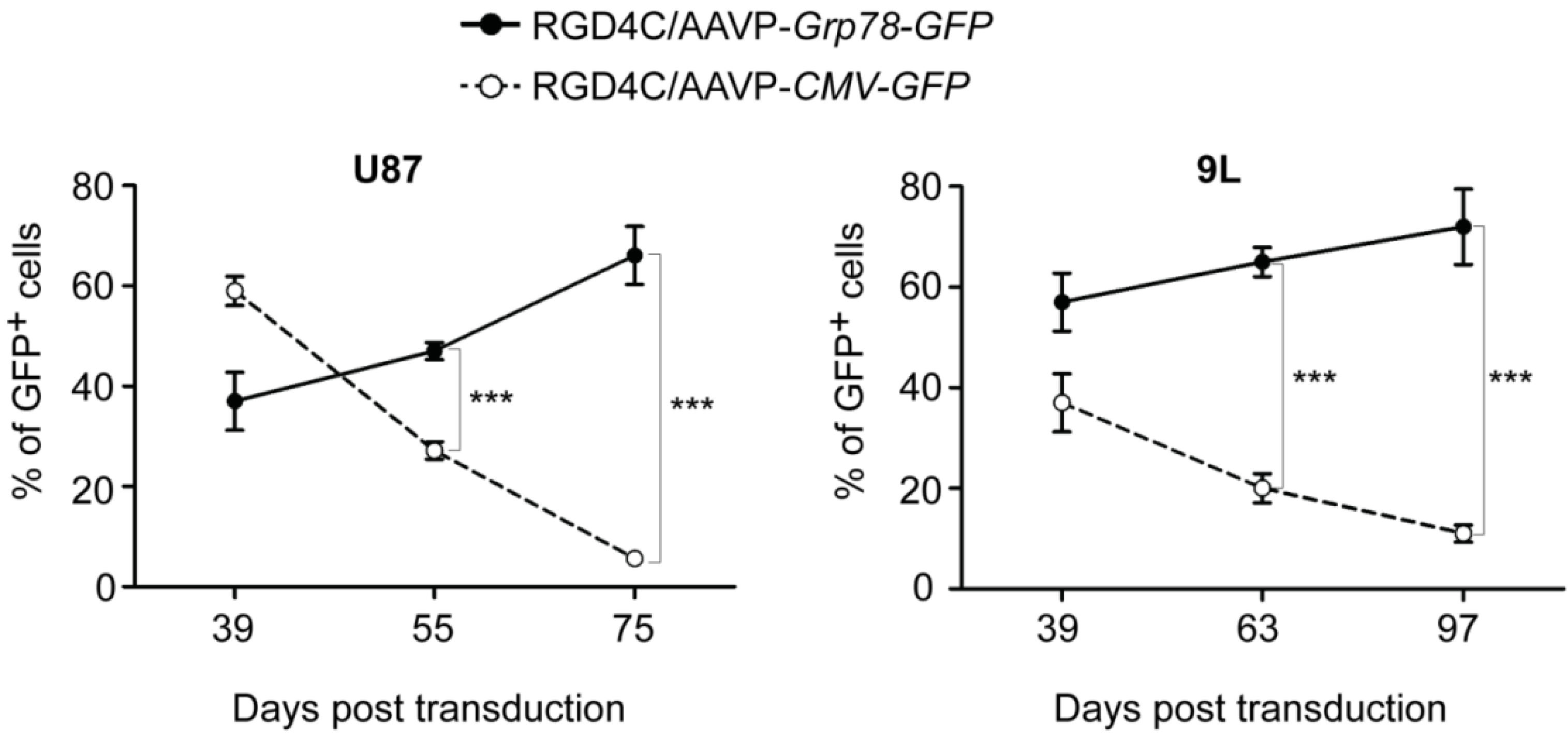
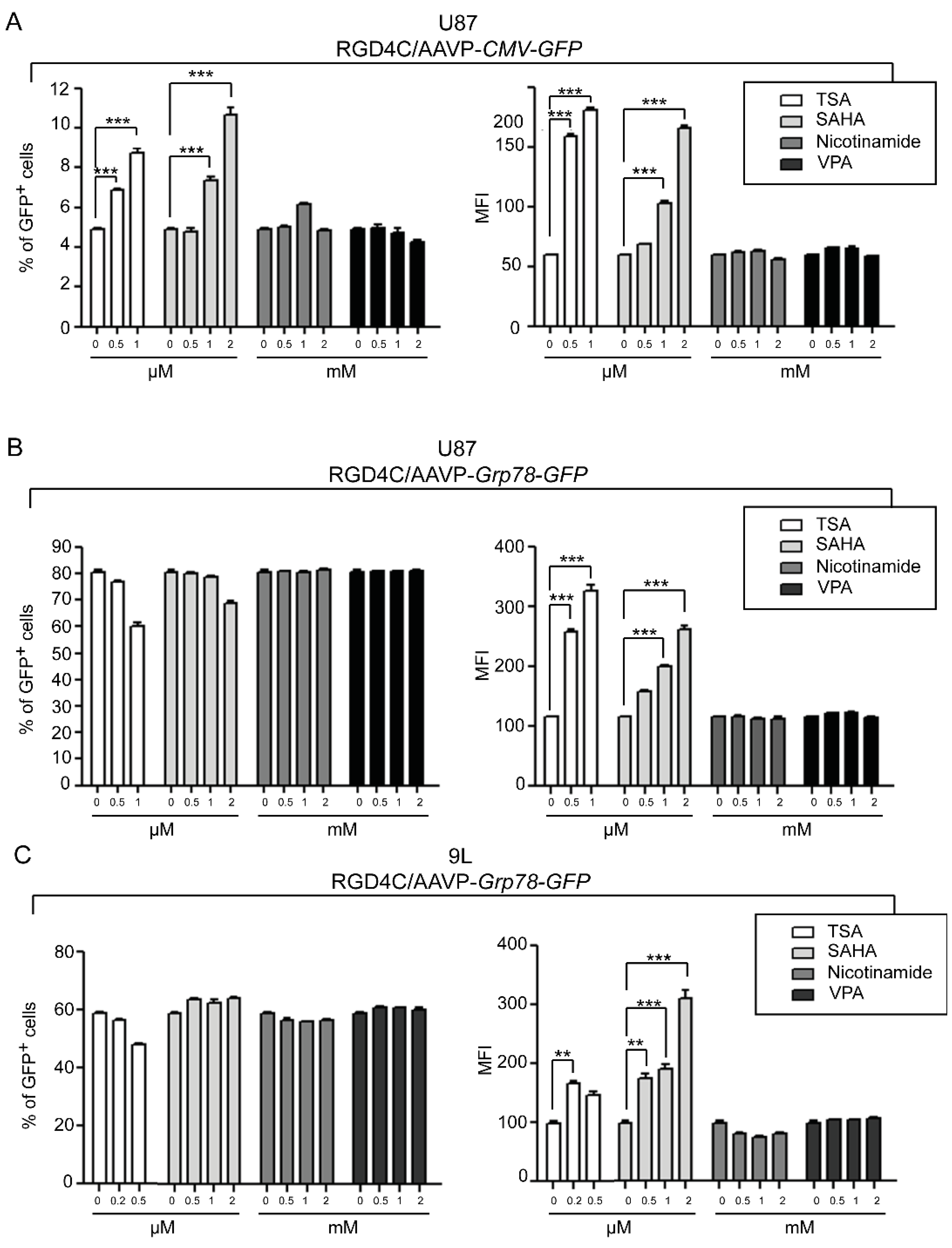
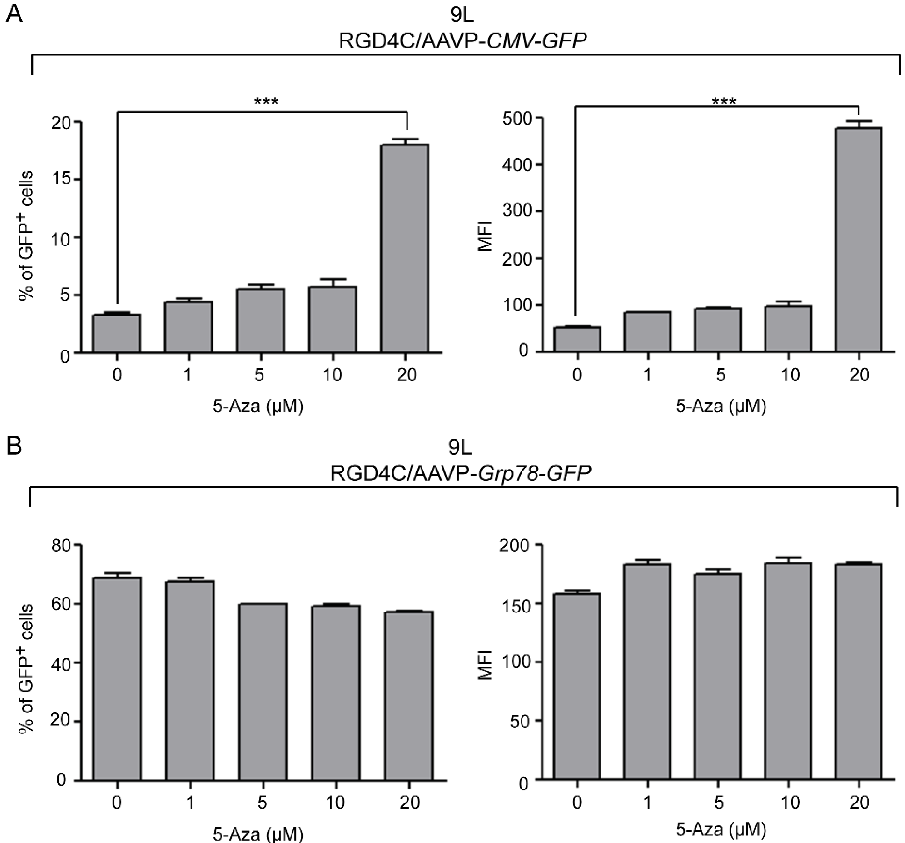
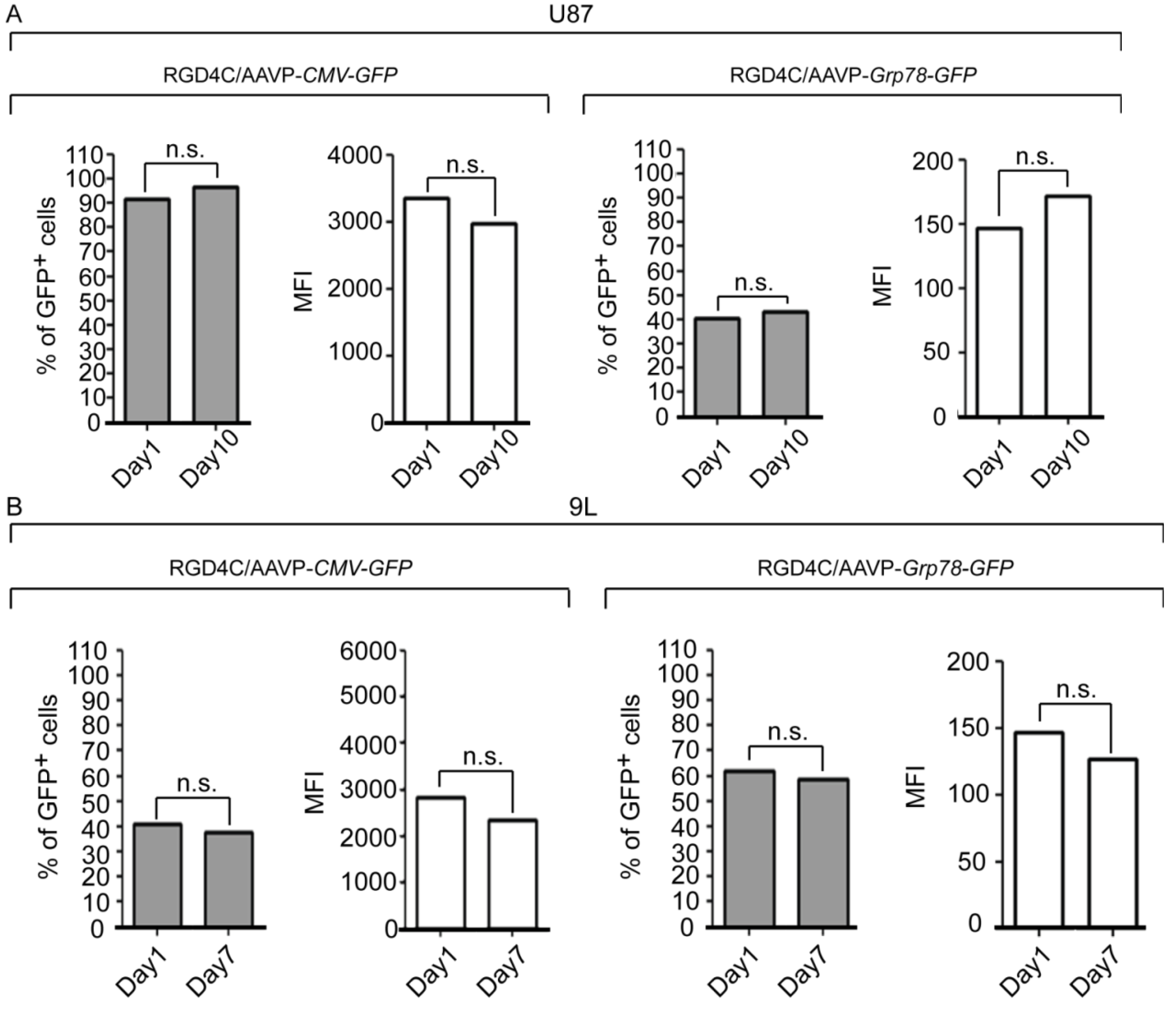
2. Results and Discussion

We monitored gene expression by AAVP in the human U87 and rat 9L glioblastoma cells over an extended time course by generating stably transduced cells with vectors carrying puroR gene that confers puromycin resistance. A marked decrease in gene expression from the RGD4C/AAVP-CMV phage vector was observed over time in U87 and 9L cells; in contrast, no silencing of Grp78-regulated gene expression was detected following cell transduction with the double-targeted RGD4C/AAVP-Grp78 phage (Figure 1).
Figure 1. Persistence of gene expression from RGD4C/AAVP-Grp78 and silencing of RGD4C/AAVP-CMV-mediated gene delivery in cancer cells. U87 and 9L glioblastoma cells were stably transduced with RGD4C/AAVP-CMV-GFP or RGD4C/AAVP-Grp78-GFP vectors. Then GFP positive cells were monitored by flow cytometry over a period of 39 to 75 days post-transduction of U87 cells, and 39 to 97 days post-transduction of 9L cells. This experiment was repeated three times with similar results, shown are data of one experiment. Statistical analyses were performed by using GraphPad Prism software (version 5.0). Error bars represent standard error of the mean (s.e.m). p-values were generated by ANOVA and denoted as follows: * p < 0.05, ** p < 0.01 and *** p < 0.001.
Viruses 05 02561 g001
Although the exact mechanisms of viral promoter silencing have remained mainly unknown, several studies have demonstrated the association of DNA methylation and histone deacetylation with inactivation of the CMV promoter [11,13,16,17]. Generally, both DNA methylation and histone acetylation statuses play major roles in the regulation of gene expression by providing transcription factors’ accessibility to gene promoters. The precise balance of acetylated and deacetylated states of histones is an important feature of gene regulation and the imbalance is found in many human cancers, often resulting from alterations in histone acetyltransferase (HATs) and histone deacetylase (HDACs) enzyme activities. Here, we quantified AAVP-mediated gene expression in the presence of HDAC inhibitors by using vectors expressing the green fluorescent protein (GFP), RGD4C/AAVP-CMV-GFP and RGD4C/AAVP-Grp78-GFP, and carrying the puroR to generate stable gene expression by stably transduced cells. Flow cytometry was used and both percentage of GFP positive cells and mean fluorescent intensity (MFI) were calculated by normalizing the results to parental non-transduced cells. As an initial experiment, we evaluated GFP expression in the human U87 cancer cells transduced with RGD4C/AAVP-CMV-GFP or RGD4C/AAVP-Grp78-GFP upon treatment with increasing concentrations of trichostatin-A (TSA), a pan-HDAC inhibitor. TSA is the first characterized organic HDAC inhibitor [18] widely utilized to study the reactivation of silenced viral constructs. In RGD4C/AAVP-CMV-GFP-transduced U87 cells, treatment with 0.5 µM and 1 µM TSA resulted in a significant increase of both percentage of GFP positive cells by 1.4- and 1.8-fold, and MFI by 2.7- and 3.0-fold, respectively (Figure 2A). These findings are consistent with previous reports demonstrating reactivation of the CMV promoter by TSA in U87 cells and other cell lines [12,19]. Interestingly, GFP expression in U87 cells stably transduced by RGD4C/AAVP-Grp78-GFP increased at the level of MFI only, upon TSA treatment, with no effect on GFP positive cells (Figure 2B). Next, we investigated additional HDAC inhibitors such as suberoylanilide hydroxamic acid (SAHA), which is structurally similar to TSA, as well as nicotinamide and valporic acid (VPA). SAHA treatment with 0.5 µM and 1 µM yielded results comparable to TSA and resulted in a dose dependent reactivation of gene expression in U87 cells transduced with RGD4C/AAVP-CMV-GFP, enhancing the percentage of GFP positive cells by 1.6- and 2.2-fold, as well as MFI by 1.8- and 2.8-fold, respectively (Figure 2A). Interestingly, in RGD4C/AAVP-Grp78-GFP-transduced cells and similar to TSA treatment, only the MFI was increased upon SAHA treatment while the percentage of GFP positive cells remained intact (Figure 2B). Finally, treatment with either nicotinamide or VPA had no effect on GFP expression of U87 cells transduced with either RGD4C/AAVP-CMV-GFP (Figure 2A) or RGD4C/AAVP-Grp78-GFP (Figure 2B). These results show that TSA and SAHA, both Zn2+ binding inhibitors of HDACs class I and II, restore GFP expression from RGD4C/AAVP-CMV in U87 cells; whereas, nicotinamide, a class III HDAC inhibitor, and VPA, an inhibitor of class I HDACs, had no effect on gene expression from the RGD4C/AAVP-CMV-GFP phage vector. Based on these observations we can postulate the involvement of HDAC class II in gene expression silencing from RGD4C/AAVP-CMV-GFP in U87 cells. Interestingly, induction of RGD4C/AAVP-Grp78-guided gene expression by TSA and SAHA treatment was also reproduced in the 9L cells transduced with RGD4C/AAVP-Grp78-GFP, and consistently with U87 cells, only MFI was increased with no effect on GFP positive cells (Figure 2C). Again, nicotinamide and VPA inhibitors had no impact (Figure 2C). In contrast, TSA, SAHA, nicotinamide, and VPA treatment of 9L cells transduced with RGD4C/AAVP-CMV-GFP had no significant effect on GFP expression (data not shown). Consequently, we investigated an additional mechanism regulating the CMV promoter silencing, the promoter methylation status, to determine whether gene expression from the RGD4C/AAVP-CMV phage in 9L cells can be rescued by DNA methylation inhibitors. Thus, treatment with various concentrations of the DNA methylation inhibitor 5-Azacytidine (5-Aza) resulted in a dose dependent increase of GFP expression in 9L cells transduced with RGD4C/AAVP-CMV-GFP (Figure 3A). Addition of 20 µM of 5-Aza increased the percentage of GFP positive cells to 17.5% compared to 3.5% of control untreated cells, and boosted the MFI by 8.9-fold. In contrast, 5-Aza treatment of 9L cells transduced with the RGD4C/AAVP-Grp78-GFP phage resulted in no significant change in GFP expression (Figure 3B). Extensive methylation of the CMV promoter sequences has previously been reported [12,13,16,20]. Chromatin structural alterations in the CMV promoter region were also described following suppression of expression of genes stably delivered by AAV vectors carrying the CMV promoter [21,22].
Figure 2. HDAC inhibitors restore gene expression from RGD4C/AAVP-CMV and boost RGD4C/AAVP-Grp78-mediated gene expression in cancer cells. U87 cells at day 75 post-transduction with (A) RGD4C/AAVP-CMV-GFP or (B) RGD4C/AAVP-Grp78-GFP were plated in 6 well plates, then treated 24 h later with various concentrations of the HDAC inhibitors TSA, SAHA, nicotinamide or VPA for three days; (C) 9L cells stably transduced with RGD4C/AAVP-Grp78-GFP were treated with various concentrations of the HDAC inhibitors at day 97 post-transduction as described above. Finally, U87 and 9L cells were collected and GFP positive cells as well as the MFI were analyzed by flow cytometry. The experiments were performed in triplicate, repeated three times with similar results, and a representative experiment is shown. Error bars represent standard error of the mean (s.e.m). p-values were generated by ANOVA and denoted as follows: * p < 0.05, ** p < 0.01 and *** p < 0.001.
Viruses 05 02561 g002
Figure 3. Treatment with the DNA methylation inhibitor 5-Aza restores gene expression from RGD4C/AAVP-CMV in 9L glioblastoma cells. 9L cells stably transduced with (A) RGD4C/AAVP-CMV-GFP or (B) RGD4C/AAVP-Grp78-GFP were kept for 97 days in culture, then plated in 6 well plates and treated 24 h later with increasing concentrations of 5-Aza. At day three, post 5-Aza treatment, cells were collected and GFP positive cells as well as the MFI were analyzed by flow cytometry. The experiments were repeated three times, in triplicate, with similar results and shown are data from a representative experiment. Statistical analyses were performed as described in Figure 1 and Figure 2.
Viruses 05 02561 g003
HDACs are upregulated in cancer. Therefore, HDAC inhibitors could be combined to restore RGD4C/AAVP-CMV efficacy or enhance RGD4C/AAVP-Grp78 in cancer cells specifically. Another interesting observation was a dose-dependent induction of the Grp78 promoter by TSA and SAHA in both 9L and U87 cells at the MFI level only, without any effect on the GFP positive cells. These data show that the GFP positive cells remain stable over time in RGD4C/AAVP-Grp78-GFP-transduced cells, confirming the absence of Grp78 promoter silencing and that treatment with HDAC inhibitors further enhance gene expression from RGD4C/AAVP-Grp78. Activation of the Grp78 promoter by TSA in various cancer cells has been reported [23]. Moreover, Grp78 induction by HDAC inhibitors was detected in xenograft models and biopsies from breast cancer patients undergoing TSA treatment [23]. HDAC inhibitors are considered potential candidates for cancer treatment [24]. For example, SAHA has recently been approved by the US Food and Drug Administration as a new class of anti-cancer drugs with potential against cutaneous T cell lymphoma [25]. SAHA has also been used in phase I and II clinical trials for hematological malignancies and solid tumors [26,27]. Nevertheless, HDAC inhibitors are considered poor drugs against solid tumors [26,28].
Grp78 is a stress-inducible gene that supports cell survival and drug resistance [29] and cancer cell chemoresistance increases with stress mediated by cell confluence. Therefore, we investigated whether cell confluence induces Grp78 activity. Transduced U87 and 9L cells were grown until 70% confluence. Subsequently, GFP expression analysis by flow cytometry over time revealed that the percentage of GFP positive cells and MFI remained constant in both U87 and 9L cells transduced with either RGD4C/AAVP-CMV-GFP or RGD4C/AAVP-Grp78-GFP vectors (Figure 4). These data provide evidence that cell confluence does not affect the RGD4C/AAVP-Grp78 activity.
Figure 4. Assessment of the effect of cell confluence on AAVP-driven gene expression in stably transduced cells. U87 and 9L populations of pooled clones stably transduced with RGD4C/AAVP-CMV-GFP or RGD4C/AAVP-Grp78-GFP, were plated at 300,000 cells/well in 6 well plates and flow cytometry analysis was performed to quantify GFP expression over the indicated times. Shown are percentage of GFP positive cells and MFI derived from transduction with either the RGD4C/AAVP-CMV-GFP or RGD4C/AAVP-Grp78-GFP. Day one represents 70% confluent cells while days 10 or 7 are the upper-limit time points at which overgrown cells could still be kept alive The experiments were repeated three times, in triplicate. n.s. = non-significant.
Viruses 05 02561 g004

3. Experimental

3.1. Cell Culture

The human U87 glioblastoma cell line was obtained from the Cancer Research UK and the rat 9L glioblastoma cell line was kindly provided by Dr. Hrvoje Miletic, University of Bergen, Norway. These cell lines were maintained in Dulbecco’s Modified Eagle Medium supplemented with Fetal Bovine Serum (FBS), l-glutamine, penicillin, and streptomycin.

3.2. Vector Construction and Phage Production

To generate the double-targeted RGD4C/AAVP-Grp78-GFP and targeted RGD4C/AAVP-CMV-GFP vectors carrying the puroR resistance gene, a puromycin resistance cassette was cloned into the SacI site of AAVP vector located next to the AAV transgene cassette. First, the 1,168 bp fragment containing the puromycin resistance gene under the control of SV40 early enhancer/promoter was released from the pGL4.20 plasmid (promega) by BamHI and SalI double restriction digestion then ligated to SacI linkers and subsequently inserted into SacI site of the RGD4C/AAVP-Grp78-GFP and RGD4C/AAVP-CMV-GFP phage plasmids. Phage viral particles were amplified as described [7] then expressed as bacterial transducing units (TU/μL).

3.3. Generation of Stably Transduced Cells

The U87 or 9L cells, 70% confluent, were transduced with either double targeted RGD4C/AAVP-Grp78-GFP or targeted RGD4C/AAVP-CMV-GFP phage encoding the GFP reporter gene and carrying the puroR, at 2 × 10⁶ TU/cell. At day three, post-vector transduction, cells were trypsinized and suspended in medium containing an appropriate dose of puromycin (1 µg/mL for U87 cells and 7 µg/mL for 9L cells). Parental non-transduced cells were used as controls. The medium was removed and replaced with fresh medium containing puromycin every two to three days. After two weeks, all control cells were killed and puromycin-resistant single cell clones, which were derived from single cells, were monitored under the microscope. When the puromycin-resistant cell clones were visible, they were pooled to produce a population of stably transduced cells. All stable selected cells were maintained in medium containing puromycin, imaged and monitored under a fluorescent microscope every two to three days, then further analyzed by FACS.

3.4. Fluorescence Activated Cell Sorting (FACS)

The percentage of cells expressing GFP following stable cell transduction with either RGD4C/AAVP-Grp78-GFP or RGD4C/AAVP-CMV-GFP was determined by using FACS. U87 and 9L cells were seeded in 6 well plates at a density of 3 × 10⁵ cells/well. After 48 h, cells were resuspended in PBS containing 0.5% FBS, then analyzed by using a FACScalibur instrument. A total of 20,000 individual cells were mounted within a manual gate in each condition.
To validate reactivation of the CMV promoter activity, stably transduced U87 and 9L cells as well as parental control cells, were seeded in 6 well plates. After 24 h, cells were treated with DNA methyltransferase or HDACs inhibitor drugs, then monitored by fluorescent microscope. Afterwards, cells were washed, trypsinized and resuspended in PBS containing 0.5% FBS then analyzed by FACS. The mean fluorescence intensity (MFI), used to define the fluorescence intensity of each cell population, and the percentage of GFP positive cells were calculated by normalizing to the control (parental cells grown in similar condition). Data were analyzed using FlowJo software.

3.5. Statistical Analyses

Statistical analyses were performed by using GraphPad Prism software (version 5.0). Error bars represent standard error of the mean (s.e.m). p values were generated by ANOVA and denoted as follows: * p < 0.05, ** p < 0.01 and *** p < 0.001.

4. Conclusions

Our findings show that histone deacetylation and DNA methylation are involved, at least in part, in the long-term gene expression silencing from the parental RGD4C/AAVP bacteriophage gene delivery vector. Specifically, we have presented a novel strategy to advance gene transfer to cancer cells by bacteriophage by combining AAVP with inhibitors of histone deacetylation and DNA methylation. Additionally, our findings prove that combination of Grp78-guided gene expression and HDAC inhibitors provides novel and most suitable strategy to enhance cancer gene therapy by the AAVP phage-derived vector, and should be considered for clinical applications against solid tumors.

Acknowledgments

We thank George Smith, Hrvoje Miletic, Renata Pasqualini and Wadih Arap for reagents; we thank Anna Ettorre for assistance with the flow cytometry experiments. AK and AH were supported by a Grant G0701159 of the UK Medical Research Council (MRC). TY was funded by a scholarship from the Thai Government. NH was funded by the MRC and Health Protection Agency (HPA).

Conflicts of Interest

The authors declare no conflict of interest.

References

  1. Waehler, R.; Russell, S.J.; Curiel, D.T. Engineering targeted viral vectors for gene therapy. Nat. Rev. Genet. 2007, 8, 573–587. [Google Scholar] [CrossRef]
  2. Larocca, D.; Witte, A.; Johnson, W.; Pierce, G.F.; Baird, A. Targeting bacteriophage to mammalian cell surface receptors for gene delivery. Hum. Gene Ther. 1998, 9, 2393–2399. [Google Scholar] [CrossRef]
  3. Hajitou, A.; Trepel, M.; Lilley, C.E.; Soghomonyan, S.; Alauddin, M.M.; Marini, F.C., 3rd.; Restel, B.H.; Ozawa, M.G.; Moya, C.A.; Rangel, R.; et al. A hybrid vector for ligand-directed tumor targeting and molecular imaging. Cell 2006, 125, 385–398. [Google Scholar] [CrossRef]
  4. Stoneham, C.A.; Hollinshead, M.; Hajitou, A. Clathrin-mediated endocytosis and subsequent endo-lysosomal trafficking of adeno-associated virus/phage. J. Biol. Chem. 2012, 287, 35849–35859. [Google Scholar] [CrossRef]
  5. Przystal, J.M.; Umukoro, E.; Stoneham, C.A.; Yata, T.; O'Neill, K.; Syed, N.; Hajitou, A. Proteasome inhibition in cancer is associated with enhanced tumor targeting by the adeno-associated virus/phage. Mol. Oncol. 2013, 7, 55–66. [Google Scholar] [CrossRef]
  6. Trepel, M.; Stoneham, C.A.; Eleftherohorinou, H.; Mazarakis, N.D.; Pasqualini, R.; Arap, W.; Hajitou, A. A heterotypic bystander effect for tumor cell killing after adeno-associated virus/ phage-mediated, vascular-targeted suicide gene transfer. Mol. Cancer Ther. 2009, 8, 2383–2391. [Google Scholar] [CrossRef]
  7. Hajitou, A.; Rangel, R.; Trepel, M.; Soghomonyan, S.; Gelovani, J.G.; Alauddin, M.M.; Pasqualini, R.; Arap, W. Design and construction of targeted AAVP vectors for mammalian cell transduction. Nat. Protoc. 2007, 2, 523–531. [Google Scholar] [CrossRef]
  8. Hajitou, A.; Lev, D.C.; Hannay, J.A.; Korchin, B.; Staquicini, F.I.; Soghomonyan, S.; Alauddin, M.M.; Benjamin, R.S.; Pollock, R.E.; Gelovani, J.G.; et al. A preclinical model for predicting drug response in soft-tissue sarcoma with targeted AAVP molecular imaging. Proc. Natl. Acad. Sci. USA 2008, 105, 4471–4476. [Google Scholar] [CrossRef]
  9. Tandle, A.; Hanna, E.; Lorang, D.; Hajitou, A.; Moya, C.A.; Pasqualini, R.; Arap, W.; Adem, A.; Starker, E.; Hewitt, S.; et al. Tumor vasculature-targeted delivery of tumor necrosis factor-alpha. Cancer 2009, 115, 128–139. [Google Scholar] [CrossRef]
  10. Paoloni, M.C.; Tandle, A.; Mazcko, C.; Hanna, E.; Kachala, S.; Leblanc, A.; Newman, S.; Vail, D.; Henry, C.; Thamm, D.; et al. Launching a novel preclinical infrastructure: Comparative oncology trials consortium directed therapeutic targeting of TNFalpha to cancer vasculature. PLoS One 2009, 4, e4972. [Google Scholar] [CrossRef]
  11. Brooks, A.R.; Harkins, R.N.; Wang, P.; Qian, H.S.; Liu, P.; Rubanyi, G.M. Transcriptional silencing is associated with extensive methylation of the CMV promoter following adenoviral gene delivery to muscle. J. Gene Med. 2004, 6, 395–404. [Google Scholar] [CrossRef]
  12. Grassi, G.; Maccaroni, P.; Meyer, R.; Kaiser, H.; D'Ambrosio, E.; Pascale, E.; Grassi, M.; Kuhn, A.; Di Nardo, P.; Kandolf, R.; et al. Inhibitors of DNA methylation and histone deacetylation activate cytomegalovirus promoter-controlled reporter gene expression in human glioblastoma cell line U87. Carcinogenesis 2003, 24, 1625–1635. [Google Scholar] [CrossRef]
  13. Prosch, S.; Stein, J.; Staak, K.; Liebenthal, C.; Volk, H.D.; Kruger, D.H. Inactivation of the very strong HCMV immediate early promoter by DNA CpG methylation in vitro. Biol. Chem. 1996, 377, 195–201. [Google Scholar]
  14. Kia, A.; Przystal, J.M.; Nianiaris, N.; Mazarakis, N.D.; Mintz, P.J.; Hajitou, A. Dual systemic tumor targeting with ligand-directed phage and Grp78 promoter induces tumor regression. Mol. Cancer Ther. 2012, 11, 2566–2577. [Google Scholar] [CrossRef]
  15. Dong, D.; Dubeau, L.; Bading, J.; Nguyen, K.; Luna, M.; Yu, H.; Gazit-Bornstein, G.; Gordon, E.M.; Gomer, C.; Hall, F.L.; et al. Spontaneous and controllable activation of suicide gene expression driven by the stress-inducible grp78 promoter resulting in eradication of sizable human tumors. Hum. Gene Ther. 2004, 15, 553–561. [Google Scholar] [CrossRef]
  16. Choi, K.H.; Basma, H.; Singh, J.; Cheng, P.W. Activation of CMV promoter-controlled glycosyltransferase and beta-galactosidase glycogenes by butyrate, tricostatin A, and 5-aza-2'-deoxycytidine. Glycoconj. J. 2005, 22, 63–69. [Google Scholar] [CrossRef]
  17. Murphy, J.C.; Fischle, W.; Verdin, E.; Sinclair, J.H. Control of cytomegalovirus lytic gene expression by histone acetylation. EMBO J. 2002, 21, 1112–1120. [Google Scholar] [CrossRef]
  18. Yoshida, M.; Kijima, M.; Akita, M.; Beppu, T. Potent and specific inhibition of mammalian histone deacetylase both in vivo and in vitro by trichostatin A. J. Biol. Chem. 1990, 265, 17174–17179. [Google Scholar]
  19. Chen, W.Y.; Bailey, E.C.; McCune, S.L.; Dong, J.Y.; Townes, T.M. Reactivation of silenced, virally transduced genes by inhibitors of histone deacetylase. Proc. Natl. Acad. Sci. USA 1997, 94, 5798–5803. [Google Scholar]
  20. Kong, Q.; Wu, M.; Wang, Z.; Zhang, X.; Li, L.; Liu, X.; Mu, Y.; Liu, Z. Effect of trichostatin A and 5-Aza-2'-deoxycytidine on transgene reactivation and epigenetic modification in transgenic pig fibroblast cells. Mol. Cell. Biochem. 2011, 355, 157–165. [Google Scholar] [CrossRef]
  21. Chen, W.Y.; Townes, T.M. Molecular mechanism for silencing virally transduced genes involves histone deacetylation and chromatin condensation. Proc. Natl. Acad. Sci. USA 2000, 97, 377–382. [Google Scholar] [CrossRef]
  22. Liu, X.F.; Yan, S.; Abecassis, M.; Hummel, M. Establishment of murine cytomegalovirus latency in vivo is associated with changes in histone modifications and recruitment of transcriptional repressors to the major immediate-early promoter. J. Virol. 2008, 82, 10922–10931. [Google Scholar] [CrossRef]
  23. Baumeister, P.; Dong, D.; Fu, Y.; Lee, A.S. Transcriptional induction of GRP78/BiP by histone deacetylase inhibitors and resistance to histone deacetylase inhibitor-induced apoptosis. Mol. Cancer Ther. 2009, 8, 1086–1094. [Google Scholar] [CrossRef]
  24. Finnin, M.S.; Donigian, J.R.; Cohen, A.; Richon, V.M.; Rifkind, R.A.; Marks, P.A.; Breslow, R.; Pavletich, N.P. Structures of a histone deacetylase homologue bound to the TSA and SAHA inhibitors. Nature 1999, 401, 188–193. [Google Scholar] [CrossRef]
  25. Grant, S.; Easley, C.; Kirkpatrick, P. Vorinostat. Nat. Rev. Drug Discov. 2007, 6, 21–22. [Google Scholar] [CrossRef]
  26. Modesitt, S.C.; Sill, M.; Hoffman, J.S.; Bender, D.P. A phase II study of vorinostat in the treatment of persistent or recurrent epithelial ovarian or primary peritoneal carcinoma: A Gynecologic Oncology Group study. Gynecol. Oncol. 2008, 109, 182–186. [Google Scholar] [CrossRef]
  27. Galanis, E.; Jaeckle, K.A.; Maurer, M.J.; Reid, J.M.; Ames, M.M.; Hardwick, J.S.; Reilly, J.F.; Loboda, A.; Nebozhyn, M.; Fantin, V.R.; et al. Phase II trial of vorinostat in recurrent glioblastoma multiforme: A north central cancer treatment group study. J. Clin. Oncol. 2009, 27, 2052–2058. [Google Scholar] [CrossRef]
  28. Luu, T.H.; Morgan, R.J.; Leong, L.; Lim, D.; McNamara, M.; Portnow, J.; Frankel, P.; Smith, D.D.; Doroshow, J.H.; Wong, C.; et al. A phase II trial of vorinostat (suberoylanilide hydroxamic acid) in metastatic breast cancer: A California Cancer Consortium study. Clin. Cancer Res. 2008, 14, 7138–7142. [Google Scholar] [CrossRef]
  29. Pyrko, P.; Schonthal, A.H.; Hofman, F.M.; Chen, T.C.; Lee, A.S. The unfolded protein response regulator GRP78/BiP as a novel target for increasing chemosensitivity in malignant gliomas. Cancer Res. 2007, 67, 9809–9816. [Google Scholar] [CrossRef]

