
Recent Progress in the Design and Medical Application of In Situ Self-Assembled Polypeptide Materials

Abstract
:1. Introduction
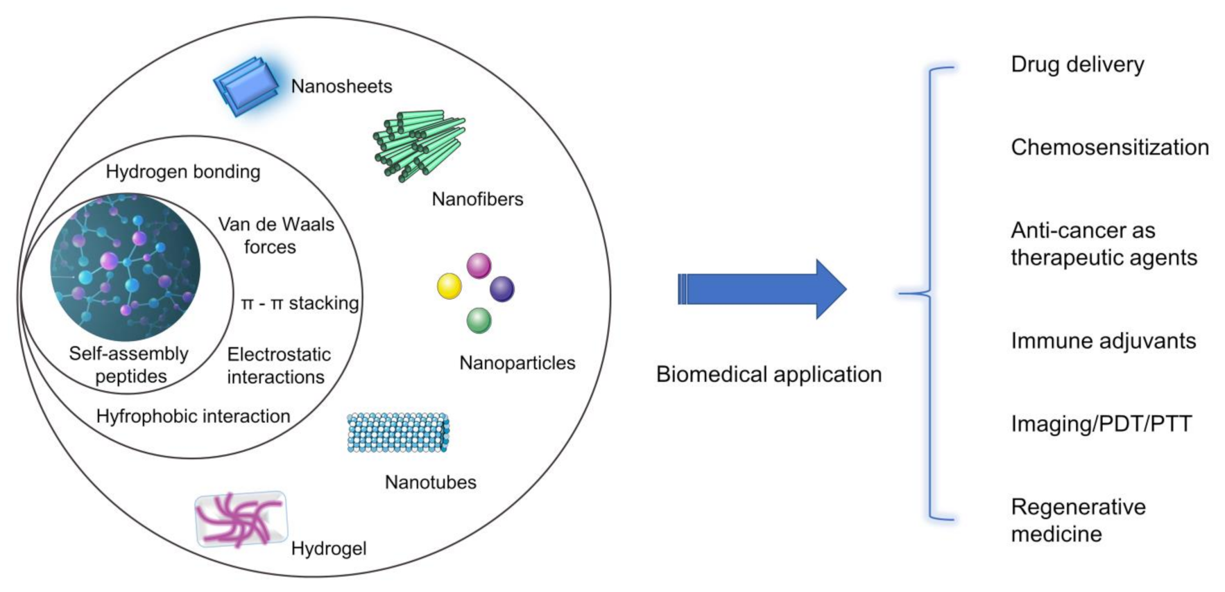
2. Classification and Assembly Principle of Self-Assembled Polypeptides
2.1. Spontaneous Self-Assembled Polypeptides
2.2. Trigger-Type Self-Assembled Polypeptides
3. Applications in Biomedicine
3.1. Drug Delivery System
3.2. Anti-Cancer as Therapeutic Agents
3.3. Immune Adjuvants
3.4. Imaging/PDT/PTT
3.5. Regenerative Medicine
3.5.1. Cell Culture
3.5.2. Bone Tissue Regeneration
3.5.3. Neuron Repair and Regenerate
3.5.4. Wound Healing
4. Expectation
Author Contributions
Funding
Acknowledgments
Conflicts of Interest
References
- Mazo, A.R.; Allison-Logan, S.; Karimi, F.; Chan, J.A.; Qiao, G.G. Ring opening polymerization of α-amino acids: Advances in synthesis, architecture and applications of polypeptides and their hybrids. Chem. Soc. Rev. 2020, 49, 4737–4834. [Google Scholar] [CrossRef]
- Colin, B. Secondary structures of synthetic polypeptide polymers. Polym. Chem. 2018, 9, 1517–1529. [Google Scholar]
- Yang, C.; Liang, G.; Lin, J.; Wang, L.; Li, Z. Toroid Formation through a Supramolecular “Cyclization Reaction” of Rodlike Micelles. Angew. Chem. Int. Ed. Engl. 2017, 129, 5546–5550. [Google Scholar] [CrossRef]
- Abbas, M.; Zou, Q.; Li, S.; Yan, X. Self-Assembled Peptide- and Protein-Based Nanomaterials for Antitumor Photodynamic and Photothermal Therapy. Adv. Mater. 2017, 29, 1605021. [Google Scholar] [CrossRef]
- Wang, W.; Ma, Z.; Zhu, S.; Wan, H.; Yue, J.; Ma, H.; Ma, R.; Yang, Q.; Wang, Z.; Li, Q. Molecular Cancer Imaging in the Second Near-Infrared Window Using a Renal-Excreted NIR-II Fluorophore-Peptide Probe. Adv. Mater. 2018, 30, e1800106. [Google Scholar] [CrossRef]
- Mendes, A.C.; Baran, E.T.; Reis, R.L.; Azevedo, H.S. Self-assembly in nature: Using the principles of nature to create complex nanobiomaterials. Wiley Interdiscip. Rev. Nanomed. Nanobiotechnol. 2013, 5, 582–612. [Google Scholar] [CrossRef]
- Mitchison, T.; Kirschner, M. Dynamic instability of microtubule growth. Nature 1984, 312, 237. [Google Scholar] [CrossRef]
- Mitchison, T.; Kirschner, M. Microtubule assembly nucleated by isolated centrosomes. Nature 1984, 312, 232–237. [Google Scholar] [CrossRef]
- Brenner, S.; Jacob, F.; Meselson, M. An Unstable Intermediate Carrying Information from Genes to Ribosomes for Protein Synthesis. Nature 1961, 190, 576–581. [Google Scholar] [CrossRef]
- Shi, Y. Apoptosome: The cellular engine for the activation of caspase-9. Structure 2002, 10, 285–288. [Google Scholar] [CrossRef]
- Martinon, F.; Burns, K.; Tschopp, J. The inflammasome: A molecular platform triggering activation of inflammatory caspases and processing of proIL-beta. Mol. Cell 2002, 10, 417–426. [Google Scholar] [CrossRef]
- Lashuel, H.A.; Hartley, D.; Petre, B.M.; Walz, T.; Lansbury, P.T., Jr. Neurodegenerative disease: Amyloid pores from pathogenic mutations. Nature 2002, 418, 291. [Google Scholar] [CrossRef]
- Mann, S. Life as a Nanoscale Phenomenon. Angew. Chem. Int. Ed. 2008, 47, 5306–5320. [Google Scholar] [CrossRef]
- Grazon, C.; Salas-Ambrosio, P.; Ibarboure, E.; Buol, A.; Bonduelle, C. Aqueous Ring-Opening Polymerization Induced Self-Assembly (ROPISA) of N-carboxyanhydrides. Angew. Chem. Int. Ed. 2019, 59, 622–626. [Google Scholar] [CrossRef]
- Ruoslahti, E. Peptides as Targeting Elements and Tissue Penetration Devices for Nanoparticles. Adv. Mater. 2012, 24, 3747–3756. [Google Scholar] [CrossRef]
- Hofmann, S.; Bellmann-Sickert, K.; Beck-Sickinger, A.G. Chemical modification of neuropeptide Y for human Y1 receptor targeting in health and disease. Biol. Chem. 2019, 400, 299–311. [Google Scholar] [CrossRef] [Green Version]
- Zhang, S. Emerging biological materials through molecular self-assembly. Biotechnol. Adv. 2002, 20, 321–339. [Google Scholar] [CrossRef]
- Ghadiri, M.R.; Granja, J.R.; Milligan, R.A.; Mcree, D.E.; Khazanovich, N. Self-assembling organic nanotubes based on a cyclic peptide architecture. Nature 1993, 366, 324. [Google Scholar] [CrossRef] [PubMed]
- Woolfson, D.N.; Bartlett, G.J.; Bruning, M.; Thomson, A.R. New currency for old rope: From coiled-coil assemblies to alpha-helical barrels. Curr. Opin. Struct. Biol. 2012, 22, 432–441. [Google Scholar] [CrossRef]
- Rhys, G.G.; Wood, C.W.; Lang, E.J.M.; Mulholland, A.J.; Brady, R.L.; Thomson, A.R.; Woolfson, D.N. Maintaining and breaking symmetry in homomeric coiled-coil assemblies. Nat. Commun. 2018, 9, 4132. [Google Scholar] [CrossRef] [Green Version]
- Veggiani, G.; Sidhu, S.S. Peptides meet ubiquitin: Simple interactions regulating complex cell signaling. Pept. Sci. 2019, 111, e24091. [Google Scholar] [CrossRef]
- Li, L.L.; Qiao, S.L.; Liu, W.J.; Ma, Y.; Wan, D.; Pan, J.; Wang, H. Intracellular construction of topology-controlled polypeptide nanostructures with diverse biological functions. Nat. Commun. 2017, 8, 1276. [Google Scholar] [CrossRef] [PubMed]
- Kuang, Y.; Shi, J.; Li, J.; Yuan, D.; Alberti, K.A.; Xu, Q.; Xu, B. Pericellular Hydrogel/Nanonets Inhibit Cancer Cells. Angew. Chem. Int. Ed. 2014, 126, 8242–8245. [Google Scholar] [CrossRef] [Green Version]
- Kalmouni, M.; Al-Hosani, S.; Magzoub, M. Cancer targeting peptides. Cell. Mol. Life Sci. 2019, 76, 2171–2183. [Google Scholar] [CrossRef]
- Saket, A.; Vikas, P.; Vandana, S. RGD Peptide as a Targeting Moiety for Theranostic Purpose: An Update Study. Int. J. Pept. Res. Ther. 2018, 25, 49–65. [Google Scholar]
- Tian, X.; Sun, F.; Zhou, X.R.; Luo, S.Z.; Chen, L. Role of peptide self-assembly in antimicrobial peptides. J. Pept. Sci. 2015, 21, 530–539. [Google Scholar] [CrossRef]
- Qi, G.B.; Gao, Y.J.; Wang, L.; Wang, H. Self-Assembled Peptide-Based Nanomaterials for Biomedical Imaging and Therapy. Adv. Mater. 2018, 30, 1703444. [Google Scholar] [CrossRef]
- Ganta, S.; Devalapally, H.; Shahiwala, A.; Amiji, M. A review of stimuli-responsive nanocarriers for drug and gene delivery. J. Control. Release 2008, 126, 187–204. [Google Scholar] [CrossRef]
- Liu, M.; Du, H.; Zhang, W.; Zhai, G. Internal stimuli-responsive nanocarriers for drug delivery: Design strategies and applications. Mater. Sci. Eng. C 2016, 71, 1267–1280. [Google Scholar] [CrossRef]
- Kim, K.K.; Siddiqui, Z.; Patel, M.; Sarkar, B.; Kumar, V.A. A self-assembled peptide hydrogel for cytokine sequestration. J. Mater. Chem. B 2020, 8, 945–950. [Google Scholar] [CrossRef]
- Jin, H.; Zhang, L.; Song, Y.; Liu, L. Modulation of structure and mechanical properties of self-assembled peptide nanofibrils and nanosheets. Mater. Lett. 2019, 255, 126540. [Google Scholar] [CrossRef]
- Calvelo, M.; Granja, J.R.; Fandino, R.G. Competitive double-switched Self-Assembled Cyclic Peptide Nanotubes: A dual internal and external control. Phys. Chem. Chem. Phys. 2019, 21, 20750–20756. [Google Scholar] [CrossRef] [PubMed]
- Cao, M.; Wang, Y.; Hu, X.; Gong, H.; Lu, J.R. Reversible Thermoresponsive Peptide-PNIPAM Hydrogels for Controlled Drug Delivery. Biomacromolecules 2019, 20, 3601–3610. [Google Scholar] [CrossRef]
- Gao, Y.; Zhang, C.; Chang, J.; Yang, C.; Liu, J.; Fan, S.; Ren, C. Enzyme-instructed self-assembly of a novel histone deacetylase inhibitor with enhanced selectivity and anticancer efficiency. Biomater. Sci. 2019, 7, 1477–1485. [Google Scholar] [CrossRef]
- Wiwanitkit, V.; Sereemaspun, A.; Rojanathanes, R. Effect of gold nanoparticles on spermatozoa: The first world report. Fertil. Steril. 2009, 91, e7–e8. [Google Scholar] [CrossRef]
- De Jong, W.H.; Borm, P.J. Drug delivery and nanoparticles: Applications and hazards. Int. J. Nanomed. 2008, 3, 133. [Google Scholar] [CrossRef] [Green Version]
- Vega-Villa, K.R.; Takemoto, J.K.; Yáñez, J.A.; Remsberg, C.M.; Forrest, M.L.; Davies, N.M. Clinical toxicities of nanocarrier systems. Adv. Drug Deliv. Rev. 2008, 60, 929–938. [Google Scholar] [CrossRef]
- Cai, C.; Lin, J.; Lu, Y.; Zhang, Q.; Wang, L. Polypeptide self-assemblies: Nanostructures and bioapplications. Chem. Soc. Rev. 2016, 45, 5985–6012. [Google Scholar] [CrossRef]
- Shi, J.; Xu, B. Nanoscale Assemblies of Small Molecules Control the Fate of Cells. Nano Today 2015, 10, 615–630. [Google Scholar] [CrossRef] [Green Version]
- Zhang, C.; Lu, J.; Tian, F.; Li, L.; Hou, Y.; Wang, Y.; Sun, L.; Shi, X.; Lu, H. Regulation of the cellular uptake of nanoparticles by the orientation of helical polypeptides. Nano Res. 2019, 12, 889–896. [Google Scholar] [CrossRef]
- Baek, K.; Noblett, A.D.; Ren, P.; Suggs, L.J. Self-assembled nucleo-tripeptide hydrogels provide local and sustained doxorubicin release. Biomater. Sci. 2020, 8, 3130–3137. [Google Scholar] [CrossRef] [PubMed]
- Xu, L.; Wang, Y.; Zhu, C.; Ren, S.; Chen, Z. Morphological transformation enhances Tumor Retention by Regulating the Self-assembly of Doxorubicin-peptide Conjugates. Theranostics 2020, 10, 8162–8178. [Google Scholar] [CrossRef]
- Wang, H.; Feng, Z.; Tan, W.; Xu, B. Assemblies of d-Peptides for Targeting Cell Nucleolus. Bioconjug. Chem. 2019, 30, 2528–2532. [Google Scholar] [CrossRef] [PubMed]
- Naito, S.; Sakamoto, N.; Kotoh, S.; Goto, K.; Kumazawa, J. Expression of P-glycoprotein and multidrug resistance in renal cell carcinoma. Eur. Urol. 1993, 24, 156–160. [Google Scholar] [PubMed]
- Wang, Z.; An, H.-W.; Hou, D.; Wang, M.; Zeng, X.; Zheng, R.; Wang, L.; Wang, K.; Wang, H.; Xu, W. Addressable Peptide Self-Assembly on the Cancer Cell Membrane for Sensitizing Chemotherapy of Renal Cell Carcinoma. Adv. Mater. 2019, 31, 1807175. [Google Scholar] [CrossRef] [PubMed]
- Wang, F.; Su, H.; Xu, D.; Dai, W.; Cui, H. Tumour sensitization via the extended intratumoural release of a STING agonist and camptothecin from a self-assembled hydrogel. Nat. Biomed. Eng. 2020, 4, 1–12. [Google Scholar] [CrossRef]
- Liu, X.; Feng, Z.; Wang, C.; Su, Q.; Song, H.; Zhang, C.; Huang, P.; Liang, X.J.; Dong, A.; Kong, D. Co-localized delivery of nanomedicine and nanovaccine augments the postoperative cancer immunotherapy by amplifying T-cell responses. Biomaterials 2020, 230, 119649. [Google Scholar] [CrossRef]
- Skoulas, D.; Stuttgen, V.; Gaul, R.; Cryan, S.A.; Heise, A. Amphiphilic Star Polypept(o)ides as Nanomeric Vectors in Mucosal Drug Delivery. Biomacromolecules 2020, 21, 2455–2462. [Google Scholar] [CrossRef]
- Schatz, C.; Louguet, S.; Meins, J.L.; Lecommandoux, S. Polysaccharide-block-polypeptide Copolymer Vesicles: Towards Synthetic Viral Capsids. Angew. Chem. Int. Ed. 2009, 48, 2572–2575. [Google Scholar] [CrossRef]
- Oliveira, H.D.; Thevenot, J.; Garanger, E.; Ibarboure, E.; Lecommandoux, S. Nano-Encapsulation of Plitidepsin: In Vivo Pharmacokinetics, Biodistribution, and Efficacy in a Renal Xenograft Tumor Model. Pharm. Res. 2014, 31, 983–991. [Google Scholar] [CrossRef]
- Salva, R.; Meins, J.; Sandre, O.; BrûLet, A.; Schmutz, M.; Guenoun, P.; Lecommandoux, S. Polymersome shape transformation at the nanoscale. ACS Nano 2013, 7, 9298–9311. [Google Scholar] [CrossRef] [PubMed]
- Klok, H.A.; Langenwalter, J.F.; Lecommandoux, S. Self-Assembly of Peptide-Based Diblock Oligomers. Macromolecules 2000, 33, 7819–7826. [Google Scholar] [CrossRef]
- Caillol, S.; Lecommandoux, S.; Mingotaud, A.F.; Schappacher, M.; Soum, A.; Bryson, N.; Meyrueix, R. Synthesis and Self-Assembly Properties of Peptide-Polylactide Block Copolymers. Macromolecules 2009, 36, 1118–1124. [Google Scholar] [CrossRef]
- Bellomo, E.G.; Wyrsta, M.; Pakstis, L.; Pochan, D.J.; Deming, T.J. Stimuli-responsive polypeptide vesicles by conformation-specific assembly. Nat. Mater. 2004, 3, 244. [Google Scholar] [CrossRef] [PubMed]
- Cameron, N.; Deming, T. Peptide-based materials for nanomedicine. Macromol. Biosci. 2015, 15, 7–8. [Google Scholar] [CrossRef] [Green Version]
- Hsieh, P.C.; Davis, M.E.; Gannon, J.; MacGillivray, C.; Lee, R.T. Controlled delivery of PDGF-BB for myocardial protection using injectable self-assembling peptide nanofibers. J. Clin. Investig. 2006, 116, 237–248. [Google Scholar] [CrossRef] [Green Version]
- Mcgovern, S.L.; Caselli, E.; Grigorieff, N.; Shoichet, B.K. A Common Mechanism Underlying Promiscuous Inhibitors from Virtual and High-Throughput Screening. J. Med. Chem. 2002, 45, 1712–1722. [Google Scholar] [CrossRef]
- Dobson, C.M. Protein Folding and Misfolding. Nature 2003, 426, 884–890. [Google Scholar] [CrossRef]
- Kayed, R. Common Structure of Soluble Amyloid Oligomers Implies Common Mechanism of Pathogenesis. Science 2003, 300, 486–489. [Google Scholar] [CrossRef] [Green Version]
- Zorn, J.A.; Wolan, D.W.; Agard, N.J.; Wells, J.A. Fibrils Colocalize Caspase-3 with Procaspase-3 to Foster Maturation. J. Biol. Chem. 2012, 287, 33781–33795. [Google Scholar] [CrossRef] [Green Version]
- Kuang, Y.; Du, X.; Zhou, J.; Xu, B. Supramolecular nanofibrils inhibit cancer progression in vitro and in vivo. Adv. Healthc. Mater. 2014, 3, 1217–1221. [Google Scholar] [CrossRef] [PubMed]
- Kuang, Y.; Long, M.J.C.; Zhou, J.; Shi, J.; Gao, Y.; Xu, C.; Hedstrom, L.; Xu, B. Prion-like Nanofibrils of Small Molecules (PriSM) Selectively Inhibit Cancer Cells by Impeding Cytoskeleton Dynamics. J. Biol. Chem. 2014, 289, 29208–29218. [Google Scholar] [CrossRef] [PubMed] [Green Version]
- Gao, Y.; Kuang, Y.; Du, X.; Zhou, J.; Chandran, P.; Horkay, F.; Xu, B. Imaging Self-Assembly Dependent Spatial Distribution of Small Molecules in a Cellular Environment. Langmuir 2013, 29, 15191–15200. [Google Scholar] [CrossRef] [PubMed] [Green Version]
- Mäe, M.; Myrberg, H.; Langel, E.A. Design of a Tumor Homing Cell-Penetrating Peptide for Drug Delivery. Int. J. Pept. Res. Ther. 2009, 15, 11–15. [Google Scholar] [CrossRef]
- Cao, J.; Qiao, B.; Luo, Y.; Cheng, C.; Yang, A.; Wang, M.; Yuan, X.; Fan, K.; Li, M.; Wang, Z. A multimodal imaging-guided nanoreactor for cooperative combination of tumor starvation and multiple mechanism-enhanced mild temperature phototherapy. Biomater. Sci. 2020, 8, 6561–6578. [Google Scholar] [CrossRef]
- Jeena, M.T.; Palanikumar, L.; Go, E.M.; Kim, I.; Kang, M.G.; Lee, S.; Park, S.; Choi, H.; Kim, C.; Jin, S.M. Mitochondria localization induced self-assembly of peptide amphiphiles for cellular dysfunction. Nat. Commun. 2017, 8, 26. [Google Scholar] [CrossRef]
- Fan, J.; Fan, Y.; Wei, Z.; Li, Y.; Wang, H. Transformable peptide nanoparticles inhibit the migration of N-cadherin overexpressed cancer cells. Chin. Chem. Lett. 2020, 31, 1787–1791. [Google Scholar] [CrossRef]
- Luo, S.; Feng, J.; Xiao, L.; Guo, L.; Deng, L.; Du, Z.; Xue, Y.; Song, X.; Sun, X.; Zhang, Z.; et al. Targeting self-assembly peptide for inhibiting breast tumor progression and metastasis. Biomaterials 2020, 249, 120055. [Google Scholar] [CrossRef]
- Yang, P.P.; Zhang, K.; He, P.P.; Fan, Y.; Wang, H. A biomimetic platelet based on assembling peptides initiates artificial coagulation. Sci. Adv. 2020, 6, eaaz4107. [Google Scholar] [CrossRef]
- Wang, T.-T.; Wei, Q.-C.; Zhang, Z.-T.; Lin, M.-T.; Chen, J.-J.; Zhou, Y.; Guo, N.-N.; Zhong, X.C.; Xu, W.-H.; Liu, Z.-X.; et al. AIE/FRET-based versatile PEG-Pep-TPE/DOX nanoparticles for cancer therapy and real-time drug release monitoring. Biomater. Sci. 2019, 8, 118–124. [Google Scholar] [CrossRef]
- Zhan, J.; Cai, Y.; He, S.; Wang, L.; Yang, Z. Tandem Molecular Self-Assembly in Liver Cancer Cells. Angew. Chem. 2017, 130, 1831–1834. [Google Scholar] [CrossRef]
- Chen, J.; Chen, Z.J. PtdIns4P on dispersed trans-Golgi network mediates NLRP3 inflammasome activation. Nature 2018, 564, 71–76. [Google Scholar] [CrossRef]
- Ajithkumar, T.; Parkinson, C.; Fife, K.; Corrie, P.; Jefferies, S. Evolving treatment options for melanoma brain metastases. Lancet Oncol. 2015, 16, e486–e497. [Google Scholar] [CrossRef]
- Couzin-Frankel, J. Breakthrough of the year 2013. Cancer immunotherapy. Science 2013, 342, 1432–1433. [Google Scholar] [CrossRef] [PubMed] [Green Version]
- Mellman, I.; Coukos, G.; Dranoff, G. Cancer Immunotherapy Comes of Age. Nature 2011, 480, 480–489. [Google Scholar] [CrossRef]
- Cai, J.; Wang, H.; Wang, D.; Li, Y. Improving Cancer Vaccine Efficiency by Nanomedicine. Adv. Biosyst. 2019, 3, 1800287. [Google Scholar] [CrossRef]
- Gotwals, P.; Cameron, S.; Cipolletta, D.; Cremasco, V.; Crystal, A.; Hewes, B.; Mueller, B.; Quaratino, S.; Sabatos-Peyton, C.; Petruzzelli, L. Prospects for combining targeted and conventional cancer therapy with immunotherapy. Nat. Rev. Cancer Clin. Oncol. 2017, 17, 286–301. [Google Scholar] [CrossRef]
- Sharma, P.; Allison, J.P. Immune checkpoint targeting in cancer therapy: Toward combination strategies with curative potential. Cell 2015, 161, 205–214. [Google Scholar] [CrossRef] [Green Version]
- Mueller, K.L. Realizing the Promise. Science 2015, 348, 54–55. [Google Scholar] [CrossRef] [Green Version]
- Bonner, J. Cancer vaccines. Chem. Ind. 2001, 9, 277–280. [Google Scholar]
- Martins, K.A.O.; Cooper, C.L.; Stronsky, S.M.; Norris, S.L.W.; Kwilas, S.A.; Steffens, J.T.; Benko, J.G.; Tongeren, S.A.V.; Bavari, S. Adjuvant-enhanced CD4 T Cell Responses are Critical to Durable Vaccine Immunity. Ebiomedicine 2016, 3, 67–78. [Google Scholar] [CrossRef] [PubMed] [Green Version]
- Annalisa, C.; Elena, P.; Fabio, F.; Gabiria, P.; Peter, A.; Gianni, P.; Donata, M. Modulation of Primary Immune Response by Different Vaccine Adjuvants. Front. Immunol. 2016, 7, 427. [Google Scholar]
- Reed, S.G.; Tomai, M.; Gale, M.J. New horizons in adjuvants for vaccine development. Curr. Opin. Immunol. 2020, 65, 97–101. [Google Scholar] [CrossRef] [PubMed]
- Munir, A.; Javier, T.; Roesler, A.S.; Pietro, M.; Billur, A.; Theall, B.P.; Haewon, S.; Mirna, P.; Margery, S.; Juraj, K. Second signals rescue B cells from activation-induced mitochondrial dysfunction and death. Nat. Immunol. 2018, 19, 871–884. [Google Scholar]
- Yang, P.; Song, H.; Feng, Z.; Wang, C.; Huang, P.; Zhang, C.; Kong, D.; Wang, W. Synthetic, Supramolecular, and Self-Adjuvanting CD8+ T-Cell Epitope Vaccine Increases the Therapeutic Antitumor Immunity. Adv. Ther. 2019, 2, 1900010. [Google Scholar] [CrossRef]
- Tran, K.A.; Partyk, P.P.; Jin, Y.; Bouyer, J.; Galie, P.A. Vascularization of self-assembled peptide scaffolds for spinal cord injury repair. Acta Biomater. 2020, 104, 76–84. [Google Scholar] [CrossRef] [PubMed]
- Siegel, R.L.; Miller, K.D.; Jemal, A. Cancer statistics, 2018. CA A Cancer J. Clin. 2018, 68, 7. [Google Scholar] [CrossRef]
- Pyonteck, S.M.; Akkari, L.; Schuhmacher, A.J.; Bowman, R.L.; Sevenich, L.; Quail, D.F.; Olson, O.C.; Quick, M.L.; Huse, J.T.; Teijeiro, V. CSF-1R inhibition alters macrophage polarization and blocks glioma progression. Nat. Med. 2013, 19, 1264. [Google Scholar] [CrossRef] [Green Version]
- Zhang, J.; Liu, J. Tumor stroma as targets for cancer therapy. Pharmacol. Ther. 2013, 137, 200–215. [Google Scholar] [CrossRef] [Green Version]
- Marusyk, A.; Almendro, V.; Polyak, K. INtra-tumour heterogeneity: A looking glass for cancer? Nat. Rev. Cancer 2012, 12, 323–334. [Google Scholar] [CrossRef]
- Maeda, H.; Nakamura, H.; Fang, J. The EPR effect for macromolecular drug delivery to solid tumors: Improvement of tumor uptake, lowering of systemic toxicity, and distinct tumor imaging in vivo. Adv. Drug Deliv. Rev. 2013, 65, 71–79. [Google Scholar] [CrossRef] [PubMed]
- Zou, Q.; Chang, R.; Xing, R.; Yuan, C.; Yan, X. Injectable self-assembled bola-dipeptide hydrogels for sustained photodynamic prodrug delivery and enhanced tumor therapy. J. Control. Release 2020, 319, 344–351. [Google Scholar] [CrossRef] [PubMed]
- Zhang, D.; Qi, G.-B.; Zhao, Y.-X.; Qiao, S.-L.; Yang, C. In Situ Formation of Nanofibers from Purpurin18-Peptide Conjugates and the Assembly Induced Retention Effect in Tumor Sites. Adv. Mater. 2015, 27, 6125–6130. [Google Scholar] [CrossRef] [PubMed]
- Ye, D.; Shuhendler, A.J.; Cui, L.; Tong, L.; Tee, S.S.; Tikhomirov, G.; Felsher, D.W.; Rao, J. Bioorthogonal cyclization-mediated in situ self-assembly of small-molecule probes for imaging caspase activity in vivo. Nat. Chem. 2014, 6, 519. [Google Scholar] [CrossRef] [Green Version]
- Zhao, X.-X.; Li, L.-L.; Zhao, Y.; An, H.-W.; Cai, Q.; Lang, J.-Y.; Han, X.-X.; Peng, B.; Fei, Y.; Liu, H.; et al. In Situ Self-Assembled Nanofibers Precisely Target Cancer-Associated Fibroblasts for Improved Tumor Imaging. Angew. Chem. Int. Ed. 2019, 58, 15287–15294. [Google Scholar] [CrossRef] [PubMed]
- Guo, H.; Song, S.; Dai, T.; Sun, K.; Dou, H. Near-Infrared Fluorescent and Magnetic Resonance Dual-Imaging Nano-Coacervates for Trypsin Mapping and Targeted Payload Delivery of Malignant Tumors. ACS Appl. Mater. Interfaces 2020, 12, 17302–17313. [Google Scholar] [CrossRef]
- Li, S.; Zhang, W.J.C.M.; Xue, H.; Xing, R.; Yan, X. Tumor microenvironment-oriented adaptive nanodrugs based on peptide self-assembly. Chem. Sci. 2020, 11, 8644–8656. [Google Scholar] [CrossRef]
- Chu, C.; Lin, H.; Liu, H.; Wang, X.; Wang, J.; Zhang, P.; Gao, H.; Huang, C.; Zeng, Y.; Tan, Y. Tumor Microenvironment-Triggered Supramolecular System as an In Situ Nanotheranostic Generator for Cancer Phototherapy. Adv. Mater. 2017, 29, 1605928. [Google Scholar] [CrossRef] [PubMed]
- Liu, J.; Shi, J.; Nie, W.; Wang, S.; Liu, G.; Cai, K. Recent Progress in the Development of Multifunctional Nanoplatform for Precise Tumor Phototherapy. Adv. Healthc. Mater. 2020, 10, 2001207. [Google Scholar] [CrossRef] [PubMed]
- Sun, W.; Zhao, X.; Fan, J.; Du, J.; Peng, X. Photodynamic Therapy: Boron Dipyrromethene Nano-Photosensitizers for Anticancer Phototherapies. Small 2019, 15, 1970167. [Google Scholar] [CrossRef] [Green Version]
- Cheng, L.; Wang, C.; Feng, L.; Yang, K.; Liu, Z. Functional Nanomaterials for Phototherapies of Cancer. Chem. Rev. 2014, 114, 10869–10939. [Google Scholar] [CrossRef] [PubMed]
- Da Silva, E.R.; Faria de Freitas, Z.M.; Brito Gitirana, L.D.; Ricci-Júnior, E. Improving the topical delivery of zinc phthalocyanine using oleic acid as a penetration enhancer:in vitropermeation and retention. Drug Dev. Ind. Pharm. 2010, 37, 569–575. [Google Scholar] [CrossRef]
- Liu, K.; Xing, R.; Zou, Q.; Ma, G.; Möhwald, H.; Yan, X. Simple Peptide-Tuned Self-Assembly of Photosensitizers towards Anticancer Photodynamic Therapy. Angew. Chem. 2016, 128, 3088–3091. [Google Scholar] [CrossRef]
- Adler-Abramovich, L.; Gazit, E. The physical properties of supramolecular peptide assemblies: From building block association to technological applications. Chem. Soc. Rev. 2014, 43, 6881–6893. [Google Scholar] [CrossRef] [Green Version]
- Lee, J.; Choe, I.R.; Kim, N.K.; Kim, W.J.; Jang, H.S.; Lee, Y.S.; Nam, K.T. Water-Floating Giant Nanosheets from Helical Peptide Pentamers. ACS Nano 2016, 10, 8263–8270. [Google Scholar] [CrossRef]
- Jang, H.S.; Lee, J.H.; Park, Y.S.; Kim, Y.O.; Park, J.; Yang, T.Y.; Jin, K.; Lee, J.; Park, S.; You, J.M. Tyrosine-mediated two-dimensional peptide assembly and its role as a bio-inspired catalytic scaffold. Nat. Commun. 2014, 5, 3665. [Google Scholar] [CrossRef] [Green Version]
- Zehnder, T.; Freund, T.; Demir, M.; Detsch, R.; Boccaccini, A.R. Fabrication of Cell-Loaded Two-Phase 3D Constructs for Tissue Engineering. Materials 2016, 9, 887. [Google Scholar] [CrossRef] [PubMed] [Green Version]
- Brown, J.H.; Das, P.; Divito, M.D.; Ivancic, D.; Poh Tan, L.; Wertheim, J.A. Nanofibrous PLGA electrospun scaffolds modified with type I collagen influence hepatocyte function and support viability in vitro. Acta Biomater. 2018, 73, 217–227. [Google Scholar] [CrossRef] [PubMed]
- Diaferia, C.; Ghosh, M.; Sibillano, T.; Gallo, E.; Stornaiuolo, M.; Giannini, C.; Morelli, G.; Adler-Abramovich, L.; Accardo, A. Fmoc-FF and hexapeptide-based multicomponent hydrogels as scaffold materials. Soft Matter 2019, 15, 487–496. [Google Scholar] [CrossRef]
- Halperin-Sternfeld, M.; Ghosh, M.; Sevostianov, R.; Grigoriants, I.; Adler-Abramovich, L. Molecular co-assembly as a strategy for synergistic improvement of the mechanical properties of hydrogels. Chem. Commun. 2017, 53, 9586–9589. [Google Scholar] [CrossRef] [PubMed]
- Song, S.; Wang, J.; Cheng, Z.; Yang, Z.; Shi, L.; Yu, Z. Directional molecular sliding movement in peptide hydrogels accelerates cell proliferation. Chem. Sci. 2020, 11, 1383–1393. [Google Scholar] [CrossRef] [Green Version]
- Restu, W.K.; Yamamoto, S.; Nishida, Y.; Ienaga, H.; Maruyama, T. Hydrogel formation by short D-peptide for cell-culture scaffolds. Mater. Sci. Eng. C 2020, 111, 110746. [Google Scholar] [CrossRef] [PubMed]
- Ghosh, M.; Halperin-Sternfeld, M.; Grigoriants, I.; Lee, J.; Nam, K.T.; Adler-Abramovich, L. Arginine-Presenting Peptide Hydrogels Decorated with Hydroxyapatite as Biomimetic Scaffolds for Bone Regeneration. Biomacromolecules 2017, 18, 3541–3550. [Google Scholar] [CrossRef] [PubMed]
- Onak, G.; Gkmen, O.; Yaral, Z.B.; Karaman, O. Enhanced Osteogenesis of Human Mesenchymal Stem Cells by Self-Assembled Peptide Hydrogel Functionalized with Glutamic Acid Templated Peptides. J. Tissue Eng. Regen. Med. 2020, 14, 1236–1249. [Google Scholar] [CrossRef]
- Koutsopoulos, S.; Zhang, S. Long-term three-dimensional neural tissue cultures in functionalized self-assembling peptide hydrogels, Matrigel and Collagen I. Acta Biomater. 2013, 9, 5162–5169. [Google Scholar] [CrossRef] [PubMed]
- Lee, J.; Zhao, T.; Peeler, D.J.; Lee, D.C.; Pun, S.H. Formulation of thrombin-inhibiting hydrogels via self-assembly of ionic peptides with peptide-modified polymers. Soft Matter 2020, 16, 3762–3768. [Google Scholar] [CrossRef] [PubMed]
- Silva, L.; Cristobal, C.D.; Lai, C.S.E.; Aranda, L.; Lee, H.K.; Hartgerink, J.D. Self-assembling multidomain peptide hydrogels accelerate peripheral nerve regeneration after crush injury. Biomater. Sci. 2020, 265, 120401. [Google Scholar] [CrossRef] [PubMed]
- Behrens, A.M.; Sikorski, M.J.; Kofinas, P. Hemostatic strategies for traumatic and surgical bleeding. J. Biomed. Mater. Res. Part A 2014, 102, 4182–4194. [Google Scholar] [CrossRef] [Green Version]
- Kang, H.J.; Chen, N.; Dash, B.C.; Hsia, H.C.; Berthiaume, F. Self-Assembled Nanomaterials for Chronic Skin Wound Healing. Adv. Wound Care 2020, 10, 221–233. [Google Scholar] [CrossRef]
- Wei, S.; Chen, F.; Geng, Z.; Cui, R.; Liu, C. Self-assembling RATEA16 peptide nanofiber designed for rapid hemostasis. J. Mater. Chem. B 2020, 8, 1897–1905. [Google Scholar] [CrossRef]
- Zhao, C.; Zhu, L.; Wu, Z.; Yang, R.; Liang, L. Resveratrol-loaded peptide-hydrogels inhibit scar formation in wound healing through suppressing inflammation. Regen. Biomater. 2019, 7, 99–107. [Google Scholar] [CrossRef] [PubMed]
- Xu, X.D.; Liang, L.; Cheng, H.; Wang, X.H.; Zhang, X.Z. Construction of therapeutic glycopeptide hydrogel as a new substitute for antiproliferative drugs to inhibit postoperative scarring formation. J. Mater. Chem. 2012, 22, 18164–18171. [Google Scholar] [CrossRef]
- Shu, J.L.; O’Brien-Simpson, N.M.; Pantarat, N.; Sulistio, A.; Qiao, G.G. Combating multidrug-resistant Gram-negative bacteria with structurally nanoengineered antimicrobial peptide polymers. Nat. Microbiol. 2016, 1, 16162. [Google Scholar]
- Salas-Ambrosio, P.; Tronnet, A.; Verhaeghe, P.; Bonduelle, C. Synthetic Polypeptide Polymers as Simplified Analogues of Antimicrobial Peptides. Biomacromolecules 2021, 22, 57–75. [Google Scholar] [CrossRef] [PubMed]


| Polypeptides | Assembled Module | Responsive Type | Aggregations | Application |
|---|---|---|---|---|
| SLaM: K–(SL)6–K–G-WKNFQTI | K–(SL)6–K | shear-responsive | nanofibrils | immunomodulation |
| KFAK | H2N-KKFAFAFAKK-COOH | — | fibril to sheet | tissue engineering |
| PNIPAM-I3K | I3K | thermo-sensitive | nanofibrils | drug delivery |
| NapGDFDFpYSV | NapGDFDFp | enzyme-responsive | hydrogels | anti-tumor |
| Ade-FFF | Ade-FFF | — | hydrogels | drug delivery |
| FDPC | KIGLFRWR | pH-sensitive | nanofibers | drug delivery |
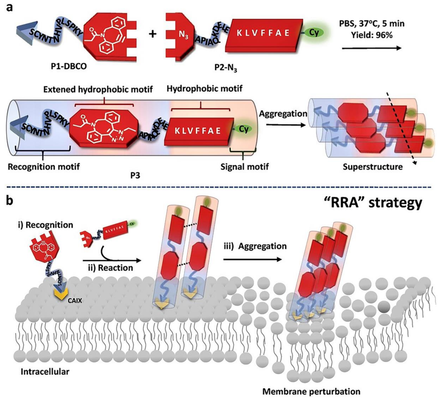
| P3 | KLVFFAE | — | nanofibers | chemosensitization |
| NF/PDGF-BB | AcN-RARADADARARADADA-CNH2 | — | nanofibers | drug delivery |
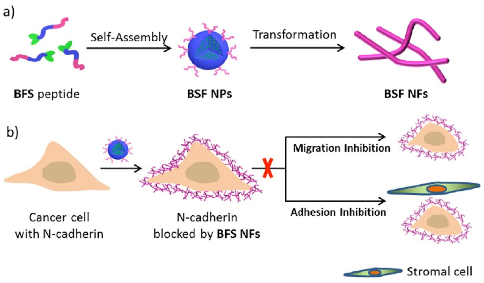
| BFS | KLVFF | — | nanofibers | anti-tumor |
| PEG-Pep-TPE | FFKY | GSH-responsive | nanofibers | chemotherapy synergy |
| Fbp-GDFDFDYD (E, S, or K)-ss-ERGD | Fbp-GDFDFDYD (E, S or K)-SH | GSH-responsive | nanofibers | immune adjuvants |
| O1/O2 | KLDLKLDLKLDL | — | hydrogels | bone tissue regeneration |
| RADA16-FRM-MP | RADA16 | — | nanofibers | neuron repair and regenerate |
| p(TEGMA-co-BM3)-FEFK | FEFK | MMP3-responsive | nanofibers | CNS regeneration |
| RADA-16I | RADA-16I | pH-sensitive | hydrogels | tissue regeneration |
| RATEA16 | RATEA16 | pH-responsive | nanofibers | wound healing |
| EAK16-II | AEAEAKAKAEAEAKAK | — | hydrogels | wound healing |
| K2MDP | K2 (SL)6K2 | — | hydrogels | wound healing |
Publisher’s Note: MDPI stays neutral with regard to jurisdictional claims in published maps and institutional affiliations. |
© 2021 by the authors. Licensee MDPI, Basel, Switzerland. This article is an open access article distributed under the terms and conditions of the Creative Commons Attribution (CC BY) license (https://creativecommons.org/licenses/by/4.0/).
Share and Cite
Wang, T.-T.; Xia, Y.-Y.; Gao, J.-Q.; Xu, D.-H.; Han, M. Recent Progress in the Design and Medical Application of In Situ Self-Assembled Polypeptide Materials. Pharmaceutics 2021, 13, 753. https://0-doi-org.brum.beds.ac.uk/10.3390/pharmaceutics13050753
Wang T-T, Xia Y-Y, Gao J-Q, Xu D-H, Han M. Recent Progress in the Design and Medical Application of In Situ Self-Assembled Polypeptide Materials. Pharmaceutics. 2021; 13(5):753. https://0-doi-org.brum.beds.ac.uk/10.3390/pharmaceutics13050753
Chicago/Turabian StyleWang, Tian-Tian, Yi-Yi Xia, Jian-Qing Gao, Dong-Hang Xu, and Min Han. 2021. "Recent Progress in the Design and Medical Application of In Situ Self-Assembled Polypeptide Materials" Pharmaceutics 13, no. 5: 753. https://0-doi-org.brum.beds.ac.uk/10.3390/pharmaceutics13050753