Article

Evaluation of the Correlation between the rs4918 Polymorphism of AHSG Gene and Coronary Artery Calcification in Patients with Coronary Artery Disease

by
Zeynab Ahmadihosseini
1,
Morteza Moeinian
1,
Saeed Nazemi
2,
Sepideh Elyasi
1,* and
Amir Hooshang Mohammadpour
1,3,*
1
Department of Clinical Pharmacy, School of Pharmacy, Mashhad University of Medical Sciences, Mashhad 9179156314, Iran
2
Department of Cardiovascular Diseases, Razavi Hospital, Mashhad 9177948954, Iran
3
Pharmaceutical Research Center, Mashhad University of Medical Sciences, Mashhad 9179156314, Iran
*
Authors to whom correspondence should be addressed.
Submission received: 29 October 2020 / Accepted: 5 November 2020 / Published: 6 November 2020

Abstract

:
Objectives: Fetuin-A is a circulating calcification inhibitor that prevents coronary artery calcification (CAC) by increasing calcium phosphate solubility and inhibiting VSMC differentiation and apoptosis. In this study, we investigated the correlation between rs4918 and CAC in patients with coronary artery disease (CAD). Methods: Forty-two healthy individuals and eighty-one CAD patients were recruited in the present study. The CAC score (CACS) was measured by CT angiography and the genotype analysis of rs4918 single-nucleotide polymorphism SNP was performed by the polymerase chain reaction-restriction fragment length polymorphism (PCR-RFLP) technique. Results: The CACS was significantly higher in CAD patients compared to healthy individuals (p < 0.001); however, there was no significant difference between the mean CACS in the presence and absence of rs4918 (p = 0.792). The mean calcium score of the left main coronary artery (LMCA) was significantly lower in carriers of the rs4918 allele (p = 0.036). The frequency of rs4918 SNP was almost similar in the control group and CAD patients (p = 0.846). Conclusions: in patients with CAD, we found no significant association between rs4918 SNP and CACS, indicating that carriers of this allele are not at increased risk of developing cardiovascular diseases compared with those without.

1. Introduction

Coronary artery disease (CAD) is one of the major causes of mortality worldwide, and atherosclerosis is the primary etiology [1]. Coronary artery calcification (CAC) is the characteristic feature of atheroma plaque, and its extent is increased with the progression of the lesion. Calcified plaques are associated with an almost 1.7-fold higher incidence of mortality, independent of other cardiovascular risk factors [2]. Although traditional Framingham risk factors, including age, sex, hyperlipidemia, hypertension, high body mass index, diabetes, and cigarette smoking, influence the severity of atherosclerosis and the extent of CAC, these environmental factors account for only 40% of the inter-individual variety, and more than 40% of the variations in the extent of CAC are due to genetic factors [3].
It is believed that the transdifferentiation of VSMCs to the osteoblastic lineage and the loss of balance between inhibitors and inducers of calcification are the prominent etiologies of ectopic calcification. In this context, fetuin-A has been identified as an important inhibitor of ectopic calcification.
Fetuin-A, also called alpha2-Heremans Schmid glycoprotein (AHSG), is a 62 kDa glycoprotein and a member of the cystatin superfamily which is synthesized and secreted from the liver and is present abundantly in the circulation and extracellular space [4]. It is a potent inhibitor of calcification; nearly 50% of blood capacity to inhibit ectopic calcification is attributed to fetuin-A activity [5]. It inhibits vascular calcification by binding to hydroxyapatite crystals and forming the fetuin mineral complex (FMC) that enhances the calcium phosphate blood solubility [6]. Furthermore, it has anti-inflammatory activity by preventing the production and release of tumor necrosis factor (TNF) α from activated macrophages, which have a central role in the pathophysiology of CAD [7].
Several in vitro and in vivo studies have demonstrated its role in the pathogenesis of CAD. Fetuin-A-deficient mice on a calcium- and vitamin D-rich diet developed ectopic calcification, which clearly confirmed its role as a calcification inhibitor [8].
It was shown that fetuin-A is directly associated with the intimal–medial thickness (IMT), a hallmark of subclinical atherosclerosis, which points to its atherogenic property [9]. In dialysis and end-stage renal disease (ESRD) patients, the reduction in this protein has been reported to correlate with the acceleration of vascular calcification and all-cause and cardiovascular death [10,11]. Genetic studies have demonstrated the impact of the AHSG gene variations on its serum level, CVD complications, and morality [4,11]. The role of AHSG polymorphisms and haplotypes in the progression of the calcified plaque was demonstrated in a group of European American diabetic patients with subclinical atherosclerosis and without advanced renal dysfunction [12]. Higher mortality rate, vascular calcification, and lower fetuin-A concentrations have been reported in ESRD patients carrying rs4918 single-nucleotide polymorphism (SNP) [11]. Additionally, this allele has been shown to be associated with arterial stiffness in patients with normal kidney function [13]. These data suggest that genetic variations in the AHSG gene may influence the extent of CAC.
To our knowledge, no clinical study has evaluated the relationship between the rs4918 allele and CAC in patients with CAD. In the present study, the correlation between the rs4918 polymorphism and coronary artery calcification was evaluated, and its frequency in CAD participants and healthy individuals was studied.

2. Methods

2.1. Study Population

Eighty-one patients diagnosed with ischemic heart disease who fulfilled the inclusion and exclusion criteria entered the study. The inclusion criteria included age above 35 years and the presence of coronary artery disease, and the exclusion criteria involved disturbed calcium and phosphorus homeostasis, acute or chronic kidney disease (CKD), malignancies, bone disorders, primary and secondary hyperparathyroidism, and active infectious diseases.
The calcium score was measured in four main coronary arteries, including the left main coronary artery (LMCA), right coronary artery (RCA), circumflex artery (CX), and left anterior descending artery (LAD) using CT angiography.
The control group consisted of forty-two subjects free of any cardiovascular diseases (including myocardial infarction, angina, stroke, transient ischemic attack, heart failure, having current atrial fibrillation, taking nitroglycerin, or undergoing angioplasty, coronary artery bypass graft, valve replacement, pacemaker or defibrillator implantation, or any surgery on the heart or arteries); any chronic diseases; and CVD risk factors including age (above 50 years for women and 45 years for men), sex, history of death due to CVD in the first-degree family, smoking, diabetes mellitus, and hypertension. Additionally, all their biochemical measurements had to be in the normal range.
Clinical examinations, biochemical assays, calcium score measurements, and blood sampling were conducted in the cardiology department of the Razavi hospital in Mashhad, Iran, from 2014 to 2018. Ethical approval was obtained from the university medical ethics committee. All the participants signed written informed consent.
Demographic and clinical data including age, sex, history of previous and current diseases and medications, weight, height, smoking status, and family history of CVD were collected in a questionnaire. Biochemical parameters of lipid profile (total cholesterol, triglycerides, high- and low-density lipoprotein), and fasting blood sugar (FBS) were determined for all the participants by standard laboratory protocols.

2.2. Genotype Analysis

Genomic DNA was extracted from whole blood samples obtained from study participants using the FavorPerp blood genomic DNA extraction mini kit (Favorprep, South Korea) according to the manufacturer’s protocol. DNA concentration and purity was determined using Nanodrop spectrophotometer (Thermofisher Scientific, Waltham, MA, USA) and DNA extracts were stored at −20 °C.
The amplification of the targeted gene (AHSG) and analysis of the polymorphism was performed by the polymerase chain reaction-restriction fragment length polymorphism (PCR-RFLP) technique. The 309 base pairs fragment of AHSG gene was amplified using forward primer 5′-TGTGAGGAAATTGGGTGCCA-3′ and reverse primer 3′-GACCACACCCATGAAGGTGT-5′ (Bioneer, Daejeon, South Korea).
The PCR reaction mixture contained 2 ng of DNA, 0.6 μM of each forward and reverse primer, and 1× Taq DNA Polymerase 2× Master Mix Red (Ampliqon PCR enzymes and reagents, city, Denmark). The cycling program for sequence amplification was the denaturation of DNA strands at 95 °C for 5 min, followed by 30 cycles of denaturation at 95 °C for 30 s, annealing at 55 °C for 25 s, and extension at 72 °C for 20 s, with a final extension at 72 °C for 5 min, using the MyCycler thermocycler (Bio-Rad, Hercules, CA, USA).
To identify the rs4918 single-nucleotide polymorphism (SNP), the amplified gene (PCR product) was treated with the restriction enzyme Sac I (SstI) (Jena Bioscience, Hannover, Germany) for 1 h at 37 °C. The digested products were electrophoresed through loading on a 1.5% (w/v) agarose gel stained with gel red (Biotium, Fremont, CA, USA), using TBE (1.1 mM Tris base, 900 mM boric acid, 25 mM EDTA) as a running buffer, and visualized under UV light transillumination (Uvitec, Cambridge, UK).

2.3. Statistical Analysis

The comparison of the coronary artery calcium scores between different groups was performed by an independent samples t-test. The difference in SNP frequency between different groups was determined by chi-squared statistics. p values of 0.05 or less were considered statistically significant for all tests. All the statistical analyses were performed using SPSS version 16.

3. Results

3.1. Patients Characteristics

Details of the study participants’ characteristics including age; family history of CVD; sex; concomitant conventional CVD risk factors including hypertension, type two diabetes mellitus, and dyslipidemia; biochemical profiles; calcium score; and genotype frequency are listed in Table 1.

3.2. Comparison of Coronary Artery Calcium Score between Different Groups

The coronary artery calcium score was significantly higher in CAD patients compared to healthy individuals (p < 0.001); however, no significant difference was observed in the CACS in the presence and absence of rs4918 SNP (p = 0.792).

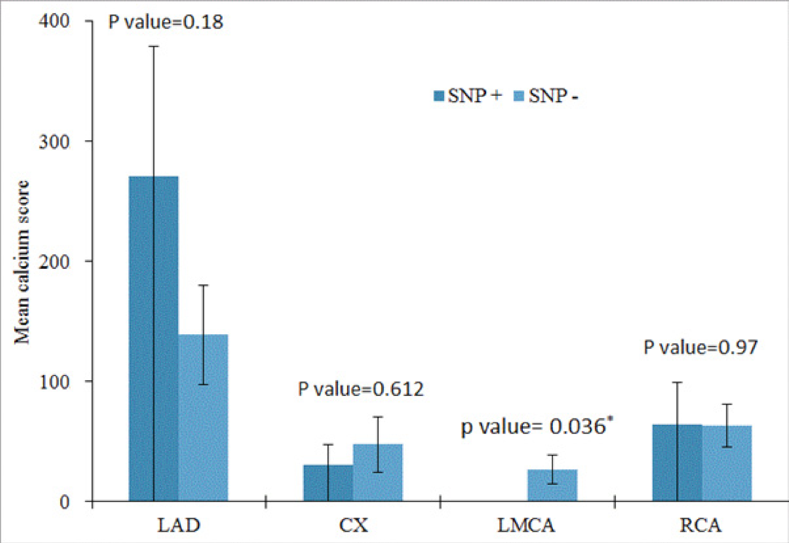
3.3. Comparison between Calcium Scores of the Main Coronary Arteries in the Presence and Absence of rs4918 SNP

The calcium score of four main coronary arteries was measured; the highest CACS was observed in the LAD artery, and the LMCA had the lowest CACS (Table 2).
The calcium score of the LMCA was significantly lower in carriers of the rs4918 allele (p = 0.036) compared with the CACS of LMCA in individuals without this allele; on the other hand, an insignificantly higher CACS was observed in the LAD artery in the presence of this SNP compared with the CACS in its absence (p = 0.120) (Figure 1).

3.4. Frequency of rs4918 SNP in Different Groups

The frequency of rs4918 SNP was similar in the control group and CAD patients (p = 0.846). The frequency of the rs4918 SNP in different calcium score groups was assessed and is presented in Table 3. Individuals were divided into three groups according to their coronary artery calcium score. Individuals with a CACS between 0 and 100 were assigned to group 1, with one between 101 and 400 were assigned to group 2, and with a CACS above 400 were assigned to group 3. There was no significant difference in the frequency of this SNP in these groups (p > 0.05).

4. Discussion and Conclusions

Coronary artery calcification has been considered as a non-traditional risk factor of cardiovascular disorders and events.
In the present study, the coronary calcium score was measured by the noninvasive and quantifiable CT angiography technique, and the correlation between the obtained coronary calcium score and the AHSG genotype was assessed. According to the results, the calcium score was significantly higher in CAD patients compared to healthy individuals, and no difference was observed between the mean calcium scores in the presence and absence of the rs4918 polymorphism.
Individuals in the control group were free of apparent coronary vasculopathy and CVD risk factors, on the other hand, while participants in the patients’ group were diagnosed with coronary artery disease and had undergone either percutaneous coronary intervention (PCI) or coronary artery bypass graft (CABG) intervention or had myocardial infarction (MI). Higher CACS in this group is a representative of the association of calcification with atheroma plaque. Moreover, patients had at least one CVD risk factor, including hypertension, diabetes, dyslipidemia, cigarette smoking, and positive family history, which triggered the formation and progression of a plaque and subsequent calcified lesion by damaging the endothelial layer.
In this project, the role of rs4918 polymorphism in the inter-individual variability of atherosclerotic calcification was studied; therefore the correlation between the rs4918 polymorphism and the coronary artery calcium score in CAD patients and healthy individuals was assessed.
Our results indicate that the rs4918 allele is distributed equally between the healthy individuals and group of CAD patients, which indicates that carriers of this allele are not at risk of developing cardiovascular diseases. Cozzolino et al. reported a similar frequency of this allele among Italian hemodialysis patients and a healthy population [14]. Moreover, we found no correlation between this SNP and coronary artery calcified plaque. This is in agreement with the results of the Lehtinen group, who analyzed 11 polymorphisms in the AHSG gene and found no association between the rs4918 allele and coronary artery calcified plaque, diabetes mellitus, BMI, and fetuin-A serum levels in a group of American diabetic patients [12]. In another study, Bellia et al. reported the association of fetuin-A levels with serum calcium and rs4918 polymorphism but not with coronary artery calcification in individuals with a low risk for developing CAD and without apparent coronary vasculopathy [15].
Although in animal studies AHSG-deficient mice developed widespread ectopic calcification of soft tissue and vasculature, which clearly confirmed its role as a calcification inhibitor [8], clinical findings regarding the impact of AHSG genotypes on CAC are still a matter of debate, and there are inconsistencies in the literature concerning the role of fetuin and its genotypes in the pathogenesis of CVD and CAC.
Fetuin-A has been studied widely in end-stage renal disease and dialysis patients, where CVD and vascular calcification are common complications and the main cause of death in these patients. Low plasma levels of fetuin-A have been reported in these patients. Furthermore, carriers of the rs4918 polymorphism had lower fetuin-A levels and were more liable to develop vascular calcification, particularly in the presence of chronic inflammation [11]. In another study, Verdujin et al. showed that, although serine allele carriers were predisposed to lower serum fetuin-A concentrations, which were associated with increased mortality risk, the Thr256Ser polymorphism had a minor effect on mortality [16].
In the general population and non-dialyzed patients, however, conflicting data are available regarding the genetic contribution of AHSG in CVD and CAC. Fisher et al. reported that AHSG polymorphisms were positively correlated with fetuin-A levels and the elevated concentration increased the risk of MI. In addition, the rs4917 allele was particularly associated with the incidence of MI [4]. In contrast, Roos et al. observed that fetuin-A levels were inversely associated with arterial stiffness, but the SerSer genotype of rs4918 SNP was significantly associated with arterial stiffness in male patients with normal renal function [13]. On the other hand, others reported no significant association between the AHSG polymorphisms and its levels and coronary artery calcification [12,17].
Analyzing the calcium score of cardiac arteries of CAD patients, the CACS of LMCA was significantly lower in carriers of the rs4918 allele compared to individuals without this allele. In contrast, an insignificant higher CACS was observed in the presence of this SNP in LAD artery compared with the mean CACS in its absence. To explain these findings, differences in ethnicity and small sample size should be taken into account.
The protein-coding rs4918 allele, also known as Thr256Ser, is situated in the coding region of the AHSG gene in exon 7 that encodes Thr256Ser amino acid substitution [18]. This protein-coding polymorphism determines the required amino acids for protein synthesis. It should be noted that it is the regulatory or promoter region of a DNA that determines the expression of mRNA and the quantity of translated protein, and the coding region determines the properties and structure of a protein and not its concentration.
However, according to the obtained reports either the structure of the protein, concentration or both can be affected by this allele especially in ESRD and dialysis patients. The transcription of AHSG gene is mediated by C/EBP and NF-1 transcription factors [19] and pro-inflammatory cytokines cause loss of transcriptional domain of C/EBP in acute inflammation, which ultimately leads to reduced serum fetuin-A concentration [20]. Furthermore, the expression of the AHSG gene can be mediated by cytokine stimulation. In vitro studies showed that TNFα [21], IL-1, and IL-6 [22] down-regulate its mRNA level in the rat and human hepatoma cell line, respectively. Chronic inflammation has been well-documented in the setting of end-stage renal disease. Low levels of fetuin-A have been reported to be associated with rs4918 SNP, increased mortality, and vascular calcification [11]. Therefore inflammation may influence the expression of mRNA and subsequently the protein level, and the mentioned mechanism may be most outstanding in the presence of this SNP, which can be an explanation for that observed in ESRD and dialysis patients.
As we did not measure the fetuin-A serum level and inflammatory proteins, the judgment is difficult and we are not sure that whether patients with rs4918 polymorphism necessarily had a lower fetuin-A serum level. The rs4918 SNP may not affect protein levels, structure, or function, and no association was observed between this SNP and the incidence of CAD and CAC in our results.
Another possible explanation for the observed inconsistency in the role of rs4918 can be attributable to the post-translational modifications and different roles of fetuin-A in CAD and CAC pathology. This protein behaves differently in the presence of various risk factors, so with a similar genotype and whatever the phenotype might be, different biological responses may be observed.
It is considered as an atherogenic protein as it inhibits the insulin receptor, which leads to insulin resistance, diabetes, metabolic syndrome, key promoters of atherosclerosis, and CAC. On the other hand, it inhibits vascular calcification by enhancing the calcium phosphate blood solubility and preventing apoptosis and VSMCs differentiation. Furthermore, it has anti-inflammatory activity by preventing the production and release of TNFα from activated macrophages [7], which has a central role in the occurrence of CAD. Hence it can be considered a protective protein. Thereby, its contribution to the pathogenesis of CAD may be dependent on the underlying pathologies and risk factors involved. Consistent with this explanation, in ischemic heart disease, high fetuin-A levels were associated with a lower risk of mortality only in patients with acute coronary syndrome where the exacerbation of inflammatory response is prevalent [23] and no association between baseline fetuin-A and CVD events was found in patients with stable angina [17]. Apart from its anti-inflammatory role and inhibition of calcification, fetuin-A inhibits insulin receptors. Jensen et al. reported that, while there was an inverse association between fetuin-A levels and the incidence of CVD among non-diabetics, an insignificant direct relation was observed in diabetic patients [24]. In another study, high concentrations of fetuin were directly correlated with the higher risk of CVD death only in older diabetic adults, whereas an inverse association was observed among non-diabetic individuals independent of CVD risk factors and renal function [25]. Consistent with these data, Chen et al. found that an inverse relation between fetuin-A and CVD, and all-cause mortality was limited to CAD non-diabetic patients only and disappeared in patients with diabetes mellitus [23].
These findings suggest that the protective effect of fetuin-A could be masked by its insulin receptor inhibitory activity [24], or excessive fetuin-A levels may result in metabolic syndrome and insulin resistance [25], leading to a positive or no association between fetuin-A levels and CVD in diabetic patients [24].
There are limitations in this study that should be considered. The number of subjects who participated in this project was limited, which reduces the statistical power. Second, because of practical and economical reasons, we did not measure the fetuin-A levels, which made it quite difficult to interpret the results and explain the observations. In addition, CRP and other inflammatory markers were not evaluated in this study. Inflammation is a major inducer of vascular calcification [26], and an inverse relationship between fetuin-A levels and inflammatory markers such as CRP in patients with CAD has been reported, which points to its anti-inflammatory activity and its role in attenuating atherosclerosis and calcification development [23]. However, it is not clear whether inflammatory factors have an impact on the transcription of a particular genotype and consequent fetuin quantity or not. Therefore, including these factors could give better insight into the activity of the rs4918 genotype specifically in situations where a flare of inflammatory response exists, such as acute coronary syndrome and MI. Finally, fetuin-A has a dual role in the pathophysiology of CVD and coronary calcification, and risk factors can influence its activity and quantity. However, in our study individuals in the control group were free of CVD and coronary risk factors, hence it would be beneficial to change the criteria for the selection of the control group and include individuals without CVD who share similar risk factors to the case group.
In conclusion, there was no association between the rs4918 SNP and the coronary artery calcification score in patients with CAD, and its frequency was almost similar in healthy individuals and CAD patients, indicating that the carriers of this allele are not at increased risk of developing cardiovascular diseases as compared with those without.

Author Contributions

Z.A.: conducting experiments, data analysis, and manuscript preparation; M.M.: planning, conducting experiments, and manuscript preparation; S.N.: conducting experiments and manuscript preparation; M.A.: conduct, data analysis, and manuscript preparation; S.E.: conceptualization, design analysis, planning, data analysis, and manuscript preparation; A.H.M.: conceptualization, design analysis, planning, data analysis, and manuscript preparation. All authors have read and agreed to the published version of the manuscript.

Funding

The authors are thankful for the funding of this study by the Research Council of Mashhad University of Medical Sciences.

Acknowledgments

This study is part of a research thesis for a Ph.D. Degree at Mashhad University of Medical Sciences.

Conflicts of Interest

The authors declare no conflict of interest.

References

  1. Lusis, A.J. Atherosclerosis. Nature 2000, 407, 233–241. [Google Scholar] [CrossRef] [PubMed]
  2. Sage, A.P.; Tintut, Y.; Demer, L.L. Regulatory mechanisms in vascular calcification. Nat. Rev. Cardiol. 2010, 7, 528–536. [Google Scholar] [CrossRef]
  3. Peyser, P.A.; Bielak, L.F.; Chu, J.S.; Turner, S.T.; Ellsworth, D.L.; Boerwinkle, E.; Sheedy, P.F. Heritability of Coronary Artery Calcium Quantity Measured by Electron Beam Computed Tomography in Asymptomatic Adults. Circulation 2002, 106, 304–308. [Google Scholar] [CrossRef] [PubMed]
  4. Fisher, E.; Stefan, N.; Saar, K.; Drogan, D.; Boeing, H.; Fritsche, A.; Joost, H.-G.; Häring, H.-U.; Hübner, N.; Boeing, H.; et al. Association of AHSG Gene Polymorphisms With Fetuin-A Plasma Levels and Cardiovascular Diseases in the EPIC-Potsdam Study. Circ. Cardiovasc. Genet. 2009, 2, 607–613. [Google Scholar] [CrossRef] [PubMed] [Green Version]
  5. Cuspidi, C.; Sala, C. Is fetuin-A a biomarker of preclinical atherosclerosis in essential hypertension? Hypertens. Res. 2012, 36, 104–106. [Google Scholar] [CrossRef]
  6. Mori, K.; Emoto, M.; Inaba, M. Fetuin-A: A multifunctional protein. Recent Pat. Endocr. Metab. Immune Drug Discov. 2011, 5, 124–146. [Google Scholar] [CrossRef]
  7. Ombrellino, M.; Wang, H.; Yang, H.; Zhang, M.; Vishnubhakat, J.; Frazier, A.; Scher, L.A.; Friedman, S.G.; Tracey, K.J. Fetuin, a negative acute phase protein, attenuates tnf synthesis and the innate inflammatory response to carrageenan. Shock 2001, 15, 181–185. [Google Scholar] [CrossRef]
  8. Schafer, C.; Heiss, A.; Schwarz, A.; Westenfeld, R.; Ketteler, M.; Floege, J.; Müller-Esterl, W.; Schinke, T.; Jahnen-Dechent, W. The serum protein alpha 2-Heremans-Schmid glycoprotein/fetuin-A is a systemically acting inhibitor of ectopic calcification. J. Clin. Invest. 2003, 112, 357–366. [Google Scholar] [CrossRef]
  9. Fiore, C.E.; Celotta, G.; Politi, G.G.; Di Pino, L.; Castelli, Z.; Mangiafico, R.A.; Signorelli, S.S.; Pennisi, P. Association of high alpha2-Heremans–Schmid glycoprotein/fetuin concentration in serum and intima-media thickness in patients with atherosclerotic vascular disease and low bone mass. Atherosclerosis 2007, 195, 110–115. [Google Scholar] [CrossRef] [PubMed]
  10. Ketteler, M.; Bongartz, P.; Westenfeld, R.; Wildberger, J.E.; Mahnken, A.H.; Böhm, R.; Metzger, T.; Wanner, C.; Jahnen-Dechent, W.; Floege, J. Association of low fetuin-A (AHSG) concentrations in serum with cardiovascular mortality in patients on dialysis: A cross-sectional study. Lancet 2003, 361, 827–833. [Google Scholar] [CrossRef]
  11. Stenvinkel, P.; Wang, K.; Qureshi, A.R.; Axelsson, J.; Pecoits-Filho, R.; Gao, P.; Barany, P.; Lindholm, B.; Jogestrand, T.; Heimberger, O.; et al. Low fetuin-A levels are associated with cardiovascular death: Impact of variations in the gene encoding fetuin. Kidney Int. 2005, 67, 2383–2392. [Google Scholar] [CrossRef] [Green Version]
  12. Lehtinen, A.B.; Burdon, K.P.; Lewis, J.P.; Langefeld, C.D.; Ziegler, J.T.; Rich, S.S.; Register, T.C.; Carr, J.J.; Freedman, B.I.; Bowden, D.W. Association of α2-Heremans-Schmid Glycoprotein Polymorphisms with Subclinical Atherosclerosis. J. Clin. Endocrinol. Metab. 2007, 92, 345–352. [Google Scholar] [CrossRef] [Green Version]
  13. Roos, M.; Richart, T.; Kouznetsova, T.; Von Eynatten, M.; Lutz, J.; Heemann, U.; Baumann, M.; Staessen, J.A. Fetuin-A and arterial stiffness in patients with normal kidney function. Regul. Pept. 2009, 154, 39–43. [Google Scholar] [CrossRef]
  14. Cozzolino, M.; Biondi, M.L.; Galassi, A.; Gallieni, M.; D’Eril, G.V.M.; Brancaccio, D. Gene Polymorphisms and Serum Alpha-2-Heremans-Schmid Levels in Italian Haemodialysis Patients. Am. J. Nephrol. 2007, 27, 639–642. [Google Scholar] [CrossRef]
  15. Bellia, C.; Agnello, L.; Sasso, B.L.; Milano, S.; Bivona, G.; Scazzone, C.; Pivetti, A.; Novo, G.; Palermo, C.; Bonomo, V.; et al. Fetuin-A is Associated to Serum Calcium and AHSG T256S Genotype but Not to Coronary Artery Calcification. Biochem. Genet. 2016, 54, 222–231. [Google Scholar] [CrossRef]
  16. Verduijn, M.; Prein, R.A.; Stenvinkel, P.; Carrero, J.J.; Le Cessie, S.; Witasp, A.; Nordfors, L.; Krediet, R.T.; Boeschoten, E.W.; Dekker, F. Is fetuin-A a mortality risk factor in dialysis patients or a mere risk marker? A Mendelian randomization approach. Nephrol. Dial. Transplant. 2010, 26, 239–245. [Google Scholar] [CrossRef] [Green Version]
  17. Roos, M.; Lutz, J.; Salmhofer, H.; Luppa, P.; Knauß, A.; Braun, S.; Martinof, S.; Schömig, A.; Heemann, U.; Kastrati, A.; et al. Relation between plasma fibroblast growth factor-23, serum fetuin-A levels and coronary artery calcification evaluated by multislice computed tomography in patients with normal kidney function. Clin. Endocrinol. 2008, 68, 660–665. [Google Scholar] [CrossRef]
  18. Osawa, M.; Umetsu, K.; Ohki, T.; Nagasawa, T.; Suzuki, T.; Takeichi, S. Molecular evidence for human alpha 2-HS glycoprotein (AHSG) polymorphism. Hum. Genet. 1997, 99, 18–21. [Google Scholar] [CrossRef]
  19. Falquerho, L.; Paquereau, L.; Vilarem, M.J.; Galas, S.; Patey, G.; Le Cam, A. Functional characterization of the promoter of pp63, a gene encoding a natural inhibitor of the insulin receptor tyrosine kinase. Nucleic Acids Res. 1992, 20, 1983–1990. [Google Scholar] [CrossRef] [Green Version]
  20. Gangneux, C.; Daveau, M.; Hiron, M.; Derambure, C.; Papaconstantinou, J.; Salier, J.P. The inflammation-induced down-regulation of plasma Fetuin-A (α2HS-Glycoprotein) in liver results from the loss of interaction between long C/EBP isoforms at two neighbouring binding sites. Nucleic Acids Res. 2003, 31, 5957–5970. [Google Scholar] [CrossRef] [Green Version]
  21. Daveau, M.; Davrinche, C.; Djelassi, N.; Lemetayer, J.; Julen, N.; Hiron, M.; Arnaud, P.; Lebreton, J. Partial hepatectomy and mediators of inflammation decrease the expression of liver α2 -HS glycoprotein gene in rats. FEBS Lett. 1990, 273, 79–81. [Google Scholar] [CrossRef] [Green Version]
  22. Daveau, M.; Davrinche, C.-; Julen, N.; Hiron, M.; Amaud, P.; Lebreton, J.-P. The synthesis of human α-2-HS glycoprotein is down-regulated by cytokines in hepatoma HepG2 cells. FEBS Lett. 1988, 241, 191–194. [Google Scholar] [CrossRef] [Green Version]
  23. Chen, X.; Zhang, Y.; Chen, Q.; Li, Q.; Li, Y.; Ling, W. Lower Plasma Fetuin-A Levels Are Associated with a Higher Mortality Risk in Patients with Coronary Artery Disease. Arter. Thromb. Vasc. Biol. 2017, 37, 2213–2219. [Google Scholar] [CrossRef] [Green Version]
  24. Jensen, M.K.; Bartz, T.M.; Mukamal, K.J.; Djoussé, L.; Kizer, J.R.; Tracy, R.P.; Zieman, S.J.; Rimm, E.B.; Siscovick, D.S.; Shlipak, M.; et al. Fetuin-A, Type 2 Diabetes, and Risk of Cardiovascular Disease in Older Adults. Cardiovasc. Health Study 2013, 36, 1222–1228. [Google Scholar] [CrossRef] [Green Version]
  25. Laughlin, G.A.; Cummins, K.M.; Wassel, C.L.; Daniels, L.B.; Ix, J.H. The Association of Fetuin-A with Cardiovascular Disease Mortality in Older Community-Dwelling Adults: The Rancho Bernardo study. J. Am. Coll. Cardiol. 2012, 59, 1688–1696. [Google Scholar] [CrossRef] [Green Version]
  26. Blake, G.J.; Ridker, P.M. C-reactive protein and other inflammatory risk markers in acute coronary syndromes. J. Am. Coll. Cardiol. 2003, 41, S37–S42. [Google Scholar] [CrossRef] [Green Version]
Figure 1. Comparison between the calcium scores of the main coronary arteries in the presence and absence of rs4918 SNP. Independent samples t-test was used for the analysis. * indicates a significant difference in the mean calcium score. CX: circumflex; LAD: left anterior descending; LMCA: left main coronary artery; RCA: right coronary artery; SNP: single-nucleotide polymorphism.
Cardiogenetics 10 00007 g001
Table 1. Characteristics of the study participants.
Characteristics Patients (n = 81)Control (n = 42)
Male, %72.8435.71
Age (Year)57.08 ± 10.5434.47 ± 10.46
Hypertension, %48.140
Dyslipidemia, % 62.960
Diabetes mellitus type II, % 18.510
Smoking status, %30.860
Family history, % 45.670
rs4918 SNP, % frequency38.2735.71
Calcium score (mean ± STD) 348.29 ± 588.756.3 ± 26.62
CX: circumflex; LAD: left anterior descending; LMCA: left main coronary artery; RCA: right coronary artery; SNP: single-nucleotide polymorphism; Std: standard. Values are the mean calcium score of four main coronary arteries. Independent samples t-test was used for the analysis.
Table 2. Calcium scores of the main coronary arteries in the presence and absence of rs4918 SNP.
Mean Calcium Score
(SNP +) ± Std. Error
Mean Calcium Score
(SNP −) ± Std. Error
p Value
LAD2.7111E+02 ± 108.121.3879E+02 ± 41.760.180
CX30.38 ± 17.0747.51 ± 22.660.612
LMCA0.14 ± 0.1427.04 ± 12.180.036
RCA64.58 ± 35.0363.24 ± 18.060.970
p values in bold indicate statistically significant—that is, p ≤ 0.05. CACS indicates coronary artery calcium score; SNP, single-nucleotide polymorphism. The frequency of genotype was expressed as a percentage.
Table 3. Comparison of the frequency of the rs4918 SNP between different calcium groups.
Calcium GroupCACSFrequency of the rs4918 SNPp Value
Group 10–10037.660.9
Group 2101–40036.36
Group 3>40034.78
Chi-squared statistic was used for the analysis. p ≤ 0.05 indicates statistically significant difference.
Publisher’s Note: MDPI stays neutral with regard to jurisdictional claims in published maps and institutional affiliations.

