Extracapsular Extension Does Not Decrease Overall Survival in Rectal Cancer Patients with Lymph Node Metastasis Following Neoadjuvant Chemoradiotherapy

 ,
,
Abstract
:1. What Does This Paper Add to the Literature?
2. Introduction
3. Methods
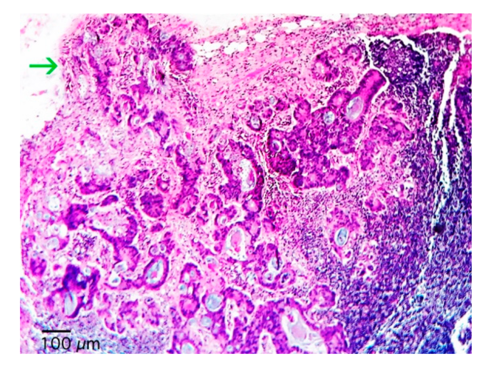
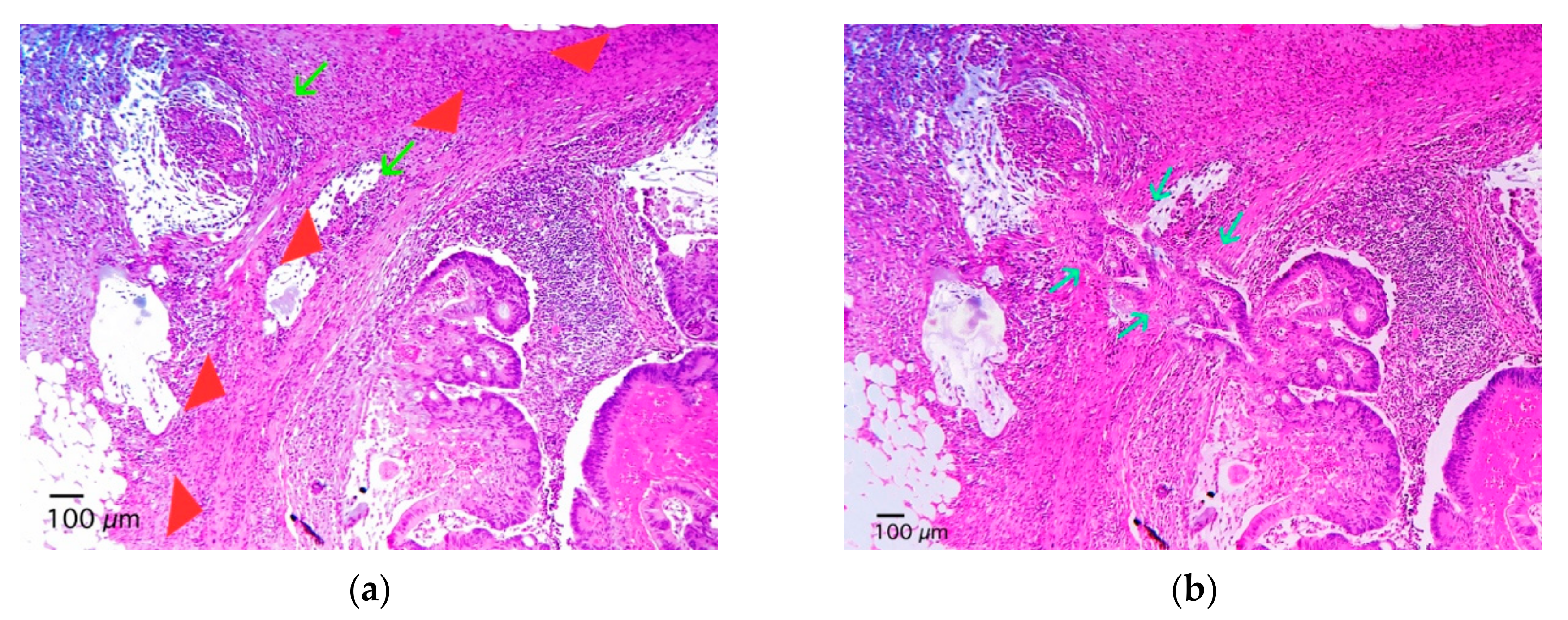
4. Results
5. Discussion
6. Conclusions
Author Contributions
Funding
Conflicts of Interest
References
- Coster, J.R.; Foote, R.L.; Olsen, K.D.; Jack, S.M.; Schaid, D.J.; DeSanto, L.W. Cervical nodal metastasis of squamous cell carcinoma of unknown origin: Indications for withholding radiation therapy. Int. J. Radiat. Oncol. Biol. Phys. 1992, 23, 743–749. [Google Scholar] [CrossRef]
- Leonard, C.; Tompkin, J.; Zhen, B.; Zehn, B.; Waitz, D.; Norton, L. Are axillary recurrence and overall survival affected by axillary extranodal tumor extension in breast cancer? Implications for radiotherapy. J. Clin. Oncol. 1995, 13, 47–53. [Google Scholar] [CrossRef] [PubMed]
- Leemans, C.R.; Tiwari, R.; Nauta, J.J.; van der Waal, I.; Snow, G.B. Regional lymph node involvement and its significance in the development of distant metastases in head and neck carcinoma. Cancer 1993, 71, 452–456. [Google Scholar] [CrossRef]
- Van der Velden, J.; van Lindert, A.C.M.; Lammes, F.B.; ten Kate, F.J.; Sie-Go, D.M.; Oosting, H.; Heintz, A.P.M. Extracapsular growth of lymph node metastasis in squamous cell carcinoma of the vulva: The impact on recurrence and survival. Cancer 1995, 75, 2885–2890. [Google Scholar] [CrossRef]
- Wind, J.; Lagarde, S.M.; Ten Kate, F.J.; Ubbink, D.T.; Bemelman, W.A.; van Lanschot, J.J.B. A systematic review on the significance of extracapsular lymph node involvement in gastrointestinal malignancies. Eur. J. Surg. Oncol. 2007, 33, 401–408. [Google Scholar] [CrossRef] [PubMed]
- Veronese, N.; Nottegar, A.; Pea, A.; Solmi, M.; Stubbs, B.; Capelli, P.; Scarpa, A. Prognostic impact and implications of extracapsular lymph node involvement in colorectal cancer: A systematic review with meta-analysis. Ann. Oncol. 2016, 27, 42–48. [Google Scholar] [CrossRef] [PubMed]
- Fujii, T.; Tabe, Y.; Yajima, R.; Yamaguchi, S.; Tsutsumi, S.; Asao, T.; Kuwano, H. Process of distant lymph node metastasis in colorectal carcinoma: Implication of extracapsular invasion of lymph node metastasis. BMC Cancer 2011, 11, 216. [Google Scholar] [CrossRef] [PubMed] [Green Version]
- Fujii, T.; Tabe, Y.; Yajima, R.; Yamaguchi, S.; Tsutsumi, S.; Asao, T.; Kuwano, H. Extracapsular invasion as a risk factor for disease recurrence in colorectal cancer. World J. Gastroenterol. 2011, 17, 2003–2006. [Google Scholar] [CrossRef] [PubMed]
- Komuta, K.; Okudaira, S.; Haraguchi, M.; Furui, J.; Kanematsu, T. Identification of extracapsular invasion of the metastatic lymph nodes as a useful prognostic sign in patients with resectable colorectal cancer. Dis. Colon Rectum 2001, 44, 1838–1844. [Google Scholar] [CrossRef] [PubMed]
- Heide, J.; Krull, A.; Berger, J. Extracapsular spread of nodal metastasis as a prognostic factor in rectal cancer. Int. J. Radiat. Oncol. Biol. Phys. 2004, 58, 773–778. [Google Scholar] [CrossRef]
- Brabender, J.; Bollschweiler, E.; Hölscher, A.H.; Strobel, K.; Gutschow, C.; Prenzel, K.; Vallböhmer, D. The prognostic impact of extracapsular lymph node involvement in rectal cancer patients: Implications for staging and adjuvant treatment strategies. Oncol. Lett. 2012, 3, 825–830. [Google Scholar] [PubMed]
- Schoenfeld, D.; Richter, J. Nomograms for calculating the number of patients needed for a clinical trial with survival as an endpoint. Biometrics 1982, 38, 163–170. [Google Scholar] [CrossRef] [PubMed]
- Amin, M.B.; Edge, S.B.; Greene, F.L.; Brookland, M.K.; Washington, M.K.; Gershenwald, J.E. (Eds.) AJCC Cancer Staging Manual, 8th ed.; Springer: New York, NY, USA, 2017. [Google Scholar]
- Alakus, H.; Hölscher, A.H.; Grass, G.; Hartmann, E.; Schulte, C.; Drebber, U.; Baldus, S.E.; Bollschweiler, E.; Metzger, R.; Mönig, S.P. Extracapsular lymph node spread: A new prognostic factor in gastric cancer. Cancer 2010, 116, 309–315. [Google Scholar] [CrossRef] [PubMed]
- Lagarde, S.M.; ten Kate, F.J.; de Boer, D.J.; Busch, O.R.; Obertop, H.; van Lanschot, J.J. Extracapsular lymph node involvement in node-positive patients with adenocarcinoma of the distal esophagus or gastroesophageal junction. Am. J. Surg. Pathol. 2006, 30, 171–176. [Google Scholar] [CrossRef] [PubMed]
- Griebling, T.L.; Ozkutlu, D.; See, W.A.; Cohen, M.B. Prognostic implications of extracapsular extension of lymph node metastases in prostate cancer. Mod. Pathol. 1997, 10, 804–809. [Google Scholar] [PubMed]
- Luchini, C.; Nottegar, A.; Solmi, M.; Sergi, G.; Manzato, E.; Capelli, P.; Scarpa, A.; Veronese, N. Prognostic implications of extranodal extension in node-positive squamous cell carcinoma of the vulva: A systematic review and meta-analysis. Surg. Oncol. 2016, 25, 60–65. [Google Scholar] [CrossRef] [PubMed]
- Knol, J.; Keller, D.S. Total Mesorectal Excision Technique-Past, Present, and Future. Clin. Colon Rectal Surg. 2020, 33, 134–143. [Google Scholar] [CrossRef] [PubMed]


| Variable | Values |
|---|---|
| Age Mean (Standard Deviation) | 57.8 (±13.76) |
| Sex - No (%) | |
| Male | 66 (52.4) |
| Female | 60 (47.6) |
| pT - No (%) | |
| pT1 | 8 (6.3) |
| pT2 | 30 (23.8) |
| pT3 | 66 (52.4) |
| pT4 | 22 (17.4) |
| pN - No (%) | |
| pN1a | 50 (39.7) |
| pN1b | 44 (34.9) |
| pN2a | 16 (12.7) |
| pN2b | 16 (12.7) |
| Grade - No (%) | |
| G1 | 20 (15.9) |
| G2 | 66 (52.4) |
| G3 | 40 (31.7) |
| Lymphovascular Invasion No (%) | |
| No | 84 (66.7) |
| Yes | 42 (33.3) |
| Venous Invasion - No (%) | |
| No | 104 (82.5) |
| Yes | 22 (17.5) |
| Perineural Invasion - No (%) | |
| No | 88 (69.8) |
| Yes | 38 (30.2) |
| Median (Interquartile Range) of Resected Lymph Nodes | 28 (12–19.7) |
| Median (Interquartile Range) of Positive Lymph Nodes | 4 (1–4) |
| Median (Interquartile Range) of Tumor Budding—Budding Count/0.785 mm2 | 8 (2–7.5) |
| Extracapsular Extension - No (%) | |
| No | 82 (65.1) |
| Yes | 44 (34.9) |
| Surgical Margins -- No (%) | |
| Negative | 104 (82.5) |
| Positive | 22 (17.5) |
| Mesorectal Quality - No (%) | |
| Incomplete | 28 (22.2) |
| Complete/near complete | 98 (77.8) |
| Ryan tumor Regression Grade - No (%) | |
| Ryan 1 | 32 (25.4) |
| Ryan 2 | 76 (60.3) |
| Ryan 3 | 9 (14.3) |
| Adjuvant Therapy - No (%) | |
| No | 14 (11.1) |
| Yes | 112 (88.9) |
| Outcome - No (%) | |
| Alive | 102 (81) |
| Dead | 24 (19) |
| Variable | No-Extracapsular Extension (n = 82) | Extracapsular Extension (n = 44) | p-Value |
|---|---|---|---|
| Sex–n (%) | |||
| Female | 36 (43.9) | 24 (54.5) | 0.420 |
| Male | 46 (56.1) | 20 (45.5) | |
| AJCC Pathologic T stage–n (%) | |||
| pT1–2 | 30 (36.6) | 8 (18.2) | 0.129 |
| pT3–4 | 52 (63.4) | 36 (81.8) | |
| AJCC Pathologic N stage–n (%) | |||
| pN1 | 70 (85.4) | 24 (54.5) | 0.007 |
| pN2 | 12 (14.6) | 20 (45.5) | |
| Histologic Grade–n (%) | |||
| G1-G2 | 58 (70.7) | 26 (59) | 0.439 |
| G3 | 24 (29.3) | 18 (41) | |
| Lymphovascular Invasion–n (%) | |||
| No | 58 (70.7) | 26 (59.1) | 350 |
| Yes | 24 (29.3) | 18 (40.9) | |
| Venous Invasion–n (%) | |||
| No | 68 (82.9) | 36 (81.8) | 0.912 |
| Yes | 14 (17.1) | 8 (18.2) | |
| Perineural Invasion–n (%) | |||
| No | 68 (80.5) | 22 (50) | 0.012 |
| Yes | 14 (17.1) | 22 (50) | |
| Mesorectal Quality–n (%) | |||
| Adequate | 66 (80.5) | 32 (72.7) | 0.769 |
| Inadequate | 16 (19.5) | 12 (27.3) | |
| Tumor budding–Median (IQR) | 4 (1–6) | 6 (2–9) | 0.103 |
| Positive Lymph Nodes–Median (IQR) | 1 (1–3) | 3 (2–5) | 0.004 |
| Resected Lymph Nodes–Median (IQR) | 13 (12–17) | 16 (13–27) | 0.035 |
| Surgical Margins–n (%) | |||
| Negative | 72 (87.8) | 32 (72.7) | 0.196 |
| Positive | 10 (12.2) | 12 (27.3) | |
| Ryan tumor Regression Score–n (%) | |||
| Ryan 1 | 20 (24.4) | 12 (27.3) | 0.312 |
| Ryan 2 | 54 (65.9) | 22 (50) | |
| Ryan 3 | 8 (9.8) | 10 (22.7) | |
| Adjuvant Therapy–n (%) | |||
| No | 6 (7.3) | 8 (18.2) | 0.191 |
| Yes | 76 (92.7) | 36 (81.8) | |
| Outcome–n (%) | |||
| Alive | 68 (82.9) | 34 (77.3) | 0.586 |
| Dead | 14 (17.1) | 10 (22.7) |
| Variable | 5-year Overall Survival (%) | Median Overall Survival (Months) | p * |
|---|---|---|---|
| Sex | |||
| Women | 98.6 | 79.7 | 0.305 |
| Men | 88.3 | 71.4 | |
| Pathologic T stage | |||
| pT1-pT2 | 100 | 113.3 | 0.006 |
| pT3-pT4 | 55.9 | 70.5 | |
| Pathologic N stage | |||
| pN1 | 84.5 | 100.5 | 0.037 |
| pN2 | 40.1 | 61.4 | |
| Histologic Grade | |||
| G1-G2 | 86.1 | 99.5 | 0.011 |
| G3 | 40.6 | 40.5 | |
| Lymphovascular Invasion | |||
| No | 77.7 | 95 | 0.789 |
| Yes | 72.6 | 76 | |
| Perineural Invasion | |||
| No | 86.3 | 102.4 | 0.006 |
| Yes | 0 | 40.6 | |
| Surgical Margin | |||
| Negative | 78.4 | N.C. | 0.283 |
| Positive | 64.3 | N.C. | |
| Tumor Deposits | |||
| No | 84.1 | 99 | 0.202 |
| Yes | 61.4 | 82.5 | |
| Mesorectal Quality | |||
| Adequate | 80.1 | 97.4 | 0.186 |
| Inadequate | 61.6 | 71.9 | |
| Adjuvant Therapy | |||
| No | 85.7 | 102.4 | 0.699 |
| Yes | 74.6 | 80.4 | |
| Tumor Regression Grade | |||
| Ryan 1 (good) | 93.8 | 103.6 | 0.404 |
| Ryan 2 (moderate) | 72.8 | 81 | |
| Ryan 3 (poor) | 54.7 | 79.3 | |
| Extracapsular Extension | |||
| No | 81.2 | 84.1 | 0.320 |
| Yes | 63.9 | 86.5 |
Publisher’s Note: MDPI stays neutral with regard to jurisdictional claims in published maps and institutional affiliations. |
© 2020 by the authors. Licensee MDPI, Basel, Switzerland. This article is an open access article distributed under the terms and conditions of the Creative Commons Attribution (CC BY) license (http://creativecommons.org/licenses/by/4.0/).
Share and Cite
Lino-Silva, L.S.; Sánchez-Acosta, C.I.; Salcedo-Hernández, R.A.; Zepeda-Najar, C. Extracapsular Extension Does Not Decrease Overall Survival in Rectal Cancer Patients with Lymph Node Metastasis Following Neoadjuvant Chemoradiotherapy. Gastroenterol. Insights 2020, 11, 11-19. https://0-doi-org.brum.beds.ac.uk/10.3390/gastroent11020004
Lino-Silva LS, Sánchez-Acosta CI, Salcedo-Hernández RA, Zepeda-Najar C. Extracapsular Extension Does Not Decrease Overall Survival in Rectal Cancer Patients with Lymph Node Metastasis Following Neoadjuvant Chemoradiotherapy. Gastroenterology Insights. 2020; 11(2):11-19. https://0-doi-org.brum.beds.ac.uk/10.3390/gastroent11020004
Chicago/Turabian StyleLino-Silva, Leonardo S., Carmen I. Sánchez-Acosta, Rosa A. Salcedo-Hernández, and César Zepeda-Najar. 2020. "Extracapsular Extension Does Not Decrease Overall Survival in Rectal Cancer Patients with Lymph Node Metastasis Following Neoadjuvant Chemoradiotherapy" Gastroenterology Insights 11, no. 2: 11-19. https://0-doi-org.brum.beds.ac.uk/10.3390/gastroent11020004