Case Report

Contrast-Enhanced Ultrasound LI-RADS LR-5 in Hepatic Tuberculosis: Case Report and Literature Review of Imaging Features

by
Antonella Forgione
1,2,
Francesco Tovoli
1,2,
Matteo Ravaioli
2,3,
Matteo Renzulli
4,
Francesco Vasuri
5,
Fabio Piscaglia
1,2 and
Alessandro Granito
1,2,*
1
Division of Internal Medicine, IRCCS Azienda Ospedaliero-Universitaria di Bologna Via Albertoni 15, 40138 Bologna, Italy
2
Department of Medical and Surgical Sciences, University of Bologna, Via Albertoni 15, 40138 Bologna, Italy
3
Department of General Surgery and Transplantation, IRCCS Azienda Ospedaliero-Universitaria di Bologna, Via Massarenti 9, 40138 Bologna, Italy
4
Radiology Unit IRCCS Azienda Ospedaliero, Department of Experimental, Diagnostic and Speciality Medicine, University of Bologna, 40138 Bologna, Italy
5
Pathology Division, IRCCS Azienda Ospedaliero, Universitaria di Bologna, 40138 Bologna, Italy
*
Author to whom correspondence should be addressed.
Gastroenterol. Insights 2021, 12(1), 1-9; https://0-doi-org.brum.beds.ac.uk/10.3390/gastroent12010001
Submission received: 7 December 2020 / Revised: 4 January 2021 / Accepted: 12 January 2021 / Published: 17 January 2021
(This article belongs to the Section Liver)

Abstract

:
Background: The liver is involved in disseminated tuberculosis in more than 80% of cases while primary liver involvement is rare, representing <1% of all cases. Hepatic tuberculosis (TB) can be treated by conventional anti-TB therapy; however, diagnosing this disease remains a challenge. The diagnosis might be particularly difficult in patients with a single liver lesion that could be misdiagnosed as a tumor or other focal liver lesions. Although computed tomography (CT) and magnetic resonance imaging (MRI) findings have been described, there is a paucity of literature on contrast-enhanced ultrasound (CEUS) features of hepatic TB. Case Summary: herein, we describe a case of a patient with tuberculous lymphadenopathy and chronic Hepatitis C Virus (HCV)-related liver disease who developed a single macronodular hepatic TB lesion. Due to the finding of a hepatocellular carcinoma (HCC) highly suggestive CEUS pattern, specifically a LR5 category according to the Liver Imaging Reporting and Data System (LI-RADS), and a good response to antitubercular therapy, a non-invasive diagnosis of HCC was made, and the patient underwent liver resection. We also review the published literature on imaging features of hepatic TB and discuss the diagnostic challenge represented by hepatic TB when occurs as a single focal liver lesion. Conclusions: this report shows for the first time that the CEUS pattern of hepatic TB might be misinterpreted as HCC and specific imaging features are lacking. Personal history and epidemiological data are mandatory in interpreting CEUS findings of a focal liver lesion even when the imaging pattern is highly suggestive of HCC.

1. Introduction

Hepatic tuberculosis (TB) is more common in men than in women, with a 2:1 ratio. It affects mostly young patients in their second decade, although isolated hepatic TB is more common in 40- to 60 year-old patients than in younger patients. Around 15% of human immunodeficiency virus (HIV)-positive patients are co-infected with tuberculosis [1].
Diagnosing hepatic TB can be challenging since it can have different imaging patterns and patients might be asymptomatic or present with non-specific constitutional symptoms such as fever, fatigue, weight loss, and night sweats, which can be present also in other systemic diseases, including solid cancers and lymphomas. Hepatic TB usually involves the liver tissue with multiple tiny miliary nodular lesions, whereas involvement as a single larger nodule is rare. Single macronodular hepatic TB is therefore at higher risk of being misdiagnosed as a neoplastic lesion, therefore, histological confirmation might be necessary [1,2].
Hepatic TB can be classified as follows: (a) primary acute pulmonary TB with liver involvement; (b) miliary TB; (c) primary TB of the liver; (d) tuberculoma (abscess); and (e) tuberculous cholangitis. Miliary TB is the most common presentation of hepatic TB and can be associated with splenic involvement. The patient can present with hepatomegaly with or without jaundice. The liver parenchyma might appear disseminated with small (<2 cm) isoechoic, hypoechoic, or rarely hyperechoic nodules on ultrasound [1,2,3,4]. Hepatic lesions could be as small as 0.5–2 mm and might not be recognizable on ultrasound, although the liver might have a course echotexture [2]. In the chronic stage, liver lesions might present with calcifications [1,2,3,4]. Computed tomography (CT) and magnetic resonance imaging (MRI) features of hepatic TB lesions are non-specific, and several imaging patterns have been described. Multiple small hypodense hepatic lesions with or without peripheral enhancement can be observed on CT scan. On MRI, the hepatic TB lesions are hypointense on T1-weighted images and hyperintense on T2-weighted images. The differential diagnosis of hepatic miliary TB includes sarcoidosis, opportunistic fungal infections, metastasis, lymphoma, or other granulomatous diseases [1,2,3,4]. Macronodular or pseudotumoral hepatic TB can be observed at the ultrasound as single or multiple nodules with a diameter greater than 2 cm. The ultrasound pattern is non-specific, ranging from hypoechoic or hyperechoic to mixed echogenicity lesions with or without anechoic areas. The margins of the lesion might be poorly or well-defined. The features of the hepatic granuloma at CT scan can vary according to the stage of the disease [1]. The peripherally enhancing rim of the hepatic TB lesions at contrast-enhanced CT or MRI represents the granulation tissue, while the hypo or non-enhancing central core constitutes the necrotic portion of the lesion. In the acute stage low attenuating lesions with central enhancement may be observed, while in the chronic stage the enhancing areas are replaced by necrotic non-enhancing tissue. The “target sign” (i.e., central calcification or punctate enhancement with surrounding hypoattenuation and ring enhancement) and the “cluster sign” (conglomeration of cystic lesions) on CT and MRI are suggestive but not pathognomonic for TB. The differential diagnosis of macronodular hepatic TB includes metastasis, pyogenic abscess, and primary liver tumors [1,5].

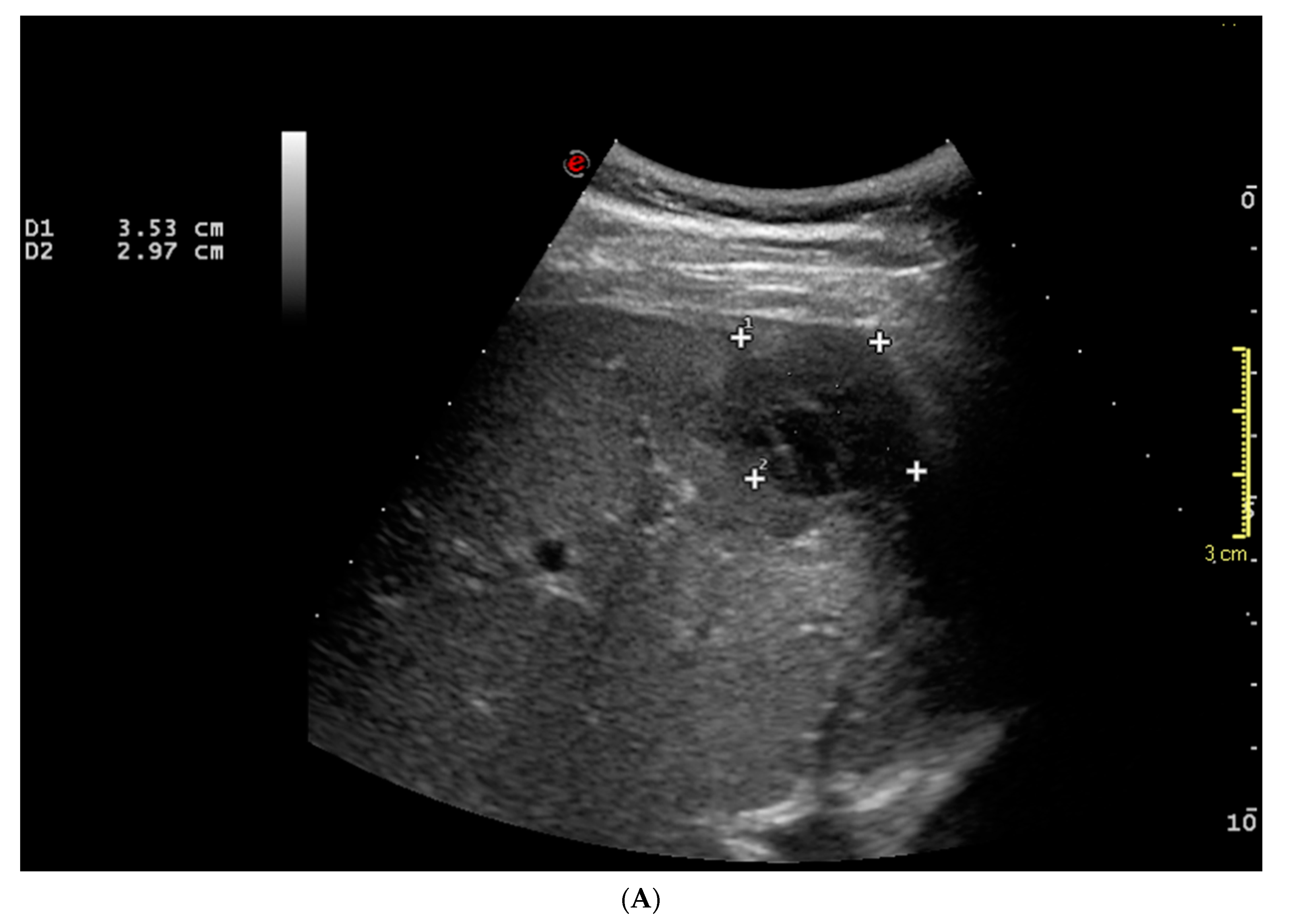
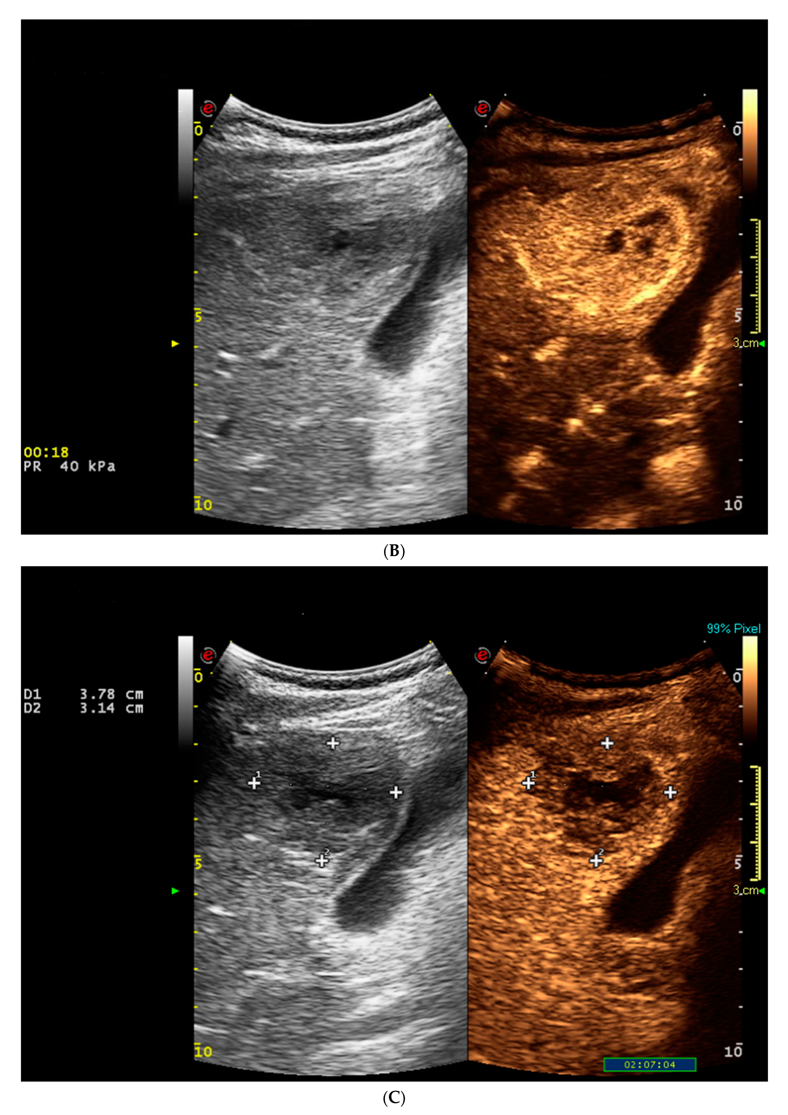
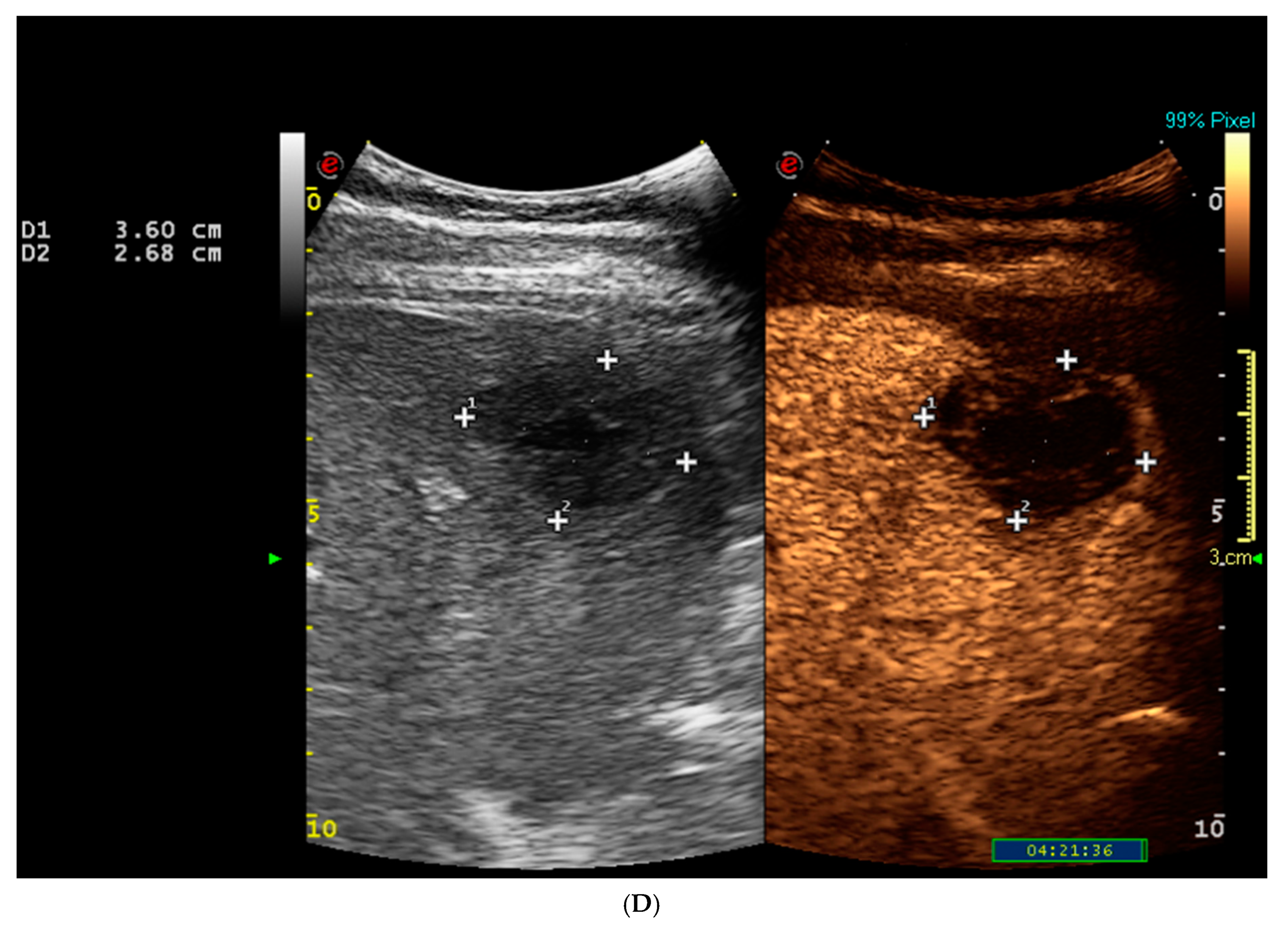
2. Case Presentation

We describe the case of a 41-year-old Pakistani man who has been living in Italy since 2009. In June 2019, the patient was admitted at our hospital because of 3-week-old fever of unknown origin associated with night sweats and unresponsive to broad spectrum antibiotics. Upon physical examination he presented with a painless, hard, and fixed mass in the right supraclavicular fossa compatible with lymphadenopathy. The supraclavicular mass had appeared a month earlier and was dimensionally stable over time. There were no other pathological findings at the physical examination. The patient had no history of weight loss, vomiting, or respiratory symptoms. A complete work-up was carried out including neck ultrasound, chest X-ray, neck and chest contrast-enhanced CT (Emotion 6, Siemens, Erlangen, Germany), and fine needle aspiration of the lymphadenopathy of the right supraclavicular fossa. On microbiological examination, Mycobacterium tuberculosis polymerase chain reaction (PCR) amplification test was positive. The patient was diagnosed with tuberculous lymphadenopathy. Antibiotic therapy with Rifampin (R) 600 mg daily, Isoniazid (H) 300 mg daily, Pyrazinamide (Z) 1500 daily, and Ethambutol (E) 800 mg daily was started. In July 2019, a neck and chest contrast-enhanced CT scan revealed a right supraclavicular lymphadenopathy of 26 × 22 mm without pulmonary lesions. In August 2019 following the detection of raised liver enzymes (AST/ALT 2-3 × upper normal levels; ALP/GGT 1.5-2 × upper normal levels), chronic Hepatitis C virus (HCV)-related liver disease was diagnosed, genotype 3 (HCV RNA viremia was 950,814 UI/mL), but liver ultrasound did not reveal focal liver lesions. The patient had no history of IV drug use and tested negative for HIV. In December 2019, a chest contrast-enhanced CT scan did not reveal mediastinal lymphadenitis, but in the inferior untargeted scans, it detected a subcapsular pluriconcamerate liver lesion of 33 × 30 × 28 mm with irregular profiles located in liver segment five (S5) of unclear origin. The liver lesion was considered suspicious for malignancy. Abdominal ultrasound confirmed a subcapsular inhomogeneous hypoechoic focal liver lesion of 3.5 cm × 2.9 cm in liver segment five (S5). (Figure 1A).
The liver stiffness determined using two-dimensional shear wave elastography (Aixplorer SuperSonic Imagine, S. A., Aix-en-Provence, France) was 10.9 kpa, suggestive of advanced chronic liver disease (F4 fibrosis stage) [6].
Contrast-enhanced ultrasound (CEUS) was performed to further characterize the liver lesion. After intravenous injection of 2.4 mL of SonoVue®, the hepatic lesion showed almost complete hyperenhancement in the arterial phase followed by late wash-out in the portal venous and late phases. (Figure 1B–D).
The CEUS pattern of the hepatic lesion, according to the American College of Radiology Liver Imaging Reporting and Data System (LI-RADS®) used to classify lesions at risk of HCC, was compatible with LI-RADS LR5 category or definite HCC [7,8]. In view of the disappearance of tuberculous lymphadenopathy and the lack of pulmonary TB features on the last CT scan, and in view of the underlying chronic HCV-related liver disease, due to the detection of a highly specific HCC pattern at CEUS, a non-invasive diagnosis of HCC was assumed according to current guidelines [9].
As the location of the lesion was close to the gallbladder, liver biopsy was not performed and the decision to perform liver resection was taken. In February 2020, the patient underwent wedge resection of liver segment five (S5). Because of the macroscopic necrotic aspect, a complete bacteriological examination (including PCR and tissue culture for Mycobacterium tuberculosis) of the lesion tissue was performed together with histological examination of the resected liver tissue and of the pericholedochal lymph node. The resected hepatic lesion was characterized as an abscess with granulomatous inflammatory reaction with giant and necrotic cells. The examined liver tissue did not reveal malignant cells nor Acid-Alcohol Resistant Bacilli (BAAR). The dissected pericholedochal lymph node was characterized as a reactive lymph node. The abscess content was BAAR negative and PCR was positive for Mycobacterium tuberculosis, sensitive to Rifampicin. The bronchial aspirate was negative for BAAR, PCR was negative for KB, and the mycobacterial culture was negative. The patient was diagnosed with hepatic tuberculosis and therapy with R 600 mg daily, H 300 mg daily, and Levofloxacin 1000 mg daily was administered.
At 8 months of follow-up, the patient is still doing fine with neither recurrence of focal liver lesions nor lymphadenopathy (Table 1).

3. Discussion

Tuberculosis is a rare disease in Western countries. However, TB remains a major global health threat with more than 10 million new cases per year of which less than two thirds are reported. Intravenous drug users, immunocompromised subjects, acquired immunodeficiency syndrome (AIDS), and diabetic patients represent the most susceptible population to infections. Pakistan, with 6% of cases, is among the eight countries accounting for two thirds of the total number of cases globally [10,11]. Despite the presence of such categories at greater risk, diagnosing hepatic TB can be challenging since it may display uncertain imaging patterns and the clinical manifestations may be quite variable.
Extrapulmonary TB represented 15% of the 7.0 million incidental cases that were notified in 2018, ranging from 8% in the Western Pacific Region to 24% in the Eastern Mediterranean Region. In northern Europe and in the USA up to 40% of all cases of TB are extrapulmonary, and lymphadenitis is the most common extrapulmonary presentation of tuberculosis [11]. Gastrointestinal TB constitutes nearly 10% of all extrapulmonary cases [12]. Hepatic TB is almost invariably associated with systemic dissemination of TB bacilli and is often unrecognized because of its relatively non-specific clinical presentations [1,3,11].
The imaging appearance of hepatic TB is considered non-specific; therefore, a histopathological or bacteriological confirmation is often required [1,2,4,5,13,14,15,16,17,18,19]. CEUS, CT, and MRI are used in clinical practice for the characterization of liver lesions. However, imaging findings might be inconclusive or misleading in the diagnosis of hepatic TB (Table 2) [1,5,16,18,19,20,21].
The diagnosis might be particularly difficult in patients with a single liver lesion, with underlying liver disease, without any history of TB nor signs of pulmonary TB. In such cases, histological and/or bacteriological confirmation might be needed to make an early diagnosis and start antibiotic therapy [13,21]. However, HCC is a unique entity as an imaging definite diagnosis can be achieved without requiring histological confirmation. For this reason, if TB occurs in patients fulfilling the criteria for non-invasive diagnosis of HCC, corresponding to a typical contrast-enhanced pattern in patients with cirrhosis; a risk of misdiagnosis could not be avoided. Therefore, clinical conditions must be carefully assessed to consider an alternative diagnosis. Early diagnosis of hepatic TB is in fact important to reduce morbidity and mortality. It has been reported that patients can recover with anti-tuberculosis therapy if treatment is administered in the early stage [14]. A delayed or wrong diagnosis can instead cause the situation to deteriorate and lead to complications, requiring invasive treatment such as percutaneous drainage or surgery [16,18,19,20].
While CT and MRI findings have been described in some detail, there is a paucity of literature on CEUS features [1,2,3,4,5,18,19]. Due to a significant overlap with other common and similar appearing hepatic lesions, hepatic TB is often either misdiagnosed or labeled as indeterminate lesions. Several cases of hepatic TB lesions resembling primary or secondary liver cancer at the imaging have been described in the literature, hence histopathologic and/or bacteriological diagnosis was needed [2,3,16,17,18,19]. However, the data on the CEUS features of hepatic TB are scarce. In the only study evaluating CEUS pattern of hepatic TB, most of the 24 hepatic TB lesions were subcapsular, located on the diaphragmatic surface of the liver, and oval-shaped [22]. Almost 80% of the lesions had an inhomogeneous hypoechoic echogenicity and more than 90% had poorly defined margins on B-mode ultrasound. Lesions usually had no or very weak intralesional flow signal on Doppler ultrasonography. After the injection of contrast medium (SonoVue®), more than half of the lesions (54.2%) presented a rapid and marked rim enhancement with a hypoenhanced or non-enhanced center in the arterial phase. About 37% of the lesions showed a transient inhomogeneous enhancement of the entire lesion during the arterial phase. Most of the lesions showed a distinct contrast wash-out in the portal phase. The enhancement patterns on CEUS correlated with the different pathological stages of the hepatic TB lesions, ranging from granulomatous lesions with or without caseation necrosis to fibrotic and calcified lesions [22].
The LI-RADS algorithm is used to stratify primary liver nodules according to their risk of malignancy from LR-1 (definitely benign) to LR-5 (definitely HCC). Although very rare, the risk of misdiagnosis should be taken into account when using contrast-enhanced ultrasound; considering the relevant clinical and epidemiological data which might justify biopsy even in case of a theoretically diagnostic pattern such as LI-RADS LR-5.

4. Conclusions

This case initially misdiagnosed as HCC complicating HCV-related chronic liver disease confirms imaging polymorphism of hepatic TB and illustrates the difficulty in reaching a correct preoperative diagnosis. Due to the lack of specific clinical manifestations and imaging features, a good knowledge of personal history and epidemiological data is mandatory in interpreting CEUS findings of a focal liver lesion and for the differential diagnosis, even when the imaging pattern would be highly suggestive of HCC.

Author Contributions

Conceptualization, A.F., A.G.; Methodology, A.F., M.R. (Mattero Renzulli), A.G.; Investigation, A.F., F.T., M.R. (Mattero Renzulli), M.R. (Matteo Ravaioli), F.V., F.P.; Data Curation, A.F., A.G.; Writing—original draft preparation, A.F., A.G.; Writing—review and editing, A.F., A.G., F.P.; Supervision, A.G., F.P. All authors have read and agreed to the published version of the manuscript.

Funding

This research received no external funding.

Informed Consent Statement

The patient provided informed consent.

Data Availability Statement

The data presented in this study are available on request from the corresponding author.

Conflicts of Interest

The funders had no role in the design of the study; in the collection, analyses, or interpretation of data; in the writing of the manuscript, or in the decision to publish the results. All the Authors do not have conflict of interest to declare.

Abbreviations

AIDSAcquired Immunodeficiency Syndrome
BAARAcid-Alcohol Resistant Bacilli
CEUSContrast-enhanced Ultrasound
CTComputed Tomography
EEthambutol
HIsoniazid
HCVHepatitis C Virus
HIVHuman Immunodeficiency Virus
LI-RADSLiver Imaging Reporting and Data System
MRIMagnetic Resonance Imaging
PCRPolymerase Chain Reaction
RRifampin
TBHepatic Tuberculosis
ZPyrazinamide

References

  1. Kakkar, C.; Polnaya, A.M.; Koteshwara, P.; Smiti, S.; Rajagopal, K.V.; Arora, A. Hepatic tuberculosis: A multimodality imaging review. Insights Imaging 2015, 6, 647–658. [Google Scholar] [CrossRef] [PubMed] [Green Version]
  2. Karaosmanoglu, A.D.; Onur, M.R.; Sahani, D.V.; Tabari, A.; Karcaaltincaba, M. Hepatobiliary Tuberculosis: Imaging Findings. Am. J. Roentgenol. 2016, 207, 1–11. [Google Scholar] [CrossRef] [PubMed]
  3. Liu, H.; Zhu, J.; Dong, H.; Wang, Y.; Ma, J.; Kou, J.; Li, X.; He, Q. Isolated hepatic tuberculosis in the caudate lobe mimicking intrahepatic carcinoma. Clin. Res. Hepatol. Gastroenterol. 2017, 41, e65–e67. [Google Scholar] [CrossRef] [PubMed]
  4. Yu, R.-S.; Zhang, S.-Z.; Wu, J.-J.; Li, R.-F. Imaging diagnosis of 12 patients with hepatic tuberculosis. World J. Gastroenterol. 2004, 10, 1639–1642. [Google Scholar] [CrossRef] [PubMed]
  5. Burrill, J.; Williams, C.J.; Bain, G.; Conder, G.; Hine, A.L.; Misra, R.R. Tuberculosis: A Radiologic Review. Radiographics 2007, 27, 1255–1273. [Google Scholar] [CrossRef] [PubMed] [Green Version]
  6. European Association for Study of Liver; Asociacion Latinoamericana para el Estudio del Higado. EASL-ALEH Clinical Practice Guidelines: Non-invasive tests for evaluation of liver disease severity and prognosis. J. Hepatol. 2015, 63, 237–264. [Google Scholar] [CrossRef] [PubMed] [Green Version]
  7. Terzi, E.; Iavarone, M.; Pompili, M.; Veronese, L.; Cabibbo, G.; Fraquelli, M.; Riccardi, L.; De Bonis, L.; SanGiovanni, A.; Leoni, S.; et al. Contrast ultrasound LI-RADS LR-5 identifies hepatocellular carcinoma in cirrhosis in a multicenter restropective study of 1006 nodules. J. Hepatol. 2018, 68, 485–492. [Google Scholar] [CrossRef] [PubMed]
  8. Schellhaas, B.; Görtz, R.S.; Pfeifer, L.; Kielisch, C.; Neurath, M.F.; Strobel, D. Diagnostic accuracy of contrast-enhanced ultrasound for the differential diagnosis of hepatocellular carcinoma: ESCULAP versus CEUS-LI-RADS. Eur. J. Gastroenterol. Hepatol. 2017, 29, 1036–1044. [Google Scholar] [CrossRef] [PubMed]
  9. European Association for the Study of the Liver. EASL Clinical Practice Guidelines: Management of hepatocellular carcinoma. J. Hepatol. 2018, 69, 182–236. [Google Scholar] [CrossRef] [PubMed] [Green Version]
  10. Tuberculosis. Available online: https://www.who.int/news-room/fact-sheets/detail/tuberculosis (accessed on 14 January 2021).
  11. Global Tuberculosis Report. 2018. Available online: https://www.who.int/tb/publications/global_report/gtbr2018_main_text_12August2019.pdf?ua=1 (accessed on 14 January 2021).
  12. Sharma, M.; Sinha, S.K.; Singh, A.K.; Samanta, J.; Sharma, A.; Kochhar, R.; Sharma, K. Challenging diagnosis of gastrointestinal tuberculosis made simpler with multi-targeted loop-mediated isothermal amplification assay. Eur. J. Gastroenterol. Hepatol. 2020, 32, 971–975. [Google Scholar] [CrossRef] [PubMed]
  13. Suri, R.; Gupta, S.; Singh, K.; Suri, S. Ultrasound guided fine needle aspiration cytology in abdominal tuberculosis. Br. J. Radiol. 1998, 71, 723–727. [Google Scholar] [CrossRef] [PubMed]
  14. Hickey, A.J.; Gounder, L.; Moosa, M.Y.; Drain, P.K. A systematic review of hepatic tuberculosis with considerations in human im-munodeficiency virus co-infection. BMC Infect. Dis. 2015, 15, 209. [Google Scholar] [CrossRef] [PubMed] [Green Version]
  15. Gambhir, S.; Ravina, M.; Rangan, K.; Dixit, M.; Barai, S.; Bomanji, J. International Atomic Energy Agency Extra-pulmonary TB Consortium. Imaging in extrapulmonary tuberculosis. Int. J. Infect. Dis. 2017, 56, 237–247. [Google Scholar] [CrossRef] [PubMed] [Green Version]
  16. Jain, R.; Sawhney, S.; Gupta, R.G.; Acharya, S.K. Sonographic appearances and percutaneous management of primary tuberculous liver abscess. J. Clin. Ultrasound 1999, 27, 159–163. [Google Scholar] [CrossRef]
  17. Ai, J.-W.; Li, Y.; Cheng, Q.; Cui, P.; Wu, H.-L.; Xu, B.; Zhang, W. Diagnosis of local hepatic tuberculosis through next-generation sequencing: Smarter, faster and better. Clin. Res. Hepatol. Gastroenterol. 2018, 42, 178–181. [Google Scholar] [CrossRef] [PubMed]
  18. Schininà, V.; Albarello, F.; Cristofaro, M.; Di Stefano, F.; Fusco, N.; Cuzzi, G.; Arend, S.M.; Goletti, D.; Rizzi, E.B. Diagnostic imaging of hepatic tuberculosis: Case series. Int. J. Tuberc. Lung Dis. 2018, 22, 779–787. [Google Scholar] [CrossRef] [PubMed]
  19. Chan, H.S.; Pang, J. Isolated giant tuberculomata of the liver detected by computed tomography. Gastrointest. Radiol. 1989, 14, 305–307. [Google Scholar] [CrossRef] [PubMed]
  20. Chen, H.-C.; Chao, Y.-C.; Shyu, R.-Y.; Hsieh, T.-Y. Isolated tuberculous liver abscesses with multiple hyperechoic masses on ultrasound: A case report and review of the literature. Liver Int. 2003, 23, 346–350. [Google Scholar] [CrossRef] [PubMed]
  21. Tan, T.C.; Cheung, A.Y.; Wan, W.Y.; Chen, T.C. Tuberculoma of the liver presenting as a hyperechoic mass on ultrasound. Br. J. Radiol. 1997, 70, 1293–1295. [Google Scholar] [CrossRef] [PubMed]
  22. Cao, B.-S.; Li, X.-L.; Li, N.; Wang, Z.-Y. The nodular form of hepatic tuberculosis: Contrast-enhanced ultrasonographic findings with pathologic correlation. J. Ultrasound Med. 2010, 29, 881–888. [Google Scholar] [CrossRef] [PubMed]
Figure 1. (A) Subcapsular inhomogeneous hypoechoic focal liver lesion of 3.5 × 2.9 cm in segment fifth (S5); (B) Arterial phase of contrast enhanced ultrasound (18 s after contrast injection) showing an almost complete enhancement of the lesion with incomplete rim-enhancement; (C) Portal venous phase (2 min after contrast injection) showing the start of wash-out; (D) Late phase (4 min after contrast injection) showing a marked wash-out of the lesion.
Gastroent 12 00001 g001aGastroent 12 00001 g001bGastroent 12 00001 g001c
Table 1. Patient history in timeline format.
TimeEventFindings
June 2019fever associated with night sweatspainless, hard, and fixed mass in the right supraclavicular fossa
July 2019Chest and Neck CTRight supraclavicular lymphadenopathy of 26 × 22 mm
Fine needle aspiration of the lymphadenopathy of the right supraclavicular fossa; tissue culture and polymerase chain reaction (PCR) amplification.Mycobacterium tuberculosis isolated from both tissue culture and PCR
July 2019Chest X-rayNo signs of pulmonary TB. Diagnosis of tuberculous lymphadenopathy.
Normal liver enzymes.
HIV negative.
July 2019Starting treatment with Rifampin (R), Isoniazid (H), Pyrazinamide (Z), and Ethambutol (E)Well tolerated
August 2019The patient was tested for viral hepatitis serological markers after detection of elevated AST/ALT, ALP, and GGTHCV-related chronic hepatitis (genotype 3)
October 2019Abdominal ultrasoundNo liver lesions
December 2019Chest contrast-enhanced CTNeither pulmonary nor lymphadenopathy TB. Subcapsular focal liver lesion in liver segment five (S5) suggestive of malignancy
January 2020Drug-induced liver injury and underlying untreated HCV-related chronic hepatitisanti-TB therapy was interrupted due to increased liver enzymes (AST 383 U/L, ALT 462 U/L), with progressive normalization of liver enzymes: AST/ALT 180/290 → 132/213 → 80/126 → 41/38 U/L in February 2020.
January 2020CEUS to further characterize the focal liver lesionFocal liver lesion with CEUS LI-RADS LR5 pattern (highly suggestive of HCC)
January 20202-dimensional shear wave elastography
(because of detection of focal liver lesion)
Liver stiffness of 10.9 kPa (suggestive of advanced chronic liver disease).
February 2020Liver wedge resection of liver segment five (S5)Abscess with granulomatous inflammatory reaction with giant and necrotic cells
February 2020Histological examination and PCR for KBNo malignant cells;
PCR positive for KB, sensitive to R;
Diagnosis of hepatic TB
March 2020Antibiotic therapy with R, H, and Levofloxacin was administered.Well tolerated
April, May, October, and December 2020Clinical and biochemical re-evaluation: no pathological findings.In December 2020 the patient was asymptomatic. Laboratory: AST 64 U/L, ALT 65 U/L; and the CT scan was negative for disease recurrence. Anti-TB therapy was stopped.
Anti-viral (HCV) therapy was scheduled.
Table 2. Imaging (CEUS, CT, MRI) features of hepatic tuberculosis.
ImagingContrast-Enhanced UltrasoundComputed TomographyMagnetic Resonance Imaging
Unenhanced scanMainly hypoechoic but also isoechoic or hyperechoic nodules *hypodense lesions °hypointense on T1-weighted images, isointense or hyperintense lesions with a peripheral hypointense rim on T2-weighted images #
Arterial phaseRim enhancement with a hypoenhanced or non-enhanced center or complete hyperenhancement of the lesion ^Peripheral contrast enhancement °Rim heterogenous contrast enhancement
Late phaseWash-outWash-outWash-out
Comparison of imaging features of hepatic tuberculosis. All the imaging techniques can reveal a macronodular single lesion or multiple small miliary nodular lesions. * The features may differ depending on the stage of the disease. ° CT imaging features of macronodular hepatic TB depends on the stage of the disease. Non-caseating granulomas appear hypodense on the unenhanced study and usually display no or minimal peripheral rim enhancement following intravenous contrast administration. Hepatic tuberculomas eventually tend to calcify, and the presence of calcified granulomas at CT in patients with relevant clinical history and in the absence of a known primary tumor should raise suspicion of tuberculosis. ^ The enhancement patterns on CEUS correlated with the different pathologic stages of the hepatic TB lesions, ranging from granulomatous lesions with or without caseation necrosis to fibrotic and calcified lesions. # Depending upon the stage of the disease.
Publisher’s Note: MDPI stays neutral with regard to jurisdictional claims in published maps and institutional affiliations.

