
Massive Gastrointestinal Bleeding Due to Jejunal Diverticula in a Community Hospital: A Case Report and Review of Diagnostic and Therapeutic Options


Abstract
:1. Introduction
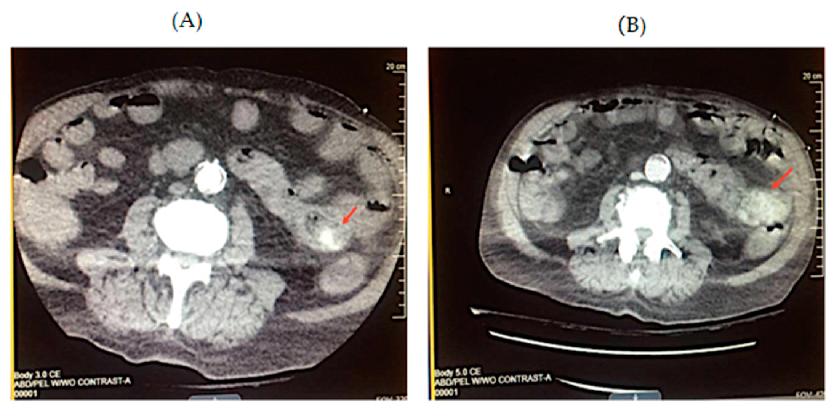
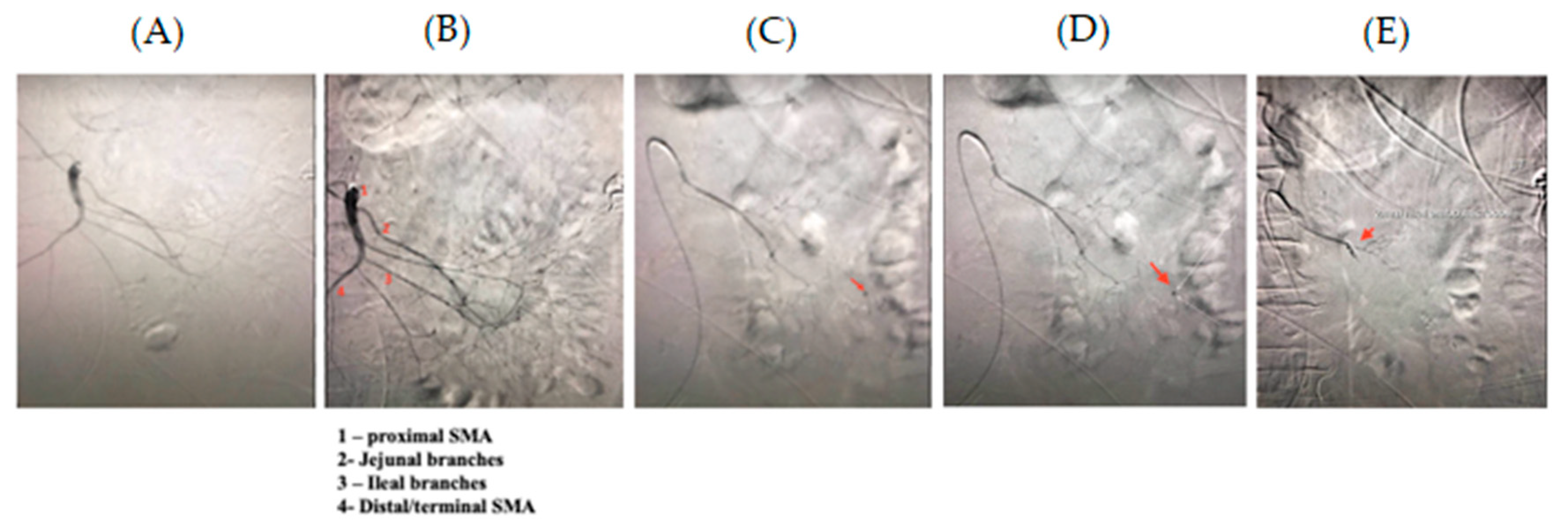
2. Case Report
3. Discussion
4. Conclusions
Author Contributions
Funding
Institutional Review Board Statement
Informed Consent Statement
Conflicts of Interest
References
- Carney, B.; Khatri, G.; Shenoy-Bhangle, A. The role of imaging in gastrointestinal bleed. Cardiovasc. Diagn. Ther. 2019, 9, S88–S96. [Google Scholar] [CrossRef] [PubMed]
- De Peuter, B.; Box, I.; Vanheste, R.; Dymarkowski, S. Small-bowel Diverticulosis: Imaging Findings and Review of Three Cases. Gastroenterol. Res. Pract. 2009, 1–3. [Google Scholar] [CrossRef] [PubMed] [Green Version]
- Lebert, P.; Millet, I.; Ernst, O.; Boulay-Coletta, I.; Corno, L.; Taourel, P.; Zins, M. Acute Jejunoileal Diverticulitis: Multicenter Descriptive Study of 33 Patients. Am. J. Roentgenol. 2018, 210, 1245–1251. [Google Scholar] [CrossRef]
- Harbi, H.; Kardoun, N.; Fendri, S.; Dammak, N.; Toumi, N.; Guirat, A.; Mzali, R. Jejunal diverticulitis. Review and treatment algorithm. Presse Médicale 2017, 46, 1139–1143. [Google Scholar] [CrossRef]
- Leigh, N.; Sullivan, B.; Anteby, R.; Talbert, S. Perforated jejunal diverticulitis: A rare but important differential in the acute abdomen. Surg. Case Rep. 2020, 6, 1–7. [Google Scholar] [CrossRef] [PubMed]
- Andersen, L.; Schjoldager, B.; Halver, B.J. Jejunal Diverticulosis in a Family. Scand. J. Gastroenterol. 1988, 23, 672–674. [Google Scholar] [CrossRef] [PubMed]
- Almaramhy, H. Jejunal atresia associated with jejunal diverticulosis. J. Pediatric Surg. Case Rep. 2018, 32, 68–71. [Google Scholar] [CrossRef]
- Yaqub, S.; Evensen, B.; Kjellevold, K. Massive rectal bleeding from acquired jejunal diverticula. World J. Emerg. Surg. 2011, 6, 17. [Google Scholar] [CrossRef] [Green Version]
- Cherian, M.; Mehta, P.; Kalyanpur, T.; Hedgire, S.S.; Narsinghpura, K.S. Arterial Interventions in Gastrointestinal Bleeding. Semin. Interv. Radiol. 2009, 26, 184–196. [Google Scholar] [CrossRef] [Green Version]
- Reis, F.; Cardia, P.; D’Ippolito, G. Computed tomography angiography in patients with active gastrointestinal bleeding. Radiol. Bras. 2015, 48, 381–390. [Google Scholar] [CrossRef]
- Wong, C.; Lin, I.; Shih, S.; Chang, W.H.; Wang, H.Y. Jejunal Diverticula Causing Unusual Massive Lower Gastrointestinal Bleeding. Int. J. Gerontol. 2008, 2, 120–123. [Google Scholar] [CrossRef] [Green Version]
- Mohan, P.; Manov, J.; Diaz-Bode, A.; Venkat, S.; Langston, M.; Naidu, A.; Howse, R.; Narayanan, G. Clinical Predictors of Arterial Extravasation, Rebleedingand Mortality Following Angiographic Interventions in Gastrointestinal Bleeding. J. Gastrointest. Liver Dis. 2018, 27, 221–226. [Google Scholar] [CrossRef] [PubMed]
- Mensel, B.; Kühn, J.; Kraft, M.; Rosenberg, C.; Partecke, L.I.; Hosten, N.; Puls, R. Selective microcoil embolization of arterial gastrointestinal bleeding in the acute situation. Eur. J. Gastroenterol. Hepatol. 2012, 24, 155–163. [Google Scholar] [CrossRef]
- Kim, B. Diagnosis of gastrointestinal bleeding: A practical guide for clinicians. World J. Gastrointest. Pathophysiol. 2014, 5, 467. [Google Scholar] [CrossRef] [PubMed]
- Bond, A.; Smith, P. British Society of Gastroenterology: Diagnosis and management of acute lower gastrointestinal bleeding. Frontline Gastroenterol. 2019, 10, 417–420. [Google Scholar] [CrossRef]
- Pérez Roldán, F.; González Carro, P.; Legaz, H.M.L.; Roncero García-Escribano, O.; Ynfante Ferrús, M.; Aoufi, S.; Sánchez-Manjavacas Muñoz, N.; Ruiz Carrillo, F. Efficacy of pediatric colonoscopy used as push enteroscopy in the management of capsule endoscopy findings. Rev. Española de Enferm. Dig. 2009, 101, 468–476. [Google Scholar] [CrossRef] [Green Version]
- Zhou, D.; Jiang, B.; Yang, X. Advances and applications of enteroscopy for small bowel. World J. Gastroenterol. 1997, 3, 205. [Google Scholar] [CrossRef]
- Zhao, L.; Lu, W.; Sun, Y.; Liang, J.; Feng, S.; Shi, Y.; Wu, Q.; Wang, J.; Wu, K. Small intestinal diverticulum with bleeding. Medicine 2018, 97, e9871. [Google Scholar]
- Abegunde, A.; Christman, E.; Hassell, L.; Kastens, D. Rare Jejunal Diverticular Bleeding. ACG Case Rep. J. 2016, 3, e146. [Google Scholar] [CrossRef]
- Mantas, D. Small intestine diverticula: Is there anything new? World J. Gastrointest. Surg. 2011, 3, 49. [Google Scholar] [CrossRef]
- Tarasconi, A.; Baiocchi, G.; Pattonieri, V.; Perrone, G.; Abongwa, H.K.; Molfino, S.; Portolani, N.; Sartelli, M.; di Saverio, S.; Heyer, A.; et al. Transcatheter arterial embolization versus surgery for refractory non-variceal upper gastrointestinal bleeding: A meta-analysis. World J. Emerg. Surg. 2019, 14, 1–13. [Google Scholar] [CrossRef] [PubMed]
- Wortman, J.; Landman, W.; Fulwadhva, U.; Viscomi, S.G.; Sodickson, A.D. CT angiography for acute gastrointestinal bleeding: What the radiologist needs to know. Br. J. Radiol. 2017, 90, 20170076. [Google Scholar] [CrossRef]
- Levy, I.; Gralnek, I. Complications of diagnostic colonoscopy, upper endoscopy, and enteroscopy. Best Pract. Res. Clin. Gastroenterol. 2016, 30, 705–718. [Google Scholar] [CrossRef]
- Maza, I.; Gralneck, I.M. Chapter 15—Obscure Gasrointestinal Bleeding. In Clinical Gastrointestinal Endoscopy, 2nd ed.; Elsevier Inc.: Amsterdam, The Netherlands, 2012; pp. 173–179. [Google Scholar]
- Teng, H.C.; Liang, H.L.; Lin, Y.H.; Huang, J.S.; Chen, C.Y.; Lee, S.C.; Pan, H.B. The Efficacy and Long-Term Outcome of Microcoil Embolotherapy for Acute Lower Gastrointestinal Bleeding. Korean J. Radiol. 2013, 14, 259. [Google Scholar] [CrossRef] [PubMed]
- Ul-Haq, T.; Idris, M.; Salam, B.; Akhtar, W.; Jamil, Y. Comparison of microcoils and polyvinyl alcohol particles in selective microcatheter angioembolization of non-variceal acute gastrointestinal hemorrhage. Park J. Med. Sci. 2015, 31, 751–756. [Google Scholar]
- Gralnek, L.M.; Neeman, Z.; Strate, L.L. Acute Lower Gastrointestinal Bleeding. N. Engl. J. Med. 2017, 376, 1054–1063. [Google Scholar] [CrossRef]
- Sengupta, N. The role of colonoscopy and endotherapy in the management of lower gastrointestinal bleeding. Best Pract. Res. Clin. Gastroenterol. 2019, 42–43, 101615. [Google Scholar] [CrossRef] [PubMed]
- Loffroy, R.; Rao, P.; Ota, S.; de Lin, M.; Kwak, B.-K.; Geschwind, J.-F. Embolization of Acute Nonvariceal Upper Gastrointestinal Hemorrhage Resistant to Endoscopic Treatment: Results and Predictors of Recurrent Bleeding. Cardiovasc. Interv. Radiol. 2010, 33, 1088–1100. [Google Scholar] [CrossRef]
- Soetikno, R.; Ishii, N.; Kolb, J.; Hammad, H.; Kaltenbach, T. The Role of Endoscopic Hemostasis Therapy in Acute Lower Gastrointestinal Hemorrhage. Gastrointest. Endosc. Clin. N. Am. 2018, 28, 391–408. [Google Scholar] [CrossRef] [PubMed]
- Feuerstein, J.D.; Ketwaroo, G.; Tewani, S.K.; Cheesman, A.; Trivella, J.; Raptopoulos, V.; Leffler, D.A. Localizing Acute Lower Gastrointestinal Hemorrhage: CT Angiography Versus Tagged RBC Scintigraphy. Gastroinetstinal Imaging 2016, 207, 578–584. [Google Scholar] [CrossRef]
- Artigas, J.M.; Marti, M.; Soto, J.A.; Esteban, H.; Pinilla, I.; Guillen, E. Multidetector CT Angiography for Acute Gastrointestinal Bleeding: Technique and Findings. Trauma Emerg. Radiol. 2013, 33, 1453–1470. [Google Scholar]
- Oakland, K.; Chadwick, G.; East, J.; Guy, R.; Humphries, A.; Jairath, V.; McPherson, S.; Metzner, M.; Morris, A.J.; Murphy, M.F.; et al. Diagnosis and management of acute lower gastrointestinal bleeding: Guidelines from the British Society of Gastroenterology. Gut 2019, 68, 776–789. [Google Scholar] [CrossRef]
- Trindade, A.; Lichtenstein, D.; Aslanian, H.; Bhutani, M.S.; Goodman, A.; Melson, J.; Navaneethan, U.; Pannala, R.; Parsi, M.A.; Sethi, A.; et al. Devices and methods to improve colonoscopy completion (with videos). Gastrointest. Endosc. 2018, 87, 625–634. [Google Scholar] [CrossRef] [PubMed]
- Strate, L.; Gralnek, I. ACG clinical guideline: Management of patients with acute lower gastrointestinal bleeding. Am. J. Gastroenterol. 2016, 111, 459–474. [Google Scholar] [CrossRef] [PubMed] [Green Version]


Publisher’s Note: MDPI stays neutral with regard to jurisdictional claims in published maps and institutional affiliations. |
© 2021 by the authors. Licensee MDPI, Basel, Switzerland. This article is an open access article distributed under the terms and conditions of the Creative Commons Attribution (CC BY) license (https://creativecommons.org/licenses/by/4.0/).
Share and Cite
Abbasi, C.; Jimenez, M.C.; Lisi, M. Massive Gastrointestinal Bleeding Due to Jejunal Diverticula in a Community Hospital: A Case Report and Review of Diagnostic and Therapeutic Options. Gastroenterol. Insights 2021, 12, 196-201. https://0-doi-org.brum.beds.ac.uk/10.3390/gastroent12020017
Abbasi C, Jimenez MC, Lisi M. Massive Gastrointestinal Bleeding Due to Jejunal Diverticula in a Community Hospital: A Case Report and Review of Diagnostic and Therapeutic Options. Gastroenterology Insights. 2021; 12(2):196-201. https://0-doi-org.brum.beds.ac.uk/10.3390/gastroent12020017
Chicago/Turabian StyleAbbasi, Cynthia, M. Carolina Jimenez, and Michael Lisi. 2021. "Massive Gastrointestinal Bleeding Due to Jejunal Diverticula in a Community Hospital: A Case Report and Review of Diagnostic and Therapeutic Options" Gastroenterology Insights 12, no. 2: 196-201. https://0-doi-org.brum.beds.ac.uk/10.3390/gastroent12020017